HAL Id: hal-01476632
https://hal.archives-ouvertes.fr/hal-01476632
Submitted on 25 Oct 2018
HAL is a multi-disciplinary open access
archive for the deposit and dissemination of
sci-entific research documents, whether they are
pub-lished or not. The documents may come from
teaching and research institutions in France or
abroad, or from public or private research centers.
L’archive ouverte pluridisciplinaire HAL, est
destinée au dépôt et à la diffusion de documents
scientifiques de niveau recherche, publiés ou non,
émanant des établissements d’enseignement et de
recherche français ou étrangers, des laboratoires
publics ou privés.
Drug releasing nanoplatforms activated by alternating
magnetic fields
Damien Mertz, Olivier Sandre, Sylvie Begin-Colin
To cite this version:
Damien Mertz, Olivier Sandre, Sylvie Begin-Colin. Drug releasing nanoplatforms activated by
alter-nating magnetic fields. Biochimica et Biophysica Acta (BBA) - General Subjects, Elsevier, 2017, 1861
(6), pp.1617-1641. �10.1016/j.bbagen.2017.02.025�. �hal-01476632�
1
Drug releasing nanoplatforms activated by
alternating magnetic fields
Damien Mertz
1,*, Olivier Sandre
2, Sylvie Begin-Colin
11
Institut de Physique et Chimie des Matériaux de Strasbourg, Université de Strasbourg, UMR 7504 CNRS, 23, rue du Loess, 67034 Strasbourg, France. E-mail: [email protected]
2
Laboratoire de Chimie des Polymères Organiques (LCPO), CNRS UMR 5629, Université de Bordeaux, Bordeaux-INP, Pessac 33607 Cedex, France
Abstract.
The use of an alternating magnetic field (AMF) to generate non-invasively and spatially a localizedheating from a magnetic nano-mediator has become very popular these last years to develop magnetic hyperthermia (MH) as a promising therapeutic modality already used in the clinics. AMF has become highly attractive this last decade over others radiations, as AMF allows a deeper penetration in the body and a less harmful ionizing effect. In addition to pure MH which induces tumor cell death through local T elevation, this AMF-generated magneto-thermal effect can also be exploited as a relevant external stimulus to trigger a drug release from drug-loaded magnetic nanocarriers, temporally and spatially. This review article is focused especially on this concept of AMF induced drug release, possibly combined with MH. The design of such magnetically responsive drug delivery nanoplatforms requires two key and complementary components: a magnetic mediator which collects and turns the magnetic energy into local heat, and a thermoresponsive carrier ensuring thermo-induced drug release, as a consequence of magnetic stimulus. A wide panel of magnetic nanomaterials/chemistries and processes are currently developed to achieve such nanoplatforms. This review article presents a broad overview about the fundamental concepts of drug releasing nanoplatforms activated by AMF, their formulations, and their efficiency in vitro and in vivo.
1. Introduction
Today, it is admitted that the combination of therapies is a way to considerably increase the efficacy of a treatment, for instance in the case of anti-cancer treatments. Besides, the follow-up of therapy by imaging is reported of paramount importance because it brings precious information spatially and in real-time to improve and optimize the therapy efficacy.[1,2] To face these challenges, nano-platforms based on nanoparticles (NPs) are very promising because NPs bring key features to overcome the major drawbacks of conventional medicines, namely poor bioavailability, insufficient target specificity, and significant side effects. There are various types of therapies offered by NPs that are mainly : drug delivery[3], gene therapy[4], photodynamic therapy (PDT) [5], photothermal therapy (PTT) [6] and magnetic hyperthermia (MH) [7]. Considering the blossoming growth of nanotechnology which has brought challenging innovations in the synthesis of such multifunctional nano-objects for therapeutic applications, the targeted and local drug delivery controlled by an external stimuli using nanoparticulate systems has emerged as a very promising strategy for nanomedicine. The concept of NP-mediated-drug delivery systems (DDS) or “nanodevices” (medical devices having simplified
2
regulatory route towards authorization in clinics as compared to medicines) involves the incorporation of a drug within nanoparticulate carriers via encapsulation, absorption, adsorption, or conjugation, for safe and stable administration in the body. Besides, an enhanced delivery and controlled release at target sites can be envisioned with such carriers through the incorporation of a cocktail of drugs aimed at combination therapy of complex diseases, such as cancer, where a single drug is rarely effective.[8,9]
For nanoplatforms used in intravenous injection, controlling the drug transport in the blood flow but also the cell targeting and the cell internalization in organs (biodistribution) are key issues.[10–12] It is now admitted and demonstrated that nanocarriers allow improving the delivery of chemotherapeutics whereas for drug alone, there are various issues such as poor water solubility of drugs, their rapid drug clearance, non-specific biodistribution, toxicity to healthy tissues, etc... Indeed, as stated by Ulbrich et al., “It has been reported that about 40% of the drugs being developed by the pharmaceutical industry are poorly water soluble molecules.”[13] Furthermore, surface functionalization of the NPs is needed to impart blood circulation and carry the drugs to the targeted tissues/cells. In addition, there are several other issues to overcome with the intravenous injections: opsonization i.e. the adsorption of plasma proteins followed by subsequent RES uptake; suitable cell internalization (i.e. cytoplasm or nucleus entry); and the relevance of active vs. passive cell targeting.
Regarding nanocarrier design, a further issue is controlling the drug release with biological or externally applied stimuli. The triggering of drug release can be achieved either by local biological stimuli (pH, redox, enzymes) or by externally applied radiation (light/electromagnetic fields, focused ultrasound).[14–16] Local triggers are encountered in various biological environments e.g. acidic pH tumor, lysosomal compartments, reductive cytosol, however in such environments there is no possibility of remote activation or dose control over drug release as the parameters are defined by the biological environment. Here only a burst or “one shot” release may be envisioned in this case. The triggering of drug release was also developed recently from polymer films by the use of mechanical forces [17–19]. The external or remote triggers present advantages to achieve drug release with spatiotemporal control. The nanoplatforms can be also designed to ensure pulsatile release i.e. the drug can be released several times on demand with control over the diffusion rates and the times of drug release. However, this requires a specific, well-controlled and/or sophisticated design.
Among the various external stimuli such as electromagnetic or acoustic waves, the use of an alternating magnetic field (AMF) is becoming highly attractive over others, as it allows a deeper penetration in the body compared with near infrared (NIR) light, that is comparable to ultrasound (US), and a less harmful ionizing effect compared with X-ray radiations. Since two decades, the conversion of the magnetic induction energy AMF into a non-invasively and spatially localized heating from a magnetic mediator is investigated to develop MH, a therapeutic modality which acts in inducing tumor apoptotic cell death through local controlled T elevation and “thermal dose” deposition. This nevertheless must be achieved within a certain range of frequency and field strength, which is called Brezovitch criterion.[20,21] Indeed, in 1988, to consider as negligible the effects due to nonspecific heating and nervous system activation by the electric field associated to the AMF, Atkins and Brezovich proposed a safety limit where the product of magnetic field and amplitude
3
frequency (H0 f) should not exceed 4.85 × 10⁸ A m-1 s-1 to avoid parasitic eddy current effects on the organism. Since recent years, this AMF-generated magneto-thermal effect can also be exploited as a relevant external stimulus to trigger a drug release from drug-loaded magnetic nanocarriers, temporally and spatially. This magnetically induced drug release [22–24]sometimes called “magneto-chemotherapy” can be thus envisioned as a complementary promising therapeutic modality to pure MH. In any case, the drug release is activated by an AMF-generated local heating (at nm-scale) which may be mutualized with MH if macroscopic T elevation is reached above a threshold, typically 43°C. In other words, magnetically induced drug release can also occur without macroscopic T elevation, provided that the local T can generate a high local structural change of the carrier, triggering the drug release from the nanoplatform. Hence this requires loading the drug in a nanoplatform composed of two key components: a magnetic mediator which collects and turns the magnetic energy into local heat, and a thermoresponsive carrier/binder ensuring thermo-induced drug release, as a consequence of magnetic stimulus. A wide panel of magnetic nanomaterials / chemistries and processes are currently developed to achieve magnetically activated drug releasing platforms.
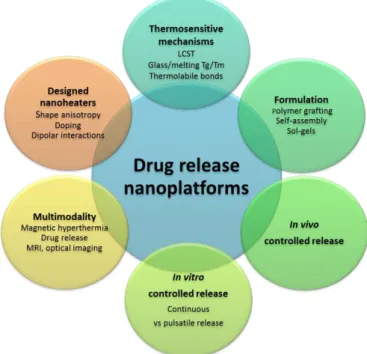
Figure 1. Concepts and challenges for the design of drug releasing nanoplatforms under AMF.
This review deals with the design of drug releasing magnetic nanoplatforms activated by external AMFs. In Part 2, we detail the fundamental concepts and the design of magnetic nanoheaters, the main mechanisms of thermo-responsive nanocarriers, the synthesis methods of thermoresponsive nanoplatforms loaded with magnetic NPs and hydrophobic or hydrophilic drugs (or both), and the experimental implementation of the in vitro assay of drug release activated upon AMF with a standardized set-up. These elements are conceptually and experimentally crucial to ensure the rational design and applications of suitable magnetic nanoplatforms displaying an efficient drug release upon AMF. Then, in Part 3, we provide a broad overview of the currently existing systems which were reported efficient to release drugs upon AMF application. For the sake of clarity, these magnetic nanoplatforms or nanomaterials are divided into four types : i) magnetic core-polymer shells,
4
ii) hollow aqueous core filled shells including vesicles (liposomes, polymersomes, hybrids…) and double emulsion or templated polymer capsules, iii) polymer-based nanoparticles including nanogels, micelles and hydrophobic NPs, and iv) hybrid inorganic nanocomposites that are essentially made of a mesoporous silica matrix. The efficiency of AMF-induced drug release and the main in vitro results with cells are presented in this part to highlight the significance of the drug release combined with hyperthermia effect. In Part 4, a focus on in vivo AMF-induced drug release is provided and the results all along this decade (since 2010) are reviewed. Indeed, this review aims at providing to the readers the main concepts of the topic but also the main performances with in vitro and in vivo results that were gathered and summarized (Figure 1). Hence, we expect that this review will hopefully provide a comprehensive and up-to-date overview of the field which may serve for future (pre)clinical purposes to chemists, physicians, biologists or clinicians. Drug releasing nanoplatforms upon AMF were never reviewed as an alone review as to date, the remote release by AMF especially in vivo being very recent (within last five years). There are reviews dealing partially with this topic or associated with other connected topics such as: Remotely Triggerable Drug Delivery Systems [25], Magnetoresponsive polymer-based materials [26,27], Remotely triggered nanovehicles actuated by the use of electromagnetic and acoustic waves [14,28], Chemothermotherapy[29], Iron oxide for magnetic resonance imaging (MRI) and therapy [29-31], and Thermoresponsive carriers [33,34], Design of organic macrocycle modified iron-oxide nanoparticles for drug delivery.[35]
2. Criteria and challenges for the design of magnetically drug releasing nanoplatforms
This part aims at describing how magnetic hyperthermia (MH) is provided (through the design of nanoheaters), how AMF-induced MH may be the trigger of controlled release (thermoresponsive mechanisms), and how MH may be combined with both drug and nanoheater encapsulation (dual loading formulation).
2.1 Mechanisms and challenges of MH: design of the magnetic nanoheaters
Iron oxide based nanoparticles (IONPs) are widely used for biological applications thanks to their outstanding balance between superpara- or ferrimagnetic properties, surface-to-volume ratio suitable for efficient functionalisation and proven biocompatibility. Their development for MRI or MH concentrates much of the attention as these nanomaterials are already used within the health system as contrast agents or iron supplement (e.g. Ferumoxytol) and heating mediators.
Indeed IONPs are commercially used as T2 (i.e. negative) contrast agents (CAs) for MRI [36,37] and are of particular interest as biodegradable and non-toxic nano-objects compared to other CA families. To be used as in vivo MRI CAs, NPs should exhibit a high saturation magnetization and be functionalized with molecules leading to biocompatible stable suspensions in physiological media with average hydrodynamic sizes smaller than 50 nm for ensuring a good in vivo pharmacokinetics (e.g. for angiography) and biodistribution (for tumor labeling).
IONPs are also developed for MH. When exposed to AMF of appropriate amplitude and frequency, these NPs release heat locally (where they are concentrated), which reduces the viability of cancer cells through apoptotic or necrotic pathways. The MH potential is demonstrated with the favorable recent results of the “nanothermotherapy” study in clinical phase II ended in 2011 led by a German company MagForce™
5
Nanotechnology (hospital Charité in Berlin) which has led to a European market authorization [38–41]. MH has been demonstrated to enhance the sensitivity of tumor cells to facilitate chemo- or radio–therapies [38,39] and to act on cell membranes or to trigger a thermally-induced release of drugs.[42]
However one of the limitations of MH is the low heating power of usual magnetic NPs, requiring a local injection of NPs in large quantities (e.g. 160 gL-1 iron oxide for MagForce™). Given the clinical importance of MH as therapy and also as possible local heater for drug release, there is thus currently a crucial need to design optimized NPs for efficient MH.[43–45] However optimization is not yet well-understood and experimental characterization methods and results on magnetic NPs vary widely. Indeed in MH, very specific physical properties are required for an optimal conversion of the supplied magnetic energy into heat. Three independent mechanisms result in thermal energy upon stimulation: Néel relaxation (internal friction of the magnetic moment with respect to the crystal lattice of NPs), Brown relaxation (external friction of the NPs in the surrounding viscous medium) and hysteresis of the DC magnetization curve (shift of domain walls for multi-domain NPs).[46,47] The relative contribution of each mechanism is strongly dependent upon size, shape, crystalline anisotropy and degree of aggregation or agglomeration of the NPs. Static hysteresis loss is encountered with large multi-domain particles. Single domain superparamagnetic NPs, which display smaller size than that of multi-domain particles, absorb much more power at biologically tolerated magnetic fields/frequencies and form more easily stable suspensions. For these small sized NPs, the Néel and Brown relaxations are the main mechanisms of magnetic moment relaxation, and their heating power is related to hysteresis of the AC magnetization curve: contrary to the static magnetization curve that is perfectly reversible for superparamagnetic NPs, AC magnetometry performed under a radiofrequency field evidences a delay between magnetization and magnetic field, ascribed to non-zero relaxation times. The heating efficiency is proportional to the surface area of this dynamic hysteresis.[48] Néel and Brown relaxation times are mainly governed by the magnetocrystalline anisotropy constant, i.e. an intrinsic property of the material which gives a preferred orientation of the magnetic moment called “easy axis”, the volume of NPs and the viscosity of the medium. Brownian relaxation will tend to dominate at larger particle volume and lower viscosities while the Néel relaxation will at smaller volumes of NPs and in viscous solutions or when the magnetic NPs are physically blocked (in frozen solvent or intra-cellular compartments). However, these relaxation rates depend also strongly on the NP anisotropy energy, which is the product of the anisotropy constant Ka (with materials, shape anisotropy and surface contributions) by the NP volume,[43,44] and this is currently a key challenge to overcome in the design of NPs with magnetic properties specifically tailored for hyperthermia related applications.[30,37,46,49,50]
The heating efficiency of an ensemble of magnetic NPs depends not only on the structural and magnetic properties of NPs but also on the magnitude and frequency of the applied AMF. Indeed the heating performance is usually assessed by determining the specific absorption rate (SAR) which is the power dissipated by magnetic NPs per unit of mass of magnetic materials [51] : SAR = C/m dT/dt with C, the water (or medium) specific heat capacity per unit of volume, m the iron oxide (or sometimes only the equivalent iron) concentration in suspension (FexOy or Fe gL-1) and dT/dt the initial increase rate of measured temperature. The best methodology for SAR measurement is still under debate, as experimental setups are rarely adiabatic and
6
thermal losses are sources or error in the determination of SAR (in Wg-1), which values depend on the structure and composition of the NPs but also on the frequency (f) and the amplitude (i.e. the maximal value of the sine function) of the magnetic field strength (H) applied during the measurements.[43,44,47] For an efficient heat treatment with minimal invasiveness for the patient, the search for new magnetic nanomaterials which show the highest SAR values at the lowest NPs dose administered and at the lowest frequency and/or magnetic field amplitude applied is of paramount relevance.[52,53]For superparamagnetic NPs, SAR values usually increase by increasing either the frequency or the magnetic field amplitude (or both) applied during the measurements. However, the variation of SAR linear in frequency and quadratic in field amplitude predicted by the “linerar relaxation model” is only valid at low field values (typically below 5 kAm-1), and recent experiments and models rather show scaling laws of different exponents, and a saturation of SAR to plateau values at large field intensity. [48] For a safe application of hyperthermia to patients, it was proposed that the product of the frequency and the magnetic field amplitude (Hf) should be smaller than 5 109 Am-1s-1, i.e. ten times the Brezovich limit, although this criterion was quite arbitrary and still under debate.[52] However, many of the SAR values reported so far for magnetic NPs are measured at frequencies between 300 and 700 kHz and fields between 10 and 30 kAm-1, resulting in Hf factors which are mostly largely above this limit (ca. 2-4 times higher). Furthermore, the lack of standard devices or established measurement protocols contributes to an increase of the variability of the SAR values reported in the literature for magnetic NPs.[39]
For frequencies of the magnetic field used in hyperthermia studies compatible with clinical use, the linear relaxation model indicates an optimal distribution of diameters centered around 14 nm for standard spherical NPs but, given the variation in the anisotropy of the different synthesized magnetite (Fe3O4) NPs, the experimental optimal diameter appears to fall between a broad range between 12 and 20 nm.[43,44,49] Indeed magnetic properties of iron oxide NPs strongly depends on their synthesis way,[54–56] and SAR values depend strongly on the measurement parameters (field frequency and amplitude, viscosity, NP concentration) which strongly vary from one paper to another. This makes that the published SAR values are very difficult to compare. Among the highest SAR values for standard spherical NPs obtained in conditions compatible with clinical uses (Hf <5 109 Am-1s-1) found up to now in the literature are those obtained for IONPs with a diameter of 30 nm synthesized by chemical methods followed by a size selection process (600 Wg-1 at 400 kHz, 13 mT), or magnetite NPs produced by bacteria (magnetosomes 960 Wg-1 at 410 kHz, 12.5 mT).[57,58] One may just conclude at this point that by imposing hyperthermia measurement conditions compatible with clinical uses (Hf<5 109 Am-1s-1), magnetocrystalline anisotropy, saturation magnetization and sizes of the NPs would be the key parameters to consider for optimizing the heating capability of NPs.
Doping of iron oxide with Co or Mn cations has been proposed as a way to improve SAR.[30,43] For example cobalt ferrite NPs were found to have very large SAR values (up to 720 Wg-1).[46] However, up to now most works were devoted to pure IONPs[23] because of their proven biocompatibility and ease of synthesis and of tuning of their size within a narrow size distribution. Despite their high potential, the development of doped ferrites was thus limited, their synthesis being generally more complex (chemical heterogeneities)[59– 61] and their biocompatibility being discussed.
7
Nevertheless, current progress in synthesis allows now synthesizing NPs with different shapes and core-shell structures that are highly promising to optimize heating efficacy. By playing on the NP shape e.g. via the synthesis of cubic iron oxide NPs[53,62] or cobalt ferrite nanocubes[61] or flower-shaped nanostructures which enable cooperative or frustrated (spin glass) magnetism[63–65], the heating power was recently considerably improved up to SAR values in the range 1000-2000 Wg-1. Besides, the capping of inorganic shells on magnetic core allows e.g. to shield non biocompatible doped-ferrites NPs by a protecting shell such as silica or to modify the magnetocrystalline anisotropy of the resulting core-shell systems (through exchange interaction between materials of different anisotropy constants Ka). Indeed, a very important anti-tumor effect induced by MH has been noticed with core-shell NPs consisting of a core with a high Ka and of a shell with a low Ka value.[45]
However, the heat released by magnetic NPs does not depend only on their intrinsic properties but also on dipolar magnetic interaction between individual NPs building up as the concentration increases.[66,67] The effect that dipolar interactions might have on SAR is not completely understood at present and has often not been properly addressed in the past years.[62,68,69] Experimental studies reported either a decrease or an increase of SAR with interactions.[62,66–68,70,71] In fact, depending on the NP anisotropy Ka, dipolar interactions may act differently on the clustering/spatial arrangement of NPs under an applied magnetic field leading either to their aggregation or to their alignment in chains. A very recent evaluation of these interaction effects [62,68] and studies on magnetosomes synthesized by bacteria[57,58] pointed out that chains of ferromagnetic NPs are ideal candidates for obtaining high SAR values, introducing a coercivity field and opening a “square hysteresis”. Furthermore, the cubic shape of NPs would favor the NPs geometrical arrangement compared to the spherical shape. Indeed chains of nanocubes can form due to the existence of strongly anisotropic dipolar forces mediating nanoparticle attachment.[62,68] Serantes et al. reported the positive effect of oriented attachment of 44 nm NPs on hyperthermia properties.[71] The formation of chains with core-shell nanocubes at low concentration was observed by TEM without applying any magnetic field[72]. These observations may thus explain the high SAR values obtained at low concentration with these nanocubes by contrast to values obtained at high concentration. Indeed at high concentration, aggregates form due to enhanced dipolar interactions and then the benefit effects of the geometric arrangement is lost. Thus MH by using iron oxide based NPs is quite complex as it depends on NP materials properties but also on dipolar interaction between them, and on the magnitude and frequency of the applied AMF which are limited by the MH equipment. Nevertheless, there are currently designed magnetic NPs (nanocubes, nanoflowers, core-shells....) with optimized MH properties in conditions compatible with clinical uses. Therefore the existence of suitable magnetic nanoheaters is promising for the release of drug triggered by locally deposited heat, itself generated internally by MH, a strategy sometimes referred to as “magneto-chemotherapy”.
2.2 Thermoresponsive bonds, polymers and nanocarriers: concepts and mechanisms
Criteria for the design. The ideal thermo-responsive nanocarriers should be designed, on the one hand, to
efficiently load a sufficient amount of drugs together with magnetic NPs for an efficient therapy, with neither burst nor sustained release at physiological T, and on the other hand to achieve a timely and spatially controlled drug release through a rapid magnetothermal response. Indeed, the drug release has to be
8
controlled in time, in location, and in dosage, with the possibility to perform sequential or pulsatile release. Designing thermosensitive carriers that can achieve such a degree of time and spatially-controlled response, i.e. at a specific upper hyperthermia temperature Tmax not damaging the healthy tissues, requires a specific thermal response with well-defined structural characteristics. To face these challenges, nanocarriers with various mechanisms of thermo-responsiveness have been explored in recent literature:
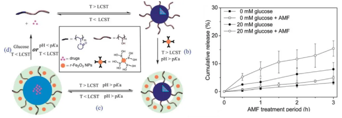
i) Polymers or copolymers displaying a lower critical solution temperature (LCST)
ii) Thermosensitive lipid bilayers or polymer matrix presenting either a melting (Tm) or a glass transition temperature (Tg)
iii) Thermolabile (non-covalent) and thermodegradable (covalent) bonds with the drug
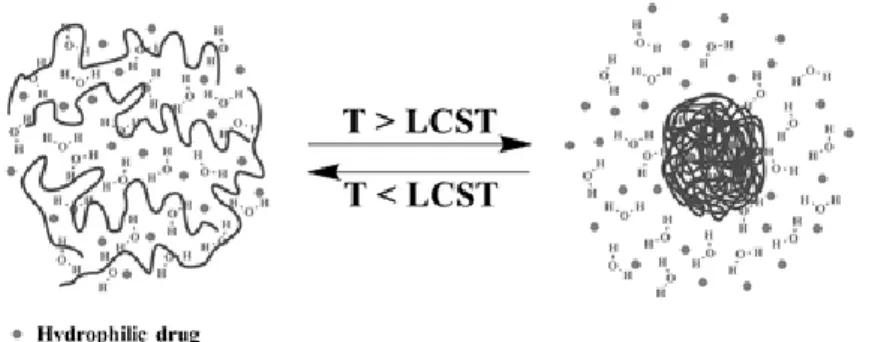
i) LCST-polymers and nanocarriers. Probably the most reported approach involved in a broad range of
thermo-responsive nanocarriers is to use the thermal response of polymers that display a dramatic volume change or coil-to-globule transition at a specific T called LCST. Below the LCST, the polymer chains are extended and fully hydrated in water through H-bonds, and when the medium T is increased above the LCST, interactions with solvent are disrupted to the benefit of inter- or intra-chain bonds, and the polymer phase segregates through H-bonds and hydrophobic interactions (Figure 2). There are various polymers or copolymers exhibiting a LCST in the physiological temperature range, which is an attractive feature for biomedical applications.[26,33,34] The main reported polymer is poly(N-isopropylacrylamide) (PNIPAM)[73] with a LCST of 32°C, but other LCST amphiphilic polymers are also used such as poly(vinlycaprolactam) (PVCL, LCST 25-35°C)[74], PEO-b-PPO-b-PEO i.e. triblock copolymers of ethylene oxide and propylene oxide (Pluronics™ F127, LCST of ca. 37°C) [75,76], hydroxypropyl cellulose (HPC, LCST 41°C) [77,78] or elastin-like polypeptides (ELPs) (LCST range of 10 to 70°C depending on the choice of guest residue X in the repetition sequence of the –VPGXG– pentapeptide)[79,80].
Figure 2. Illustration of the volume phase transition obtained with LCST polymers leading to a coil-to-globule
conformation change. Above LCST, the aqueous content (drug) is released through polymer chain collapse. [81] Reprinted with permission (Publisher name)
However to ensure a suitable drug release at a macroscopic temperature reachable by MH without damaging healthy surrounding tissues (typically 42°C), the thermo-responsive polymer should be in a hydrated state at physiological T (37°C) and its LCST should be above ca. 41-42°C but not higher than 43°C. Co-polymerization methods especially controlled radical polymerizations (CRP) including atom transfer radical polymerization (ATRP)[82,83], ring opening polymerization (ROP)[84,85], reversible addition-fragmentation chain transfer (RAFT)[86,87] are particularly well-suited to adjust finely the required LCST. Indeed, the LCST will depend on
9
the composition between the hydrophobic and the hydrophilic parts of the thermosensitive block copolymer.[13,28,33] Introducing a hydrophilic part allows shifting the LCST towards higher value while increasing the hydrophobic part favors lower LCST values. Furthermore, a narrow thermal transition requires copolymers with well-defined architecture rather than statistical copolymers that not only shift but also broaden the LCST range.
LCST polymers are key components in the design of various thermo-responsive nanocarriers and are used as polymer brushes, or coatings at the surface of inorganic NPs (preferentially as a dense brush rather than in dilute “mushroom” regime), or as building blocks of nanogels or micelles. Thus, LCST polymer shells can be anchored at the surface of IONPs, and be loaded with drugs via hydrophilic interactions and ensure the sneezing out of the drugs when T reaches the LCST, i.e. when the polymer becomes hydrophobic.[27,88] LCST polymers have also been grafted at the surface of mesoporous silica (MS), and play the role of thermo-responsive gatekeepers controlling the pore aperture of MS NPs with T and then the diffusion of the initially embedded drugs towards the outside medium of the silica NPs.[89]Thus, Liu, Yang et al. [90] have shown that Zn(II) phthalocyanine used as a PDT sensitizing drug could be efficiently loaded (drug loading content, DLC=18%,) in magnetic MS cores coated with a thermo-sensitive P(NIPAM-NHMA)polymer shell that was co-polymerized from the silica surface. The authors have shown that the LCST could be adjusted from 38 to 44°C with the composition of the copolymer, P(NIPAM-co-NHMA) acting as a gatekeeper. In a very recent work, Baeza, Vallet et al.,[91] grafted P(NIPAM-co-NHMA) copolymers at the surface of magnetic MS composite NPs loaded with fluorescein according to two different polymerization pathways: a cross-linked or a hairy P(NIPAM-co-NHMA) copolymer shell. The triggered release of fluorescein was studied at 37 and 50°C, and opposite controlled release behaviors were obtained according to the polymerization pathway. Indeed, the hairy polymer layer collapsed and blocked the drug diffusion upon T increase above LCST while the cross-linked polymer shell ensured an enhancement of the drug diffusion when it shrunk.
LCST polymers are also part of nanogels which are physically- or chemically-crosslinked systems in a hydrated or swollen state below the LCST that undergo an important volume phase transition (VPT) (i.e. gel shrinkage) above the LCST. Above the LCST, the important polymer shrinking effect ensures the diffusion of water out of the gel, together with its solubilized content. Hence in a work by Rubio-Retama et al.,[92] thermosensitive PNIPAM polymer was used to form monodisperse submicron microgels decorated with γ-Fe2O3 NPs through a surfactant-free radical polymerization. The authors have shown that a loading content of 18% IONPs allowed changing the LCST from 36 to 40°C, and to induce a submicron gel size shrinking from ca. 600 to 300 nm indicating the broad potential of magnetic nanogels for drug delivery.
In another study by Zaher et al[93],composite membranes made of blends of ethyl cellulose or cellulose acetate, PNIPAM microgels and IONPs exhibited enhanced permeability by application of an AMF (62 mT, 50 kAm-1, 450 kHz). With a thickness around 20 µm, such membrane was used as magnetically controllable valve to tune the effusion rate of a drug model (rhodamine B) between the donor and acceptor chambers of an osmotic pump.
ii) Nanocarriers with melting (Tm) or glass transition (Tg) temperatures. Aside from the LCST polymer-based
10
permeability or membrane fluidity, associated with a thermal transition that can be a Tm (1st order transition) or a Tg (2nd order or pseudo-transition). For instance, thermosensitive liposomes (TSLs) which were introduced almost forty years ago[94,95]usually respond upon T increase by a thermal destabilization of their hydrophobic bilayer membrane which becomes leaky /more permeable and allows the transfer of molecules from the inner aqueous core to the external solution. Various works and reviews reported that such effects were attributed to the main chain melting (Tm) temperature transition where the lipid bilayer turns from a gel phase (L) to a liquid crystalline state (L), the boundary line between the two phases coexisting at the transition Tm acting as defects enhancing solute permeation trough the membrane. This phenomenon allows a reversible permeation of the bilayer membrane ensuring a time-controlled release of the drugs. Dipalmitoyl-sn-glycerophosphocholine (DPPC, Tm of 41.5 °C), often combined with cholesterol was historically the first lipid to be used to form TSLs with a Tm close to the clinically relevant hyperthermia T.[28,96,97] Other lipids e.g. distearoyl-sn-glycero-phosphocholine (DSPC, Tm of 55°C) were used to improve the amount and rates of drugs released.[98] It is interesting to note that TSLs encapsulating doxorubicin in their lumen have been patented 20 years ago and commercialized under the brand name Thermodox™, leading to both pre-clinical [99] and clinical trials [100] on various cancers.
With much longer molecules than lipids like polymers, a soft and reversible temperature transition can arise either at a true Tm for semi-crystalline polymers, typically short and stereo-regular chains, or at the glass transition Tg also encountered for longer or more rigid chains. This is in particular the case with polymer vesicles named polymersomes [101,102], polymer emulsion nanocapsules [27,103] or poly(D,L-lactide-co-glycolide) (PLGA)-based NPs.[104,105] In these various works,a reversibly thermally controlled-permeability ensuring the drug release without structural alteration effects on the polymer matrix (3D) or membrane (2D) was achieved. Such a response is particularly suitable to ensure a pulsatile drug release. However, one main issue with such reversible Tm or Tg transitions which is also encountered with LCST-based nanosystems, is the non-negligible leakage of the drugs that often occurs in the absence of hyperthermia. Hence, tightest encapsulation/binding methods are thus required to ensure a “zero premature release”.
iii) Thermolabile (non-covalent) and thermodegradable (covalent) bonds. A very limited and even zero
premature release can be reached when drugs are bound via strong non-covalent or covalent thermosensitive bonds in nanocarriers. For instance, the H-bond pairing by DNA hybridization[106–109] affords a strong non-covalent pairing of complementary DNA strands in which the number of DNA base pairs determines the labile transition T in the range of 40-90°C depending on the AT/GC composition. Thermally responsive nanovalves made of pseudorotaxane complexes formed between cucurbituryl molecules complexed with grafted molecular thread at the surface of MS were also used as gatekeepers controlling the diffusion of embedded drugs.[110] The T of un-coordination was assumed to be around 52°C.
Regarding thermo-degradable covalent bonds strategies: diazo-linkers cleavage and retro Diels-Alder (DA) de-cyclization and intramolecular lactamisation are the two strategies that were developed these last years. Hence, thermo-labile diazo group linkers have been used in some recent works either to directly conjugate polymer-coated IONPs with anti-tumoral drugs[111] or as thermo-degradable moieties composing PEG polymers gatekeeping the diffusion of embedded drugs in magnetic MS.[112] The thermo-reversible DA
11
reaction was reported by N’Guyen et al.[113] in aqueous solution which involved maleimide-conjugated rhodamine, as a drug model, to furan group coupled to a phosphonated PEG grafted at IONPs surface. The local T ensuring the DA cycle opening being in the range of 90−110 °C, it was hypothesized that such high temperature above water boiling point can nevertheless be reached upon AMF application, at least in the close (nanoscale) vicinity of the NPs (ascribed to huge Laplace pressure needed to nucleate gas bubbles). [113,114] Very recently a novel thermally responsive nanoparticle system was proposed and patented, by using a tethering approach linking an alcohol- or amine-terminated drug to an ester or amide group and responding to an AMF-generated local heating by an intramolecular lactamization cyclization reaction ensuring the drug release. [115] Worthy to note that contrary to nanoplatforms displaying LCST, Tm or Tg responses which occurred typically around mild hyperthermia T (42°C), thermo-labile bonds including DNA base pairing, pseudorotaxane nanovalves and thermodegradable covalent bonds respond at a higher local T (T ≥ 80-90°C). This means that to actuate these systems, a very high local T should be reached to ensure the efficient thermal cleavage of the bonds and efficient release of the drugs. This underlines also the importance of the nanoheater design and understanding the effect of heat diffusion at the nanoscale. Furthermore, a potential drawback associated with the covalent drug binding is the decrease of the colloidal stability of the NPs in the blood stream, and also of the biological activity of the drug.
2.3 Formulation of drugs and magnetic NPs in thermally sensitive nanocarriers
The development of suitable processes to formulate drug-loaded magnetic nanoplatforms is of paramount importance. Such methods should ensure the efficient assembly of the nanoplatforms and the dual loading of the drug(s) and of IONPs without premature release in the absence of AMF while promoting the destabilization of the drug-nanotplatform bond or interaction under AMF application. This is actually still even more challenging when considering interactions with biological media in vivo.
There are various classes of antitumoral drugs that can be loaded in such platforms. The most encountered anti-tumoral agents are hydrophilic such as the anthracycline doxorubicine (DOX, which is water-soluble at mild acidic and neutral pH) or hydrophobic such as paclitaxel derivatives (e.g. Tamoxiphen™, Taxol ™), camptothecin (CPT), curcumin... The hydrophobic/hydrophilic balance of these drugs will dictate their interactions within the magnetic carriers: payloads, location in the structure, and mechanism of thermal release. Key parameters characterizing the drug loading in a nanomedicine carrier are the drug loading efficiency (DLE) and the drug loading capacity or loading content (DLC). They are defined according to the following expressions, respectively:
Evaluation of DLC and DLE is usually measured by HPLC, UV visible or fluorescence methods after establishing an etalon-curve and measuring the amount of drug remaining in the supernatant. For instance, the extinction coefficient of DOX at 485 nm and pH=7.4 is 6631 cm-1M-1 which corresponds to an absorbance value of only 0.0122 for a 1 µgmL-1 DOX solution (1 cm). [101]
12
i) Polymer grafting methods (grafting-to vs. grafting-from) at the surface of magnetic NPs ii) Self-assembly methods involving the design of magnetic liposomes, micelles, capsules
iii) Sol-gel methods ensuring the synthesis of magnetic silica nanocomposites where drug is embedded in the carrier either by in situ sequestration or by impregnation and use of polymer or wax gatekeeping.
i) Polymer grafting methods. Surface modification of magnetic NPs with a polymer coating ensuring drug
loading/release upon thermal response can be done using “grafting-to” and “grafting-from” methods. Grafting-to strategies involve the functionalization of IONPs by polymers either by non-covalent interactions[88,116] or by strong interactions (coordination/electro-covalent) using anchoring groups such as phosphonate[72,117,118], carboxylate[119] or alkoxysilane[120–123]. In these different cases of chemisorption, multivalency is an important way to increase the binding strength e.g. with bis- or tri-phosphonates[124,125] or with polyacids such as phosphonate end-terminated sidechains[126] or polyacrylates. The grafting-to method provides a great versatility of the surface modification as well with synthetic and natural polymers. One issue which is usually reported with the grafting-to method is the low polymer surface coverage density, not reaching the brush regime.[127–129] Alternatively, the grafting-from method is performed to create polymer brushes through a surface-initiated CRP polymerization such as ATRP, RAFT etc... In surface-initiated CRP[130], the initiator is usually grafted at the surface of the IONP and the monomers are brought in solution (together with an excess of “sacrificial initiator”) to ensure an effective controlled polymerization from the NP surface. Such CRPs are suitable methods to design block copolymers with controlled growth, tunable chain length, high grafting density and an adjustable LCST response.[13,127] Additionally, free radical polymerization was also reported to produce tight and thick polymer shells around IONPs.[131,132] For both grafting-to/-from schemes to get polymer shells and brushes, the thermo-sensitive drug binding may be ensured by electrostatic or hydrogen bonds, hydrophobic interaction or covalent coupling.
ii) Self-assembly methods. The design of drug-loaded magnetic liposomes, polymer capsules, polymer vesicles
(polymersomes) and micelles involve various processes of self-assembly. The drug is usually held in these nanostructures via hydrophilic or hydrophobic non-covalent bonds as described above. The non-covalent encapsulation of drugs in such magnetic nanoplatforms is advantageous as it neither changes the original drug bioactivity, nor requires any extra lab-work chemical modification, and also it simplifies the regulatory approval for clinical use compared to drug conjugates.
Magnetoliposomes. For the formation of magnetoliposomes (MLs), there are two main techniques described:
the reverse phase evaporation (RPE) and the thin film (TF) rehydration methods. In the RPE method, typically an aqueous solution of hydrophilic iron oxide NPs is added to the lipids dissolved in an organic solvent to form a reverse water in oil (w/o) micro-emulsion, and the MLs self-assemble upon solvent evaporation accelerated by sonication. The RPE is well suited to incorporate a high payload of IONPs in the aqueous core.[133–135] In the TF-rehydration process sometimes called “Bangham method”, the lipids are first dispersed in a volatile organic solvent and a thin multi-lamellar lipid film is formed onto the walls of the container upon solvent evaporation under reduced pressure: the TF is subsequently rehydrated by an aqueous solution. The lipid TF method allows both the incorporation of hydrophilic IONPs in the aqueous core[136,137] or hydrophobic NPs, yet limited in size to 5 nm, inside or on (i.e. decorating) the liposome bilayer[138–141] To encapsulate
13
hydrophilic drugs in the liposomes, the ammonium sulfate gradient method[142,143] is typically employed with a high efficacy as it was done for the formulation of Doxil™.[13] As found by Barenholz et al., DOX gets trapped inside the aqueous compartment because the molecule can cross membrane in its neutral (deprotonated) form while its sulfate salt inside the cavity precipitates (unlike its acidic DOX-HCl form that is water soluble), therefore avoiding the reverse crossing of the membrane.
Magnetic polymersomes. For the synthesis of magnetic polymersomes which can be seen as the equivalent of
liposomes for the self-assembly of amphiphilic polymers, nanoprecipitation is the main reported method ensuring the self-assembly of vesicles of sub-micron vesicles. Also called “solvent-displacement” method, nanoprecipitation consists in adding progressively an aqueous solution to an organic solution of the block copolymer or the reverse, water acting as a selective solvent (i.e. good solvent for one block, bad solvent for the other one). Vesicles being out-of-equilibrium structures, their structural properties are very process-dependent, and nanoprecipitation is a mean to orient towards preferred sizes and decrease size-dispersity as compared to direct dissolution (which is also impossible when the copolymers are glassy or semi-crystalline). The IONPs and the drugs depending on their hydrophobicity / hydrophilicity can be encapsulated within the polymersomes either in the polymer shell[101,102] or in the aqueous core.[144] The dual incorporation of the drugs and magnetic NPs can be done simultaneously during the self-assembly or the drugs can also be post-loaded by permeation of through the membrane via electrostatic attraction.[144]
Magnetic polymer capsules. Magnetic polymer capsules are usually formed according to “soft templating”
when using reverse micro-emulsion droplets as templates or “hard templating” when using rigid preformed colloids as templates. “Soft templating” allows forming polymer capsules typically by the double emulsion (DE) technique. In this technique, a first w/o reversed nanoemulsion stabilized by an amphiphilic polymer is added to a large volume of aqueous solution of a complementary polymer (PVA, proteins etc..) yielding to the formation of a w/o/w double emulsion. The polymer double emulsion capsules (DEC) are then formed after organic solvent evaporation. Synthetic polymers[103] or biomacromolecules[145] can be used to stabilize the capsule. Poly-addition reactions can also be applied at the emulsion interface[146] to create the DEC polymer shell. As for the liposomes and polymersomes, drugs and IONPs can be incorporated either in the core or within the shell during the self-assembly process depending on their hydrophobic / hydrophilic ratios. IONPs can be also formed in situ by co-precipitation in the aqueous core by initial loading of iron(II) and iron(III) ions followed by a basification step at pH=10.
Magnetic capsules made by the “hard templating” approach involve the use of either inorganic colloidal templates such as calcium carbonate[147,148]or polymer rigid organic colloidal templates such as melamine formaldehyde.[149,150] The polymer shell is usually assembled on the template by the layer-by-layer (LbL) method via electrostatic interactions typically by the sequential layering of oppositely charged polyelectrolytes. It is however worth to note that there have been recently a wide range of “one pot” chemical processes used to assemble polymer or protein capsules through hard templating.[151–157] The magnetic NPs are incorporated in the shell either by adsorption during the film build-up or by in situ coprecipitation of ferric and ferrous ions at the surface the template. The magnetic capsules are then formed after dissolution of the
14
templating core by the use of EDTA for carbonate calcium removal, or HCl for melamine formaldehyde removal.
Magnetic micelles. Regarding the preparation of magneto-micelles, it is usually performed by mixing together
the hydrophobic magnetic NPs (typically oleic acid-coated IO nanocrystals), hydrophobic drugs with the blocks amphiphilic copolymer. In the case of doxorubicin which is an amphipathic base (i.e. its protonated form is water soluble), its incorporation in the hydrophobic core of copolymer micelles can be achieved by the Kataoka method [158] consisting in fist deprotonating DOX overnight in a 1:2 mixture of triethylamine (TEA) and DMSO. The resulting drug loaded magnetic micelles are formed by the self-assembly of the amphiphilic species and the drug and IONPs are concomitantly held via hydrophobic interactions within the hydrophobic micelle core; There are different procedures allowing to prepare self-assembled magnetic micelles (i) emulsion−solvent evaporation, (ii) nanoprecipitation, (iii) dialysis, and (iv) thin film rehydration.[13] For LCST thermo-responsive polymer micelles, these are usually made by the self-assembly of di-block copolymers containing a LCST polymer moiety especially PNIPAM[159–162] In such thermo-responsive micelles, the hydrophobic part of the copolymer self-assembles into a reservoir core for the loading of hydrophobic drugs and the hydrophilic part of the copolymer forms an outer hydrophilic shell ensuring colloidal stability in physiological media. As such, the thermal stability of the micelle is mainly determined by the LCST response of the di-block copolymer, shifting from a hydrophilic to a hydrophobic state upon temperature change past the LCST. Magnetic polymer micelles were first introduced quite concomitantly by Ai et al.[163] for poly(-caprolactone)-b-poly(ethylene glycol) and by Lecommandoux et al. [164] for poly(butadiene)-b-poly(L-glutamic acid) (PB-b-PGA). Later on, magnetic polymer micelles with a thermosensitive shell were designed e.g. with the rod-coil copolymer poly( -benzyl-L-glutamate)-b-poly[2-(dimethylamino)ethyl methacrylate] (PBLG-b-PDMAEMA, LCST=39°C).[165]
iii) Sol-gel methods. Sol-gel methods are the most encountered processes allowing the formulation of
magnetic nanocomposites. There are mainly two ways to prepare drug loaded-magnetic nanocomposites. A first approach is the direct encapsulation of the drugs (and the magnetic NPs or their precursors) within the silica matrix network during the sol-gel process. Previously, Barbé et al.[166] shown for silica that Orange II, rhodamine dyes and DOX could be co-encapsulated during a sol-gel process. The sustained drug release over 20 days from the silica xerogel was explained by the combination of dye diffusion and silica dissolution processes in aqueous solutions. Later Hu, Liu et al.[167] reported the synthesis of ibuprofen-loaded silica magnetic composites NPs (size 50-60 nm) through a one step process by reacting the drugs, iron salts and silica precursors during the sol-gel process reaching a DLC=1% and a DLE=23%. In another report in the same group[168], the authors have tightly encapsulated a fluorescent dye in a PVP-coated silica core during a sol-gel process. They surrounded the dye-loaded silica core by a further layer of iron oxide on the surface as a way to provide magneto-thermal response upon AMF and a zero release in the absence of the magnetic field. In these examples, the tight magnetic silica matrix structure requires a very high local T to induced drug release by local de-structuration. The performances upon AMF for these two latter examples are described in Part 3.4.
The second approach is the formation of porous silica shells at the surface of single or multicore magnetic NPs followed by drug impregnation/adsorption within the porous matrixes. Such porous shells act as efficient reservoirs with high loading capacity. The drug release can be controlled by a further wax, polymer or
15
supramolecular capping ensuring drug gatekeeping as described in the previous paragraph in Part 2.2 Mesoporous silica (MS) shells are usually formed at the surface of hydrophilic or hydrophobic magnetic NPs by reacting a silica source precursor (TEOS) in the presence of a surfactant (e.g. CTAB) assembled in micelles.[169– 172] The surfactant acts as a pore structuring templating agent by the formation of micelles arranged in hexagonal phase and also ensures the water-phase transfer in the case of hydrophobic IONPs. The silica hence polymerizes in aqueous basic conditions around the magnetic core and the micelles, to form the magnetic ordered MS structures. After the sol-gel process, the surfactant is extracted from the pores by an acidic treatment or by the use of ammonium nitrate solutions. [173,174]The drug loading in MS porosity (whether magnetic or not) depends on several parameters: the nature of drugs (hydrophilic vs. hydrophobic), type of solvents (protic/polar/apolar) or the internal surface modification.[89] For instance, a high drug loading is typically achieved by electrostatic interactions between DOX, positively charged in water at pH 6 and the negatively charged bare MS.[175] DOX can also be adhered at the surface of bare MS in polar solvent such as ethanol by H-bonds.[176] Loading of hydrophobic drugs such as camptothecin (CPT) or paclitaxel were also achieved on bare MS in DMSO yet lower DLC were obtained compared with DOX.[177,178] Modifications of MS surface either with charged polymers [8,179] or aromatic hydrophobic molecules[180] have been also reported to influence the loading capacity by increasing respectively the electrostatic or hydrophobic interactions with the drugs.
2.4 Releasing drugs upon magnetic field: the experimental set-up
The typical setup for studying an in vitro drug release triggered by an applied AMF comprises the high power current generator feeding an application coil of sufficient diameter to hold the IONP-doped drug carrier sample (1-5 mL). The IO NPs /drug carrier sample is contained inside a dialysis bag itself immersed inside a much larger volume (at least 10 times) of buffer. The molecular weight cut-off (MWCO) of the dialysis membrane is chosen as large as possible (yet not letting the NPs go through) so that the drug release kinetics is limited by the permeability of the nanocarriers, not by the membrane porosity. Such set-up is sketched on Figure 3.
The follow-up and quantification of the drug release is performed by picking up at successive times aliquots of buffer in the reservoir, every time filling back the tank with the same volume of fresh buffer in order to maintain “sink boundary conditions”. The drug within each aliquot is titrated compared to a calibration curve using the most appropriate analysis method (by UV-visible absorbance, fluorescence or HPLC), and the result is converted into a “cumulative release amount”, the asymptotic value of 100% being obtained by disruption of the nanocarriers (e.g. with a surfactant or a solvent for liposomes). It is recommended also to record the temperature curve all along the process using a fiber optics T probe rather than a metallic thermocouple that would heat up by itself under application of the AMF. Most biological buffers having high salinity, the parasitic heating of water in the reservoir under the AC magnetic field by eddy current should also be recorded (without magnetic sample).
16
Figure 3. Setup for model drug release experiment through drug diffusion across semi-permeable membrane.
1: Solenoid and calculated magnetic field lines; 2: Dialysis bag (e.g. Spectra/Por Float-A-Lyzer™, 50000 gmol-1 MWCO, 10 mm diameter, 5 mL volume); 3: Cylinder (50 mL) filled with aqueous buffer. Usually the drug is titrated in the outer medium, in “sink” and thermostatic (37°C) conditions.
Most experiments published so far present cumulative drug release profiles measured manually. One drawback is the limited number of data points, with blanks in the kinetics ascribed to the overnight periods. The magnetic field must also be switched off when pipetting the aliquots (especially when using metallic needles!). In pharmaceutical sciences, the release curves can be much more precise by a continuous “on-line” automatic measurement during many hours. Most pharmaceutical companies use indeed the Sotax™ USP-4 flow-through system initially developed for tablet dissolution kinetics. This system was adapted to monitor drug release from nanocarriers by incorporation of a 2 mL volume dialysis bag in the loop and validation on liposomal formulation of dexamethasone.[181] Instead of taking aliquots at discrete times, it would be very benefic for data interpretation and modelling of AMF-induced drug release to monitor the drug release profile continuously, while keeping the intensity of the magnetic field ON (or applying pulsatile ON/OFF magnetic field profiles). However there is no commercial device yet coupled to MH, although several providers of MH generators are currently working on such a product. Another advantage of the Sotax™ USP-4 flow-through system is that the buffer is not static as sketched on Figure 3, but it circulates around the dialysis bag, thanks to peristaltic pump beating at 120 rpm, mimicking to some extents blood circulation. The optimal design of in vitro drug release experiments also requires that the nanocarriers are maintained at physiological temperature during all the process (whatever the increased local temperature, in close vicinity to the IONPs), e.g. with a cylindrical reservoir that is made of a double-wall jacket with water circulating through a controlled temperature bath at 37°C.
One has to remember indeed that the aim of the in vitro release curve measurements is to assess the potential of a nanoparticle-drug formulation to respond significantly to a stimulus, here the application of an AMF, whereas only preclinical in vivo assays can give truly relevant pharmacokinetic data (like halftime in blood circulation). Nevertheless, the experimental release profiles (measured manually or continuously) can be fitted to semi-empirical laws like a power law (Korsmayer-Peppas), Higuchi model for highly water insoluble drugs, or Weibull law (sigmoid profile),[182] and the resulting kinetic parameters can be compared with or without
17
applied AMF, in order to evidence “proof-of-concept” results. Ideally, these in vitro release data need to be completed by cellular experiments (internalization in relevant biological cells and cytotoxicity assays at drug concentration below the IC50) and/or in vivo data (bio-distribution, pharmacokinetics, tumor regression…). In that case the in vitro drug release profiles with model diffusion experiments under AMF can help choosing the appropriate parameters (magnetic field strength, frequency, and application time), although other physical parameters like the IONP concentration and the spatial distribution of the sample need also to be chosen as close as possible to the case of a real tumor in order to be truly biologically relevant.
3. Efficacy of magnetic nanoplatforms releasing drugs upon AMF and in vitro biological
results
This part 3 is an overview of the current magnetic nanoplatforms that were used to trigger drug release upon AMF. For sake of clarity, we have divided the nanosystems into four types:
i) Magnetic core-polymer shells
ii) Polymer or lipid vesicles having hollow aqueous compartments
iii) Polymer nanospheres (NS) including hydrophilic nanogels, hydrophobic NSs and nanomicelles iv) Inorganic nanocomposites mainly with silica matrix
For each example of nanosystem described, we provide briefly the structural properties and focus on the salient features and efficacy of the AMF-induced drug release along with the in vitro data gathered on biological cells. A broad updated overview of in vivo results will be presented in Part 4.
3.1 Activable magnetic core-polymer shells
Such nanoplatforms consist in a magnetic core surrounded by a polymer shell. As seen in Part 2, there are different ways to trigger the drug release from the polymer matrix by an applied AMF: via LCST response, non-covalent thermolabile bonds or thermodegradable non-covalent bonds.
3.1.1. Magnetothermal release by the LCST response of a polymer coating
The drugs are released through the thermoresponsive LCST behavior of grafted polymer on IONP surface via a mechanism which is detailed in Part 2. For instance, Yao et al. [131] used an emulsion free polymerization to form a dense and thick P(NIPAAM-co-Am) shell (85% wt) at the surface of IO cores which was thus loaded with the hydrophilic vitamin B12 (DLE= 65%) in the copolymer shell. The authors applied an AMF (H=6.5 kAm-1, f=60 kHz) either continuously (6h) or intermittently (61h every 3h on 24h). They showed a better cumulative AMF-induced drug release in the intermittent application (almost 100%) vs. the continuous regime (69 %). For both AMF regimes, they measured a T rise of the solution up to 46°C after 1h AMF application and the LCST of P(NIPAAM-co-Am) shell was measured at 42°C from the UV-vis transmittance, explaining such magnetically induced release behaviors.
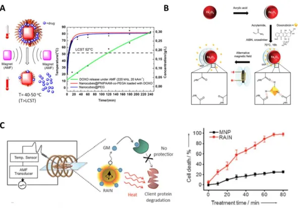
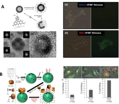
The ’grafting-from’ and ’grafting-to’ methods detailed in Part 2 are most importantly developed to prepare magnetic core-polymer shell nanostructures exhibiting AMF-induced drug release. Very recently, Kakwere, Pelegrino et al.[183]formed at the surface of IO nanocubes (19-22 nm) PNIPAAM-co-PEGA brushes by RAFT living radical polymerization with a LCST of 52°C (Figure 4.A). High DOX loading content (23%) was reached by simple impregnation 24h at room T. Application of an AMF (H=20 kAm-1, f=220 kHz, 240 min) ensured the
18
efficient DOX in vitro release (ca. 25%) whereas a negligible release was found at 37°C without applying AMF. The authors have shown that during AMF application, T of the solution rose up to 80°C in 15 min. Cell viability studies (without AMF) performed on KB cells showed cell uptake and no toxicity up to 1 gL-1 iron of the incubated core-shell NPs. In a recent study, Hervault, Thanh et al.[122] used a ‘grafting to’ approach where a concomitant pH and thermoresponsive copolymer was priory synthesized and then attached by trimethoxysilane anchoring endgroup onto the surface of the IO cores formed by microwave-assisted co-precipitation. Polymer grafting was estimated up to 8.1 % weight relatively to IO by TGA. The DOX loading (DLC = 7.6 %, DLE=82.3%) in the polymer shell was done by the formation of the pH-responsive covalent imine bond between the amine group of DOX and the aldehyde group of the copolymer. Concomitant effect of pH in media mimicking acidic tumor pH (5.7) and MH (T 50°C) allowed 85.2 % of DOX release over 48h while a lower DOX amount was released (39.4%) at 37°C within the same period.
Figure 4. A. PNIPAAM-co-PEGA brushes grafted on IO nanocubes (19-22 nm) by RAFT living radical
polymerization. Reprinted with permission from ref. [183]. Copyright 2015 American Chemical Society B. Preparation and DOX loading in MIP γ-Fe2O3 NPs. Reproduced from ref. [184] with permission of The Royal Society of Chemistry. C. In vitro release of geldanamycine with cells upon AMF application. Reproduced from ref. [185] with permission of Wiley.
3.1.2 Magnetothermal release by H bonds and π -stacking disruption
The magnetothermal release of drugs bound by weak interactions (H-bonds, electrostatic, π-π stacking) is also reported. As underlined in Part 2, an advantage is that the required thermal energy is often lower compared with the one needed for the cleavage of thermo-sensitive covalent bonds. However the molecules are more loosely attached and hence they may experience more premature release in the absence of AMF. H-bonds are mostly exploited to bind drugs to a polymer shell matrix. Hence, Fluorouracyl (5-FU) drugs could be efficiently
19
loaded in. PolyA15 polynucleotide grafted to poly(styrene-alt-maleic acid) coated Fe3O4 core (22 nm) through 5-FU/adenine H-bond interactions (5425 FU molecules per NP).[186]A cell targeting function was added to the thermally responsive core-shell NPs by grafting anti-HER2 monoclonal antibody to target overexpressed human epidermal growth factor receptor (HER) receptors in mouse bladder tumor (MBT2) cells. The authors have shown that the combination of cell targeting and application of AMF (H=33 kAm-1, f=1.3 MHz, 15 min) on the MBT-2 cells allowed to reduce efficiently the cell viability at 50%, whereas the same treatment but with a non-specific targeting ligand (IgG) or a free 5FU treatment did not reduce as much the cell viability.
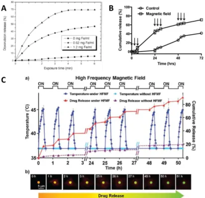
The magnetothermal H-bond release of DOX was also performed with molecularly imprinted poly(acrylamide) shell around γ-Fe2O3 core further cross-linked with ethyleneglycoldimethacrylate (EGDMA) [184]. The DOX was loaded in situ during the polymerization process and was held via H-bonding by the poly(acrylamide) mesh. (Figure 4.B) The authors investigated the “athermal” AMF induced drug released by performing AMF pulsatile sequences (B=9 mT, f=335 kHz, 52 min separated by 30 s intervals) on regular points over 8 hours and compared it with the drug release at a macroscopic T maintained at 37°C. They have shown that 4 times higher drug quantity was released with applied AMF compared to the externally applied T at 37°C (60% vs. 15% cumulative release respectively). PC 3 cancer cells internalized these core-shell NPs and were treated at higher field parameters (700 kHz, B=25 mT) in isothermal conditions for t= 30 min, 1 h 30 min, and 2 h 30 min AMF treatment. The results indicated a 40% cell viability reduction after 1h30 of AMF treatment compared with a negligible toxicity without AMF.
DNA hybridization is also a relevant way to tune the thermal response as the base pair (bp) length and thus the force of the H-bonds between the complementary DNA strands can be tuned. Thus Derfus, Bhatia et al.[106] hybridized simultaneously fluorescently labelled 12 and 24 bp single strands DNA on a 30 bp single strand DNA conjugated to a dextran shell coated at the surface of IONPs. They investigated the respective thermal response according to the power of the AMF applied (400 kHz, P=0.55-3 kW, 5 min pulses every 40 min). They showed that low power at 0.55 kW allowed the selective release of only the 12 bp DNA whereas a higher power at 3 kW allowed the simultaneous release of both 12 and 24 bp DNA demonstrating the possibility to modulate the thermal release response through DNA hybridization approaches.
Recently, an original approach was developed by Xu, Biris et al.[187] by binding aromatic drugs to iron NPs coated with a graphitic surface. DOX anthracycline and Erlotinib inhibitor drugs were bound (respectively at 0.07 and 0.11 mg per mg of Fe) by π-stacking to the carbon shell of NPs. These drugs could be efficiently released by rupturing the non-covalent bond with the carbon-coated IONPs. The AMF application (350 kHz, P=1.2 kW, 10 min) treatment in PBS at pH 7.4 was shown to enhance the DOX and Erlotinib release rates in the first hour. The release was at about 4.8 and 1.8 fold higher respectively compared to the release without AMF treatment. Trypan blue viability assays on Panc1 cells was also studied. Overall, application of the AMF was shown to decrease the cell viability of Panc-1 cells incubated with both DOX- and Erlotinib-bound C/Fe NPs. For instance, Erlotinib-bound C/Fe NPs reduced the cell viability from 50.4% to 25.4 % upon AMF application.
3.1.3. Magnetothermal release of drugs by covalent bond cleavage.
One of the most suitable ways of releasing drugs ’on demand’ from a magnetic core and with zero premature release in the absence of AMF consists in grafting the drug to the NP with a covalent thermo-cleavable bond. In