THE JOURNAL OF INFECTIOUS DISEASES • VOL. 145, NO.5. MAY 1982 © 1982 by the University of Chicago. All rights reserved. 0022-1899/82/4505-0005$01.09
Complement-Mediated Opsonic Activity in Normal and Infected Human
Cerebrospinal Fluid: Early Response During Bacterial Meningitis
Andre Zwahlen, Urs E. Nydegger, Pierre Vaudaux, Paul-Henri Lambert, and Francis A. Waldvogel
From the Infectious Disease Division and the World Health Organization Immunology Research and Training Center, University Hospital, Geneva, Switzerland A local defense mechanism in bacterial meningitis was evaluated in humans by
measur-ing complement-mediated opsonic activity (CMOA) in normal and infected cere-brospinal fluid (CSF) with a complement-dependent phagocytic bactericidal assay. CMOA was absent in normal untreated CSF and remained undetectable in 20 samples of CSF from patients with viral meningitis and five samples from patients with acute meningococcemia. In contrast, 15 of 27 samples of CSF from patients with acute bacterial meningitis had a measurable CMOA, which was correlated with protein con-centrations (P< 0.01) and C4 hemolytic activity (P< 0.001) in the CSF. A favorable out-come of bacterial meningitis was associated with the presence of CMOA in CSF (P
<
0.005). Recovery was also correlated with higher levels of C4 (P<
0.01) and C3 (P<
0.05) in CSF and with lower concentrations of microorganisms in the sample of CSF collected at the time of admission (P< 0.01). Thus, CMOA, although absent in normal CSF, can appear in CSF during acute bacterial meningitis, particularly in patients who recover completely.Mortality and morbidity from acute bacterial meningitis are still high despite substantial prog-ress in diagnosis and treatment [1-3]. The devel-opment and course of meningeal infection are governed by the complex interaction of many
vari-ables [4], including (1) microbial virulence factors
such as encapsulation [5, 6], (2) clearance of
bac-teria from the blood [7-9], (3) antibacterial
mech-anisms in the subarachnoid space [10], (4)
anatomic and functional changes resulting from
inflammation [11, 12], and (5) antibiotic therapy.
Because bacterial meningitis is in essence a
closed-Received for publication June 16, 1981, and in revised form September 15, 1981.
Portions of this work appeared as an abstract (no. 141) in the
Proceedings of the 20th Interscience Conference on Antimicro-bial Agents and Chemotherapy and in Clinical Research
27:361A, 1979.
Dr. Zwahlen is the recipient of fellowship no. 83.562.077 from the Swiss National Research Foundation.
We thank Anneliese Kahr, Sonia Grosjean, and Rachel Ballard for technical assistance, Dr. Luc H. Perrin for advice in the complement study, Drs. Gerard Gauthier, Marcelle Megret, Kaplan Riffat, Gregory Szappanyos, and Annie Bellinazzo and Else Budry for help in the collection of cere-brospinal fluid samples, Dr. Andre Assimacopoulos for tech-nical evaluation of the data, and Francoise Michaud for manuscript preparation.
Please address requests for reprints to Dr. Francis A. Wald-vogel, Infectious Disease Division, University Hospital, 1211 Geneva 4, Switzerland.
635
space infection, local defense mechanisms may be important factors that limit bacterial growth. Nor-mal cerebrospinal fluid (CSF) can be considered as a site of a "local defect in host defense," because it contains no phagocytic cells, has a low protein concentration with a predominance of albumin [13], contains no IgM, and has low levels of C3 and C4 [14-16]. Furthermore, recent studies have
suggested that neither CH50 (the amount of
com-plement required to lyse 50070 of sensitized ery-throcytes) nor overall bactericidal and opsonic activities are detectable in normal CSF [17, 18]. As
a result, seeding of the CSF with> 103 organisms
overcomes the clearance mechanisms of the CSF and leads to a rapid multiplication of the micro-organisms [19]. Thus, CSF from patients with meningitis often yields initial bacterial counts of
1 OL 1 08 bacterial ml; the higher values are
asso-ciated with clinical complications [20]. Once established, bacterial meningitis initiates inflam-matory reactions in the CSF that result in the gen-eration of chemotactic factors [21, 22] and in the
appearance of CH50 activity [18], various classes of
immunoglobulins [23, 24], and polymorphonu-clear leukocytes (PMNLs), all of which would be expected to improve the local host defense mechanisms.
Effective opsonization of bacteria by comple-ment and immunoglobulin is an essential step for phagocytosis by PMNLs [25]. Thus, in susceptible
Table 1. Clinical data of 27 patients with bacterial meningitis and five patients with acute meningococcemia and positive cultures of cerebrospinal fluid (CSF). CSF findings No. of cells/mml Glucose Patient no. Blood Protein ratio (CSF: (sex/age [years]) Outcome Bacteria cultured (cfu/ml) culture Leukocytes PMNLs Erythrocytes (mg/l00 ml) plasma) 1 (F/7) Complete recovery Neisseria meningitidis group B 10,000 8,000 0 363 0.09 (3 x 10 3) 2 (F/18) Cauda equina syndrome N. meningitidis group B + 800 800 0 270 0.005 (1.5 x 10 7) 3 (MI27) Complete recovery N. meningitidis group B 800 700 0 775 0 (2.7 x 10 6) 4 (M/45) Complete recovery N. meningitidis (NO) NO 26,900 26,000 0 852 0.17 5 (M/50) Complete recovery N. meningitidis group C + 9,800 9,800 170 592 0.26 (8 x lOl) 6 (F/18) Complete recovery N. meningitidis (7 X 10 4) 12,500 12,500 0 776 0.03 7 (F/14) Complete recovery* N. meningitidis group B 3,530 3,415 0 42 0.44 (3 x 10 4) 8 (M/18) Complete recovery Gram-negative diplococci 1,870 1,755 53 62 0.47 (NO) 9 (M/54) Complete recovery Gram-negative diplococci NO 4,625 4,525 0 336 0.43 (ND) 10 (M/52) Complete recovery Streptococcus pneumoniae + 7,130 6,630 170 700 ND (3.7 x 10 6) 11 (M/53) Complete recovery S. pneumoniae (ND) 1,000 900 0 240 0.17 12 (MI26) Complete recovery S. pneumoniae (NO) + 17,500 17,150 0 650 NO 13 (MI2) Hydrocephalus S. pneumoniae (10 7) + 517 437 0 236 0.003 14 (M/19) Complete recovery S. pneumoniae (1.3 x 10 6) + 570 570 0 64 0.37 15 (MI2) Hydrocephalus, ataxia Haemophilus injluenzae + 789 731 200 133 0.13 type b (1.4 x 10 6) 16 (F/3) Complete recovery H. injluenzae type b (NO) 7,575 6,950 0 128 0.53 17 (M/3) Hemiparesis, ataxia H. inJluenzae type b ND 2,275 2,150 25 133 0.34 (3.7 x 10 7) 18 (M/67) Complete recovery Listeria monocytogenes NO 195 104 14 238 0.37 (ND) 19 (M171) Complete recovery L. monocytogenes 4,300 4,128 0 301 0.11 (4.1 x IOl) 20 (F/61) Complete recovery L. monocytogenes 400 380 105 345 0.12 (1,3 x 10 4) 21 (FI25) Complete recovery* Gram-positive rods (ND) 8,000 7,360 0 112 0 22 (F/81) Complete recovery Escherichia coli (4 x IOl) + 1,680 1,616 70 481 0.09 continued
Opsonic Activity in CSF 637
subjects, complement-mediated opsonization may playa crucial role during the early phase of men-ingitis when specific antimicrobial antibodies are lacking in the serum and CSF.
In the present study we evaluate qualitative and quantitative changes of complement-mediated op-sonic activity (CMOA) in normal and infected CSF and try to establish the relation of CMOA to the course and outcome of bacterial meningitis. CMOA and complement levels were measured in the CSF and plasma from uninfected control pa-tients and from papa-tients with viral meningitis. MaterialsandMethods
Patients. Patients who had a diagnostic spinal tap at the University Hospital, Geneva, were pro-spectively included in the study. Conditions for
ac-ceptance were(1)an unequivocal diagnosis based on
clinical findings and a CSF sample; (2) availability of a CSF sample from a spinal tap immediately after admission; (3) absence of contamination of the CSF sample by blood, as demonstrated by an erythrocyte count of <200 cells/mm ' and/or a negative hemoglobin test (Labstix'"; Diethelm Co., Zurich) that detected an admixture of blood in
CSF at a dilution of 1:25,000; (4) delay of <6 hr
between lumbar puncture and freezing of the CSF
(median, 1.5hr; range, 1-6hr); and (5) absence of
degenerative or demyelinating disease of the cen-tral nervous system and absence of spinal sub-arachnoid block. The patients were divided into four groups.
Group 1 consisted of patients with normal CSF. Samples of CSF were taken from 10 patients who had an acute, self-limited, febrile illness and from
15afebrile patients with suspected disk herniation
who underwent diagnostic gas myelography and had normal neuroradiologic findings. Each sam-ple of CSF in this group had a normal protein con-centration and a normal cell count. A CSF pool of
15ml was obtained from a third cohort of 24
nor-mal patients who had spinal anesthesia for
surgery.
Group 2 consisted of 20 patients who had viral meningitis; these patients all had a febrile, self-limited disease with signs of meningeal infection that subsided without antibiotic treatment. The patients ranged in age from three to 88 years (me-dian, 29 years). The delay between the onset of symptoms and the diagnostic lumbar puncture
>.1 ...
.... t1S 41) ....5
U ~U 41) t1S .... .0 '0 CIS • ..c:: .~ r- .-:= N OIl I C.:; '5
g e
",'0 ~ 41) C ~ 41) CIS • .... 41) ~.
tIS ~ 1l.,Q ~] f-o •08:
Z *c
41) ;> o Ue
~ + + + i .~ U g U o gf 'S 41) E '0 t1S ..c:: N· M I 00 N V t:: o '0o
c: II 0 0°
10 Z ~\OIO - M N V"loO -r:t-:z 0 0 O - N M M M -~~~ I"- ... N '",:,'~6~ ~ ~ 41) 41) o...c::o.e
~e
0 4 1 ) 0uou
v; 41) >. g .:.: ;j1"--1"-1-:
t1S 41) U ;j c: o ..c::0-o
E >. o ~ N ...-
....
8-1\ j Z ~ c, + + \0 ~ M 00 00 M OM zt-: o .;g >. ~ t ~ ;> t1S 0 ~ U '" 41) "ti) """ 41) 41) ~ '0 .9-0.e e
41) 0xu
0\00 N M§O
--~~-"
~6 000\ N N ++~ I~ 0 0 0 0 0V"l .... _ OgONII)zoZd;;;
11)\011)00_ M O O l - O \ <"!M-~~S<88 ~~\O\OOO ~ r-:..:N'I"
-- "i=!6
666
c ·9 z z z Z Z-t::l.. ---Oe
cece
ceceu
~bQ.. 0..0.. 0..Cl.0.. _ 'i=! '" c.i
5 5
5 5 5
0 .. 0 ' 0 0 : : : 1 - · · . . . . _ E.~Oi:lOi:lO OIli:lOi:lO ~ 41) 0 Z .~"~ .~.~"~ x _ ~.2: g - ~ ~ ~ ~ ~ ~ ~ 0 ~ ~ ~ E'~'~2;'~'~'~g
- Z ~ ~ ~ .::! r.::: r.::: _ r.::: r.::: r.::: '" ~~~ c ' ,.~.~ .~.~.~ ~ :.:::: :.::::.~ I ti ~ ~ r.::: x ~ ~ ~5
8 8
~§
~] E ~ ~ E E E .~ ~~l<o~ ~~ ~~~ ~ ';j '60 t:: 'S8
c
*cE
41) 41) CIS ;> ;> 0.. 0 0 4 1 ) g g;>.
.... .... ~ ~ ~ ~ ~t\1 ..c:: o....c:: 0.. ... ~e
~ E t:: 41) 0 41) 0 ~ououu
§
0 0 0 00 l/")I"-N\O MMV"l1l) ror) r-..~... -...I"
--
-0\ - - N ~-"O\lI')"-"" ,00~,M "':' ... "':" 6~~6~ ~~~~~ 41) E ~;jo
-E
<, .2 ~ '0 41)E
:; U t1S 'C ~ U t1Sce
-
'".... CIS .~ 0 ... t:: 41) ~ OIl t:: t1S 4 1 ) , ".= ~ t1S 41) Cl.~ '0 41) o • o 3 ii3""§ -t::E
'B 8 0-. ,
~ i:lO5
'" 41) >.U o • ..c:: >..
~ '"s
~ o .:.: ;j 41) .J~I
'"
, ...J ==~ Z~ ' - ~ o ci Z ~-41) en t1S ~ U E U - " , ;j 0 t1Sa ''::;
0. ~ ~ en U '" OIl t:::e
t:: <+:::"""
~ :E tIS po..~
:::l ·5... s::: c ~ranged from 18 to 240 hr (mean, 96 hr). Viral cul-tures and serologic studies were not routinely per-formed, but bacterial cultures of CSF and blood were consistently negative. CSF in this group was characterized by a median leukocyte count of 120 cells/mm3 with a predominance of lymphocytes, a normal glucose level, and a moderate increase in protein concentration (mean, 45 mg/loo ml; range, 20-76 mg/l00 ml).
Group 3 consisted of five patients (nos. 28-32) who had fulminant meningococcemia - that is, sep-tic shock, petechiae, disseminated intravascular
coagulation, and presence of Neisseria
meningi-tidis both in blood and CSF (table 1). One patient
died and one was discharged with permanent neurologic sequelae - that is, ataxia and left-sided hemiparesis. The patients' ages ranged from seven to 64 years. Despite CSF cultures that were
posi-tive for N. meningitidis, these patients had
mini-mal inflammation of CSF, as evidenced by a low leukocyte count (mean, 38 cells/mm3), normal protein concentration, and normal glucose level.
Group 4 consisted of 27 patients (nos. 1-27) who had bacterial meningitis (table 1). Their ages ranged from two to 81 years (median, 25 years). The delay between the onset of symptoms and hospital admission ranged from 12 to 120 hr (mean, 39 hr). Samples of CSF included in the study were always obtained during the first lum-bar puncture and were stored within a median time of 90 min. Pertinent clinical and bacterio-logic data and CSF findings are listed in table 1. Of this group, 20 patients recovered completely, five survived with neurologic sequelae (cauda equinea syndrome, hydrocephalus, ataxia, and palsy due to cranial nerve damage), and two died of extensive brain damage.
CSF and blood. To minimize in vitro
comple-ment activation, all CSF samples were collected and mixed in tubes that contained EDT A (final concentration, 5 mM) and were centrifuged at
1,500 g for 15 min; the supernatants were stored
immediately at -70 C. To obtain serum, blood was allowed to clot at room temperature (1"\.120 C) for 60 min, and serum was separated by
centrifu-gation at 1,500 g for 15 min and stored at - 70 C.
To obtain plasma, blood was collected in tubes that contained EDT A (final concentration, 20 mM) and centrifuged; the supernatant was imme-diately stored at -70 C. Protein concentrations were measured in all of the samples of CSF and sera by the Bio-Rad protein assay (Bio-Rad
Labo-ratories, Munich). A pool of normal human serum that was obtained from 20 donors was used as a standard. All samples were tested in duplicate. Samples of blood and CSF were cultured, and the microorganisms were identified by standard bac-teriologic techniques [26]. Quantitative cultures were obtained from the CSF of 15 patients with bacterial meningitis and one patient with menin-gococcemia according to the method described by Feldman [27].
Measurements of CMOA in CSF and serum. Preparation of CSF and serum samples. Before
being used in the phagocytic assay, each sample of CSF or serum was diluted and reconcentrated three times in an ultrafiltration device with an ex-clusion limit of 10,000 daltons (model no. PTGC 001-K5; Millipore Corp., Kloten, Switzerland). This procedure, carried out at 4 C, was used to eliminate EDT A and to concentrate the immuno-globulins and complement components in Dulbec-co's phosphate-buffered saline (pH 7.4; Grand Island Biological Co., Basel, Switzerland).
CMOA was not eliminated by this ultrafiltra-tion procedure as demonstrated by the following experiment: six samples of 1 % normal human serum were submitted either to three consecutive dilution-concentration cycles or to no manipula-tion; we observed a slight and reproducible loss of
CMOA of 44% ± 120/0 (mean ± sD)-that is, less
than one dilution - when CMOA was evaluated in a phagocytic assay. Similarly, for one patient (no. 22) with bacterial meningitis, a sample of CSF that was obtained before the onset of antibiotic therapy was studied for CMOA before and after the dilution-concentration procedure; the samples
showed 50% killing (K50) opsonic titers of 19.8
and 11. 9 units/ml, respectively - that is, a loss of 40% in opsonic activity after manipulation. Fur-thermore, no increase of C3d was detected in ul-trafiltered plasma. There was no residual anti-biotic activity in ultrafiltered samples of serum or CSF as tested by agar diffusion [26] against
Staphylococcus aureus variant strain Wood 46. Preparation of PMNLs. Leukocytes from
healthy volunteers were separated from citrated blood by dextran sedimentation. The leukocytes were washed twice and suspended in
phosphate-buffered saline at a concentration of 1.25 x 10⁷
PMNLs/ml.
Phagocytic bactericidal assay. The CMOA of
the samples was evaluated by a modified phagocy-tic bactericidal assay [28] using the Wood 46 strain
Opsonic Activity in CSF
of S. aureus. As outlined by Lew et al. [29], strain
Wood 46 depends entirely on complement for its opsonization, and no significant phagocytosis or killing occurs in serum that has been inactivated by heating to 56 C for 30 min. Ingestion and killing of the test strain, however, are optimal (>90070 of the inoculum) in the presence of either normal, agammaglobulinemic, C3-deficient, or
Mg2EGTA-treated 10070 serum. For each 0.5-ml
assay, an aliquot of 0.05 ml of bacteria (3 x 10⁶
bacteria/ml) was added to a mixture of 0.4 ml of
purified leukocytes (5 x 10⁶ PMNLs/ml) and 0.05
ml of serially diluted CSF or serum. The cell suspensions were incubated at 37 C in a shaking water bath, and duplicate samples of the test sus-pension were drawn at time 0 and after incubation for 30 min. The samples were diluted, and the PMNLs were osmotically lysed with distilled sterile water and plated on Mueller-Hinton agar; the number of residual bacterial colonies was counted after incubation for 24 hr. Each of the
samples was tested with two controls: (1)
heat-inactivated serum or CSF with PMNLs and (2) serum or CSF without PMNLs.
Definition of the K50 opsonic titers. The Kso opsonic titer (expressed in units/ml) was defined as the inverse of the dilution of serum or CSF that
promoted 50070 killing of S. aureus strain Wood 46.
Measurements of complement factors in CSF and plasma. CHso was measured in a continuous flow system [30]. C4 hemolytic activity was also tested in a continuous flow system using the plasma of C4-deficient guinea pigs as a source of the other complement components in excess amount [31]. The activity of plasma factor B was
assessed by lysis of 51 Cr-Iabeled rabbit
erythro-cytes in the presence of serum devoid of functional factor B activity [32] and was expressed in units/ml. C3 and C3d levels were measured by im-munodiffusion [33, 34], using monospecific anti-sera (Behringwerke, Marburg-Lahn, Federal Re-public of Germany). The levels of the various complement components were measured in dupli-cate and were expressed as percentages of the levels obtained with a pool of reference standard plasma.
Samples of normal or infected CSF, which were stored in EDT A, were assessed for C4 hemolytic activity as well as for levels of C3 and C3d. The techniques that were used to test plasma were used for CSF measurements, but the sensitivity of the assays was increased by using lower dilutions of
639
the samples. The levels of the CSF complement factors were expressed as percentages of those of the pool of standard plasma.
Statistical evaluation. Statistical tests were chosen according to Meddis [35]. Differences be-tween groups were assessed by Student's unpaired
t-test. Linear correlation coefficients were calculated by a standard program. Mean dose re-sponse curves (figure 1) were calculated using the equation described by Wagner [36].
Results
CMOA in normal CSF and sera. Normal CSF
had to be concentrated in order to exhibit CMOA,
100 50 -;. OJ C
....
~ 0 C1 12,5 25 50 100 200 400 Qj U C1 m 100 12,5 25 50 100 200 400 Protein concentration (mg 1100ml)Figure 1. Heat-labile opsonic activity in (0) human
cerebrospinal fluid (CSF) and (.) serum. The results show the effects of protein concentration in the opsonic source (untreated, concentrated, or diluted CSF and
di-luted sera) on the percentage of killing of
Staphylococ-cus aureus variant strain Wood 46. Mean dose-response curves were calculated for ( - - - ) CSF and (-) serum:
(top) 15 samples of CSF and paired sera from subjects
without meningitis; (bottom) 15 samples of CSF and
paired sera from patients who had acute bacterial men-ingitis and measurable opsonic activity.
because neither diluted nor undiluted samples of
CSF were able to opsonize S. aureus strain Wood
46 (table 2). Addition of purified IgO (3 mg/ml) or 100/0 heat-inactivated serum to untreated CSF did not promote opsonization of the test bacteria. In contrast, a 10-fold concentration of 15 samples of normal CSF obtained during gas myelography
(when tested at a final mean [± SD] protein
con-centration of 354 ± 135 mg/100 ml) allowed for
detection of a heat-labile opsonic activity compa-rable to that of 100/0 normal serum. Further evi-dence for the complement-mediated nature of this opsonic activity was obtained by testing the sam-ple of CSF that was taken at the time of admission from an agammaglobulinemic patient who had acute meningococcal meningitis (patient no. 5). When tested at a protein concentration of 296 mg/ 100 ml, this sample of CSF promoted 650/0 bacterial killing.
The relationship between CMOA, expressed as the percentage of bacteria that were killed, and protein concentration in CSF was further studied in the group of 15 samples of normal concentrated CSF referred to above. A sigmoidal relationship was observed when the 16-fold concentrated
sam-Table 2. Opsonization of Staphylococcus aureus
variant strain Wood 46 by human cerebrospinal fluid (CSF) and serum in a phagocytic bactericidal assay.
Source of opsonic activity, treatment CSF Normal, 10% dilution Normal, 500/0 dilution Normal, untreated
Normal, 1O-fold concentration Normal, 1O-fold concentration,
heatedt Agammaglobulinemic, 50% Percentage of bacterial No. of killing * experiments o o o 66-97 0-2 10 25 15 15 3 dilutiont 65 Serum Normal, 10% dilution Normal, 10% dilution, heatedt Agammaglobulinemic, 10% 90-99 0-5 dilution 72 10 10
* Percentage of initial inoculum killed after 30 min of in-cubation. Data are ranges when more than one experiment were performed.
t Heated at 56 C for 30 min.
t This sample of CSF was obtained from a patient with acute meningitis and was tested at a protein concentration similar to that of normal CSF at a 1O-fold dilution.
~
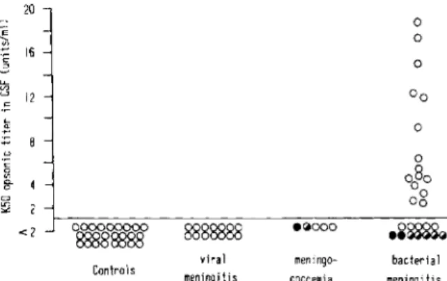
<:: ::> )j <:: .... '" 20 16 12 :;::; 8 o 8- 4 CJ !!2 2 <2 Controls 8888888 vira 1 meningiti s .~ooo meningo-coccemia o o o o bacterial meningi tisFigure 2. Complement-mediated opsonic activity
measured in the cerebrospinal fluid (CSF) of 25 control patients and patients with viral meningitis (14 patients), meningococcemia (five), or bacterial meningitis (27). Clinical outcome was reported as (0) complete recovery, (~) recovery with neurologic sequelae, or (.) death. The horizontal line represents the- detection level of opsonic activity with the assay used; the first CSF dilution tested was 1 :2. Kso opsonic titers were defined as the inverse of the dilution of serum or CSF that pro-moted 50070 killing of Staphylococcus aureus variant strain Wood 46 (see Materials and Methods).
pIes of CSF were serially diluted to the normal range of protein concentration in the CSF (figure 1, top). A similar sigmoidal curve was also ob-served when sera from the same patients were serially diluted, but the levels of bacterial killing increased at lower protein concentrations when
compared with the samples of CSF (P
<
0.001).Normal CSF had to be concentrated an average of 4.2 times to kill 50% of the test bacteria. Thus, the
mean (± sn) Kso opsonic titer of the 15 samples of
CSF was estimated at 0.242 ± 0.122 units/ml of
untreated CSF. By comparison, the mean Kso
op-sonic titer in the paired sera was 151 ± 38
units/m!. When adjusted for protein
concentra-tion, 1 mg of CSF protein corresponded to 0.82 ±
0.33 Kso opsonic units as compared with 2.12 ±
0.64 units in 1 mg of serum protein (P
<
0.001).When CMOA was expressed in Kso units, we
found a linear correlation of this parameter with
the CSF protein concentration (r = 0.68; P
<
0.001). CHso was also assayed in 10 of the 15 samples of concentrated CSF and was found to bepositively correlated with the Kso opsonic titers (r
= 0.70; P
<
0.02).CMOA in inflammatory CSP. The most
strik-ing variations in the CMOA of CSF were observed in group 4 (bacterial meningitis) patients (figure 2). Because of the small volumes of the samples from patients with meningitis, CMOA could only
Opsonic Activity in CSF
be estimated at an initial CSF dilution of 1 :2. At this dilution, group 1 (control patients), group 2 (patients with viral meningitis), and group 3 (pa-tients with meningococcemia) showed complete absence of CMOA. In contrast to these results, 15 of 27 patients in group 4 had evidence of CMOA in their CSF. When further tested at serial
dilu-tions, the CMOA-positive samples of CSF had K50
opsonic titers ranging from 2.4 to 18.5 units (mean
± SD, 8.13 ± 5.45). These Kso titers of CSF were
not significantly correlated with Kso titers in the
corresponding sera but were correlated with the
protein concentrations in inflammatory CSF (r =
0.56; P< 0.01) (figure 3). Several samples of CSF, however, had undetectable levels of CMOA in spite of a marked increase in their protein tration. The relationship between protein concen-tration and CMOA was further tested in the 15 CMOA-positive samples of CSF from patients with bacterial meningitis and exhibited a sigmoi-dal dose-response curve (figure 1, bottom) as noted above for normal CSF (figure 1, top). In contrast to the normal curve, there was no shift of the inflammatory CSF curve. Because 1 mg of
CSF protein contained 1.81 ± 0.84 (mean ± SD)
Kso opsonic units vs. 2.0 ± 0.94 units/mg of
serum protein (difference not significant), we con-cluded that the inflammatory process observed in 15 of 27 patients with bacterial meningitis restored
E 20 ' - 0 '" 0 c 16 :> 0 ~ 12 0 0 c::: 0 .... '" 0 0 0 u 4 0 00 c::: 0 0 '" 2 0 0 g-o <2 0 <2>o~.cg ~~
•
I n "" I 1 1 I I I 25 50 100 200 400 800 1600 Protein concentration (rng/IO(Jnl)Figure 3. Opsonic activity measured in the
cerebrospi-nal fluid (CSF) of 27 patients who had bacterial men-ingitis; results are expressed as a function of the protein concentration in CSF. Clinical outcome is reported as
(0) complete recovery, «(j) recovery with neurologic
se-quelae, or ( • ) death. The horizontal line represents the detection level of opsonic activity with the assay used; the first CSF dilution tested was 1 :2.
641
i
2,5 ;::;-os; o~ 1,5:
-u.
~ 0 E 0,5 -=:-Q) ..c.
-.1._ u<t ---;iii;-.:::.
---.....
- *11 5 ~ 3 (') ~.!~ u I .~\ :---:.-
...
-1:::::-uc::- .. I: .. ~..----30 55·' ~ 20 u (') u 10 -In=s~==- -:n::::::- ~.\""-...
controls viral meningo~ abacteri~ meningitis coccemia
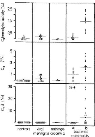
meningitis Figure 4. Levels of complement components in the
cerebrospinal fluid (CSF) of control patients and pa-tients who had viral meningitis, meningococcemia, or bacterial meningitis. The percentages of activity or con-centration of complement in pooled normal plasma are
shown for (top) C4 hemolytic activity, (middle) C3
con-centrations, and (bottom) C3d concentrations. Patients
with bacterial meningitis were divided into (a) those who had detectable and (b) those who had undetectable levels of opsonic activity in the CSF. Detectable levels of
op-sonic activity were defined as K~o opsonic titers (the
in-verse of the dilution of CSF that promoted 500/0 killing
of Staphylococcus aureus variant strain Wood 46) of ~2
units/ml. Horizontal lines show averages of comple-ment levels for each group of patients.
the levels of CMOA observed in samples of unin-fected CSF to those observed in serum.
CSF complement factors were assayed in the four groups of patients (figure 4); patients from group 4 were divided into two subgroups accord-ing to whether their CSF was CMOA-positive or -negative. A striking increase of C4 hemolytic ac-tivity was observed in the CMOA-positive samples of CSF from patients with bacterial meningitis as compared either with CMOA-negative CSF or with
CSF obtained in the other three groups (P
Table 3. Heat-labile opsonic activity (K50 ), hemolytic activity of complement, and concentrations of complement
components in sera and plasma from normal control patients and patients with acute meningitis.
Control patients Viral Meningococcemia Bacterial
Complement factors (n = 20) meningitis (n = 18) (n = 5) meningitis (n = 26)
K50 opsonic titers*
Experimental valuest 151 ± 38 156 ± 92 52 ± 34t 104 ± 46+
Values adjusted for
protein concentration§ 2.12 ± 0.64 2.63 ± 1.7 0.96 ± 0.88+ 2.00 ± 0.96 Hemolytic activity of cH5oll# 96 ± 20 124 ± 27t 52 ± 20+ 110 ± 24 C4# 99 ± 27 85 ± 22 77 ± 39 91 ± 46 Factor Bt 58 ± 18 54 ± 18 39 ± 19 59 ± 21 Concentration of C3** 96 ± 17 100 ± 13 54 ± 34+ 121 ± 22+ C3d** 5 ± 8 13 ± 7 33 ± 22+ 31 ± 25+ Total proteintt 73 ± 9 65 ± 7t 54 ± 9t 56 ± 9t
NOTE. Data are means ± SD; n = no. of patients tested.
* The inverse of the dilution that promoted 500/0 killing of Staphylococcus aureus variant strain Wood 46. t Expressed as units/ml.
t Differs significantly from control values (P
<
0.01). § Expressed as units/mg.II Total hemolytic activity of complement.
# Expressed as the percentage of activity in pooled normal serum. ** Expressed as the percentage of concentration in pooled normal serum. tt Expressed as g/liter.
group 4 showed a close correlation with CSF K50
opsonic titers (r = 0.79; P< 0.001) and with CSF
protein concentrations (r = 0.59; P
<
0.005).CMOA-positive samples of CSF also showed an increase in their immunochemically measured levels of C3 as compared with CMOA-negative
samples of CSF (P
<
0.02). C3d, the breakdownproduct of C3, was demonstrated in eight CMOA-positive samples and in one CMOA-negative sam-ple of CSF but was never detected in the CSF from patients in groups 1, 2, or 3. Levels of C3d in CSF, when expressed per milligram of protein, were three to 48 times higher than the protein-adjusted levels of C3d in plasma obtained from the same patients. Thus, the presence of measurable levels of CMOA in CSF of patients with bacterial men-ingitis is associated with an increase of C4 activity and C3 level and with evidence of local activation of C3.
Serum CMOA and plasma complement levels were measured in the four groups of patients to learn whether the local changes in the CSF during bacterial infection resulted from simultaneous modifications in the plasma (table 3). Definite changes were observed in the plasma of patients who had fulminant meningococcemia: lower levels
of K50 opsonic titers (P
<
0.01), CHso (P<
0.001),and factor B activities (P
<
0.05) and higher levelsof C3d (P< 0.01) when compared with control
pa-tients (group 1). Levels of C3d also increased in
the plasma of patients with bacterial meningitis
when compared with group 1 subjects (P
<
0.01).No correlation was found between the levels of either component in paired samples of CSF and plasma.
Correlation between opsonic activity of CSF and clinical course of bacterial meningitis. Among
the 27 patients with bacterial meningitis, 20
re-covered completely, two died from the disease, and five developed permanent neurologic se-quelae. All of the 15 patients who had measurable levels of CMOA in their CSF recovered complete-ly from the disease, whereas all seven patients who had neurologic sequelae or a lethal outcome were among the 12 patients who had undetectable levels of opsonic activity in their CSF (table 4). There-fore, the presence of CMOA in the CSF from pa-tients with bacterial meningitis was correlated with
a favorable clinical outcome (P
<
0.005; Fisher'sexact test). Complete recovery was also associated with a higher mean C4 hemolytic activity in CSF
(P
<
0.001), with higher C3 levels (P<
0.05), and with a lower mean bacterial count in the sample ofCSF taken at the time of admission (P
<
0.001)(table 5). No significant differences could be ob-served in the mean protein concentration or in the
Opsonic Activity in CSF
Table 4. Clinical outcome in patients with bacterial meningitis and the presence of measurable opsonic ac-tivity in cerebrospinal fluid (CSF).
Opsonic activity Present Absent Total Clinical outcome (no. of patients) Complete Sequelae recovery or death 15 0 5 7 20 7 Total no. of patients 15 12 27 NOTE. Samples of CSF were tested first at a dilution of 1 :2, which corresponded to an opsonic activity of ~2 Kso (50nJo killing) units/ml. The association between complete recovery and the presence of opsonic activity in CSF is statistically significant (P
<
0.005 by Fisher's exact test).mean PMNL counts in CSF between the two groups of patients. Patient groups with a favorable or unfavorable clinical course had
similar plasma K50 opsonic titers and complement
levels; further analysis of the various clinical data failed to reveal a significant association of neuro-logic complications or death with age, sex, dura-tion of symptoms prior to the start of antibiotic therapy, bacteremia on admission, or presence of a particular etiologic agent.
Discussion
The rationale for studying CMOA in normal and infected CSF resides in the fact that in nonimmune patients, opsonically active complement is one of the first defense mechanisms against pathogenic organisms [37, 38] and might contribute to host
643
defense at an early phase of bacterial meningitis when specific antibodies are still lacking. The data presented above demonstrate that CMOA was un-detectable in normal untreated CSF but was mea-surable after concentration; levels of CMOA also remained undetectable in the unconcentrated CSF of patients who had viral meningitis or fulminant meningococcemia at an early stage of the disease when noninflammatory CSF was already culture-positive. In contrast, CMOA appeared in 15 of 27 samples of CSF obtained during the early phase of bacterial meningitis, and its presence was corre-lated with a favorable clinical outcome. Such a favorable clinical evolution was also associated with an increase in complement levels and with lower concentrations of infecting microorganisms in CSF.
Several methodologic aspects of the technique used in this study should be discussed. In our pha-gocytic assay we did not use encapsulated bacteria causing meningitis as biological effectors; we used S. aureus strain Wood 46 because we wanted to measure early, nonspecific, functional CMOA in CSF, rather than individual immune response to specific microorganisms in nonimmune patients. Furthermore, it is generally accepted that bacterial meningitis develops only in patients who lack
type-specific, functional serum antibodies [39]. It
is therefore likely that the study of early, specific immune response in CSF to the offending orga-nisms would have given negative results. For ethical reasons, the phenomenon of the time-dependent appearance of specific antibodies can only be ex-amined in an animal model. Previous studies in our laboratory have shown that the opsonization
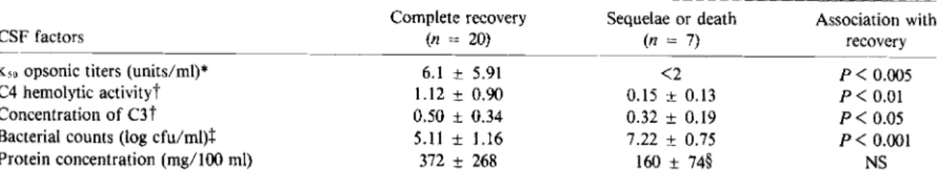
Table 5. Correlation of clinical outcome in patients with bacterial meningitis and K50 (50070 killing) opsonic titers,
complement levels, and bacterial counts in cerebrospinal fluid (CSF).
Complete recovery Sequelae or death Association with
CSF factors (n = 20) (n = 7) recovery
Kso opsonic titers (units/ml)* 6.1 ± 5.91 <2 P< 0.005
C4 hemolytic activityt 1.12 ± 0.90 0.15 ± 0.13 P
<
0.01Concentration of C3 t 0.50 ± 0.34 0.32 ± 0.19 P< 0.05
Bacterial counts (log cfu/mI)t 5.11 ± 1.16 7.22 ± 0.75 P
<
0.001Protein concentration (mg/100 ml) 372 ± 268 160 ± 74§ NS
NOTE. Data are means ± SD and probabilities of association with recovery calculated by Fisher's exact test. NS = not significant.
* Kso = the inverse of the dilution of CSF that promoted 50070 killing of Staphylococcus aureus variant strain Wood 46. t Expressed as the percentage of activity or concentration in pooled normal serum.
t Performed in samples from 10 patients who recovered and from five patients who either died or had sequelae.
§ Mean calculated for six of the seven patients who either died or had sequelae; patient no. 23 was excluded because of an unusually high concentration of protein in the CSF (1,230 mgllOO mt).
of S. aureus strain Wood 46 by human serum depends mainly on the complement system, and the role played by antibody is minimal [29]. The results of the present studies, which use CSF as the source of opsonic activity, confirm the earlier data (table 2). The correlation found between CMOA and CHso in 10 samples of normal concentrated CSF (P
<
0.02) and between CMOA and C4 ac-tivities in samples of CSF from patients withmen-ingitis (P
<
0.001) provides further evidence thatthe heat-labile opsonic activity in CSF that was measured by our phagocytic assay was indeed complement-mediated.
The method used to obtain and store CSF samples for measurement of complement levels or opsonic activity turned out to be a crucial metho-dologic point; spontaneous decay of complement occurs in untreated frozen CSF [15] and may even be accelerated when CSF that is taken from pa-tients with systemic lupus erythematosus and cen-tral nervous system involvement is stored at - 45 C [40]. To minimize this problem, all samples of CSF were drawn into vials containing EDT A and were immediately frozen at -70 C. Blood con-tamination of the CSF, which would have resulted in falsely high levels of complement activities, was eliminated by excluding all CSF samples with an
admixture of blood of ~1:25,000.
We started our study with the hypothesis that the phagocytic test developed by Lew et al. [29] could be adapted to untreated CSF because the
mean Kso opsonic titers measured in pooled serum
(236 units/ml) approximated the protein ratio found between normal untreated CSF and serum (1:250). However, CMOA was not detected in fresh undiluted CSF with this assay. By concen-trating large volumes of normal CSF obtained during gas myelography, we were able to show that normal untreated CSF was devoid of CMOA. The absence of CMOA in CSF was evidenced by the low protein concentration in CSF; moreover, CMOA, expressed per milligram of protein, could be demonstrated to be lower in the CSF than in the serum from the same patient (figure 1, top). These findings are consistent with the data of Tofte et al. [17], who described an absence of opsonization of various encapsulated bacteria by normal CSF, whereas one sensitive strain of S. aureus (strain 502A) was opsonized by esp. Our results are also in agreement with those published by Simberkoff et al. [18], who used various pathogenic and
non-pathogenic test bacteria and showed no bacterici-dal and no opsonic activity in normal CSF.
Our experiments with concentrated CSF suggest that the opsonic defect is not qualitative but mere-ly quantitative. Sensitization of the phagocytic test by ultrafiltration had the additional advantages of eliminating all potential traces of antibiotics and EDT A from the samples and allowing us to test CSF under native conditions but in a well-buffered system. To minimize any error introduced by manipulating the CSF, we systematically prepared each sample of CSF and serum by ultrafiltration shortly before use. Control experiments showed that the ultrafiltration-concentration procedure resulted in a minimal, reproducible loss of CMOA and was not accompanied by any evidence of com-plement activation.
Early appearance of CMOA in CSF during bac-terial meningitis - as opposed to viral men-ingitis - was paralleled by increases in C3 activity and C3 levels, which confirmed the presence of
CHso [18] and various other complement
com-ponents [24, 41, 42] in the CSF of patients with meningitis. The early appearance of CMOA in CSF was further correlated with changes in the protein concentrations in CSF early in the course
of the disease. Kso opsonic titers in the CSF,
however, when adjusted for protein
concentra-tion, never exceeded Kso values in serum, which
were also adjusted for protein concentration. It is
therefore probable that the appearance of CMOA in CSF is one of the numerous changes induced by local inflammation of the subarachnoid space. The appearance of CMOA may be the result of an increase in the permeability of the blood-brain barrier for large molecules, as suggested by the ap-pearance of IgM in infected CSF [23]. A part of complement is probably activated in the eSF dur-ing mendur-ingitis, as evidenced by the presence of C3d, the breakdown product of C3, in the CSF of nine patients.
The clinical relevance of the correlation found between the presence of CMOA in the CSF and a favorable clinical outcome for patients with bacte-rial meningitis needs to be put in proper perspec-tive. First, CMOA - as assessed by the phagocytic assay with S. aureus strain Wood 46 - does not measure possible specific opsonins in eSF and serum or the bactericidal defense mechanisms that each patient produces in response to the particular microorganism that causes the infection.
How-Opsonic Activity in CSF
ever, these factors are unlikely to be present ini-tially and were undetectable in the CSF of patients with acute bacterial meningitis [18]. In contrast, complement activity - as measured in our assay-is a nonspecific but immediately available host de-fense factor [38], and its level in the CSF may be important in limiting the rate of bacterial growth, as shown under other conditions [25, 37, 43]. Also, it should be stressed that the association be-tween the presence of CMOA in the CSF and a favorable outcome for patients with meningitis is purely descriptive and that the present data do not prove a causal relationship. In fact, we cannot ex-clude the possibility that a common mechanism (for example, greater permeability of the blood-CSF barrier for both complement components and antibiotics) simultaneously influences several factors. However, patients who recovered
com-pletely from meningitis not only had higher K50
opsonic titers but also had lower mean initial bac-terial counts in their CSF than did patients with complications, as previously observed by Feldman [20]. Thus, any mechanism that is able to moder-ate bacterial growth in the CSF may be an impor-tant determinant of the course of the disease and may allow the patient to receive medical therapy before complications arise. The possibility that CMOA in CSF might not merely reflect the per-meability of the blood-meningeal barrier but may express an important biologic role of complement that is independent of the inflammatory process is illustrated by one subject in our study. Patient no. 23 had fatal post-traumatic meningitis due to
E. coli; his initial sample of CSF contained over
108 cfu of bacteria/ml and had undetectable levels
of C4, C3, and opsonic activity, in spite of a mas-sive increase in protein concentration in the CSF. This situation was associated with very high levels of C3d, the breakdown product of C3, in the CSF. Thus, bacterial infection of the subarachnoid space may occasionally lead to a state of local con-sumption of opsonins and complement as pre-viously demonstrated for pleural empyema [29, 44, 45].
Our results show that despite its barely detect-able concentration in normal CSF, CMOA rapidly appears during the course of bacterial meningitis. The current results suggest that CMOA in CSF might be one of the numerous factors that influ-ence the outcome of the disease. Increasing the level of opsonic activity in the CSF had already
645
been suggested in the preantibiotic era [46] as a means of influencing the outcome of bacterial meningitis, and this therapeutic strategy can now be evaluated in animal models.
References
1. Dodge, P. R., Swartz, M. N. Bacterial menmgltls-a review of selected aspects. II. Special neurologic prob-lems, postmenigitic complications and clinicopathologi-cal correlations. N. Eng!. J. Med. 272:954-960, 1965. 2. Hodges, G. R., Perkins, R. L. Acute bacterial meningitis:
an analysis of factors influencing prognosis. Am. J. Med. Sci. 270:427-440, 1975.
3. Feigin, R. D., Dodge, P. R. Bacterial meningitis: newer concepts of pathophysiology and neurologic sequelae. Pediatr. Clin. North Am. 23:541-556, 1976.
4. ScheId, W. M. Pathophysiologic correlates in bacterial meningitis. Journal of Infection 3(Supp!. 1):5-20, 1981. 5. Robbins, J. B., McCracken, G. H., Jr., Gotschlich, E. c.,
ct>rskov, F., ~rskov, I., Hanson, L. A. Escherichia coli
Kl capsular polysaccharide associated with neonatal meningitis. N. Eng!. J. Med. 290:1216-1220,1974. 6. Moxon, E. R., Murphy, P. A. Haemophilus injluenzae
bacteremia and meningitis resulting from survival of a single organism. Proc. Natl. Acad. Sci. U.S.A. 75: 1534-1536, 1978.
7. Petersdorf, R. G., Swarner, D. R., Garcia, M. Studies on the pathogenesis of meningitis. II. Development of men-ingitis during pneumococcal bacteremia. J. Clin. Invest. 41:320-327,1962.
8. Moxon, E. R., Ostrow, P. T. Haemophilus injluenzae
meningitis in infant rats: role of bacteremia in pathogen-esis of age-dependent inflammatory responses in cere-brospinal fluid. J. Infect. Dis. 135:303-307,1977. 9. Scheifele, D. W., Daum, R. S., Syriopoulou, V. P., Averill,
D. R., Smith, A. L. Haemophilus injluenzae bacteremia and meningitis in infant primates. J. Lab. Clin. Med. 95:450-462, 1980.
10. Scheid, W. M., Park, T.-S., Dacey, R. G., Winn, H. R., Jane, J. A., Sande, M. A. Clearance of bacteria from cerebrospinal fluid to blood in experimental meningitis. Infec. Immun. 24:102-105, 1979.
11. McAllister, C. K., O'Donoghue, J. M., Beaty, H. N. Experimental pneumococcal meningitis. II. Characteri-zation and quantitation of the inflammatory process. J. Infect. Dis. 132:355-360, 1975.
12. ScheId, W. M., Dacey, R. G., Winn, H. R., Welsh, J. E., Jane, J. A., Sande, M. A. Cerebrospinal fluid outflow resistance in rabbits with experimental meningitis: alter-ations with penicillin and methylprednisolone. J. Clin. Invest. 66:243-253, 1980.
13. Tibbling, G., Link, H., Ohman, S. Principles of albumin and IgG analyses in neurological disorders. I. Establish-ment of reference values. Scand. J. Clin. Lab. Invest. 37:385-390, 1977.
14. Propp, R. P., Jabbari, B., Barron, K. Measurement of the third component of complement in cerebrospinal fluid by modified electroimmunodiffusion. J. Lab. Clin. Med. 82:154-157, 1973.
15. Petz, L. D. Measurement of spinal fluid complement in immunologically mediated neurologic disorders. In W. Opferkuch, K. Rother, and D. R. Schultz [ed.]:Clinical aspects of the complement system. Georg Thieme, Stutt-gart, Federal Republic of Germany, 1978, p. 125-128. 16. Cova, 1. L., Propp, R. P., Barron, K. D. Quantitative
relationships of the fourth complement component in human cerebrospinal fluid. 1. Lab. Clin. Med. 89:615-621, 1977.
17. Tofte, R. W., Peterson, P. K., Kim, Y., Quie, P. G. Op-sonic activity of normal human cerebrospinal fluid for selected bacterial species. Infec. Immun. 26: 1093-1098, 1979.
18. Simberkoff, M. S., Moldover, N. H., Rahal, 1. 1., 1r. Absence of detectable bactericidal and opsonic activities in normal and infected human cerebrospinal fluids: a regional host defense deficiency. 1. Lab. Clin. Med. 95: 362-372, 1980.
19. Petersdorf, R. G., Luttrell, C. N. Studies on the pathogen-esis of meningitis. I. Intrathecal infection. 1. Clin. In-vest. 41:311-319, 1962.
20. Feldman, W. E. Relation of concentrations of bacteria and bacterial antigen in cerebrospinal fluid to prognosis in patients with bacterial meningitis. N. Engl. 1. Med. 296: 433-435, 1977.
21. Nolan, C. M., Clark, R. A., Beaty, H. N. Experimental pneumococcal meningitis. III. Chemotactic activity in cerebrospinal fluid. Proc. Soc. Exp. BioI. Med. 150: 134-136, 1975.
22. Greenwood, B. M. Chemotactic activity of cerebrospinal fluid in pyogenic meningitis. 1. Clin. Pathol. 31 :213-216, 1978.
23. Ganrot-Norlin, K. Relative concentrations of albumin and IgG in cerebrospinal fluid in health and in acute menin-gitis. Scand. 1. Infect. Dis. 10:57-60, 1978.
24. Whittle, H. c., Greenwood, B. M. Cerebrospinal fluid immunoglobulins and complement in meningococcal meningitis. 1. Clin. Pathol. 30:720-722, 1977. 25. Stossel, T. P. Phagocytosis [part 1]. N. Engl. 1. Med. 290:
717-723, 1974.
26. Lennette, E. H., Balows, A., Hausler, W. 1., Jr., Truant, J. P. [ed.]. Manual of clinical microbiology. 3rd ed. American Society for Microbiology, Washington, D.C., 1980, p. 1-440.
27. Feldman, W. E. Concentrations of bacteria in cerebrospinal fluid of patients with bacterial meningitis. J. Pediatr. 88:549-552, 1976.
28. Hirsch, J. G., Strauss, B. Studies on heat-labile opsonin in rabbit serum. 1. Immunol. 92:145-154, 1964.
29. Lew, P. D., Zubler, R., Vaudaux, P., Farquet, 1. 1., Waldvogel, F. A., Lambert, P.-H. Decreased heat-labile opsonic activity and complement levels associated with evidence of C3 breakdown products in infected pleural effusions. 1. Clin. Invest. 63:326-334, 1979.
30. Nydegger, U. E., Achermann, L. M., Lambert, P.-H., Miescher, P. A. A simple automated method for com-plement estimation in a continuous flow system. 1. Im-munol. 109:910-913, 1972.
31. Frank, M. M., May, J., Gaither, T., Ellman, L. In vitro studies of complement function in sera of C4-deficient guinea pigs. J. Exp. Med. 134:176-187, 1971. 32. Aguado, M. T., Perrin, L. H., Ramirez, R., Miescher,
P. A., Lambert, P.-H. Evaluation of alternative path-way and factor B haemolytic activities in patients with systemic lupus erythematosus: correlations with the alternative pathway regulatory proteins. Clin. Exp. Im-munol. 42:495-505, 1980.
33. Perrin, L. H., Lambert, P.-H., Nydegger, U. E., Miescher, P. A. Quantitation of C3PA (properdin factor B) and other complement components in diseases associated with a low C3 level. Clin. Immunol. Immunopathol. 2:16-27, 1973.
34. Perrin, L. H., Lambert, P.-H., Miescher, P. A. Comple-ment breakdown products in plasma from patients with systemic lupus erythematosus and patients with mem-branoproliferative or other glomerulonephritis. 1. Clin. Invest. 56:165-176, 1975.
35. Meddis, R. Statistical handbook for non-statisticians. McGraw-Hill, Maidenhead, Berkshire, England, 1975, p.48-56.
36. Wagner, J. G. Fundamentals of clinical pharmacokinetics. Drug Intelligence Publications, Hamilton, Ill., 1975, p.316-317.
37. Winkelstein, 1. A., Smith, M. R., Shin, H. S. The role of C3 as an opsonin in the early stages of infection. Proc. Soc. Exp. BioI. Med. 149:397-401, 1975.
38. Fearon, D. T., Austen, K. F. The alternative pathway of complement - a system for host resistance to microbial infection. N. Engl. 1. Med. 303:259-263, 1980. 39. McGee, Z. A., Kaiser, A. B. Acute meningitis. In G. L.
Mandell, R. G. Douglas, 1r., and 1. E. Bennett [ed.]. Principles and practice of infectious diseases. Vol. 1. Wiley, New York, 1979, p. 738-760.
40. Hadler, N. M., Gerwin, R. D., Frank, M. M., Whitaker, 1. N., Baker, M., Decker, 1. L. The fourth component of complement in the cerebrospinal fluid in systemic lupus erythematosus. Arthritis Rheum. 16:507-521, 1973.
41. Fothergill, L. D. Observations on the presence of comple-ment in the cerebrospinal fluid in various pathologic conditions of the central nervous system. 1. Pediatr. 6:374-381, 1935.
42. Spicer, S., Appelbaum, E., Rutstein, D. D. Complement and its component fractions in cerebrospinal fluid in in-flammatory cerebrospinal diseases. J. Clin. Invest. 28: 389-393, 1949.
43. Forsgren, A., Quie, P. G. Influence of the alternate com-plement pathway on opsonization of several bacterial species. lnfec. Immun. 10:402-404, 1974.
44. Suter, S., Nydegger, U. E., Roux, L., Waldvogel, F. A. Cleavage of C3 by neutral prot eases from granulocytes in pleural empyema. 1. Infect. Dis. 144:499-508, 1981. 45. Lew, D. P., Despont, 1.-P., Perrin, L. H., Aguado, M.-T., Lambert, P.-H., Waldvogel, F. A. Demonstration of a local exhaustion of complement components and of an enzymatic degradation of immunoglobulins in pleural empyema: a possible factor favouring the persistence of local bacterial infections. Clin. Exp. Immunol. 42:506-514, 1980.
46. Fonde, E. C. The use of fresh human serum (complement) in combination with the antiserum in the treatment of meningococcic meningitis. J.A.M.A. 105:110-112, 1935.