Introduction
Fractures and dislocations of the cranio-cervical junction represent one-third of all injuries to the cervical spine. They are usually caused by high-energy trauma such as traffic accident or fall from a great height [3, 9]. Atlanto-occipital dislocation (AOD), because of its accompanying injuries to the brain stem and lesions to the vascular struc-tures of the neck, is mostly a lethal injury [11, 32, 98]. The diagnosis is often overlooked [74]. Recognition of AOD and therapeutic procedures are hampered by the fre-quent combination of AOD with traumatic brain injury and/or polytrauma. In this article four adult patients with AOD are presented and two showed a long-term survival and were surgically treated.
Case reports Case 1
A 22-year-old man fell 12 m from a roof, was unconscious and had a Glasgow Coma Scale (GCS) of three points for an unknown time. Cardiac arrest and apnea occurred after arrival of the rescue team. Following successful resuscita-tion and intubaresuscita-tion, the patient was transferred to the emergency ward of the University Hospital of Zurich without relaxation or sedation. The GCS was unchanged, pupillary light reactions and vital signs were normal. Ex-cept for a generalized brain edema in the computed to-mography (CT) scan of the head, there were no patholog-ical findings in the clinpatholog-ical and radiologpatholog-ical work-up. Af-ter insertion of a subdural catheAf-ter for intracranial pres-sure (ICP) monitoring, the patient was transferred to the intensive care unit (ICU). On the first posttraumatic day, clinical neurological examination showed bilateral cranial Abstract Traumatic
atlanto-occipi-tal dislocation (AOD) is a rare cervi-cal spine injury and in most cases fa-tal. Consequently, relatively few case reports of adult patients surviving this injury appeared in the literature. We retrospectively report four pa-tients who survived AOD injury and were treated at our institution. A young man fell from height and a woman was injured in a traffic acci-dent. Both patients survived the in-jury but died later in the hospital. The third patient had a motorcycle accident and survived with incom-plete paraplegia. The last patient, a man involved in a working accident, survived without neurological deficit
of the upper extremities. Rigid poste-rior fixation and complete reduction of the dislocation were applied in last two cases using Cervifix to-gether with a cancellous bone graft-ing. Previously reported cases of pa-tients surviving AOD are reviewed, and clinical features and operative stabilisation procedures are dis-cussed.
Keywords Atlanto-occipital dislocation · Cervical spine injury · Posterior cervical fusion
Ludwig Labler Karim Eid Andreas Platz Otmar Trentz Thomas Kossmann
Atlanto-occipital dislocation:
four case reports of survival in adults
and review of the literature
Received: 18 February 2003 Revised: 2 September 2003 Accepted: 7 November 2003 Published online: 13 December 2003 © Springer-Verlag 2003
L. Labler (✉) · K. Eid · A. Platz ·
O. Trentz
Division of Trauma Surgery, Department of Surgery, University Hospital Zurich,
Rämistrasse 100, 8091 Zurich, Switzerland Tel.: +41-1-2554188,
Fax: +41-1-2554406,
e-mail: ludwig.labler@chi.usz.ch T. Kossmann
Department of Surgery,
Monash University, The Alfred Hospital, Commercial Road,
nerve palsies, absent gag reflex and flabby tetraparesis. Electroencephalogram (EEG) showed a suppression burst pattern and there were no reactions in somatosensory evoked potentials, which was consistent with a high spi-nal paralysis. Intracranial pressure remained normal all the time. Control CT scan of the head showed a decreased cerebral edema without intracerebral ischaemia or con-cussions. After an attempt of a wake up, the patient showed a hypoxic aphalic syndrome suspicious of a high cervical spine lesion. Magnetic resonance imaging (MRI) confirmed the diagnosis of an AOD with traumatic lesions of the upper cervical spinal cord and of the lower brain stem. The patient died on tenth day due to severe lesion in the transition from medulla oblongata to spinal cord, ver-ified by autopsy as a severe myelomalacia (total necrosis) in the described anatomic region.
Case 2
A 68-year-old woman was hit by a tram. On arrival of the rescue team, the patient showed a gasping breathing and a GCS of three points with no pupillary light reaction on the left eye. In the emergency ward of our hospital, the patient was orally intubated, sedated, relaxated and developed a
haemodynamic instability. After cardiopulmonary resus-citation, clinical and radiological work-up revealed AOD together with a traumatic brain injury and subarachnoidal bleeding. Furthermore, a chest trauma with serial rib frac-tures, lung contusions on both sides, a pelvic fracture, a floating elbow with an open distal fracture of the humerus, a closed forearm fracture and a fracture of right radius were diagnosed. After immediate insertion of chest tubes, a subdural catheter was placed for ICP monitoring and the left fractured humerus was stabilized by an external fixa-tor. The patient was then transferred to the ICU, where she died the same day due to severe brain and thoracic in-juries.
Case 3
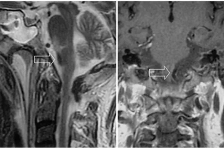
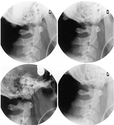
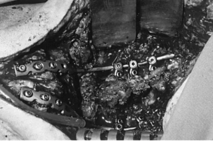
A 51-year-old man fell from a motorcycle and collided with a crash barrier. The patient was resuscitated by para-medics until the ambulance arrived. He was orally intu-bated at the scene showing an GCS of 3 points and no pupillary light reaction on the left eye. Clinical and radio-logical assessment by conventional X-ray and CT scan of the head revealed a severe brain injury with haemorrhagic concussions on the left temporal side, multiple subarach-noidal haemorrhages, further “shearing injuries” and a lig-amentous instability of the right elbow and knee. The CT scan did not show any signs of injuries in the occipito-cer-vical junction. The ratio according to Powers et al. [73] was less than 0.9. A ventricular catheter for ICP monitor-ing was inserted and the patient was transferred to ICU. Despite a constantly low intracranial pressure, a failing at-tempt to wake the patient was a sign of a lesion in the pon-tomedullary junction. An MRI scan confirmed the diag-nosis of an ischaemic lesion in this region (Fig. 1). The in-jury pattern, the incomplete flabby tetraplegia, the pres-ence of a “locked in” syndrome, bilateral palsy of the N. abducens and absent gag reflex indicated AOD. An ex-amination under the image intensifier confirmed the diag-nosis (Fig. 2). The neurological findings did not change during hospitalisation. The AOD was stabilized by a dor-sal fusion between the occiput and C5 by Cervifix (Stratec, Oberdorf, Switzerland; Fig. 3). Due to patient’s neurolo-gene dysphagia, tracheotomy and endoscopically assisted
Fig. 2 Functional examination
under image intensifier demon-strating atlanto-occipital dislo-cation (AOD)
Fig. 1 Sagittal and frontal T2-weighted MRI sequences. Lesion in
percutaneous gastroenterostomy (PEG) were performed. Postoperative complications were not observed. Early phys-iotherapy was applied according to the Bobath scheme. The patient was transferred to other hospital for further neuro-rehabilitation where he progressively recovered from his neurological deficits. Sixteen months after the accident, he was fully ambulatory without medical aids. The pa-tient, besides an incomplete paraplegia, has a persistent hoarseness and a bilateral palsy of the abducens nerve, for which he has undergone an operative correction.
Case 4
A 35-year-old patient tipped over with a forklift and his shoulder was wedged in by the roof of the engine. At the scene, the initial GCS was 15 points and the patient was able to move his extremities with exception of the right arm. Because of increasing shortness of breath and an in-spiratory stridor, he was intubated at the scene by the res-cue ambulance and transferred to the University Hospital of Zurich. Clinical work-up revealed a massive swelling at the neck, a subtotal tear of the auricle and a cut at the forearm. Radiological examinations showed fractures of the right clavicle, the left scapula and gave a hint on an in-jury in the occipito-cervical junction (Fig. 4). The ratio
ac-cording to Powers et al. [73] was 1.1. The diagnosis of AOD was confirmed by functional X-rays (Fig. 5). During the wake-up, bilateral sensomotor deficiency with palsies of shoulder girdle and the upper extremities were ob-served. An MRI scan showed a lesion of the vertebral artery and the presence of an epidural haematoma (Fig. 6). The AOD was then stabilised by a dorsal fusion of C0–C5 with a Cervifix (Fig. 7). Postoperative complications were not observed and the neurological deficits of the upper ex-tremities receded. The patient was transferred for further neuro-rehabilitation. Three months after the accident, the patient was fully ambulatory without aids. He still com-plains of a persistent sensomotor shortage of the right arm at C4–C6. The patient was discharged for ambulatory treatment.
Discussion
A recent study reports an incidence of cervical injuries of 4.3% [39]. The incidence of cervical spine injuries fol-lowing blunt trauma in children is estimated to be 1.3– 1.5% [71, 72]. In 30% of 300 analysed patients with cer-vical spine injuries the localisation is in the upper region, i.e. between C0 and C2 [9]. Radiological injuries of the upper cervical spine, of which 93% were in the first two Fig. 3 a Positioning of the
pa-tient, in prone position, with fixation of the head by a May-field clamp. b Postoperative X-rays showing stabilization of the AOD by dorsal fusion, from the occiput to C5, with Cervifix and cancellous bone
segments, were found in 24.4% of 312 deceased traffic victims [3]. Solely ligamentous injuries to the cervical spine were found in only 14 patients of 14,577 with blunt trauma [16]. Injuries to the upper cervical spine are fre-quent in traffic accidents with the highest prevalence for pedestrians and motorcyclists [86].
Atlanto-occipital dislocation, described for the first time by Blackwood in 1908, is a rare injury [8]. Among 155 examined traffic victims, 12 patients with AOD were found with additional various fractures in the occipito-cervical junction [1]. Others report an incidence of AOD of 8% up to 31% in fatal traffic accidents [11, 98] and of nearly 10% in fatal cervical spine injuries [3]. The AOD may be complete or incomplete. Three different types (ventral, dorsal, axial) of complete AOD were described [88]. The ventral dislocation (type I) is the most common case, the axial dislocation (type II) the most unstable one and the dorsal dislocation (type III) the rarest [42]. In our four cases we found two of type-I (cases 2 and 4) and two
of type-II (cases 1 and 3) dislocations. Additionally, lat-eral dislocations of AOD have been described [90]. In-complete forms of AOD represent subluxations of the at-lanto-occipital junction [42]. The atat-lanto-occipital, the lat-eral atlanto-axial and the median atlanto-axial articula-tions are a functional unit. The overall effect of all joints equals a spherical joint. The rigidity depends mostly on the surrounding ligamentous structures. Among these, the membrana tectoria and the bilateral ligamenta alaria are the main stabilizer [92]. Children and young adults suffer more AOD [11, 24, 64, 79], because of a disparity between the occipital condyles and the articular surfaces of the at-las, a more horizontal plane of the articular surfaces and an increased laxity of the ligamentous structures [12, 45, 52, 80]. Additionally, there are reports that airbag deploy-ment may cause AOD in children [4, 35, 36].
Neurological deficits in up to 33% [32] and vascular complications lead to high mortality. The immediate cause of death may be due to vascular lesions to carotid arteries Fig. 4 a Atlanto-occipital
dis-sociation in the CT scan demonstrating the subluxation with a rotationary component after first reduction (open
ar-row) and avulsion fracture of
the ligaments (solid arrows).
b A 3D reconstruction with an
anteroposterior view (arrows).
c Subluxation viewed cranially
through the foramen magnum (arrows)
or vertebral arteries [25, 27, 33, 58, 68, 73] as well as to direct injury to the spinal cord or brain stem [11, 25, 52]. Our patient in case report 4 showed a lack of a vertebral artery due to AOD. Three of our four patients showed signs of direct injuries of the spinal cord or the brain stem. Subdural haematoma as accompanying injury of AOD is
described in 16% of cases [1]. A combination with aortic lesions is reported in 25% and with basal skull fractures in 21% [86]. Except our case 4, all patients suffered from se-vere head injuries. Injuries to the brain nerves, particu-larly the abducens nerve (N VI) as for example, in our case 4, are occasionally reported in the literature [15, 27, 31, 33]. Out of a group of eight patients with AOD, one patient sustained a rupture of the membrana tectoria with a lethal outcome, and in the remaining seven cases, MRI showed a characteristic stretching with a simultaneous el-evation from the clivus and the tip of the dens and a fluid collection [84]. Fourteen of the 79 surviving (18%) pa-tients showed no neurological deficits, and 8 (10%) showed isolated deficits of brain nerves [74].
Absence of neurological deficits has also been reported by other authors [26, 29, 31, 33, 52, 66]. Hemiplegia oc-curred in 34% and tetraplegia in 38% in patients surviving AOD [74]. Patients with AOD have been reported to show an improvement of neurological deficits [22, 27, 31, 33, 38, 41, 51, 52, 54, 56, 58, 66, 85, 91, 93] and this was also the case with our two patients with long-term sur-vival who both showed an improvement of neurological deficits. Rupture of the trachea [67] and retropharyneal pseudomeningocele [63] were reported as rare complica-tions following AOD.
The majority of patients with an AOD are either im-mediately dead or survive for few hours only [3, 9, 11]; therefore, this injury was considered potentially lethal [26, 27, 29, 31, 33, 66, 73, 77]. Due to improved pre-hospital care, rapid rescue and shorter transportation time, the number of patients surviving AOD and arriving alive in the hospital is increasing [2, 4, 6, 7, 10, 12, 13, 15, 17, 18, 19, 20, 22, 23, 24, 25, 28, 30, 34, 35, 38, 40, 41, 43, 44, 45, 46, 47, 49, 50, 52, 54, 55, 56, 57, 58, 60, 61, 62, 63, Fig. 5 Functional tests of the atlanto-occipital junction under
im-age intensifier
Fig. 6 a An MRI scan of
AOD shows an epidural haem-atoma (arrows) with b associ-ated lesion of the left vertebral artery (arrow)
64, 65, 67, 69, 70, 71, 75, 76, 78, 79, 80, 81, 82, 83, 84, 85, 87, 89, 90, 91, 93, 94, 95, 96]. Meta-analysis of the lit-erature between 1948 and present showed 211 patients surviving AOD including 131 (62%) children [2, 4, 9, 12, 13, 15, 17, 18, 20, 22, 23, 24, 25, 27, 28, 29, 30, 31, 34, 35, 38, 41, 44, 45, 46, 47, 52, 54, 56, 60, 61, 62, 64, 65, 68, 69, 71, 73, 74, 75, 77, 78, 79, 80, 81, 82, 83, 84, 88, 89, 93, 94, 96, 97] and 56 (27%) adults (Table 1). For the remaining 24 (11%) cases, the age of the patients was not specified [32, 37, 57].
A complete AOD is accompanied by massive swelling of the head and neck, and radiological diagnosis is, as in our fourth case, usually undemanding. In the presence of a respiratory insufficiency, an irregular cardiac activity up to a cardiac arrest or a hypotension, caused by traumatic lesions of the brain stem, AOD should be suspected [2]. Less prominent dislocations or spontaneously reduced AOD are easily overlooked despite the comprehensive ra-diological work-up [18, 22, 25, 30, 37, 45, 54, 56]. An analysis of 79 patients with AOD found that in 19 of 50 (38%) children and in 17 of 29 (59%) adults, the diagno-sis of an AOD, particularly of an axial dislocation, was missed [74]. We too encountered the same problem in two of our four cases. Diagnostic work-up for AOD consists of a lateral view of the upper cervical spine and the as-sessment of the ratio according to Powers et al. [73],
Har-ris et al. [42], Dublin et al. [25] or Kaufmann et al. [53]. Functional examination under image intensifier is de-scribed [51], combined, if necessary, with monitoring of the somato-sensory potentials [5]. Computer tomography with sagittal, coronary, and three-dimensional reconstruc-tion [21, 40, 60], MRI [13, 14, 21, 37] and angiography [58] are the instruments for the diagnosis and for the as-sessment of additional injuries in AOD. Avulsion frac-tures from the occipital condyles or the tip of the dens and a retropharyngeal haematoma may lead to the diagnosis of an AOD [21]. In the CT scan and the MRI in three of our four cases, we found the described radiological signs. Early diagnosis of AOD is mandatory to prevent further impair-ment of the spinal cord by manipulations during position-ing, diagnostic work-up or therapeutic procedures.
After establishing vital functions, particularly of the air-ways, reposition and splinting are achieved by either a stiff neck, traction in a Gardner–Wells, or a Halo-fixator [7, 27, 29, 33, 56, 62, 67]. For axial dislocation of AOD (type II) traction is not recommended and should be avoided [52]. Complete retention is sometimes impossible due to interposition of fragments or ligamentous struc-tures. Conservative treatment with extension or halo-fixa-tor as further management is recommended in children and in the case of little instability of AOD [2, 28, 49, 56, 63, 73, 83, 84, 93]; however, from the aforementioned lit-erature it is not evident how much instability may be tol-erated. In children, a fibrous ankylosis is expected after months of conservative treatment [49, 56, 83, 93]. In adults and with increasing instability, a short dorsal spondylode-sis should be performed [15, 34, 68, 82, 93]. The dorsal fusion is achieved by an occipito-cervical tension wire band and augmentation by autologous cancellous bone with an immobilisation in a minerva cast [5, 15, 85]. Fu-sion by a bone graft from the iliac crest with additional plate fixation (Y-plate, Roy-Camille plate, reconstruction plate) and transpedicular screws allows early mobilization without a cast or with a soft neck for 6 weeks to 4 months [7, 18, 22, 27, 31, 46, 47, 68, 69, 78, 88, 93, 97]. A new technique for stabilization of AOD was described using transarticular fixation with additional occipito-cervical Y-plate fixation [38]. Fusion with the Cervifix system with a bone graft from the iliac crest with or without can-cellous bone is an alternative dorsal stabilization proce-dure [51, 55] and was also applied in two of our four pa-tients. The implant is made from titanium allowing post-operative CT scans with fewer artefacts. The implant de-sign consists of a reconstruction plate in the cranial part and a rod system in the caudal part. An implant removal is not advisable even after complete neurological remission, because mobility from C0 to C2 is lost because of the bony consolidation.
Table 1 Literature review of adult patients surviving
atlanto-oc-cipital dislocation
No. of Reference No. of
patients cases 1 [3, 5, 6, 7, 8, 9, 14, 19, 26, 33, 40, 43, 47, 32 48, 49, 50, 51, 55, 62, 63, 66, 67, 73, 76, 78, 79, 85, 87, 90, 91, 93, 95 2–5 2, 10, 15, 25, 30, 74 17 >5 23] 7 Total 56
Conclusion
It can be concluded that, due to improved rescue services, decreased transportation time, and increased awareness of the injury, the number of patients surviving AOD in-creases and that therefore longtime observations are pos-sible [59, 69, 88]. Reviewing the literature, there are 211 documented patients surviving AOD initially and 108 (51%) cases, mainly children and young adults, showed a long-term survival. Diagnosis may be hampered and is frequently delayed, in part due the lack of clinical or
ra-diological evidence, in part due to the additional injuries where a clinical examination is impossible. The extent of neurological deficits in AOD varies from no deficits up to severe neurological deficits caused either by brain stem or spinal cord injuries and by vascular injuries (carotid and vertebral arteries). The therapeutic options range from con-servative measures, usually in children, to operative fusion of the cervical spine. The prognosis of primary neurological deficits remains unfavourable; however, there are sporadic reports of neurological improvements after AOD [22, 27, 31, 33, 38, 41, 51, 52, 54, 56, 58, 66, 85, 91, 93].
1. Adams VI (1992) Neck injuries: Part I. Occipitoatlantal dislocation – a patho-logic study of twelve traffic fatalities. J Forensic Sci 37:556–564
2. Ahuja A, Glasauer FE, Alker GJ Jr, Klein DM (1994) Radiology in sur-vivors of traumatic atlanto-occipital dislocation. Surg Neurol 41:112–118 3. Alker GJ Jr, Oh YS, Leslie EV (1978)
High cervical spine and craniocervical junction injuries in fatal traffic acci-dents: a radiological study. Orthop Clin North Am 9:1003–1010
4. Angel CA, Ehlers RA (2001) Images in clinical medicine. Atlanto-occipital dislocation in a small child after airbag deployment. N Engl J Med 345:1256 5. Babbitz JD, Kim KD (2001) Unknown
case. Spine 26:1401–1403
6. Banna M, Stevenson GW, Tumiel A (1983) Unilateral atlanto-occipital dis-location complicating an anomaly of the atlas: a case report. J Bone Joint Surg Am 65:685–687
7. Belzberg AJ, Tranmer BI (1991) Sta-bilisation of traumatic atlanto-occipital dislocation: case report. J Neurosurg 75:478–482
8. Blackwood NJ (1908) Atlanto-occipital dislocation: a case of fracture of the at-las and axis, and forward dislocation of the occiput on the spinal column, life being maintained for thirty-four hours and forty minutes by artificial respira-tion, during which a laminectomy was performed upon the third cervical ver-tebra. Ann Surg 47:654–658
9. Bohlmann HH (1979) Acute fractures and dislocations of the cervical spine: an analysis of three hundred hospital-ized patients and review of the litera-ture. J Bone Joint Surg Am 61:1119– 1142
10. Bools JC, Rose BS (1986) Traumatic atlantooccipital dislocation: two cases with survival. AJNR 7:901–904 11. Bucholz RW, Burkhead WZ (1979)
The pathological anatomy of fatal at-lanto-occipital dislocations. J Bone Joint Surg Am 61:248–250
12. Bulas DI, Fitz CR, Johnson DL (1993) Traumatic atlanto-occipital dislocation in children. Radiology 188:155–158 13. Bundschuh CV, Alley JB, Ross M,
Porter IS, Gudeman SK (1992) Mag-netic resonance imaging of suspected atlanto-occipital dislocation: two case reports. Spine 17:245–248
14. Chaljub G, Singh H, Gunito FC Jr, Crow WN (2001) Traumatic atlanto-occipital dislocation: MRI and CT. Neuroradiology 43:41–44
15. Chattar-Cora D, Valenziano CP (2000) Atlanto-occipital dislocation: a report of three patients and a review. J Orthop Trauma 14:370–375
16. Chiu WC, Haan JM, Cushing BM, Kramer ME, Scalea TM (2001) Liga-mentous injuries of cervical spine in unreliable blunt trauma patients: inci-dence, evaluation, and outcome. J Trauma 50:457–464
17. Cohen A, Hirsch M, Katz M, Sofer S (1991) Traumatic atlanto-occipital dis-location in children: review and report of five cases. Pediatr Emerg Care 7: 24–27
18. Collato PM, Demuth WW, Schwentker EP, Boal DK (1986) Traumatic atlanto-occipital dislocation. J Bone Joint Surg Am 68:1106–1109
19. Colnet G, Chabannes J, Commun C, Rigal MC, Alassaf M (1989) Luxation occipito-atloidienne et syringomyelie deux complications rares de la trauma-tologie cervicale. Incidences diagnos-tiques et therapeudiagnos-tiques. A propos d’un cas. Neurochirurgie 35:58–63
20. Dallek M, Meenen NM, Jungbluth KH, Bentele KH, Grzyska U (1995) Trau-matic dislocations of cranial spine in childhood: clinical description of 2 cases. Unfallchirurgie 21:40–44 21. Deliganis AV, Baxter AB, Hanson JA,
Fisher DJ, Cohen WA, Wilson AJ, Mann FA (2000) Radiologic spectrum of craniocervical distraction injuries. Radiographics 20:237–250
22. DiBenedetto T, Lee CK (1990) Trau-matic atlanto-occipital instability: a case report with follow-up and a new diagnostic technique. Spine 15:595– 597
23. Dickmann CA, Papadopoulos SM, Sonntag VKH, Spetzler RF, Rekate HL, Drabier J (1993) Traumatic occipi-toatlantal dislocation. J Spinal Disord 6:300–313
24. Donahue DJ, Muhlbauer MS, Kauf-mann RA, Warner WC, Sanford RA (1987) Childhood survival of atlanto-occipital dislocation: underdiagnosis, recognition, treatment, and review of the literature. Pediatr Neurosurg 21: 105–111
25. Dublin AB, Marks WM, Weinstock D, Newton TH (1980) Traumatic disloca-tion of the atlanto-occipital articuladisloca-tion (AOA) with short-term survival. With a radiographic method of measuring AOA. J Neurosurg 52:541–546 26. Eismont FJ, Bohlmann HH (1978)
Pos-terior atlanto-occipital dislocation with fractures of the atlas and odontoid process: report of a case with survival. J Bone Joint Surg Am 60:397–399 27. Evarts CM (1970) Traumatic
occipito-atlantal dislocation: report of a case with survival. J Bone Joint Surg Am 52:1653–1660
28. Farley FA, Graziano GP, Hensinger RN (1992) Traumatic atlanto-occipital dislocation in a child. Spine 17:1539– 1541
29. Farthing JW (1948) Atlantocranial dis-location with survival: a case report. NC Med J 9:34–36
30. Ferrera PC, Bartfield JM (1996) Trau-matic atlanto-occipital dislocation: a potentially survivable injury. Am J Emerg Med 14:291–296
31. Fruin AH, Pirotte TP (1977) Traumatic atlanto-occipital dislocation: case re-port. J Neurosurg 46:663–666
32. Fujimura Y, Nishi Y, Chiba K, Koba-yashi K (1995) Prognosis of neurologi-cal deficits associated with upper cervi-cal spine injuries. Paraplegia 33:195– 202
33. Gabrielsen TO, Maxwell JA (1966) Traumatic atlanto-occipital dislocation with case report of a patient who sur-vived. AJR 97:624–629
34. Georgopoulos G, Pizzutillo PD, Soo Lee M (1987) Occipito-atlantal insta-bility in children. J Bone Joint Surg Am 69:429–436
35. Giguere JF, St. Vil D, Turmel A, Lorenzo M di, Pothel C, Manseau S, Mercier C (1998) Airbags and chil-dren: a spectrum of C-spine injuries. J Pediatr Surg 33:811–816
36. Gossmann W, June RA, Wallace D (1999) Fatal atlanto-occipital disloca-tion secondary to airbag deployment. Am J Emerg Med 17:741–742 37. Grabb BC, Frye TA, Hedlund GL,
Vaid YN, Grabb PA, Royal SA (1999) MRI diagnosis of suspected atlanto-oc-cipital dissociation in childhood. Pedi-atr Radiol 29:275–281
38. Grob D (2001) Transarticular screw fixation for atlanto-occipital disloca-tion. Spine 26:703–707
39. Grossman MD, Reilly PM, Gillett T, Gillett D (1999) National survey of in-cidence of cervical spine injury and ap-proach to cervical spine clearance in U.S. trauma centers. J Trauma 47: 684–690
40. Guigui P, Milaire M, Morvan G, Las-sale B, Deburge A (1995) Traumatic atlanto-occipital dislocation with sur-vival: case report and review of the lit-erature. Eur Spine J 4:242–247 41. Harmanli O, Koyfman Y (1993)
Trau-matic atlanto-occipital dislocation with survival: a case report and review of the literature. Surg Neurol 39:324–330 42. Harris JH, Carson GC, Wagner LK,
Kerr N (1994) Radiologic diagnosis of traumatic occipitovertebral dislocation: comparison of three methods of detect-ing occipitovertebral relationships on lateral radiographs of spine subjects. AJR 162:887–892
43. Henry MB, Angelastro DB, Gillen JP (1998) Unrecognized traumatic atlanto-occipital dislocation. Am J Emerg Med 16:406–408
44. Hladky JP, Lejeune JP, Leclercq F, Dhellemmes P, Christiaens JL (1991) La dislocation occipito-atloidienne traumatique. Neurochirurgie 37:312– 317
45. Hosono N, Yonenobu K, Kawagoe K, Hirayama N, Ono K (1993) Traumatic anterior atlanto-occipital dislocation: a case report with survival. Spine 18: 786–790
46. Houle P, McDonnell DE, Vender J (2001) Traumatic atlanto-occipital dis-location in children. Pediatr Neurosurg 34:193–197
47. Hummel A, Plaue R (1988) Diagnostik und Behandlung atlantookzipitaler Rup-turen. Unfallchirurgie 14:311–319 48. Isaeff SA, Siegel A (2000) Brain
scintigraphy of cerebellar infarction secondary to atlantooccipital disloca-tion. Clin Nucl Med 25:1031–1032 49. Jevitch V (1989) Traumatic lateral
at-lanto-occipital dislocation with sponta-neous bony fusion: a case report. Spine 14:123–124
50. Jones DN, Knox AM, Sage MR (1990) Traumatic avulsion fracture of the oc-cipital condyles and clivus with associ-ated unilateral atlanto-occipital distrac-tion. AJNR 11:1181–1183
51. Junge A, Krueger A, Petermann J, Gotzen L (2001) Posterior atlanto-oc-cipital dislocation and discoligamen-tous C3–C4 instability with survival. Spine 26:1722–1725
52. Kaufmann RA, Dunbar JS, Botsford JA, McLaurin RL (1982) Traumatic longitudinal atlanto-occipital distrac-tion injuries in children. AJNR 3:415– 419
53. Kaufmann RA, Carroll CD, Buncher CR (1987) Atlantooccipital junction: standards for measurement in normal children. AJNR 8:995–999
54. Kenter K, Worley G, Griffin T, Fitch RD (2001) Pediatric traumatic atlanto-occipital dislocation: five cases and a review. J Pediatr Orthop 21:585–589 55. Kohler H, Vock B, Hochstein P,
Wentzensen A (1998) Die Fusion des craniocervicalen Üebergangs mit dem “Cervifix” nach überlebter atlanto-oc-cipitaler Dislokation. Chirurg 69:677– 681
56. Labbe JL, Leclair O, Duparc B (2001) Traumatic atlanto-occipital dislocation with survival in children. J Pediatr Or-thop B 10:319–327
57. Lee C, Woodring JH, Goldstein SJ, Daniel TL, Young AB, Tibbs PA (1987) Evaluation of traumatic at-lantooccipital dislocation. AJNR 8: 19–26
58. Lee C, Woodring JH, Walsh JW (1991) Carotid and vertebral artery in-jury in survivors of atlanto-occipital dislocation: case reports and literature review. J Trauma 31:401–407
59. Levine AM, Edwards CC (1989) Trau-matic lesions of the occipitoatlantoax-ial complex. Clin Orthop 239:53–68 60. Matava MJ, Whitesides TE, Davis PC
(1993) Traumatic atlanto-occipital dis-location with survival: serial computer-ized tomography as an aid to diagnosis and reduction. A report of three cases. Spine 18:1897–1903
61. Maves CK, Souza A, Prenger EC, Kirks DR (1991) Traumatic atlanto-oc-cipital disruption in children. Pediatr Radiol 21:504–507
62. Montane I, Eismont FJ, Green BA (1991) Traumatic occipitoatlantal dis-location. Spine 16:112–116
63. Naso WB, Cure J, Cuddy BG (1997) Retropharyngeal pseudomeningocele after atlanto-occipital dislocation: re-port of two cases. Neurosurgery 40: 1288–1290
64. Nichols J, West JS (1994) Traumatic atlanto-occipital dislocation injury in children. AORN J 60:544–554 65. Nischal K, Chumas P, Sparrow O
(1993) Prolonged survival after at-lanto-occipital dislocation: two case re-ports and review. Br J Neurosurg 7: 677–682
66. Page CP, Story JL, Wissinger JP, Branch CL (1973) Traumatic atlanto-occipital dislocation: case report. J Neurosurg 39:394–397
67. Palmer MT, Turney SZ (1994) Tra-cheal rupture and atlanto-occipital dis-location: case report. J Trauma 37: 314–317
68. Pang D, Wildberger JE Jr (1980) Trau-matic atlanto-occipital dislocation with survival: case report and review. Neu-rosurgery 7:503–508
69. Papadopoulos SM, Dickmann CA, Sonntag VKH, Rekate HL, Spetzler RF (1991) Traumatic atlanto-occipital dis-location with survival. Neurosurgery 28:574–579
70. Park JB, Ha KY, Chang H (2001) Traumatic posterior atlanto-occipital dislocation with Jefferson fracture and fracture/dislocation of C6–C7: a case report with survival. Eur Spine J 10: 524–528
71. Partrick DA, Bensard DD, Moore EE, Calkins CM, Karrer FM (2000) Cervi-cal spine trauma in injured child: a tragic injury with potential for salvage-able functional outcome. J Pediatr Surg 35:1571–1575
72. Patel JC, Tepas JJ III, Mollitt DL, Pieper P (2001) Pediatric cervical spine injuries: defining the disease. J Pediatr Surg 36:373–376
73. Powers B, Miller MD, Kramer RS, Martinez S, Gehweiler JA (1979) Trau-matic anterior atlanto-occipital disloca-tion. Neurosurgery 4:12–17
74. Przybylski GJ, Clyde BL, Fitz CR (1996) Craniocervical junction sub-arachnoid hemorrhage associated with atlanto-occipital dislocation. Spine 21: 1761–1768
75. Putnam WE, Stratton FT, Rohr RJ, Stitzell W, Roat G (1986) Traumatic atlanto-occipital dislocation: value of the Powers ratio in diagnosis. J Am Osteopath Assoc 86:798–804
76. Ramsay AH, Waxman BP, O’Brien JF (1986) A case of traumatic atlanto-oc-cipital dislocation with survival. Injury 17:412–413
77. Rockswold GL, Seljeskog EL (1979) Traumatic atlantocranial dislocation with survival. Minn Med 62:151–154 78. Roy-Camille R, Benazet JP, Saillant G,
Henry P, Mamoudy P, Leonard P (1986) Luxation traumatique occipi-toatloidienne. Interet de nouveaux signes radiologiques (a propos de deux cas). Rev Chir Orthop 72:303–309 79. Saeheng S, Phuenpathom N (2001)
Traumatic occipitoatlantal dislocation. Surg Neurol 55:35–40
80. Shamoun JM, Riddick L, Powell RW (1999) Atlanto-occipital
subluxation/dislocation: a “survivable” injury in children. Am Surg 65:317– 320
81. Shirakawa K, Iwanaga M, Baba H, Yonekura M, Teramoto S, Iwamot K (1996) Traumatic atlanto-occipital dis-location: a case report with survival. No Shinkei Geka 24:451–454 82. Sponseller PD, Cass JR (1997)
At-lanto-occipital fusion for dislocation in children with neurologic preservation. Spine 22:344–347
83. Steinmetz MP, Verres M, Anderson JS, Lechner RM (2002) Dual-strap aug-mentation of halo orthosis in the treat-ment of atlanto-occipital dislocation in infants and young children. J Neuro-surg 96:346–349
84. Sun PP, Poffenbearger GJ, Durham S, Zimmerman RA (2000) Spectrum of occipitoatlantoaxial injury in young children. J Neurosurg 93:28–39 85. Takayasu M, Hara M, Suzuki Y,
Yoshida J (1999) Treatment of trau-matic atlanto-occipital dislocation in chronic phase. Neurosurg Rev 22: 135–137
86. Tepper SL, Fligner CL, Reay DT (1990) Atlanto-occipital disarticula-tion: accident characteristics. Am J Forensic Med Pathol 11:193–197 87. Tomasini A, Berlot G, Randino A,
Vi-viani M (2002) Atlanto-occipital trau-matic dislocation. Am J Emerg Med 20:133–135
88. Traynelis VC, Marano GD, Dunker RO, Kaufmann HH (1986) Traumatic atlanto-occipital dislocation: case re-port. J Neurosurg 65:863–870 89. Vedantam R, Crawford AH (1997)
Multiple noncontiguous injuries of the spine in child: atlanto-occipital disloca-tion and seat-belt injury of the lumbar spine. Acta Orthop Belg 63:23–27 90. Watridge CB, Orrison WW, Arnold H,
Woods GA (1985) Lateral atlanto-oc-cipital dislocation: case report. Neuro-surgery 17:345–347
91. Watteau N, Roger R, Besson M, Roulot B, Alison D (2001) Traumatic atlanto-occipital dislocation with re-gressive tetraparesia. J Radiol 82: 1005–1007
92. Werne S (1957) Studies in spontaneous atlas dislocation. Acta Orthop Scand (Suppl) 23:1
93. Woodring JH, Selke AC, Duff DE (1981) Traumatic atlanto-occipital dis-location with survival. AJR 137:21–24 94. Yamaguchi N, Ikeda K, Ishise J,
Ya-mashita J (1996) Traumatic atlanto-oc-cipital dislocation with long-term sur-vival. Neurol Med Chir 36:36–39 95. Zampella EJ, Duvall ER, Langford KH
(1988) Computed tomography and magnetic resonance imaging in trau-matic locked-in syndrome. Neuro-surgery 22:591–593
96. Zigler JE, Waters RL, Nelson RW, Capen DA, Perry J (1986) Occipito-cervico-thoracic spine fusion in a pa-tient with occipito-cervical dislocation and survival. Spine 11:645–646 97. Zilch H (1977) Traumatische
atlanto-occipitale Verrenkung. Chirurg 48: 417–421
98. Zivot U, Di Maio VJ (1993) Motor vehicle–pedestrian accidents in adults: relationship between impact speed, injuries and distance thrown. Am J Forensic Med Pathol 14:185–186