Publisher’s version / Version de l'éditeur:
ACS Applied Materials & Interfaces, 9, 1, pp. 740-749, 2016-11-29
READ THESE TERMS AND CONDITIONS CAREFULLY BEFORE USING THIS WEBSITE. https://nrc-publications.canada.ca/eng/copyright
Vous avez des questions? Nous pouvons vous aider. Pour communiquer directement avec un auteur, consultez la première page de la revue dans laquelle son article a été publié afin de trouver ses coordonnées. Si vous n’arrivez pas à les repérer, communiquez avec nous à PublicationsArchive-ArchivesPublications@nrc-cnrc.gc.ca.
Questions? Contact the NRC Publications Archive team at
PublicationsArchive-ArchivesPublications@nrc-cnrc.gc.ca. If you wish to email the authors directly, please see the first page of the publication for their contact information.
NRC Publications Archive
Archives des publications du CNRC
This publication could be one of several versions: author’s original, accepted manuscript or the publisher’s version. / La version de cette publication peut être l’une des suivantes : la version prépublication de l’auteur, la version acceptée du manuscrit ou la version de l’éditeur.
For the publisher’s version, please access the DOI link below./ Pour consulter la version de l’éditeur, utilisez le lien DOI ci-dessous.
https://doi.org/10.1021/acsami.6b13164
Access and use of this website and the material on it are subject to the Terms and Conditions set forth at
Reduced ensemble plasmon line widths and enhanced two-photon
luminescence in anodically formed high surface area Au–TiO2 3D
Nanocomposites
Farsinezhad, Samira; Banerjee, Shyama Prasad; Bangalore Rajeeva,
Bharath; Wiltshire, Benjamin D.; Sharma, Himani; Sura, Anton;
Mohammadpour, Arash; Kar, Piyush; Fedosejevs, Robert; Shankar, Karthik
https://publications-cnrc.canada.ca/fra/droits
L’accès à ce site Web et l’utilisation de son contenu sont assujettis aux conditions présentées dans le site
LISEZ CES CONDITIONS ATTENTIVEMENT AVANT D’UTILISER CE SITE WEB.
NRC Publications Record / Notice d'Archives des publications de CNRC:
https://nrc-publications.canada.ca/eng/view/object/?id=a69e0472-62d4-4f2a-a4d9-4e466c58bc3d https://publications-cnrc.canada.ca/fra/voir/objet/?id=a69e0472-62d4-4f2a-a4d9-4e466c58bc3dReduced Ensemble Plasmon Line Widths and Enhanced Two-Photon
Luminescence in Anodically Formed High Surface Area Au−TiO
2
3D
Nanocomposites
Samira Farsinezhad,
†Shyama Prasad Banerjee,
†Bharath Bangalore Rajeeva,
†Benjamin D. Wiltshire,
†Himani Sharma,
†Anton Sura,
†Arash Mohammadpour,
†Piyush Kar,
†Robert Fedosejevs,
†and Karthik Shankar
*
,†,‡†
Department of Electrical and Computer Engineering, University of Alberta, 9211−116 Street, Edmonton, Alberta T6G 1H9, Canada
‡
NRC National Institute for Nanotechnology, 11421 Saskatchewan Drive NW, Edmonton, Alberta T6G 2M9, Canada
*
S Supporting InformationABSTRACT: Localized surface plasmon resonances (LSPR) in TiO2nanorod and nanotube arrays decorated by gold
nano-particles can be exploited to improve photocatalytic activity, enhance nonlinear optical coefficients, and increase light har-vesting in solar cells. However, the LSPR typically has a low quality factor, and the resonance is often obscured by the Urbach tail of the TiO2 band gap absorption. Attempts to
increase the LSPR extinction intensity by increasing the density of gold nanoparticles on the surface of the TiO2
nano-structures invariably produce peak broadening due to the effects of either agglomeration or polydispersity. We present a new class of hybrid nanostructures containing gold nanoparticles (NPs) partially embedded in nanoporous/nanotubular TiO2 by
performing the anodization of cosputtered Ti−Au thin films containing a relatively high ratio of Au:Ti. Our method of anodizing thin film stacks containing alternate layers of Ti and TiAu results in very distinctive LSPR peaks with quality factors as high as 6.9 and ensemble line widths as small as 0.33 eV even in the presence of an Urbach tail. Unusual features in the anodization of such films are observed and explained, including oscillatory current transients and the observation of coherent heterointerfaces between the Au NPs and anatase TiO2. We further show that such a plasmonic NP-embedded nanotube structure dramatically
outperforms a plasmonic NP-decorated anodic nanotube structure in terms of the extinction coefficient, and achieves a strongly enhanced two-photon fluorescence due to the high density of gold nanoparticles in the composite film and the plasmonic local field enhancement.
KEYWORDS: electrochemical anodization, titania nanotubes, heterogeneous catalysis, internal electric fields, solar fuels, nonlinear optical media
1. INTRODUCTION
The anatase phase of TiO2is a versatile large bandgap
semicon-ductor prominently employed in a range of catalytic, optical, electronic, and optoelectronic applications.1−3 Consequently,
there exists a strong motivation to construct nanostructured TiO2−noble metal composites that can provide an LSPR-induced improvement in function and performance in devices such as dye-sensitized solar cells, ordered bulk heterojunction photovoltaics, photocatalysts, and optical sensors through the enhancement of local electromagnetic fields and is therefore a much pursued objective.4−12 The use of noble metal−semiconductor hybrid
substrates in matrix-assisted laser desorption and ionization-time-of-flight (MALDI-TOF) mass spectrometry is receiving increas-ing attention for integration with surface enhanced Raman scattering (SERS)13 and also for obtaining a plasmonic enhancement of molecular yields while reducing interference from large proteins.14 A much less widely studied application is the generation of substrates for nonlinear plasmonics.
The conventional methods of decorating high surface area metal oxide nanotube and nanorod arrays by gold nanoparticles are (i) vacuum deposition followed by thermal dewetting,15 (ii) photocatalytic deposition,16(iii) thermal decomposition of
surface-adsorbed gold salts,17(iv) surface anchoring of colloidal Au NPs,18and (v) various wet impregnation techniques.19The maximum achievable LSPR extinction coefficients per unit film thickness are limited in the conventional method of noble metal decoration by polydispersity and agglomeration, due to which weak and/or broadened resonances partially obscured by the Urbach absorption tail of TiO2 are seen,20,21 instead of the
sharp and distinct LSPR peaks observed for Au NPs in aqueous solutions. Even when the Au nanoparticles are used to decorate photonic crystal-like periodic TiO2nanotube arrays, extremely
Received: October 16, 2016
Accepted: November 29, 2016
Published: November 29, 2016
Research Article
www.acsami.org
© 2016 American Chemical Society 740 DOI:10.1021/acsami.6b13164
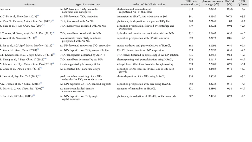
Table 1. Champion Q-Factors Obtained for the LSPR Peaks in One-Dimensional Tio2Nanostructures Decorated By Gold Nanoparticles
reference type of nanostructure method of Au NP decoration wavelength (nm)LSPR peak plasmon resonanceenergy (eV) FWHM(eV) Q-FactorLSPR
this work Au NP-decorated TiO2nanorods,
nanotubes and nanopores electrochemical anodization ofcosputtered Au−Ti thin films 533 2.3253 0.337 6.9
Y.-C. Pu et al., Nano Lett. (2013)33 Au NP-decorated TiO2nanowires immersion in HAuCl4and calcination at 300 541 2.2940 *0.72 ∼3.2
Y. Tian, T. Tatsuma, J. Am. Chem. Soc. (2005)34 TiO
2film loaded with Au NPs photocatalytic deposition In a porous TiO2film 560 2.2148 1.03 ∼2.2
Z. Bian et al., J. Am. Chem. Soc. (2014)35 TiO
2mesocrystals modified with Au NPs deposition-precipitation, followed by centrifuge and
evaporation 539 2.3022 0.92 ∼2.5
J. Thomas, M. Yoon, Appl. Cat. B: Env. (2012)36 TiO2nanofibers doped with Au NPs hydrothermal reaction and sonication with Au NPs 552 2.2447 0.56 ∼4.0
Y. Wen et al., Nanoscale (2013)37 anatase/rutile mixed TiO
2nanotubes
precipitated with Au NPs deposition-precipitation with HAuCl4and urea 559 2.2173 0.86 ∼2.6
Z. Jin et al., ACS Appl. Mater. Interfaces (2016)23 Au NP-decorated monolayer TiO2nanotubes anodic oxidation and photoreduction of HAuCl4 582 2.1292 0.80 ∼2.7
A. Zhu et al., Anal. Chem. (2009)38 Au NPs deposited on TiO
2nanoneedle film 12−15H immersion in Au NP suspension 539 2.2997 0.51 ∼4.5
S.T. Kochuveedu et al., J. Phys. Chem. C (2012)39 TiO
2nanospheres decorated by Au NPs TiO2beads dispersed in citrate-capped Au NP solution 535 2.3458 0.64 ∼3.7
Z. Zhang et al., J. Phys. Chem. C (2013)40 TiO2nanofibers decorated by Au NPs electrospinning with postcalcination using HAuCl4 574 2.1619 0.46 ∼4.7
A. Primo et al., Phys. Chem. Chem. Phys.(2011)41 titania supported gold nanoparticles sol−gel based thin films decorated by spin-coating 539 2.2988 0.75
∼3.1 Y. Chen et al., Dalton Trans. (2012)42 Au-decorated TiO2nanotube arrays deposition of Au seeds In HAuCl4and in situ seed
growth 504 2.4583 0.41 ∼6.0
S. Luo et al., Sep. Pur. Tech.(2011)43 gold nanotubes consisting of Au NPs embedded In TiO2nanotube arrays
electrodeposition of Au NPs using HAuCl4 516 2.4032 0.66 ∼3.6
A.G. Dosado et al., J. Catal.. (2015)44 Au NPs deposited on TiO
2nanorod supports deposition-precipitation with urea using HAuCl4 558 2.2233 0.46 ∼4.8
R. Ma et al., J. Am. Chem. Soc. (2004)45 Au nanocrystal-loaded titanate
nanotube suspension reduction of nanotubes in HAuCl4 521 2.3801 0.51 ∼4.7
L. Bu et al., RSC Adv. (2015)46 Au NPs deposited on TiO
2single
crystal nanorods photocatalytic reduction of HAuCl4by the nanorods 507 2.4455 0.93 ∼2.6
ACS Applied Materials & Interfaces Research Article DOI: 10.1021/acsami.6b13164 ACS Appl. Mater. Interfaces 2017, 9, 740 −749 741
broad or indistinct LSPR peaks were still observed in the optical spectra.22,23 Because the quality factor of the LSP resonance is inversely proportional to the peak-width, many of the reports on high-activity Au−TiO2plasmonic photocatalysts
that exhibit very broad resonances in their UV−vis spectra,24in actuality have very small local electric field enhancements. The use of Au-doped sols and RF cosputtering have also been used to form Au NPs completely embedded within TiO2thin films
typically for nonlinear optics applications,25,26but the effective surface area of these planar film architectures is rather low thus limiting applications in surface enhanced Raman scattering (SERS), photocatalysis and sensing.
We present here a method based on electrochemical anod-ization of cosputtered TiAu films that produces high surface area nanostructures simultaneously exhibiting strong and distinct LSPR peaks unobscured by the extinction of the TiO2nanoscaffolds. Depending on the fabrication conditions
gold nanoparticle-decorated titania nanorods, nanotubes, or nanopores were obtained. We collectively term these nano-structures Anodic PLasmonic Au−TiO2 Engineered
Nano-composites (A-PLATENs). A-PLATENs allowed us to achieve a Quality factor (Q) for the plasmonic resonance that is considerably higher than previously published reports as shown inTable 1. The A-PLATENs also exhibited a strong enhance-ment of the two-photon luminescence when excited by femtosecond laser pulses at 800 nm. As we demonstrate later in this report, A-PLATENs have unique features such as the fact that the gold NPs are partially embedded in the TiO2and
also the formation of select coherent heterointerfaces between
Au and TiO2. Our electrochemical method of synthesis does
not result in surfactant-coated Au NPs unlike purely chemical methods and shares many features with physical methods of preparation of Au NPs such as laser ablation,27which preclude the change in surface reactivity and undesirable modification of functional properties due to the presence of the surfactants. A-PLATENs are expected to extend the usage of TiO2
nano-tubes and nanorods in biosensing applications through enhanced fluorescence, refractive index sensitivity of the LSPR and higher conductivity in electrochemical sensors.28−32 A number of
unusual features were observed in the anodization process due to the high gold content of the cosputtered films, which could provide key insights into the anodic synthesis of metal oxide thin films and nanostructures from high concentration mix-tures of valve metals and nonvalve metals. These aspects are discussed in more detail in theResults and Discussionsection.
2. RESULTS AND DISCUSSION
There is one prior report of anodizing Ti−Au bimetallic com-posites. Schmuki and colleagues fabricated TiO2 nanotubes
decorated with 5 nm Au nanoparticles by the electrochemical anodization of Ti−Au alloy foils containing 0.02 at%, 0.2 at%, and 1 at% Au.47They reported high activity for photocatalytic H2production from ethanol solutions but did not report optical
spectra for the Au NP-decorated nanotubes perhaps because their substrates were opaque. Our report differs from the aforementioned paper in a number of wayswe use bimetallic films with much higher gold content (typically 4−5 atom % of Au), we form the Ti−Au films by sputtering, we use a multilayer Figure 1.(a, b) TEM cross-sectional image and schematic illustration of the thin film stack formed by sputtering alternate layers of Ti and Au−Ti on a glass substrate; (c) TEM image following anodization of the thin film stack showing bands of gold nanoparticles concentrated in regions corresponding to the Au−Ti layers; and (d) current transients recorded during the electrochemical anodization of the film stack at 40 V in an ethylene glycol-based electrolyte containing F−species. An electrolyte used for the very first time to perform anodization is “fresh” while if the
electrolyte has been previously used to perform anodization, and therefore contains (TiF6)2−ions from the anodization process, then the electrolyte
is “aged”.
ACS Applied Materials & Interfaces Research Article
DOI:10.1021/acsami.6b13164
ACS Appl. Mater. Interfaces 2017, 9, 740−749
film stack containing alternate layers of undoped- and Au-doped Ti instead of a homogeneously doped Au−Ti film (seeFigure 1b) and we collect optical spectra which are of paramount impor-tance in this report. Through a series of experiments, we deter-mined that homogeneously doped Au−Ti films were unsuitable because the less reactive gold inhibited field assisted oxidation and etching for mild anodization conditions (low anodization voltage and low fluoride content in electrolyte), and was leached away by the electrolyte for stronger anodization condi-tions. However, we found an alternating film stack (Figure 1a) to both retain gold and generate a nanostructured anodic oxide provided that the both the initial and terminal films in the stack were undoped Ti films.
A distinctive feature of the anodization of the alternating Ti/Ti−Au film stack is the observation of oscillations in the anodization current (Figure 1d) with the number of current maxima corresponding to the number of Au-doped Ti layers. This is in contrast to the regular anodization of Ti where a single peak in the current is typically observed. Two other differences are (i) the amplitude of the current maxima for the Ti/Ti−Au film stack is much higher than that observed for the anodization of Ti foils and films and (ii) the current minima are roughly constant or at least nondecreasing until the last maximum passes. When the anodic process begins, the topmost Ti layer is first subjected to anodization and the current drops due to the formation of an insulating TiO2barrier layer. At this
point, chemical- and field-assisted etching of the barrier layer reopen conduction paths in the barrier layer leading to the more conductive Ti−Au layer underneath and thus resulting in the current increasing. In the Ti−Au layer, Ti transforms to TiO2 while Au nanoparticles are formed in tight bands (see
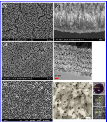
Figure 1c) with significant diffusion into the surrounding regions, particularly nearly the top of the film stack. Au is not anodized and consequently, as the field-assisted oxidation process for the second undoped Ti layer proceeds to comple-tion, the anodization current decreases again producing a peak (the first current maximum). Likewise, current maxima are produced corresponding to the anodic oxidation of each of the undoped Ti layers below the surface layer. Due to the diffusion of the gold into the surrounding regions, the conductivity of the anodized film stack, as a whole, is higher than that of a regular Figure 2.A-PLATEN morphologies obtained: (a) and (b) FESEM
top-view and cross-sectional image of Au NP-decorated TiO2
nanorod arrays formed by anodizing a thin film stack identical to that shown in Figure 1; (a, b) the anodization was performed potentiostatically at 40 V in an EG-based electrolyte containing 2% H2O and 0.3% NH4F (c) FESEM top-view and cross-sectional TEM
image of Au NP-decorated TiO2 nanorod arrays with a ∼200
nm-thick nanoporous capping layer formed by anodizing a thin film stack identical to that shown inFigure 1; the anodization was performed potentiostatically at 40 V in an EG-based electrolyte containing 1% H2O and 0.1% NH4F; the total Au content in the thin film stacks
used to prepare the samples shown in (a) through (d) was 4.1% (e) FESEM top-view of nanotube arrays formed by anodizing a thin film stack containing a smaller amount of Au in the Au-doped Ti layers. The total Au content in the deposited film was 1.4%; (f) HRTEM image of sample in (a) showing the lattice spacings of the TiO2host
and the Au NPs.
Figure 3.(a) Raman spectra of A-PLATEN formed by anodizing a thin film stack at 40 V in an EG based electrolyte containing 2% water. The black, red, and blue curves correspond to ammonium fluoride concentrations in the anodization electrolyte of 0.1%, 0.2%, and 0.3%, respectively, and the inset is a magnified view of the spectra region from 100 to 200 cm−1. (b) X-ray diffractogram of A-PLATEN formed by anodizing a thin film stack
identical to that shown inFigure 1; the inset shows a cross-sectional FESEM image of the sample showing the nanorod structures.
ACS Applied Materials & Interfaces Research Article
DOI:10.1021/acsami.6b13164
ACS Appl. Mater. Interfaces 2017, 9, 740−749
TiO2barrier layer and accounts for the plateauing of the
cur-rent minima inFigure 1d.Figure 2shows the morphologies of the Au−TiO2 nanocomposites obtained subsequent to
anod-ization of the multilayer film stacks shown in Figure 1b in EG-based electrolytes containing fluoride ions.
The Raman spectra of the films resulting from the anod-ization of the TiAu thin film stack following annealing are shown inFigure 3a. The anatase Egabsorption mode that
nor-mally occurs at 144 cm−1is shifted by 8 cm−1to higher energies
(see inset of Figure 3), consistent with a previous report for Au-doped mesoporous TiO2.48The higher degree of disorder
in the anatase crystal structure that produces the 8 cm−1
positive peak shift indicates that the Au NPs do not merely decorate the titania nanotubes but are partially embedded within them. Likewise, the B1gmode of anatase (397 cm−1)49is
blue-shifted to 402 cm−1in the A-PLATEN. The presence of a
peak at 612 cm−1inFigure 3may be indicative of the presence
of a small amount of rutile whose A1gmode typically occurs as a
major peak at 610 cm−1. A minor peak inFigure 3at 836 cm−1
is attributed to the B2g mode of rutile.49 These observations
find strong support in the X-ray diffractogram of the same type of sample in Figure 3b, wherein several of the peaks cor-responding to the anatase phases are slightly shifted from their regular positions, which we attribute to disorder and stress due to the gold doping.
It is known that anodization of Ti in EG-based electrolytes containing 1−2% HF results in TiO2 nanotube arrays with a
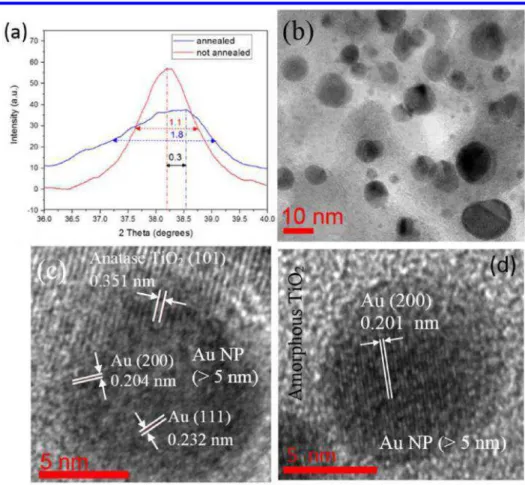
strong crystallographic texture oriented to the (004) plane of anatase subsequent to thermal annealing.50The X-ray diffracto-gram inFigure 3b indicates this to be the case even for the anodization of Ti−Au thin film stacks. Applying Scherrer’s equation51to the Au (111) XRD peaks in Figure 4a, a
post-annealing average Au NP size of 8.26 nm and a prepost-annealing average Au NP size of 13.51 nm were obtained. The TEM lattice spacing of 0.351 nm inFigure 4c,d is consistent with the (110) reflection of anatase TiO2.52Au NPs of two size regimes,
i.e., smaller and larger than 5 nm, are observed in the Au−TiO2
3D nanocomposites (Figure 4b−d and Figure 5). TEM lattice images of the larger Au nanoparticles (Figure 4c), indicate more than one facet, Au (111) and Au (200), in the annealed Au NPs. In contrast TEM lattice images of smaller Au NPs (Figure 5) show Figure 4.(a) XRD peak of the Au (111) in the Au−TiO2in pre- and postannealing states; (b) low magnification TEM image showing Au NPs in the
annealed A-PLATEN; and (c) and (d) HRTEM lattice fringe images of the Au NPs in annealed and unannealed A-PLATENs, respectively.
Figure 5. HRTEM image of Au−TiO2 interface illustrating the
coherent heterointerface between Au (111) and TiO2 (004) that
support the Au NPs in the TiO2lattice.
ACS Applied Materials & Interfaces Research Article
DOI:10.1021/acsami.6b13164
ACS Appl. Mater. Interfaces 2017, 9, 740−749
Au (111) as the only facet. The smaller size and positive shift in the Bragg angle of about 0.3° in the postannealed Au NPs (Figure 4a) imply a compressive stress in the postannealed Au NPs when compared with the same before annealing.
Figure S4 indicates that Au NPs are not present in the as-deposited Au−Ti thin film stack. Larger Au NPs in the anodized and annealed A-PLATENs show secondary fringes (observed inFigure 4c) due to various grain orientations. This is in contrast with the smaller Au NPs (less than 5 nm), where the grain orientations are more uniform with a single crystal plane. The observed variance in lattice orientations of larger Au NPs are likely due to their gradual displacement or rotation, which occurs as the Au NP accommodates to stresses in the TiO2matrix.53We found evidence of coherent Au−TiO2
het-eroepitaxial interfaces for a portion of the Au NPs, as evident from quasi-matching lattices (Figure 5) of the Au (111) and anatase TiO2(112) crystal planes. The angle between crystal
planes in the tetragonal Bravais lattice of anatase titania is given by ϕ = + + + + + +
(
) ( )
cos h h k k a l l c h k a l c h k a l c 1 2 1 2 2 1 2 2 12 12 2 12 2 22 22 2 22 2 (1)The HRTEM image in Figure S7 shows an angle of 51.9° between the Au NP’s (111) and anatase TiO2’s (100) crystal
planes which have d-spacings of 0.237 and 0.372 nm, respec-tively in the A-PLATENs. Since the (112) plane of anatase is also at an angle of 52° from the (100) anatase plane, the (112), and (111) crystal planes of gold and anatase, respectively, are most likely to be parallel and a planar heteroepitaxial interface is indicated. While coherent heterointerfaces between gold nanoparticles and anatase nanostructures remain uncommon, the heteroepitaxy of Au on TiO2has been reported in a handful
of reports.53−56
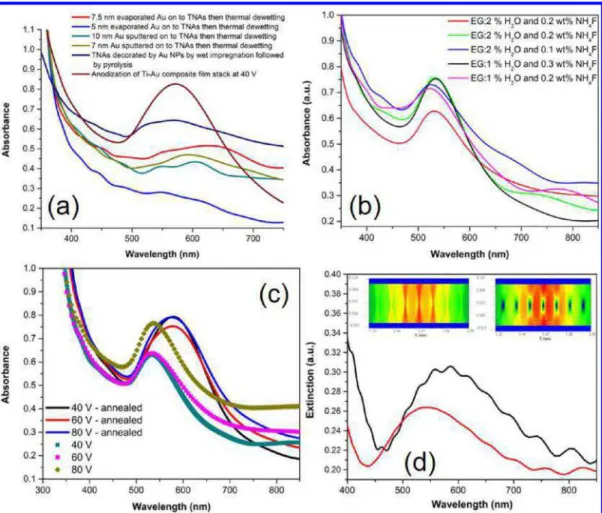
When we began studying Au−TiO2nanostructures formed
by the anodization of TiAu thin film stacks, it was evident early on that these samples exhibited unusually intense LSPR bands in comparison to nanotube and nanorod array thin films of similar thickness that were decorated by Au NPs by more con-ventional methods such as wet impregnation, photocatalytic reduction of Au salts, vacuum deposition followed by thermal dewetting, etc. This comparison is made graphically inFigure 6a. Subsequently, we tuned the parameters of the anodization process in order to maximize the quality factor of the LSPR peak. During this process, we found that the water content and fluoride content in the electrolyte were key factors that enabled control of the full-width at half-maximum (fwhm) of the LSPR Figure 6.(a) Comparison of the optical spectra of A-PLATENs formed by anodizing a thin film stack at 40 V using an unoptimized recipe with anodic TiO2nanotube array samples decorated with Au NPs using other methods such as wet impregnation and vacuum deposition followed by
dewetting; (b) A-PLATENs formed by using electrolyte recipes optimized to achieve sharper resonances; (c) effect of anodization voltage and postanodization thermal annealing on the LSPR peak of Au NP-decorated TiO2nanorod arrays; and (d) optical spectra of Au NP-decorated TiO2
nanotube arrays from FDTD simulations. The black curve corresponds to Au NPs wedged between adjacent nanotubes with no air gaps, while the red curve accounts for a finite air gap between the Au NPs and the nanotube wall, and the insets show the magnitude of the simulated Poynting vector off-resonance (left) and at resonance (right).
ACS Applied Materials & Interfaces Research Article
DOI:10.1021/acsami.6b13164
ACS Appl. Mater. Interfaces 2017, 9, 740−749
peak (Figure 6b) while the anodization potential did not exercise a significant influence on the peak width (Figure 6c). The sharpest resonances were achieved using EG-based elec-trolytes containing 1% H2O and 0.3 wt % NH4F, and 2% H2O
and 0.2 wt % NH4F.Figure 4c also shows that the induction of
crystallinity through a high temperature anneal does affect the optical spectra significantly, red-shifting the peak wavelength by ∼45 nm from 535 to 580 nm. HRTEM imaging indicated a smaller void fraction in the annealed samples. Since the surface plasmon resonance of gold nanoparticles is extremely sensi-tive to the dielectric constant of the local environment, we hypothesized that in the as-anodized state, the Au NPs were mostly surrounded by air due to a more hollow structure while in the annealed state, the voids collapsed and the Au NPs were partially surrounded by, and in contact with, TiO2whose
dielectric constant is much larger than that of air. FDTD simulations provided support for this hypothesis. Thus, in Figure 6d, we observe that a model with the Au NPs directly wedged betweeen adjacent TiO2 nanotubes has a more
red-shifted LSPR band peaking at ∼590 nm while a model where air gaps are present between the Au NPs and the TiO2
nano-tubes, resulted in the more commonly observed LSPR peak at ∼535 nm. The inset ofFigure 6d shows the magnitude of the Poynting vector which provides a measure of the electric field intensity in the plane of the vertically oriented nanostructures (parallel to the substrate). Close to the surface plasmon reso-nance (plane wave simulation at 600 nm, right inset inFigure 6d), the gold nanoparticles have a large extinction cross-section and
therefore the blue dots corresponding to the location of the Au nanoparticles decorating the TiO2 nanotubes manifest much
larger dimensions in the Poynting vector magnitude plot than the actual physical size of the Au NPs. However, off resonance (plane wave simulation at 660 nm, right inset inFigure 6d), the blue dots are not observable since the extinction cross-section bears more resemblance to the physical size of the Au NPs.
The quality factor (Q) of a resonator is given by the ratio of the resonance frequency to the line width, expressed as the full-width at half-maximum (fwhm) of the corresponding peak. It is highly desirable for sensing, photocatalysis, nonlinear optics, and other applications for an ensemble of nanoparticles exhib-iting LSPR to have a low fwhm and a high Q. For a single plasmonic nanoparticle, Q is directly proportional to the local electric field enhancement (L(ω)) while the fwhm is inversely proportional to the plasmon lifetime. For an ensemble of nanoparticles, the Q represents the lower threshold of L(ω) achievable in the structure.
The measurement setup for two-photon fluorescence studies of the A-PLATENs, and the resulting data obtained are shown in Figure 7. Undecorated anodically synthesized TiO2
nano-tubes exhibit a peak at ∼400 nm (orange curve inFigure 7b, which we attribute to second harmonic generation (SHG) in the high surface area porous architecture. The SHG signal at ∼400 nm of bare TiO2is not enhanced any more than expected
from estimates of the overall surface area. Essentially, there is no local field enhancement effect for the SHG signal because the SHG peak is significantly distant in energetic terms from Figure 7.(a) Experimental setup used to obtain the two-photon luminescence spectra, (b) two-photon luminescence spectra obtained from A-PLATENs, (c) two-photon fluorescence images obtained in a confocal microscope using 800 nm laser excitation contrasting the enhancement obtained with A-PLATENs with regular anodic titania nanotubes as well as anodic TiO2nanotubes decorated with Au NPs using conventional
techniques, under identical excitation and imaging conditions, and (d) the two-photon luminescence intensity plotted as a function of the fluence of the exciting laser for TiAu film stacks anodized at 40 V.
ACS Applied Materials & Interfaces Research Article
DOI:10.1021/acsami.6b13164
ACS Appl. Mater. Interfaces 2017, 9, 740−749
the LSPR. The photoluminescence quantum yield (ΦPL) in
smooth Au films is ∼10−10due to multiple nonradiative
trans-itions and increases to 10−5in 2−25 nm spherical Au NPs on
account of the plasmonic enhancement of radiative transi-tions.57The PL in smooth and roughened thin films of Au has been reported to have emission peaks in two bands: 410− 430 nm and 530−570 nm, which are commonly attributed to direct radiative recombination of sp-band electrons at the Fermi level with holes in the d band.58However, the PL in spherical Au NP suspensions was attributed to the radiative decay of particle plasmons emitted by d-band holes combining non-radiatively with sp band electrons.57For all the samples in this study, we observe two-photon luminescence peaks in the range 415−430 nm and 530−560 nm in addition to the SHG signal at 400 nm, consistent with prior reports but also observe a minor peak at 505 nm, whose origin is unclear to us (Figure S5). We also observe a shoulder at 350 nm for the A-PLATENs while the same feature is absent in the PL spectra of the TiO2
nano-tube arrays decorated by Au NPs using the wet impregnation +pyrolysis (i.e., themal) method and by using vacuum deposi-tion followed by thermal dewetting. The most intense PL is exhibited by the as-anodized TiAu thin film stacks (unannealed A-PLATENs) whose emission intensities are greater by a factor of 2.5−3.5 than the emission intensities of anodic TiO2
nano-tubes decorated by Au NPs using more conventional techniques. Annealing reduces the PL intensity of the A-PLATENs and also red-shifts the two major emission peaks from 415 to 430 nm and 535 to 560 nm, respectively. While the intensities of the multiphoton luminescence for smooth and roughened gold films are reported to increase monotonically with increasing emission wavelength (decreasing energy),58 the two-photon luminescence spectra of the gold nanoparticle decorated nanostructured TiO2 samples in this study decrease
signifi-cantly at longer wavelengths. This coupled with the previously mentioned observations of a similar red-shift upon annealing in both the optical spectra and the luminescence spectra are strongly suggestive of the participation of particle plasmons in the emission process, as opposed to the purely radiative recom-bination of d-band holes with sp-band electrons.57 However, systematic studies of the luminescence quantum yield as a func-tion of the particle size are needed to confirm this suggesfunc-tion, which are outside the scope of the present work.
The dependence of the integrated luminescence signal fol-lowing 800 nm laser excitation was measured as a function of laser fluence, and a quadratic dependence was found within the limits of experimental error, which implies the initiation of the excitation by two-photon absorption. However, the high-energy cutoff in the two-photon luminescence spectra does not occur close to 400 nm as would be expected from a process dom-inated purely by two-photon absroption but instead occurs at ∼300 nm, limited in this case by the near-zero transmission of the laser optic below 300 nm. The combination of the emission dependence on the square of the excitation intensity coupled with a cutoff energy approaching that of three photons suggest to us that a portion of the excited states rapidly populated (fs) by two-photon absorption absorb a third photon during the reminder of the pulse duration. We therefore expect two sequential processestwo photon absorption followed by single photon absorption by the excited state.
3. CONCLUSIONS
We present a novel synthetic methodology to generate high surface area 3D plasmonic nanocomposites containing gold
nanoparticles partially embedded in the walls of vertically oriented TiO2 nanorod, nanotube, and nanopore arrays. The
methodology consists of vacuum depositing a thin film stack consisting of alternating layers of Ti and high atom fraction Ti−Au thin films, and then anodizing the stack. The resulting 3D nanocomposite exhibits very prominent localized surface plasmon resonances with ensemble line widths as small as 0.33 eV and Q-factors as high as 6.9, and also achieves a strongly enhanced two-photon photuminescence in comparison to more typical plasmonic nanostructures consisting of TiO2nanorod or
nanotube arrays decorated with gold nanoparticles prepared through conventional vacuum- and solution-based synthetic routes. Coherent heterointerfaces between the (112) planes of the anatase phase of TiO2and the (111) planes of the partially
embedded gold nanoparticles were observed coupled with a more red-shifted plasmon resonance due to the higher per-mittivity of the TiO2host matrix. High surface area metal
oxide-noble metal 3D nanocomposites reported here constitute a new platform to exploit plasmonic phenomena in optical and SERS sensors, in substrates for MALDI-TOF mass spectrometry and in light harvesting devices such as solar cells and photocatalysts.
■
ASSOCIATED CONTENT*
S Supporting InformationThe Supporting Information is available free of charge on the ACS Publications websiteat DOI:10.1021/acsami.6b13164.
Materials and methods, supplementary transmission electron micrographs, and 2-PL spectra peak fits (PDF)
■
AUTHOR INFORMATION Corresponding Author *E-mail:kshankar@ualberta.ca(K.S.). ORCID Karthik Shankar:0000-0001-7347-3333 Author ContributionsS.F., H.S., and B.B.R. performed the nanomaterials growth, optical spectroscopy, and the morphological and structural studies. S.P.B. and A.M. performed the data collection and analysis for the two-photon photoluminescence spectroscopy. S.F. performed FDTD simulations. A.S. calculated Q-factors and FWHMs and compared them to prior work. B.D.W. and P.K. performed the structural analysis of the thin films and hybrid nanostructures. K.S. prepared and edited the paper. K.S. and R.F. provided guidance for the experiments.
Notes
The authors declare no competing financial interest.
■
ACKNOWLEDGMENTSThis project was funded through grants from the National Research Council of Canada (NRC) and NSERC. S.F., B.D.W., and A.M. acknowledge scholarship support from Alberta Innovates Technology Futures while B.B.R. held a Canadian Commonwealth Fellowship during the conduct of this research. Some device fabrication and testing used research infrastructure made possible by a Leaders Opportunity Fund grant to K.S. from the Canada Foundation for Innovation (CFI) and the Alberta Small Equipment Grants Program (SEGP). Equipment use and staff assistance at the UofA NanoFab is acknowledged along with the funding obtained for user fees from CMC Microsystems. A.M. thanks Dr. Xuejun Sun at the Cell Imaging
ACS Applied Materials & Interfaces Research Article
DOI:10.1021/acsami.6b13164
ACS Appl. Mater. Interfaces 2017, 9, 740−749
Facility, while S.F. and P.K. thank Kai Cui at the NINT electron microscopy facility.
■
REFERENCES(1) Kapilashrami, M.; Zhang, Y.; Liu, Y.-S.; Hagfeldt, A.; Guo, J. Probing the Optical Property and Electronic Structure of TiO2 Nanomaterials for Renewable Energy Applications. Chem. Rev. 2014,
114, 9662−9707.
(2) Chen, W.; Kuang, Q.; Wang, Q. X.; Xie, Z. X. Engineering a High Energy Surface of Anatase TiO2 Crystals Towards Enhanced Performance for Energy Conversion and Environmental Applications.
RSC Adv. 2015, 5, 20396−20409.
(3) De Angelis, F.; Di Valentin, C.; Fantacci, S.; Vittadini, A.; Selloni, A. Theoretical Studies on Anatase and Less Common TiO2 Phases: Bulk, Surfaces, and Nanomaterials. Chem. Rev. 2014, 114, 9708−9753. (4) Awazu, K.; Fujimaki, M.; Rockstuhl, C.; Tominaga, J.; Murakami, H.; Ohki, Y.; Yoshida, N.; Watanabe, T. A Plasmonic Photocatalyst Consisting of Silver Nanoparticles Embedded in Titanium Dioxide. J.
Am. Chem. Soc. 2008, 130, 1676−1680.
(5) Standridge, S. D.; Schatz, G. C.; Hupp, J. T. Distance Dependence of Plasmon-Enhanced Photocurrent in Dye-Sensitized Solar Cells. J. Am. Chem. Soc. 2009, 131, 8407−8409.
(6) Brown, M. D.; Suteewong, T.; Kumar, R. S. S.; D’Innocenzo, V.; Petrozza, A.; Lee, M. M.; Wiesner, U.; Snaith, H. J. Plasmonic Dye-Sensitized Solar Cells Using Core-Shell Metal-Insulator Nanoparticles.
Nano Lett. 2011, 11, 438−445.
(7) Liu, Z. W.; Hou, W. B.; Pavaskar, P.; Aykol, M.; Cronin, S. B. Plasmon Resonant Enhancement of Photocatalytic Water Splitting Under Visible Illumination. Nano Lett. 2011, 11, 1111−1116.
(8) Feuz, L.; Jonsson, M. P.; Hook, F. Material-Selective Surface Chemistry for Nanoplasmonic Sensors: Optimizing Sensitivity and Controlling Binding to Local Hot Spots. Nano Lett. 2012, 12, 873− 879.
(9) Da, P. M.; Li, W. J.; Lin, X.; Wang, Y. C.; Tang, J.; Zheng, G. F. Surface Plasmon Resonance Enhanced Real-Time Photoelectrochem-ical Protein Sensing by Gold Nanoparticle-Decorated TiO2 Nano-wires. Anal. Chem. 2014, 86, 6633−6639.
(10) Liu, K.; Bi, Y.; Qu, S. C.; Tan, F. R.; Chi, D.; Lu, S. D.; Li, Y. P.; Kou, Y. L.; Wang, Z. G. Efficient Hybrid Plasmonic Polymer Solar Cells with Ag Nanoparticle Decorated TiO2 Nanorods Embedded in the Active Layer. Nanoscale 2014, 6, 6180−6186.
(11) Farsinezhad, S.; Sharma, H.; Shankar, K. Interfacial band alignment for photocatalytic charge separation in TiO2 nanotube arrays coated with CuPt nanoparticles. Phys. Chem. Chem. Phys. 2015,
17, 29723−29733.
(12) Kar, P.; Farsinezhad, S.; Mahdi, N.; Zhang, Y.; Obuekwe, U.; Sharma, H.; Shen, J.; Semagina, N.; Shankar, K. Enhanced CH4 yield by photocatalytic CO2 reduction using TiO2 nanotube arrays grafted with Au, Ru, and ZnPd nanoparticles. Nano Res. 2016, 9, 3478−3493. (13) Alessandri, I.; Vassalini, I.; Bertuzzi, M.; Bontempi, N.; Memo, M.; Gianoncelli, A. “RaMassays”: Synergistic Enhancement of
Plasmon-Free Raman Scattering and Mass Spectrometry for Multimodal Analysis of Small Molecules. Scientif ic Reports 2016, 6, Art. No. 34521.
(14) Li, X.; Tan, J.; Yu, J.; Feng, J.; Pan, A.; Zheng, S.; Wu, J. Use of a Porous Silicon−Gold Plasmonic Nanostructure to Enhance Serum Peptide Signals in MALDI-TOF Analysis. Anal. Chim. Acta 2014, 849, 27−35.
(15) Nitta, S.; Yamamoto, A.; Kurita, M.; Arakawa, R.; Kawasaki, H. Gold-Decorated Titania Nanotube Arrays as Dual-Functional Platform for Surface-Enhanced Raman Spectroscopy and Surface-Assisted Laser Desorption/Ionization Mass Spectrometry. ACS Appl. Mater. Interfaces 2014, 6, 8387−8395.
(16) Yu, J. G.; Dai, G. P.; Huang, B. B. Fabrication and Characterization of Visible-Light-Driven Plasmonic Photocatalyst Ag/AgCl/TiO2 TiO2 Nanotube Arrays. J. Phys. Chem. C 2009, 113, 16394−16401.
(17) Elmoula, M. A.; Panaitescu, E.; Phan, M.; Yin, D.; Richter, C.; Lewis, L. H.; Menon, L. Controlled Attachment of Gold Nanoparticles
on Ordered Titania Nanotube Arrays. J. Mater. Chem. 2009, 19, 4483− 4487.
(18) Ampelli, C.; Genovese, C.; Lanzafame, P.; Perathoner, S.; Centi, G. A Sustainable Production of H-2 by Water Splitting and Photo-Reforming of Organic Wastes on Au/TiO2Nanotube Arrays. In Pres
2014, 17th Conference on Process Integration, Modelling and Optimisation for Energy Saving and Pollution Reduction, Pts 1−3; Varbanov, P. S.,
Klemes, J. J., Liew, P. Y., Yong, J. Y., Stehlik, P., Eds.; 2014; Vol. 39, pp 1627−1632.
(19) Zhu, B. L.; Sui, Z. M.; Wang, S. R.; Chen, X.; Zhang, S. M.; Wu, S. H.; Huang, W. P. Alternative Approaches to Fabrication of Gold-Modified TiO2 Nanotubes. Mater. Res. Bull. 2006, 41, 1097−1104.
(20) Sudhagar, P.; Song, T.; Devadoss, A.; Lee, J. W.; Haro, M.; Terashima, C.; Lysak, V. V.; Bisquert, J.; Fujishima, A.; Gimenez, S.; Paik, U. Modulating the Interaction Between Gold and TiO2 Nanowires for Enhanced Solar Driven Photoelectrocatalytic Hydrogen Generation. Phys. Chem. Chem. Phys. 2015, 17, 19371−19378.
(21) Xu, Z.; Lin, Y.; Yin, M.; Zhang, H.; Cheng, C.; Lu, L.; Xue, X.; Fan, H. J.; Chen, X.; Li, D. Understanding the Enhancement Mechanisms of Surface Plasmon-Mediated Photoelectrochemical Electrodes: A Case Study on Au Nanoparticle Decorated TiO2
Nanotubes. Adv. Mater. Interfaces 2015, 2, Art. No. 1500169.10.1002/admi.201570062
(22) Zhang, Z.; Zhang, L.; Hedhili, M. N.; Zhang, H.; Wang, P. Plasmonic Gold Nanocrystals Coupled with Photonic Crystal Seamlessly on TiO2 Nanotube Photoelectrodes for Efficient Visible Light Photoelectrochemical Water Splitting. Nano Lett. 2013, 13, 14− 20.
(23) Jin, Z.; Wang, Q.; Zheng, W.; Cui, X. Highly Ordered Periodic Au/TiO2 Hetero-Nanostructures for Plasmon-Induced Enhancement of the Activity and Stability for Ethanol Electro-Oxidation. ACS Appl.
Mater. Interfaces 2016, 8, 5273−5279.
(24) Wang, H.; You, T.; Shi, W.; Li, J.; Guo, L. Au/TiO2/Au as a Plasmonic Coupling Photocatalyst. J. Phys. Chem. C 2012, 116, 6490− 6494.
(25) Li, T.; Moon, J.; Morrone, A. A.; Mecholsky, J. J.; Talham, D. R.; Adair, J. H. Preparation of Ag/SiO2 Nanosize Composites by a Reverse Micelle and Sol−Gel Technique. Langmuir 1999, 15, 4328− 4334.
(26) Garcia, H.; Kalyanaraman, R.; Sureshkumar, R. Nonlinear Optical Properties of Multi-Metal Nanocomposites in a Glass Matrix. J.
Phys. B: At., Mol. Opt. Phys. 2009, 42 10.1088/0953-4075/42/17/
175401.
(27) Intartaglia, R.; Rodio, M.; Abdellatif, M.; Prato, M.; Salerno, M. Extensive Characterization of Oxide-Coated Colloidal Gold Nano-particles Synthesized by Laser Ablation in Liquid. Materials 2016, 9 10.3390/ma9090775.
(28) Mun, K.-S.; Alvarez, S. D.; Choi, W.-Y.; Sailor, M. J. A Stable, Label-free Optical Interferometric Biosensor Based on TiO2 Nano-tube Arrays. ACS Nano 2010, 4, 2070−2076.
(29) Liu, S.; Chen, A. Coadsorption of Horseradish Peroxidase with Thionine on TiO2 Nanotubes for Biosensing. Langmuir 2005, 21, 8409−8413.
(30) An, Y.; Tang, L.; Jiang, X.; Chen, H.; Yang, M.; Jin, L.; Zhang, S.; Wang, C.; Zhang, W. A Photoelectrochemical Immunosensor Based on Au-Doped TiO2 Nanotube Arrays for the Detection of α-Synuclein. Chem. - Eur. J. 2010, 16, 14439−14446.
(31) Kar, P.; Pandey, A.; Greer, J. J.; Shankar, K. Ultrahigh Sensitivity Assays for Human Cardiac Troponin I Using TiO2 Nanotube Arrays.
Lab Chip 2012, 12, 821−828.
(32) Farsinezhad, S.; Mohammadpour, A.; Dalrymple, A. N.; Geisinger, J.; Kar, P.; Brett, M. J.; Shankar, K. Transparent Anodic TiO2Nanotube Arrays on Plastic Substrates for Disposable Biosensors
and Flexible Electronics. J. Nanosci. Nanotechnol. 2013, 13, 2885− 2891.
(33) Pu, Y. C.; Wang, G.; Chang, K. D.; Ling, Y.; Lin, Y. K.; Fitzmorris, B. C.; Liu, C. M.; Lu, X.; Tong, Y.; Zhang, J. Z.; Hsu, Y. J.; Li, Y. Au Nanostructure-Decorated TiO2 Nanowires Exhibiting
ACS Applied Materials & Interfaces Research Article
DOI:10.1021/acsami.6b13164
ACS Appl. Mater. Interfaces 2017, 9, 740−749
Photoactivity Across Entire UV-Visible Region for Photoelectrochem-ical Water Splitting. Nano Lett. 2013, 13, 3817−3823.
(34) Tian, Y.; Tatsuma, T. Mechanisms and Applications of Plasmon-Induced Charge Separation at TiO2 Films Loaded with Gold Nanoparticles. J. Am. Chem. Soc. 2005, 127, 7632−7637.
(35) Bian, Z.; Tachikawa, T.; Zhang, P.; Fujitsuka, M.; Majima, T. Au/TiO2 Superstructure-Based Plasmonic Photocatalysts Exhibiting Efficient Charge Separation and Unprecedented Activity. J. Am. Chem.
Soc. 2014, 136, 458−465.
(36) Thomas, J.; Yoon, M. Facile Synthesis of Pure TiO2(B) Nanofibers Doped with Gold Nanoparticles and Solar Photocatalytic Activities. Appl. Catal., B 2012, 111−112, 502−508.
(37) Wen, Y.; Liu, B.; Zeng, W.; Wang, Y. Plasmonic Photocatalysis Properties of Au Nanoparticles Precipitated Anatase/Rutile Mixed TiO2 Nanotubes. Nanoscale 2013, 5, 9739−9746.
(38) Zhu, A.; Luo, Y.; Tian, Y. Plasmon-Induced Enhancement in Analytical Performance Based on Gold Nanoparticles Deposited on TiO2 Film. Anal. Chem. 2009, 81, 7243−7247.
(39) Kochuveedu, S. T.; Kim, D.-P.; Kim, D.-H. Surface-Plasmon-Induced Visible Light Photocatalytic Activity of TiO2 Nanospheres Decorated by Au Nanoparticles with Controlled Configuration. J. Phys.
Chem. C 2012, 116, 2500−2506.
(40) Zhang, Z.; Wang, Z.; Cao, S. W.; Xue, C. Au/Pt Nanoparticle-Decorated TiO2 Nanofibers with Plasmon-Enhanced Photocatalytic Activities for Solar-To-Fuel Conversion. J. Phys. Chem. C 2013, 117, 25939−25947.
(41) Primo, A.; Corma, A.; García, H. Titania Supported Gold Nanoparticles as Photocatalyst. Phys. Chem. Chem. Phys. 2011, 13, 886−910.
(42) Chen, Y.; Tian, G.; Pan, K.; Tian, C.; Zhou, J.; Zhou, W.; Ren, Z.; Fu, H. In Situ Controlled Growth of Well-Dispersed Gold Nanoparticles in TiO2 Nanotube Arrays as Recyclable Substrates for Surface-Enhanced Raman Scattering. Dalton Trans. 2012, 41, 1020− 1026.
(43) Luo, S.; Xiao, Y.; Yang, L.; Liu, C.; Su, F.; Li, Y.; Cai, Q.; Zeng, G. Simultaneous Detoxification of Hexavalent Chromium and Acid Orange 7 by a Novel Au/TiO2 Heterojunction Composite Nanotube Arrays. Sep. Purif. Technol. 2011, 79, 85−91.
(44) Dosado, A. G.; Chen, W. T.; Chan, A.; Sun-Waterhouse, D.; Waterhouse, G. I. N. Novel Au/TiO2 photocatalysts for hydrogen production in alcohol-water mixtures based on hydrogen titanate nanotube precursors. J. Catal. 2015, 330, 238−254.
(45) Ma, R.; Sasaki, T.; Bando, Y. Layer-By-Layer Assembled Multilayer Films of Titanate Nanotubes, Ag- or Au-Loaded Nano-tubes, and Nanotubes/Nanosheets with Polycations. J. Am. Chem. Soc. 2004, 126, 10382−10388.
(46) Bu, L.; Yang, W.; Ming, H. Low Temperature Synthesis of Rutile TiO2 Single Crystal Nanorods with Exposed (002) Facets and Their Decoration with Gold Nanoparticles for Photocatalytic Applications. RSC Adv. 2015, 5, 45122−45128.
(47) Lee, K.; Hahn, R.; Altomare, M.; Selli, E.; Schmuki, P. Intrinsic Au Decoration of Growing TiO2 Nanotubes and Formation of a High-Efficiency Photocatalyst for H2 Production. Adv. Mater. 2013, 25, 6133−6137.
(48) Li, H.; Bian, Z.; Zhu, J.; Huo, Y.; Li, H.; Lu, Y. Mesoporous Au/ TiO2 Nanocomposites with Enhanced Photocatalytic Activity. J. Am.
Chem. Soc. 2007, 129, 4538−4539.
(49) Tompsett, G. A.; Bowmaker, G. A.; Cooney, R. P.; Metson, J. B.; Rodgers, K. A.; Seakins, J. M. The Raman Spectrum of Brookite, TiO2 (Pbca, Z = 8). J. Raman Spectrosc. 1995, 26, 57−62.
(50) Lee, S.; Park, I. J.; Kim, D. H.; Seong, W. M.; Kim, D. W.; Han, G. S.; Kim, J. Y.; Jung, H. S.; Hong, K. S. Crystallographically Preferred Oriented TiO2 Nanotube Arrays for Efficient Photovoltaic Energy Conversion. Energy Environ. Sci. 2012, 5, 7989−7995.
(51) Langford, J. I.; Wilson, A. Scherrer After Sixty Years: A Survey and Some New Results in the Determination of Crystallite Size. J.
Appl. Crystallogr. 1978, 11, 102−113.
(52) Powder Diffraction File Card No. 21−1272 JCPDS-International
Centre for Dif f raction Data, Swarthmore 1997.
(53) Shibata, N.; Goto, A.; Matsunaga, K.; Mizoguchi, T.; Findlay, S.; Yamamoto, T.; Ikuhara, Y. Interface Structures of Gold Nanoparticles on TiO2 (110). Phys. Rev. Lett. 2009, 102
10.1103/PhysRev-Lett.102.136105.
(54) Giorgio, S.; Henry, C. R.; Pauwels, B.; Van Tendeloo, G. Au Particles Supported on (110) Anatase-TiO2. Mater. Sci. Eng., A 2001,
297, 197−202.
(55) Akita, T.; Tanaka, K.; Tsubota, S.; Haruta, M. Analytical High− Resolution TEM Study of Supported Gold Catalysts: Orientation Relationship between Au Particles and TiO2 Supports. J. Electron
Microsc. 2000, 49, 657−662.
(56) Nagamatsu, D.; Nemoto, T.; Kurata, H.; Jiu, J.; Adachi, M.; Isoda, S. Interface Structure of Gold Particles on TiO2 Anatase. Mater.
Trans. 2011, 52, 280−284.
(57) Dulkeith, E.; Niedereichholz, T.; Klar, T.; Feldmann, J.; Von Plessen, G.; Gittins, D.; Mayya, K.; Caruso, F. Plasmon Emission in Photoexcited Gold Nanoparticles. Phys. Rev. B: Condens. Matter Mater.
Phys. 2004, 70, 205424.
(58) Boyd, G.; Yu, Z.; Shen, Y. Photoinduced Luminescence from the Noble Metals and Its Enhancement on Roughened Surfaces. Phys.
Rev. B: Condens. Matter Mater. Phys. 1986,
3310.1103/Phys-RevB.33.7923.
ACS Applied Materials & Interfaces Research Article
DOI:10.1021/acsami.6b13164
ACS Appl. Mater. Interfaces 2017, 9, 740−749