Impairment of both languages
in late bilinguals with dementia
of the Alzheimer type
∗
M É L A N I E M A N C H O N
Neurology Unit, Department of Medicine, Faculty of Sciences, University of Fribourg, Fribourg, Switzerland
K A R I N B U E T L E R
Neurology Unit, Department of Medicine, Faculty of Sciences, University of Fribourg, Fribourg, Switzerland
F R A N Ç O I S E C O L O M B O
Neuropsychology Unit, Hôpitaux Fribourgeois, Fribourg, Switzerland
L U C A S S P I E R E R
Neurology Unit, Department of Medicine, Faculty of Sciences, University of Fribourg, Fribourg, Switzerland
F R É D É R I C A S S A L
Department of Clinical Neurosciences, Geneva University and Hospital, Geneva, Switzerland
J E A N - M A R I E A N N O N I
Neurology Unit, Department of Medicine, Faculty of Sciences, University of Fribourg, Fribourg, Switzerland
(Received: May 6, 2013; final revision received: April 4, 2014; accepted: April 7, 2014)
Neuropsychological theories raise the question if in late bilinguals with dementia of the Alzheimer type (DAT),
the second language (L2) may be more impaired than the first (L1). We compared language performance in different tasks of oral comprehension (semantic and syntactic) and production (naming, repetition and fluency) in L1 and L2 in a group of 13 late proficient bilinguals wit DAT immersion, and a matched control group of 12 healthy late bilinguals. Two-way mixed repeated-measure ANOVAs with factors Language and Group revealed main effects of Group (p< .05) indicating that DAT affects all aspects of language. There was no Group× Language interaction, suggesting that DAT affects both languages similarly. Our study thus shows that neurodegenerative diseases affect L1 and L2 in a parallel manner, particularly at the levels of semantic, lexical and syntactic processing. These results speak in favour of a shared L1 and L2 network in late bilinguals.
Keywords: Alzheimer, late bilinguals, language breakdown
Introduction
In bilingual individuals, the first (L1) and the second (L2) language have been shown by behavioral and neurophysiological data to share partly overlapping brain representations (Chee, Tan & Thiel,1999; Illes, Francis, Desmond, Gabrieli, Glover, Poldrack, Lee & Wagner,
1999; Kim, Relkin, Lee & Hirsch,1997; Perani & Cappa,
1998), with the degree of overlap of each language depending on the age of acquisition (Kim et al.,1997), immersion and proficiency (Abutalebi, 2008; Perani & Abutalebi, 2005). Neuroimaging studies of language organization in bilinguals suggest that the earlier the age of acquisition and the higher the proficiency in the ∗ This work was supported by grant #325130_138497 from the Swiss
National Science Foundation to J.-M. Annoni. We thank Sylvie Blatter and Mme Risse (Famille au Jardin Foundation) for their help in collecting and analyzing the data. We also wish to thank the anonymous reviewers for their helpful comments on earlier drafts of this paper.
Address for correspondence:
Mélanie Manchon, Laboratory for Cognitive and Neurological Sciences, Chemin du Musée, 1700 Fribourg, Switzerland
second language, the larger will be the overlap of the brain representations of L1 and L2 in various aspects of comprehension and production (Chee, Hon, Lee & Soon,
2001; Hernandez, Martinez & Kohnert, 2000; Klein, Milner, Zatorre, Meyer & Evans,1995; Klein, Zatorre, Milner, Meyer & Evans,1994; Perani, Paulesu, Galles, Dupoux, Dehaene, Bettinardi, Cappa, Fazio & Mehler,
1998). The brain regions supporting both L1 and L2 include the left and, occasionally, the bilateral frontal and temporal areas (Abutalebi & Green,2007; Fabbro,2001b). This overlap of L1 and L2 representations is supported by studies of post-stroke aphasia in bilinguals. Patients often display parallel modes of impairment and recovery in their first and second language (Fabbro,
2001a; Tschirren, Laganaro, Michel, Martory, Di Pietro, Abutalebi & Annoni,2011). However, some biographical factors may affect the level of overlap and functioning of L1 and L2. For example, L2 semantic processing can be affected by the level of proficiency in L2, and syntactic processing seems to depend on the age of second-language
acquisition (Wartenburger, Heekeren, Abutalebi, Cappa, Villringer & Perani,2003; Weber-Fox & Neville,1996). These findings support the idea that, while grammatical processing of a late second language relies on explicit processing, the processing of the first language’s grammar relies on implicit processing (Ullman,2001).
Studying the elderly and populations with neurodegen-erative disease may also allow the testing of predictions on these language models. Normal aging seems to affect both languages equally (Juncos-Rabadan & Inglesias,
1994). In DAT, different patterns of language impairment have been found, however, the dominant trend suggests that neurodegenerative diseases impact more on the non-dominant language. According to the above-mentioned neuropsychological theories, it has been proposed that cortical dementia, such as dementia of the Alzheimer type or progressive aphasia, would more importantly impair explicit semantic and syntactic L2 processing (Paradis,
2008), while implicit L1 processing predominantly in-volving the basal ganglia would be spared (Zanini, Angeli & Tavano, 2011). Further evidence for different neural networks for L1 and L2 processing come from studies showing that late bilinguals do not achieve native-like competence in L2 syntax (Long,1990). Studies of epilep-tic patients showing that brain stimulation can impair specifically one language also suggest that some cortical areas may be more bound to one than the other language (e.g. Giussani, Roux, Lubrano, Gaini & Bello,2007).
However, more recent studies better controlled for biography of bilingualism have failed to show such differential impairment of L1 and L2 (Costa, Calabria, Marne, Hernández, Juncadella, Gascón-Bayarri, Lleó, Ortiz-Gil, Ugas, Blesa & Reñé, 2012). Costa and colleagues suggest that the “neural substrates of the lexico-semantic representations are largely shared (at least at the macroscopic level) between the two languages” (ibid., p.749). Support for a shared bilingual neural substrate also comes from stroke studies (Tschirren et al.,
2011), as well as experimental priming studies observing shared lexicons (Dijkstra,2005) and syntax (Hartsuiker, Pickering & Veltkamp, 2004; Kantola & van Gompel,
2011).
DAT is a neurodegenerative disease evolving in dementia syndromes and is characterized by a progressive decline of episodic and working memory, followed by language deficits (Faber-Langendoen, Morris, Knesevich, LaBarge, Miller & Berg,1988). Language deficits in DAT have been the focus of numerous studies, but how DAT affects both languages of bilinguals, and in particular late bilinguals, has so far not been investigated systematically. In monolinguals, one of the first symptoms of linguistic decline related to DAT are word-retrieval difficulties (Cardebat, Demonet, Puel, Agniel, Viallard & Celsis,
1998; Gomez & White, 2006). Later, DAT patients typically exhibit deficits at the level of oral production,
semantic dissociations (Ulatowska, Allard, Donnell, Bristow, Haynes, Flower & North, 1988) and both quantitative and qualitative discourse impoverishment (Heller, Dobbs & Rule,1992). As the disease progresses, DAT affects written production and comprehension (Cardebat et al.,1998).
However, language impairments in monolinguals with DAT remain heterogeneous, and their temporal dynamics show a high degree of variability. Language deficits may be manifest at the onset of DAT, or appear later as the disease develops (Cummings, Benson, Hill & Read,
1985). Neuropathologically, DAT manifests at the levels of several language-related brain areas. In the initial stage of the disease, neurofibrillar degeneration occurs within all polymodal cortical regions, including the superior temporal cortex, the frontal pole and the parietal cortex. Typically, the degeneration then further extends to the subcortical nuclei (Delacourte,2000).
Collectively, previous evidence concerning the organization of language in bilinguals and brain lesion patterns in DAT patients suggests that global L1 and L2 deficits should manifest in late, proficient bilingual DAT patients. To our knowledge, whether language deficits in bilinguals with DAT will manifest similarly in each language remains unknown, as this has rarely been tested systematically. Indeed, previous studies of language in bilingual patients with DAT focused on the analysis of a single language function or theme (e.g. Fama, Sullivan, Shear, Stein, Yesavage, Tinklenberg & Pfefferbaum, 2000). For instance, Salvatierra, Rosselli, Acevedo and Duara (2007) investigated semantic and phonemic fluency in bilinguals with DAT and showed that while healthy participants produced more words in the semantic than the phonemic condition, the DAT patients’ performances were the same in both tasks, in both languages. The authors interpreted this finding in terms of a general deficit in semantic processing which only indirectly impairs semantic fluency.
Further, some case studies on the impact of bilingualism on language functions in dementia patients focused on discursive and conversation analyses. Hyltenstam and Stroud (1989) showed that bilinguals with DAT have difficulty in selecting the appropriate language during daily conversations. More recent studies, which have focused more specifically on L1 and L2 impairment in DAT, suggest a parallel progressive impairment of L1 and L2 for early bilinguals (Gómez-Ruiz, Aguilar-Alonso & Espasa, 2012), and for naming capacities in more distant languages, such as English and Spanish (Gollan, Salmon, Montoya & da Pena, 2010). However, this last study considered L2 as the less proficient language and did not look at the age of acquisition.
In the current study, we used a mixed design to compare language production performance in the first and second languages of bilinguals with DAT to healthy
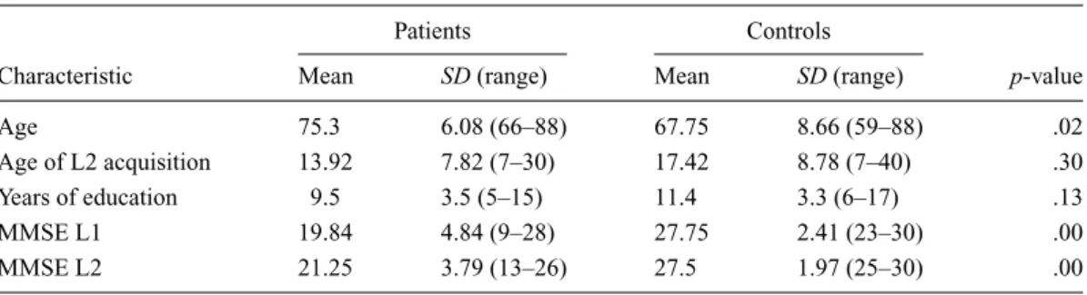
Table 1. Demographic characteristics of the Patient Group (n= 13) and the Control Group (n = 12).
Patients Controls
Characteristic Mean SD (range) Mean SD (range) p-value
Age 75.3 6.08 (66–88) 67.75 8.66 (59–88) .02
Age of L2 acquisition 13.92 7.82 (7–30) 17.42 8.78 (7–40) .30
Years of education 9.5 3.5 (5–15) 11.4 3.3 (6–17) .13
MMSE L1 19.84 4.84 (9–28) 27.75 2.41 (23–30) .00
MMSE L2 21.25 3.79 (13–26) 27.5 1.97 (25–30) .00
SD: standard deviation; MMSE: Mini-Mental State Evaluation; L1: first language; L2: second language. p-value: uncorrected independent
sample t-tests, p-value refers to difference between controls and patients.
participants matched by age and bilingualism at the level of proficiency, age of acquisition and immersion. We focused on several tasks including semantic, lexical and grammatical aspects, as well as automatisms and transcoding.
Materials and methods
The study was approved by the local Ethics Committee of Fribourg and Geneva, Switzerland, and all participants provided written informed consent in accordance with the Declaration of Helsinki.
Participants
Inclusion criteria for patients were: (i) a diagnosis of DAT (probable or possible Alzheimer’s disease) according to good clinical practice; (ii) a Mini-Mental State Examination (MMSE; Gollan et al.,2010)ࣘ 25 and ࣙ 10; and (iii) a Clinical Dementia Rating (CDR) of 1 or 2 (Morris,1993).
Exclusion criteria were: (i) unaided sensory disorders; (ii) history of stroke, vascular dementia, Parkinson’s disease, psychiatric disorders, major illness (e.g. cancer) and any other neurological disorders; (iii) a score> 7 on the Hachinski scale (Hachinski, Oveisgharan, Romney & Shankle, 2012); (iv) extrapyramidal rigidity of the upper limbs with a score > 2, based on item 22 of the Unified Parkinson’s Disease Rating Scale (UPDRS) motor score (Movement Disorder Society Task Force on Rating Scales for PD, 2003); and (v) widespread deep white-matter hyper-intense lesions and/or extensive, irregular periventricular hyper-intense lesions, and/or micro-haemorrhage.
The inclusion criterion for the control subjects was an MMSE > 25. Exclusion criteria were a history of neurological, medical or psychiatric disorders and abnormal neurological examinations. The control population was matched to the patients by age, education level and age of acquisition of L2 (seeTable 1).
For both groups, bilingualism-related inclusion criteria were: (i) French as a second language (L2) with late acquisition (> 7 years); (ii) immersion in L2 French > 20 years; (iii) mother tongue (L1) German, Spanish or Italian. The total score for L2 proficiency (speech comprehension, writing and reading) was 75.9± 4.6% for the patients and 75.4± 3.6% for the controls (t = 0.09; p = .92). Moreover, a previous medical history of language/speech difficulties (e.g. developmental dyslexia) was an exclusion criterion for both groups.
Patients
The patients were recruited from the neurology departments at the hospitals of Fribourg, Lausanne and Geneva and underwent a complete neuropsychological assessment. Out of 20 screened bilingual patients, 13 right-handed bilingual patients with dementia were included in the study (seven females). All the patients had French as L2. Three patients had Italian, two had Spanish and eight had German as L1. Their dementia was diagnosed by an experienced neurologist, trained on the basis of clinical criteria for dementia as defined by CDR > 0.5 (Morris, Ernesto, Schafer, Coats, Leon & Sano,1997), the diagnosis of possible or probable DAT according to NINCDS-ADRDA (McKhann, Drachman, Folstein, Katzman, Price & Stadlan,1984) and the Swiss Consensus for Dementia Diagnosis (Monsch, Hermelink, Kressig, Fish, Grob, Hiltbrunner, Martensson, Rüegger-Frey & von Gunten,2008). Eleven patients had probable DAT and two possible DAT. See Table 1 for detailed demographic information.
Controls
Twelve right-handed healthy participants (five females) were included as a control group. All participants had French as L2. Three had Italian, two had Spanish and seven had German as L1 (Table 1).
Procedure
The tests were conducted in two sessions with an interval of one to ten days between each session. One session was
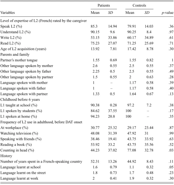
Table 2. Comparison between Patients and Controls on variables influencing bilingualism.
Patients Controls
Variables Mean SD Mean SD p-value
Level of expertise of L2 (French) rated by the caregiver
Speak L2 (%) 85.3 14.94 79.91 14.03 .36
Understand L2 (%) 90.15 9.6 90.25 8.4 .97
Write L2 (%) 53.15 33.86 60.17 34.89 .61
Read L2 (%) 75.23 27.07 71.25 25.69 .71
Age of L2 acquisition (years) 13.92 7.81 17.42 8.78 .30
Parents and family
Partner’s mother tongue 1.55 0.69 1.55 0.82 1
Other language spoken by mother 2.6 0.55 2.5 0.55 .37
Other language spoken by father 2.25 0.5 2.5 0.55 .49
Other language spoken by partner 1.5 0.55 2 0.63 .28
Language spoken with mother 1 – 1.17 0.58 .39
Language spoken with father 1 – 1.17 0.58 .40
Language spoken with partner 1.33 0.5 1.64 0.67 .13
Childhood before 6 years
L1 taught at school (%) 90.38 0.28 97.2 7.2 .38
L1 spoken by students (%) 84.62 37.55 100 – .17
L1 spoken at home (%) 94.23 20.8 100 – .35
Frequency of L2 use in adulthood, before DAT onset
At workplace (%) 30.77 25.32 29.17 23.44 .87
Watching television (%) 48.08 31.39 47.92 31 .99
Speaking with friends (%) 38.46 19.41 43.75 33.92 .63
Reading a book (%) 53.92 33.2 43.75 35.56 .52
Counting in head (%) 44.23 37.02 77.08 32.78 .03
History
Number of years spent in a French-speaking country 52.31 13.26 44.92 8.43 .11
Language learnt at school 1.6 0.79 1.1 0.32 .05
Language learnt on the street 1.8 0.73 1.7 0.48 .23
Language learnt at work 2 0.41 1.9 0.32 .30
SD: standard deviation; L2: second language; Partner’s mother tongue: 1= German, 2 = French, 3 = other; p-value: uncorrected independent
sample t-tests, p-value refers to difference between controls and patients.
in French (L2) and the other in the participant’s native language (L1: Italian, Spanish, or German). The order of the L1 and L2 sessions and of the tests within each session was counterbalanced across participants.
Materials
Evaluation of bilingualism
Proficiency and immersion in L2 were assessed using internally-developed questionnaires consisting of a visual analogue scale, which were scored by the examiner based on the patient’s answers and validated by the family. The questionnaires evaluated L2 proficiency at the level of oral and written expression, oral comprehension and reading in L2 French, levels of exposure to and use of L1 and
L2 as a percentage frequency of the languages used in childhood and languages currently used in learning and spoken within the family (Tschirren et al.,2011).
The patient group matched the control group in all the criteria evaluating the level of bilingualism.Table 2
presents the mean values, standard deviations and ranges for these variables for each population and the statistical assessment of the degree of matching between the two groups.
Evaluation of oral language
Comprehension and expression of oral language were assessed in both L1 and L2 using the following tests:
From the Boston Diagnostic Aphasia Evaluation (BDAE; Mazeaux, Orgogozo, Goodglass & Kaplan,1983;
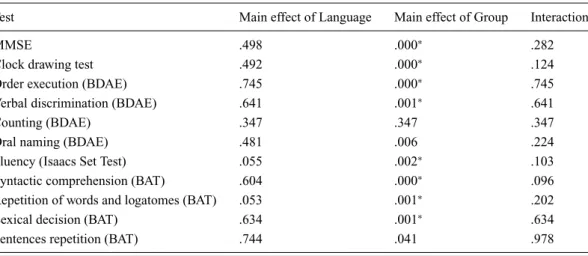
Table 3. p-values of the effects obtained in the different language test scores.
Test Main effect of Language Main effect of Group Interaction
MMSE .498 .000∗ .282
Clock drawing test .492 .000∗ .124
Order execution (BDAE) .745 .000∗ .745
Verbal discrimination (BDAE) .641 .001∗ .641
Counting (BDAE) .347 .347 .347
Oral naming (BDAE) .481 .006 .224
Fluency (Isaacs Set Test) .055 .002∗ .103
Syntactic comprehension (BAT) .604 .000∗ .096
Repetition of words and logatomes (BAT) .053 .001∗ .202
Lexical decision (BAT) .634 .001∗ .634
Sentences repetition (BAT) .744 .041 .978
Note: Correction for multiple tests was achieved using the Bonferroni method (11 tests, alpha set to .005). The significant values are marked
with an∗.
Naeser & Hayward, 1978), the following subtests were selected: (i) Verbal discrimination: associating images of six objects, six actions, six shapes, six symbols, six colours and six figures to orally pronounced words; (ii) Order execution: performing an oral command; (iii) Oral naming: naming of six objects, six actions, two shapes, six symbols, six colours, six figures and three body parts; (iv) Performance in automatic language (Counting): counting aloud to 21.
From the Bilingual Aphasia Test (BAT; Paradis,
2011), the following subtests were selected: (i) Syntactic comprehension: selecting the image that corresponds to the orally-presented sentence; (ii) Repetition of words and non-words: a lexical decision task is associated with each word, the patient repeats an orally-presented word, and then has to say whether the word exists or not; (iii) Repetition of sentences: repeating orally presented sentences.
Finally, for verbal fluency, the Isaacs Set Test (Isaacs & Kennie,1973) was used. The patient was asked to produce as many nouns as possible from four semantic categories (colours, towns, animals and fruits) which alternated every 15 seconds.
The choice of the BDAE and the BAT were based on the fact that these tests have been validated in French and German, which were the main languages of our groups. The tests in each language were performed on two different days with an interval of 1 to 10 days. The evaluator was different for each language and was always a native speaker of the language of interest, except in the case of a Spanish–French patient, whose examiner was a highly proficient bilingual in both Spanish and French.
Statistical analysis
Bilingual competence and demographic factors were compared between the patients and the healthy
controls using uncorrected independent sample t-tests.
Language performance between the patients and the healthy controls was analysed using a 2× 2 mixed analysis of variance (ANOVA) design, with Language (L1; L2) as within-subject factor and Group (controls; patients) as between-subject factor. Correction for multiple tests was achieved using the Bonferroni method. All data analyses were performed using SPSS (version 19; SPSS Inc.).
Results
All results are summarized inTable 3(see alsoFigure 1). We only report the essentials here. As was expected from the inclusion criteria, the groups differed significantly in their MMSE scores (p< .000). The Language × Group ANOVA revealed a significant (p< .005) main effect of Group for all the tests (all p-values< .004; seeTable 3), except for Counting (p= .347), Oral naming (p = .006) and Sentence repetition (p= .041). There was no main effect of Language and no interaction (all p-values are reported inTable 3).
Discussion
To identify the impact of DAT on both languages of the bilinguals, we assessed language comprehension and oral production in a group of 13 DAT patients and 12 matched controls using 2× 2 mixed designs with First Language (L1) and Second Language (L2) as within-subject factor and Group (DAT patients and controls) as between-subject factor. For all tests except Sentence repetition, Oral naming and Counting, there were main effects of the factor Group, driven by the poorer performance of the DAT patients compared to the controls. There were no main effects of Language, and, critically, we did not
Figure 1. (Colour online) Group-averaged scores in two expressive language tasks (BDAE Oral naming task – here scores are divided by two for graphic purposes – and Isaacs Set Test (Verbal fluency task)), and in two receptive language tasks (BDAE Order execution and BAT Syntactic comprehension task) in the first (L1) and second (L2) language. DAT patients performed worse than the controls in Fluency, Order execution and Syntactic comprehension tasks. No differences were found between languages, and there were no Language× Group interactions (see text for details).
find evidence of interaction between the factors Group and Language, suggesting that DAT impaired L1 and L2 similarly.
Main effect of Group
As expected, the main effects of Group revealed that the DAT patients performed less well than the control group in terms of language, despite having the same biography of bilingualism.
The DAT patients performed worse in all language tests except Counting, Oral naming and Sentence repetition. This result corroborates previous findings that linguistic automatisms are preserved in the early stages of DAT (Gomez & White, 2006). The tests employed for both languages in the DAT group are those involving the lexico-semantic system (for instance, complex comprehension (Order execution) or verbal fluency) because these abilities are impaired very early on in the course of DAT (Ullman, 2004). In contrast, other tasks in our neuropsychological battery involved aspects such
as automatic counting and repetitions, which rely on automatic mechanisms and subcortical brain regions usually spared in early DAT (Arroyo-Anllo, Bellouard, Ingrand & Gil,2011).
No main effect of Language
We did not find any main effect of Language, confirming that language performance was similar in both L1 and L2 in our sample. The global rating of L2 proficiency in the DAT patients provided by their families, and the self-ratings by the controls in oral comprehension and oral expression were higher or equal to 80% of the proficiency in L1. The study of populations with comparable L1 and L2 proficiency was important to the present study because most of our language tasks focused on oral language and were thus strongly influenced by the proficiency of the participants. In our sample, the high proficiency of elderly people in both L1 and L2 is in line with previous evidence for a limited impact of aging on L2. Schrauf (2009), for example, demonstrated that in elderly immigrants, L2 is
well preserved in healthy subjects, particularly in those that have children and are well integrated into their L2 community.
There was no main effect of Language for the Syntactic comprehension tasks of the BAT, corroborating that aspects of language acquired late are well preserved in late bilingual immigrants as those included in the current study (Schrauf,2009).
It should be noted that a marginally significant effect of Language was found in fluency (Isaacs Set Test) and the repetition of words and logatomes (BAT). Marginal uncorrected effects in the repetition tests call for further studies with larger groups and patients with more advanced DAT, to test for preservation of L1 phonological processing. Phonological processes are generally found to be less impaired in DAT (Bayles, Tomoeda & Trosset,
1992; Glosser & Deser, 1991; Nebes, 1992). Glosser and colleagues (1997) report that for word and non-word repetition, monolingual DAT patients show worse performance than controls. The results of the patients were not correlated with the severity of DAT. The patients produced the same types of errors as the controls and repeated words better than non-words. The authors conclude that the DAT patients use the same phonologic system for the repetition of the two types of stimuli, i.e. independently of their meaning. In the present study, the slight main effect of language at the level of the Repetition task may be linked to better preservation of phonological capacities in L1.
No Language× Group interaction
The main result of the present study is the absence of an interaction between the factors Group and Language in any of the language tasks. This pattern of results supports the argument that degenerative processes in DAT impact equally on L1 and L2 and that L1 is not specifically spared in DAT.
This pattern corroborates previous literature on the effect of DAT on language and on the organization of language in the bilingual brain. Thus, current evidence suggests that the cortical representations of L1 and L2 overlap at the level of the aspects impaired by DAT, and therefore DAT should provoke the same deficits in L1 and L2.
Indeed, the convergence hypothesis of language representation in bilinguals (Abutalebi & Green, 2007) posits an overlap of L1 and L2 representations for semantico-lexical aspects including, for example, naming (Hernandez, Dapretto, Mazziotta & Bookheimer, 2001) or semantic judgment (Illes et al., 1999). The specific characteristics of our patient group (late bilinguals) allowed us to test Ullman’s hypothesis differentiating between implicit and explicit syntactic/grammatical learning. Generally, early L1 would be subserved by
fronto-subcortical loops involving left frontal and basal ganglia structures, while explicit processing associated to L2 would be associated with bilateral temporal (particularly lateral) lobes structures (Ullman,2001).
Greater syntactic impairments in L1 than L2 in bilingual Parkinson’s disease patients have been proposed as evidence for this theory (Zanini, Tavano, Vorano, Schiavo, Gigli, Aglioti & Fabbro,2004). Basal ganglia are not early targets of either Alzheimer’s disease or normal aging, in contrast with temporal lobe atrophy in DAT. Temporal lobe atrophy would predict an interaction in syntactic performance, due to stronger impairment of L2, which is not the case here. Thus our data do not support divergent syntactic representation for L1 and L2.
To summarize, our data support the convergent representation, but marginal Language effects in repetition and verbal fluency raise the question of some cortical specialization. Larger studies which include L2 proficiency as covariate may help to answer this point. Furthermore, while L2 requires supplementary cognitive resources when its proficiency is lower than L1, the convergence of brain representations for L1 and L2 increases with L2 proficiency (Abutalebi,2008; Stein, Federspiel, Koenig, Wirth, Lehmann, Wiest, Strik, Brandeis & Dierks,2009; Wartenburger et al.,2003).
Perani et al. (1998) tested in a PET study whether the age of acquisition or the proficiency determined the degree of convergence of the brain region involved in processing L1 and L2. Their population were Italian– English highly proficient bilinguals with late age of acquisition (> 10 years) and a group of Spanish–Catalan bilinguals with early age of acquisition (< 4 years). The late acquisition group listened to stories in Italian, English and Japanese (an unfamiliar language), while the early acquisition group listened to stories in L1 and L2. The authors showed that even if the age of acquisition was late, oral comprehension involved the same areas in L1 as in L2, if L2 was highly proficient.
We hypothesize that the two languages of our patients were similarly affected by DAT as those of the controls were affected by normal aging because there was no difference between the groups in L2 proficiency or in age of L2 acquisition. Our results suggest that late bilinguals living in a French-speaking environment reach a high level of immersion and proficiency in L2, which is maintained during aging and even neurodegenerative disorders. Our patients arrived in French-speaking Switzerland and began to learn their second language between the ages of 7–30 years of age. They have been living in their second country for 50 years, have developed their social skills in L2, and use their second language for a large part of their activities (watching television, reading books, even mental arithmetic), suggesting that their L2 actually has become their preferred way of communicating. Another possible reason is that the region where the patients live is
partly bilingual, thus promoting the preservation of both languages. Thus, 12 out of the 13 patients live in Fribourg, a strongly French–German bilingual environment, and one in Geneva, a predominantly French-speaking region. Costa et al. (2012) and Gómez-Ruiz, Aguilar-Alonso and Espasa (2012) studied early bilinguals whose languages were linguistically very close. They found a difference only in spontaneous speech, where DAT patients produced more words and sentences in Catalan. In the BAT subtests that they used, only the Verbal fluency subtest showed slightly higher mean performances in Spanish (mean= 19.9) than in Catalan (mean = 13.6). Thus, in their population, L2 was slightly better preserved than L1 in verbal fluency tasks, a result which supports the role of social immersion in language preservation. We selected participants from various mother tongue languages (Italian, German and Spanish). The linguistic families of these languages differ, thus enabling us to be sure that the lack of Group× Language interaction did not arise from the fact that our patients and controls spoke specific types of languages.
Our data do not support the declarative/procedural theoretical model proposed by Paradis (2008), i.e. that in the L1, grammatical knowledge depends mostly on procedural memory processes supported by the left fronto/basal-ganglia structures (Ullman, 2001; Ullman,
2004). In this model, the L2 of late bilinguals relies on more explicit knowledge, which is mostly supported by the cortical area. Consequently, cortical degeneration diseases, such as DAT, should lead to decreased L2 performances in syntactical scores. In our DAT group (and in Gomez-Ruiz’s group of patients), performances in syntactic tasks did not differ between L1 and L2.
The lack of difference in the impact of DAT on L1 and L2 is also in line with previous data on stroke-induced language impairment in bilinguals, which show that lesions impair L1 and L2 similarly (Tschirren et al.,2011). A meta-analysis by Paradis (1995) also showed that L1 and L2 improve to a similar extent and concurrently in 61% of patients during post-lesion recovery. To further support the argument that brain damage similarly affects L1 and L2, growing evidence indicates that after brain lesions there is active transfer from one language to the other, which was observed in about half of the stroke patients who received monolingual speech therapy (Faroqi-Shah, Frymark, Mullen & Wang,2010). Moreover, such transfer seems more effective if the languages have more structural similarities, as in the current study (Goral, Levy & Kastl,
2010).
An important limitation of the present study is that our interpretations are based on negative results (i.e. a lack of Group× Language interaction). Negative results should be interpreted with caution since a finding of no interaction does not provide definitive evidence that there are no interactions. This issue is particularly relevant in the
current study since we have limited statistical power and thus the probability of type II errors is increased (Moher, Dulberg & Wells, 1994). Post-hoc power analyses were conducted using G∗Power 3.1.5 (Faul, Erdfelder, Buchner & Lang,2009) to compute the number of patients needed per group to reach a significant p-value (p< .05), with an effect size of 0.8 when comparing the mean of the two groups using an independent sample t-test. The results indicate that, to reach a power of 0.8, each group should be of 26 participants. Further studies with larger sample sizes are thus required to confirm our finding that DAT affects L1 and L2 similarly in bilinguals.
The tendency for a Language × Group interaction for the Syntactic comprehension task should be further explored in patients with more severe DAT than that in our group. Previous studies comparing semantic and syntactic performance between monolingual DAT to controls showed no difference at the level of syntactic processing, suggesting that the structural language of DAT patients remained rich (Kave & Levy,2003; Lai, Pai & Lin,2009). In contrast, at the semantic level, DAT patients produced a less informative discourse and more semantic errors than the controls.
The marginally significant effect of Language in the Isaacs Set Test with better scores in L1 than L2 may result from the fact that in verbal fluency tasks, bilingual individuals produce more intrusion errors in their non-dominant language. This tendency may not be present in other tasks, for example naming, because they are less sensitive to interference (Gollan, Montoya, Cera & Sandoval, 2008; Gollan, Montoya, Fennema-Notestine & Morris, 2005). Alternatively such results may be consistent with the hypothesis suggested by Costa (2012, p. 749) for a “largely–but not totally” shared network between the two languages, with a larger implication of subcortical structures for L1 being less sensitive both to aging (Joanette, Ali-Cherif, Delpuech, Habib, Pellissier & Poncet, 1983) and cortical degeneration (Zanini et al.,2011). Prestia, Baglieri, Pievani, Bonetti, Rasser, Thompson and Frisoni (2013) showed that aging impacted on the cortex of healthy individuals at the level of fronto-temporo-parietal regions. This hypothesis should be explored with larger studies.
In addition, the use of questionnaires to rate premorbid proficiency in patients and current proficiency in controls may also be a limiting factor in the present study, since the patients’ families may have misjudged the true level of patients’ premorbid L2 proficiency. There is, however, no other possible way to evaluate the patients’ previous language knowledge (except through access to previous language examinations, which is very rare). Moreover, a number of studies have demonstrated the validity of this questionnaire in assessing language proficiency (e.g. Schrauf, 2009), and have shown that self-ratings correlate with objective measures of proficiency in highly
proficient subjects (Marian, Blumenfeld & Kaushanskaya,
2007).
The mean MMSE of our patients group were respectively 19.8 for L1 and 21.2 for L2. Moreover, we restricted our DAT group to MMSEs higher than 10. Because of this exclusion criterion, the patient group was in the mild to moderate severity range of DAT, and our data must be interpreted for this group. We cannot rule out that Group× Language interactions may appear in more severe DAT cases. However, in a recent study of 71 Catalan–Spanish bilinguals, the severity of the dementia as measured by the MMSE could not explain the differences in the performances between L1 and L2 (Costa et al.,2012), which suggests that this issue is not necessarily critical.
Another limitation of the present study is the difference in age between our two populations (patients: mean age= 75.3 years; controls: mean age= 67.7 years), which may have confounded differences related to the presence of DAT and may explain some of the main effects of Group in the language tests. The decrease in language performance in normal aging is moderate and generally manifests after 70 years of age. Most probably, the main effect of Group in tasks with high attentional demand (Oral naming, Isaacs Set Test and Syntactic comprehension) may be accounted for by the difference in age (Eustache,1993). However, we did not find a significant main effect of Group for Oral naming, suggesting that the age difference cannot be advanced as the only cause of the between-group differences we have found. This finding calls for further studies with perfectly age-matched populations.
In addition to the effect of normal aging on language, another problem related to age differences between patients and controls is the putative modification of language control with age, which may affect L1 or L2 production indirectly. However, recent findings suggest that age-related inhibition decline concerns mostly balanced bilinguals and late bilinguals to a lesser degree (Goral, Campanelli & Spiro, published online April 4,
2013). We would also note that our primary interest was the interaction between the two factors Group and Language, not the main effect of the groups. In this regard, the difference in age most likely impacted similarly on L1 and L2 and thus did not influence the interaction.
In addition to assessing language performance, future study should focus on language control, a key aspect in language impairment and recovery in neurologically impaired bilingual patients. We did not assess language control in the present study because the DAT population showed a high degree of fatigability, and we thus had to choose a limited number of tests of interest among all possibilities. Language control has indeed been advanced as a potential factor influencing whether and how L1 and L2 recover after stroke (e.g. Abutalebi, Della Rosa, Tettamanti, Green & Cappa,2009). Such mechanisms may
also influence language performance in bilingual DAT populations.
Our results also provide insights into whether language assessment in dementia cases should be conducted in L1 or L2 in bilingual individuals (Faroqi-Shah et al.,
2010; Gollan et al., 2010). On the basis of our results, it would appear that at present there is no reason to choose one language over the other for the purposes of neuropsychological assessment of DAT.
References
Abutalebi, J., & Green, D. (2007). Bilingual language production: The neurocognition of language representation and control. Journal of Neurolinguistics, 20, 242–275. Abutalebi, J. (2008). Neural aspects of second language
representation and language control. Acta Psychologica,
128, 466–478.
Abutalebi, J., Della Rosa, P. A., Tettamanti, M., Green, D. W., & Cappa, S. F. (2009). Bilingual aphasia and language control: A follow-up fMRI and intrinsic connectivity study.
Brain & Language, 109, 141–156.
Arroyo-Anllo, E. M., Bellouard, S., Ingrand, P., & Gil, R. (2011). Effects of automatic/controlled access processes on semantic memory in Alzheimer’s disease. Journal of
Alzheimer’s Disease, 25, 525–533.
Bayles, K. A., Tomoeda, C. K., & Trosset, M. W. (1992). Relation of linguistic communication abilities of Alzheimer’s patients to stage of disease. Brain & Language,
42, 454–472.
Cardebat, D., Demonet, J. F., Puel, M., Agniel, A., Viallard, G., & Celsis, P. (1998). Brain correlates of memory processes in patients with dementia of Alzheimer’s type: A SPECT Activation Study. Journal of Cerebral Blood Flow &
Metabolism, 18, 457–462.
Chee, M. W. L., Hon, N., Lee, H. L., & Soon, C. S. (2001). Relative language proficiency modulates BOLD signal change when bilinguals perform semantic judgments.
NeuroImage, 13, 1155–1163.
Chee, M. W. L., Tan, E. W., & Thiel, T. (1999). Mandarin and English single word processing studied with functional magnetic resonance imaging. Journal of Neuroscience, 19, 3050–3056.
Costa, A., Calabria, M., Marne, P., Hernández, M., Juncadella, M., Gascón-Bayarri, J., Lleó, A., Ortiz-Gil, J., Ugas, L., Blesa, R., & Reñé, R. (2012). On the parallel deterioration of lexico-semantic processes in the bilinguals’ two languages: Evidence from Alzheimer’s disease.
Neuropsychologia, 50, 740–753.
Cummings, J. L., Benson, F., Hill, M. A., & Read, S. (1985). Aphasia in dementia of the Alzheimer type. Neurology, 35, 394–397.
Delacourte, A. (2000). Natural and molecular history of Alzheimer disease. Annales de Biologie Clinique, 58, 350– 355.
Dijkstra, T. (2005). Bilingual visual word recognition and lexical access. In J. F. Kroll & A. M. B. de Groot (eds.) Handbook
of bilingualism: Psycholinguistic approaches, pp. 178–201.
Eustache, F. (1993). Langage, vieillissement et démences. In F. Eustache & B. Lechevalier, (eds.). Langage et aphasie, pp. 207–227. Bruxelles: DeBoeck Université.
Fabbro (2001a). The bilingual brain: Bilingual aphasia. Brain &
Language, 79, 201–210.
Fabbro (2001b). The bilingual brain: Cerebral representation of languages. Brain & Language, 79, 211–222.
Faber-Langendoen, K., Morris, J. C., Knesevich, J. W., LaBarge, E., Miller, J. P., & Berg, L. (1988). Aphasia in senile dementia of the Alzheimer type. Annals of Neurology, 23, 365–370.
Fama, R., Sullivan, E. V., Shear, P. K., Stein, M., Yesavage, J. A., Tinklenberg, J. R., & Pfefferbaum, A. (2000). Extent, pattern, and correlates of remote memory impairment in Alzheimer’s disease and Parkinson’s disease.
Neuropsychology, 14, 265–276.
Faroqi-Shah, Y., Frymark, T., Mullen, R., & Wang, B. (2010). Effect of treatment for bilingual individuals with aphasia: A systematic review of the evidence. Journal of
Neurolinguistics, 23, 319–334.
Faul, F., Erdfelder, E., Buchner, A., & Lang, A.-G. (2009). Statistical power analyses using G∗Power 3.1: Tests for correlation and regression analyses. Behavior Research
Methods, 41, 1149–1160.
Giussani, C., Roux, F.-E., Lubrano, V., Gaini, S. M., & Bello, L. (2007). Review of language organisation in bilingual patients: What can we learn from direct brain mapping?
Acta Neurochirurgica, 149, 1109–1116.
Glosser, G., & Deser, T. (1991). Patterns of discourse production among neurological patients with fluent language disorders.
Brain & Language, 40, 67–88.
Gollan, T. H., Montoya, R. I., Cera, C., & Sandoval, T. C. (2008). More use almost always means a smaller frequency effect: Aging, bilingualism, and the weaker links hypothesis.
Journal of Memory and Language, 58, 787–814.
Gollan, T. H., Montoya, R. I., Fennema-Notestine, C., & Morris, S. K. (2005). Bilingualism affects picture naming but not picture classification. Memory & Cognition, 33, 1120– 1234.
Gollan, T. H., Salmon, D. P., Montoya, R. I., & da Pena, E. (2010). Accessibility of the nondominant language in picture naming: A counterintuitive effect of dementia on bilingual language production. Neuropsychologia, 48, 1356–1366.
Gomez, R. G., & White, D. A. (2006). Using verbal fluency to detect very mild dementia of the Alzheimer type. Archives
of Clinical Neuropsychology, 21, 771–775.
Gómez-Ruiz, I., Aguilar-Alonso, A., & Espasa, M. A. (2012). Language impairment in Catalan–Spanish bilinguals with Alzheimer’s disease. Journal of Neurolinguistics, 25, 552– 566.
Goral, M., Campanelli, L., & Spiro, A. III. Lan-guage dominance and inhibition abilities in bilingual older adults. Bilingualism: Language and Cognition, doi:10.1017/S1366728913000126. Published online by Cambridge University Press, April 4, 2013.
Goral, M., Levy, E. S., & Kastl, R. (2010). Cross-language treatment generalization: A case of trilingual aphasia.
Aphasiology, 24, 170–187.
Hachinski, V., Oveisgharan, S., Romney, A. K., & Shankle, W. R. (2012). Optimizing the Hachinski Ischemic Scale. Archives
of Neurology, 69, 169–175.
Hartsuiker, R. J., Pickering, M. J., & Veltkamp, E. (2004). Is syntax separate or shared between languages? Cross-linguistic syntactic priming in Spanish–English bilinguals.
Psychological Science, 15, 409–414.
Heller, R. B., Dobbs, A. R., & Rule, B. G. (1992). Communicative function in patients with questionable Alzheimer’s disease. Psychology and Aging, 7, 395–400. Hernandez, A. E., Dapretto, M., Mazziotta, J., & Bookheimer, S.
(2001). Language switching and language representation in Spanish–English bilinguals: An fMRI study. NeuroImage,
14, 510–520.
Hernandez, A. E., Martinez, A., & Kohnert, K. (2000). In search of the language switch: An fMRI study of picture naming in Spanish–English bilinguals. Brain & Language, 73, 421– 431.
Hyltenstam, K., & Stroud, C. (1989). Bilingualism in Alzheimer’s dementia: Two case studies. In K. Hyltenstam & L. Obler (eds.), Bilingualism across the lifespan: Aspects
of acquisition, maturity, and loss, pp. 202–226. Cambridge:
Cambridge University Press.
Illes, J., Francis, W. S., Desmond, J. E., Gabrieli, J. D. E., Glover, G. H., Poldrack, R., Lee, C. J., & Wagner, A. D. (1999). Convergent cortical representation of semantic processing in bilinguals. Brain & Language, 70, 347–363.
Isaacs, B., & Kennie, A. T. (1973). The Set Test as an aid to the detection of dementia in old people. British Journal of
Psychiatry, 123, 467–470.
Joanette, Y., Ali-Cherif, A., Delpuech, F., Habib, M., Pellissier, J. F., & Poncet, M. (1983). Évolution de la séméiologie aphasique avec l’âge. Discussion à propos d’une observation anatomo-clinique [Development of aphasic symptomatology with age. Discussion apropos of an anatomo-clinical case]. Revue Neurologique, 139, 657– 664.
Juncos-Rabadan, O., & Inglesias, F. (1994). Decline in the elderly’s language: Evidence from cross-linguistic data.
Journal of Neurolinguistics, 8, 183–190.
Kantola, L., & van Gompel, R. P. G. (2011). Between- and within-language priming is the same: Evidence for shared bilingual syntactic representations. Memory & Cognition,
39, 276–290.
Kave, G., & Levy, Y. (2003). Morphology in picture descriptions provided by persons with Alzheimer’s disease. Journal of
Speech, Language, and Hearing Research, 46, 341–352.
Kim, K. H., Relkin, N. R., Lee, K. M., & Hirsch, J. (1997). Distinct cortical areas associated with native and second languages. Nature, 388, 171–174.
Klein, D., Milner, B., Zatorre, R. J., Meyer, E., & Evans, A. C. (1995). The neural substrates underlying word generation: A bilingual functional-imaging study. Proceedings of the
National Academy of Sciences of the United States of America, 92, 2899–2903.
Klein, D., Zatorre, R. J., Milner, B., Meyer, E., & Evans, A. C. (1994). Left putaminal activation when speaking a second language: Evidence from PET. Neuroreport, 5, 2295– 2297.
Lai, Y.-h., Pai, H.-h., & Lin, Y.-t. (2009). To be semantically-impaired or to be syntactically-semantically-impaired: Linguistic patterns in Chinese-speaking persons with or without dementia. Journal of Neurolinguistics, 22, 465–475. Long, M. (1990). Maturational constraints on language
development. Studies in Second Language Acquisition, 12, 251–285.
Marian, V., Blumenfeld, H. K., & Kaushanskaya, M. (2007). The Language Experience and Proficiency Questionnaire (LEAP-Q): Assessing language profiles in bilinguals and multilinguals. Journal of Speech, Language, and Hearing
Research, 50, 940–967.
Mazeaux, J., Orgogozo, J., Goodglass, H., & Kaplan, E. (1983). Boston Diagnostic Aphasia Examination (BDAE).
Adaptation française. Philadelphia: Lea and Febiger.
McKhann, G., Drachman, D., Folstein, M., Katzman, R., Price, D., & Stadlan, E. M. (1984). Clinical diagnosis of Alzheimer’s disease: Report of the NINCDS-ADRDA Work Group under the auspices of Department of Health and Human Services Task Force on Alzheimer’s Disease.
Neurology, 34, 939–944.
Moher, D., Dulberg, C. S., & Wells, G. A. (1994). Statistical power, sample size, and their reporting in randomized controlled trials. Journal of the American Medical
Association, 272, 122–124.
Monsch, A., Hermelink, M., Kressig, R., Fisch, H., Grob, D., Hiltbrunner, B., Martensson, B., Rüegger-Frey, B., & von Gunten, A. (2008). Consensus sur le diagnostic et la prise en charge des patients atteints de démence en Suisse. Forum
Médical Suisse, 8, 144–149.
Morris, J. C. (1993). The Clinical Dementia Rating (CDR): Current version and scoring rules. Neurology, 43, 2412– 2414.
Morris, J. C., Ernesto, C., Schafer, K., Coats, M., Leon, S., Sano, M., Thal, L. J., & Woodbury, P. (1997). Clinical Dementia Rating training and reliability in multicenter studies: The Alzheimer’s Disease Cooperative Study experience.
Neurology, 48, 1508–1510.
Movement Disorder Society Task Force on Rating Scales for PD. 2003. The Unified Parkinson’s Disease Rating Scale (UPDRS): Status and recommendations. Movement
Disorders, 18, 738–750.
Naeser, M. A., & Hayward, R. W. (1978). Lesion localization in aphasia with cranial computed tomography and the Boston Diagnostic Aphasia Exam. Neurology, 28, 545–551. Nebes, R. D. (1992). Cognitive dysfunction in Alzheimer’s
disease. In F. I. M. Craik & T. A. Salthouse (eds.) The
handbook of aging and cognition, pp. 373–448. Hillsdale,
NJ: Lawrence Erlbaum.
Paradis, M. (1995). Aspects of bilingual aphasia. Oxford: Pergamon Press.
Paradis, M. (2008). Language and communication disorders in multilinguals. In B. Stemmer & H. Whitaker (eds.),
Handbook of the neuroscience of language, pp. 341–349.
Amsterdam: Elsevier Science.
Paradis, M. (2011). Principles underlying the Bilingual Aphasia Test (BAT) and its uses. Clinical Linguistics and Phonetics,
25, 427–443.
Perani, D., & Abutalebi, J. (2005). The neural basis of first and second language processing. Current Opinion in
Neurobiology, 15, 202–206.
Perani, D., & Cappa, S. F. (1998). Neuroimaging methods in neuropsychology. In G. Denes & L. Pizzamiglio (eds.),
Handbook of clinical and experimental neuropsychology,
pp. 69–94. London: Psychology Press.
Perani, D., Paulesu, E., Galles, N. S., Dupoux, E., Dehaene, S., Bettinardi, V., Cappa, S. F., Fazio, F., & Mehler, J. (1998). The bilingual brain. Proficiency and age of acquisition of the second language. Brain, 121, 1841–1852.
Prestia, A., Baglieri, A., Pievani, M., Bonetti, M., Rasser, P. E., Thompson, P. M., Marino, S., Bramanti, P., & Frisoni, B. (2013). The in vivo topography of cortical changes in healthy aging and prodromal Alzheimer’s disease. Supplements to Clinical Neurophysiology, 62, 67– 80.
Salvatierra, J., Rosselli, M., Acevedo, A., & Duara, R. (2007). Verbal fluency in bilingual Spanish/English Alzheimer’s disease patients. American Journal of Alzheimer’s Disease
and Other Dementias, 22, 190–201.
Schrauf, R. W. (2009). English use among older bilingual immigrants in linguistically concentrated neighborhoods: Social proficiency and internal speech as intracultural variation. Journal of Cross-Cultural Gerontology, 24, 157– 179.
Stein, M., Federspiel, A., Koenig, T., Wirth, M., Lehmann, C., Wiest, R., Strik, W., Brandeis, D., & Dierks, T. (2009). Reduced frontal activation with increasing 2nd language proficiency. Neuropsychologia, 47, 2712– 2720.
Tschirren, M., Laganaro, M., Michel, P., Martory, M. D., Di Pietro, M., Abutalebi, J., & Annoni, J.-M. (2011). Language and syntactic impairment following stroke in late bilingual aphasics. Brain & Language, 119, 238–242.
Ulatowska, H. K., Allard, L., Donnell, A., Bristow, J., Haynes, S. M., Flower, A., & North, A. J. (1988). [Use of discourse analysis for evaluation of the condition of patients with dementia of the Alzheimer type]. Neurologia i
Neurochirurgia Polska [Polish Journal of Neurology and Neurosurgery], 22, 34–37.
Ullman, M. T. (2001). A neurocognitive perspective on language: The declarative/procedural model. Nature
Reviews Neuroscience, 2, 717–726.
Ullman, M. T. (2004). Contributions of memory circuits to language: The declarative/procedural model. Cognition, 92, 231–270.
Wartenburger, I., Heekeren, H. R., Abutalebi, J., Cappa, S. F., Villringer, A., & Perani, D. (2003). Early setting of grammatical processing in the bilingual brain. Neuron, 37, 159–170.
Weber-Fox, C. M., & Neville, H. J. (1996). Maturational constraints on functional specializations for language processing: ERP and behavioral evidence in bilingual speakers. Journal of Cognitive Neuroscience, 8, 231– 256.
Zanini, S., Angeli, V., & Tavano, A. (2011). Primary progressive aphasia in a bilingual speaker: A single-case study. Clinical
Linguistics and Phonetics, 25, 553–564.
Zanini, S., Tavano, A., Vorano, L., Schiavo, F., Gigli, G. L., Aglioti, S. M., & Fabbro, F. (2004). Greater syntactic impairments in native language in bilingual Parkinsonian patients. Journal of Neurology, Neurosurgery & Psychiatry,