Reply to the Letter to the Editor
on “Severe hepatic sinusoidal
obstruction and oxaliplatin-based
chemotherapy in patients with
metastatic colorectal cancer: a real
entity?”, by M. Sebagh, M. Plasse,
F. Le´vi & R. Adam (Ann Oncol
2005; 16: 331)
We are pleased to learn that another group also involved for several years in the study of neoadjuvant treatment of colorec-tal liver metastases share many of our views on the develop-ment of hepatic vascular lesions induced by oxaliplatin-based chemotherapy. The letter of Sebagh et al. raises several inter-esting questions about the histological interpretation of the hepatic lesions. In the planning phases of the study, the parti-cipating pathologists reviewed together more than half of the slides and reached a consensus on the definition and coding of the lesions. Although this joint session may not have elimi-nated all interobserver variation, they are likely to have reduced it considerably. Our goal was to describe the lesions most likely to be related to the use and the type of neoadju-vant chemotherapy, rather than providing an exhaustive assessment of the histopathological changes in the livers examined. Since neither prevalence nor severity of portal fibrosis and steatosis were significantly different between patients receiving chemotherapy or not, we opted not to report these data. The discrepancy between Sebagh’s and our group concerning the type of vascular lesions may be more related to the definition of the term than to actual differences.
Traditionally, the term ‘hepatic veno-occlusive disease’ refers to a form of toxic liver injury characterised clinically
by the development of hepatomegaly, ascites and jaundice, and histologically by diffuse damage in the centrilobular zone of the liver. Recent studies suggest that the primary site of the toxic injury is initially the sinusoidal endothelial cells, fol-lowed by a series of biological processes that lead to circula-tory compromise of centrilobular hepatocytes, fibrosis, obstruction of liver blood flow and eventually to alterations of the centrilobular vein. The cardinal histological features of this injury are marked sinusoidal dilatation and congestion, necrosis of pericentral hepatocytes, narrowing and, only even-tually, fibrosis of centrilobular veins. Based on these data, DeLeve et al. [1] recently proposed a more appropriate name for veno-occlusive disease, namely ‘sinusoidal obstruction syndrome’ (SOS).
At the present time, making the diagnosis of vascular toxic injury based exclusively on the presence of fibrotic veno-occlusive lesion would appear too restrictive. The discrepancy in frequency of the lesions detected by our two groups is prob-ably less pronounced than suggested by Sebagh et al. In fact, if four out of five of our patients receiving oxaliplatin-based chemotherapy developed sinusoidal lesions, these were, as specified in our paper, of variable degree and semi-quantitat-ively evaluated. We observed 28% of severe vascular lesions and 36% of moderate or mild lesions, but no mild lesions were seen in patients who did not receive chemotherapy. In Sebagh’s unpublished series, 38% of the 52 oxaliplatin-treated patients developed severe vascular lesions. The frequency of severe lesion is thus at least equivalent, if not greater, than the frequency we detected. Their data appear to confirm that vas-cular lesions are common in post-chemotherapy liver speci-mens. This is interesting also because it suggests that the chromomodulated infusion of chemotherapy in their patients does not seem to reduce liver toxicity. As we stated in the article, our ability to evaluate the clinical relevance of our findings was limited by the retrospective nature of this study. While we agree that the key end point is the clinical impact of SOS lesions, an increase in operative mortality appears to be
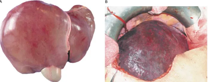
Figure 1. (A) Macroscopically normal liver. (B) Post neoadjuvant oxaliplatin-based chemotherapy. The surface of the liver shows extensive areas of congestion.
too restrictive a parameter. Indeed, severe SOS can be ident-ified macroscopically (Figure 1) or histologically in preopera-tive frozen sections. By making the surgeon aware of this lesion, a large resection can be avoided or more extensive por-tal embolisation can be used before surgery to reduce the risk of post-operative liver failure.
Since the recent publication of our paper we have become aware of several cases of patients developing complications (with long hospitalisations in intensive care unit) because of the delay in liver regeneration and liver insufficiency after limited hepatectomy (unpublished data). A patient has devel-oped acute fatal portal hypertension after oxaliplatin-based adjuvant chemotherapy for colorectal carcinoma [2]. The pub-lication of such observations in the near future will support the concept that SOS is a real and relevant clinical entity in oxaliplatin-based chemotherapy.
In our experience, oxaliplatin is a very useful drug that yields excellent responses in hepatic colorectal metastases, and we believe that its use will continue to increase. There-fore, far from intending to question the efficacy of oxaliplatin in the treatment of colorectal metastasis, we suggest that the occurrence of clinically significant secondary effects on the liver need to be recognised to increase the safety of liver sur-gery. The scope of these observations may further expand as oxaliplatin is becoming used as adjuvant therapy in the treat-ment of several other malignancies.
L. Rubbia-Brandt1*, G. Mentha2, B. Dousset3& B. Terris4 1
Division of Clinical Pathology and2Division of Digestive Surgery, Uni-versity Hospital, Geneva, Switzerland;3Service of Surgery, Hoˆpital Cochin, Paris, France;4Service d’Anatomie Pathologique, Hoˆpital Cochin,
Paris, France (*E-mail: laura.rubbiarandt@hcuge.ch)
References
1. DeLeve LD, Shulman HM, McDonald GB. Toxic injury to hepatic sinusoids: sinusoidal obstruction syndrome (veno-occlusive disease). Semin Liver Dis 2002; 22(1): 27 – 42.
2. Tisman G, MacDonald D, Shindell N et al. Oxaliplatin toxicity mas-querading as recurrent colon cancer. J Clin Oncol 2004; 22(15): 3202 – 3204.
doi:10.1093/annonc/mdi038
Obesity may decrease the
amenorrhea associated with
chemotherapy in premenopausal
breast cancer patients
We read with great interest the article by Berclaz et al. [1], which stated that body mass index (BMI) is an independent prognostic factor for overall survival in patients with breast cancer, especially among pre-/perimenopausal patients treated
with chemotherapy without endocrine therapy. These authors proposed different explanations for this association. We would like to mention another explanation.
Amenorrhea is also an important prognostic factor for pre-dicting the efficacy of chemotherapy in premenopausal patients [2]. It is known that obese women have a longer reproductive life and that longer exposure to estrogen increases breast cancer risk. Although there were insufficient data to demonstrate that chemotherapy is associated with a decreased incidence of amenorrhea in obese patients compared with lean counterparts, Mehta et al. [3] showed that 71% of obese patients develop amenorrhea after receiving chemother-apy, compared with 80% of non-obese breast cancer patients. In light of the above information, we propose that obesity itself, by suppressing the amenorrhea associated with che-motherapy, may result in poorer prognosis in premenopausal breast cancer patients with a high BMI.
K. Altundag1*, O. Altundag1, P. Morandi2& M. Gunduz3 1
Department of Medical Oncology, Hacettepe University Faculty of Medicine, Ankara, Turkey;2Department of Medical Oncology, S. Bortolo General Hospital, Vicenza, Italy;3Department of Oral Pathology and
Medicine, Graduate School of Medicine and Dentistry, Okayama University, Japan (*E-mail: drkadri@usa.net)
References
1. Berclaz G, Li S, Price KN et al. Body mass index as a prognostic fea-ture in operable breast cancer: the International Breast Cancer Study Group experience. Ann Oncol 2004; 15: 875 – 884.
2. Poikonen P, Saarto T, Elomaa I et al. Prognostic effect of amenorrhea and elevated serum gonadotropin levels induced by adjuvant che-motherapy in premenopausal node-positive breast cancer patients. Eur J Cancer 2000; 36: 43 – 48.
3. Mehta RR, Beattie CW, Das Gupta TK. Endocrine profile in breast cancer patients receiving chemotherapy. Breast Cancer Res Treat 1992; 20: 125 – 132.
doi:10.1093/annonc/mdi044
Reply to the Letter to the Editor
on “Obesity may decrease the
amenorrhea associated with
chemotherapy in premenopausal
breast cancer patients”, by
K. Altundag et al. (Ann Oncol
2005; 16: 333)
Altundag and colleagues suggest that obese women have an increased breast cancer risk due to a longer reproductive life.
A meta-analysis of 23 studies providing information on body mass index incidence in premenopausal breast cancer 333
