HAL Id: hal-02922106
https://hal.archives-ouvertes.fr/hal-02922106
Submitted on 25 Aug 2020
HAL is a multi-disciplinary open access
archive for the deposit and dissemination of sci-entific research documents, whether they are pub-lished or not. The documents may come from teaching and research institutions in France or abroad, or from public or private research centers.
L’archive ouverte pluridisciplinaire HAL, est destinée au dépôt et à la diffusion de documents scientifiques de niveau recherche, publiés ou non, émanant des établissements d’enseignement et de recherche français ou étrangers, des laboratoires publics ou privés.
Neurotrophin & synaptogenesis
Freddy Jeanneteau, Margarita Arango-Lievano, Moses Chao
To cite this version:
Freddy Jeanneteau, Margarita Arango-Lievano, Moses Chao. Neurotrophin & synaptogenesis.
John Rubenstein; Pasko Rakic; Bin Chen; Kenneth Y. Kwan; Hollis T. Cline; Jessica Cardin. Synapse Development and Maturation (2nd edition), Elsevier, pp.167-192, 2020, 978-0-12-823672-7. �10.1016/B978-0-12-823672-7.00007-7�. �hal-02922106�
Chapter
Neurotrophin & synaptogenesis
Freddy Jeanneteau1, Margarita Arango-Lievano1 & Moses V. Chao 2
1. Institut de genomique fonctionelle, departments of Physiology, Neuroscience, Inserm, CNRS, University of Montpellier, Monpellier, France
2. Skirball Institute of Biomolecular Medicine, Departments of Cell Biology, Physiology & Neuroscience and Psychiatry, New York University Langone Medical Center, 540 First Avenue, New York, NY 10016, USA
Correspondence:
Outline. Summary
I. Introduction
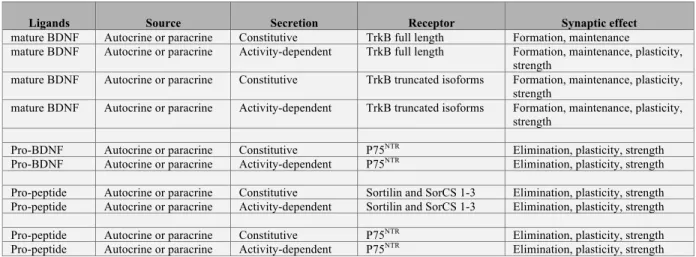
II. One neurotrophin, three ligands at the synapse II.1. Pro-BDNF
II.2. Cleavage of Pro-BDNF II.3. The prodomain
III. Which side of the synapse produce neurotrophins? III.1. Presynaptic sources
III.2. Postsynaptic sources
III.3. Non-neuronal sources at the synapse
IV. What are the modes of neurotrophin synaptic release? IV.1. Secretion of pro-BDNF
IV.2. Secretion of mature BDNF IV.3. Secretion of the prodomain V. Receptors for neurotrophin ligands
V.1. Receptors for proBDNF V.2. Receptors for mature BDNF V.3. Receptors for the prodomain VI. Signaling mechanisms of Trk and p75NTR
VI.1. Presynaptic responses VI.2. Postsynaptic responses VI.3. Rapid and slow responses
VI.4. Balancing act on excitation and inhibition VII. Specificity of neurotrophin actions at the synapse VIII. Conclusion and perspectives
Summary
Synaptogenesis is encoded by multiple genes that program the assembly of neural networks in the immature brain during development and later in life, in the experienced brain that must respond to changes of the external world and of proprioception. Processing of neural network activity cannot solely rely on the activity of established synapses as their plasticity can saturate. For this reason, synaptogenesis is reversible. Assembly and disassembly of synapses depend on the exchange of signals between the pre- and post-synaptic terminals. Synaptic trophic factors are paramount for neural network remodeling, homeostasis and survival because they are present in limited supply. Neurotrophins present the necessary attributes to operate as synaptic trophin. It is speculated that many diverse and pleiotropic actions of neurotrophins on the synapse depend on the sources of neurotrophin ligands, its modes of secretion and signaling, all influenced by their locations at the synapse. What information is encoded when synaptic neurotrophin signaling is retrograde or anterograde, paracrine or autocrine? Answers emerged from the characterization of the cells that secrete neurotrophins, the cells that respond to neurotrophins, the various modes of secretion, receptor-signaling pathways, and the temporal constraints imposed by synaptic transmission. Failure to communicate the appropriate synaptic trophic signals impairs neuronal networks maintenance and updated wiring.
Keywords:
BDNF, Autocrine-paracrine, anterograde-retrograde signaling, P75NTR, Sortilin-related receptors, TrkB, LTP-LTD, mTOR, Actin, Val66met polymorphism
I. Introduction
Exchanges of anterograde and retrograde signals between synaptic connections serve to adjust input-to-output. In the absence of bi-directional trans-synaptic signaling, the changes of synaptic strength would decay and information lost (Harris, 2008). As a result, the anterograde signaling in target post-synaptic cells should be consolidated by back-propagating signals in the target pre-synaptic cells. To be efficient, trans-synaptic signals shall present specific features: (i) it shall be produced in a limited amount; (ii) it shall be dependent on synaptic activity; (iii) it shall be poorly diffusible; (iv) it shall be emitted when pre- and post-synaptic activities are correlated; (v) receptors shall emit specific signaling on every sides of the synapse via anterograde and retrograde pathways.
For decades, evidence were collected to demonstrate that neurotrophins operate in a trans-synaptic and bi-directional fashion for regulating synaptic plasticity and strength within active but not resting neuronal networks (Autry and Monteggia, 2012; Bennett and Lagopoulos, 2014; Castren and Antila, 2017; Choi, 2018; Cohen-Cory et al., 2010; Hao et al., 2018; Lessmann, 1998; Lu and Chow, 1999; Panja and Bramham, 2014; Park and Poo, 2013; Poo, 2001; Tyler et al., 2002). Two examples: (i) anterograde spread of the trans-synaptic neurotrophin rescued the lack of input activity on post-synaptic cells (Chen et al., 2016; Du et al., 2009), and (ii) retrograde transport of target-derived neurotrophin via signaling endosomes can extend from axon terminals to dendrites where it instructs molecular composition of postsynaptic densities (Sharma et al., 2010). Of all the neurotrophins, brain derived neurotrophic factor (BDNF) is best characterized. BDNF is available in limited supply at the synapse. Bulk BDNF is extrasynaptic (Swanwick et al., 2004), secreted upon synaptic activity (Nagappan and Lu, 2005), and captured by receptors lacking the tyrosine kinase domain on astrocytes upmost and neurons (Bergami et al., 2008; Biffo et al., 1995; Stahlberg et al.). Yet, the synapse is its primary site of actions on neurons (Song et al., 2017). BDNF packed in dense core vesicles of the pre-synapse ensures anterograde signaling (Dieni et al., 2012) whereas dendritic BDNF in the post-synapse and in microglia contribute to the anterograde signaling (Hedrick et al., 2016; Parkhurst et al., 2013).
The paucity of neurotrophins results from an active process with temporal and spatial resolutions that permits synaptogenesis and competition between synapses of
different projecting axons (Cohen-Cory et al., 2010; Snider and Lichtman, 1996). This is essential for synaptic scaling of both excitatory and inhibitory neurons to maintain homeostasis (Gottmann et al., 2009) but also for synaptic potentiation, including the induction, maintenance and consolidation phases (Poo, 2001). Therefore, it is interesting that synaptic priming with BDNF can convert weak synaptic activity into robust potentiation by post-synaptic mechanisms (Figurov et al., 1996; Kovalchuk et al., 2002; Wierenga et al., 2005).
One way to restrict neurotrophin responsiveness to active synapses is to pair neurotrophin signaling with neuronal activity (Boulanger and Poo, 1999; Tanaka et al., 2008). Neuronal activity originating from both pre- and post-synaptic sides can stimulate the secretion of neurotrophins, which may result in strengthening and maintenance of active synapses (Castren and Antila, 2017; Jakawich et al., 2010). In contrast, synapses with unsynchronized activity between the pre-and post-synaptic terminals do not receive neurotrophin support and are weakened (Jakawich et al., 2010; Poo, 2001; Snider and Lichtman, 1996).
This chapter will enumerate the attributes that permit BDNF to instruct circuit-specific responses devised to enforce circuit adaptation to changing environment (e.g. behavior, perception-anticipation, drugs, diseases). Neurotrophin responses rely on a trans-synaptic molecular system consisting of multiple ligands and receptors, which specificity, sensitivity and location matter (Song et al., 2017).
II. One neurotrophin, three ligands at the synapse
Neurotrophins are synthetized as precursors (proneurotrophins) with a N-terminal prodomain and a C-terminal mature domain. Classically, neurotrophins must be cleaved off their prodomains to be operational. That is to promote synaptic and non-synaptic growth and differentiation via rapid signaling routes and long lasting changes in gene transcription (see other reviews (Gonzalez et al., 2016; Hempstead, 2006; Woo et al., 2005). This view came with the assumption that the proforms of all neurotrophins are inactive. But proforms are signaling molecules as well (Hempstead, 2015; Mizui et al., 2017).
II.1 Pro-BDNF
Like all neurotrophins, BDNF is synthetized as a large precursor called proBDNF. This precursor is approximately a 32kDa peptide that is glycosylated in the N-terminal prodomain region (Mowla et al., 2001; Mowla et al., 1999). Following synthesis in the endoplasmic reticulum (ER), proBDNF is directed to the secretory pathway. ProBDNF can be cleaved to prodomain and mature BDNF in the Golgi apparatus or in secretion vesicles but, during early development, substantial amounts of proBDNF can escape processing. Trafficking occurs in the trans Golgi network (TGN) where proBDNF interact with the type 1 receptor sortilin (Chen et al., 2005b). Sortilin is a member of a group of receptors containing a vacuolar sorting protein 10 (VPS10) domain that acts like chaperones to target their cargo protein to different cellular compartments (Nykjaer and Willnow, 2012). By interacting with the prodomain, sortilin acts as a chaperone for intracellular trafficking and directs proBDNF into dense-core vesicles destined to the activity dependent secretory pathway (Dieni et al., 2012; Mowla et al., 1999).
Yet, an interaction with sortilin is also necessary to engage BDNF toward the lysosomal pathway (Evans et al., 2011). Two mechanistic frameworks explain how proBDNF is sorted toward secretory or degradation routes. First, targeting of sortilin and its proBDNF cargo to the lysosomal pathway relies on the cleavage of sortilin by ADAM 10 (Evans et al., 2011). Second, targeting of sortilin and its proBDNF cargo to the secretory pathway required physical interaction with the huntingtin interacting protein 1 (HAP1) (Yang et al., 2011).
A separate mechanism involves a common human single nucleotide polymorphism (SNP) on the prodomain of BDNF. The Val66Met polymorphism is the result of a nucleotide change from a guanine to an adenine at position 196 (G196A) substituting a valine (Val) to methionine (Met) at codon 66. The SNP was reported to alter the transport of BDNF mRNA transcripts to dendrites for local synthesis of BDNF (Chiaruttini et al., 2009). The Val66Met substitution also disrupts the interaction between the proBDNF and sortilin, resulting in defects of transport, maturation and subcellular sorting (Chen et al., 2005b; Egan et al., 2003; Sasi et al., 2017).
Mature BDNF is a 14KDa peptide with well-characterized trophic and plasticity abilities that have been extensively reviewed elsewhere (Binder and Scharfman, 2004; McAllister et al., 1999). Mature BDNF results from proteolytic cleavage of proBDNF. Following synthesis in the ER, proBDNF can undergo processing directly in the Golgi apparatus by furin or within secretory vesicles of the regulated pathway by other proconvertases (Mowla et al., 1999). The cleavage of proneurotrophins by furin occurs at the consensus sequence R–X–K/R–R, (RVRR in proBDNF) to produce mature neurotrophins and prodomains. Furin cleaves proBDNF at Arg 128 of the consensus site. The mutation of this consensus site from RVRR to RVAA (Yang et al., 2014) or MVLR (Koshimizu et al., 2009) has been instrumental to produce a cleavage resistant proBDNF insensitive to furin and plasmin, and has been exploited to produce recombinant uncleavable proBDNF to demonstrate its biological activity in vitro and in vivo (Koshimizu et al., 2009; Pang et al., 2004; Teng et al., 2005; Yang et al., 2014). Other proconvertases, such as PACE4, PC5, and PC7, have been shown to process proBDNF in mature BDNF and prodomain utilizing the same RVRR site (Marcinkiewicz et al., 1998; Seidah et al., 1996; Wetsel et al., 2013).
When proBDNF escape intracellular cleavage by furin or other proconvertases, it can be found in the dense-core vesicles destined to the secretory pathway. The existence of secreted proBDNF was the subject of an intense debate that we will discuss later in this chapter, but if secreted, proBDNF can be converted into mature BDNF and prodomain by extracellular proteases. The most prominent of these proteases is plasmin, which is the product of the activation of the inactive plasminogen by tissue-type plasminogen activator (tPA). The specific plasmin cleavage site of proBDNF was identified and found it to be located within the consensus furin-cleavage sequence of BDNF, but occurring after Arg125 rather than Arg128 of the RVRR sequence (Gray and Ellis, 2008). Interestingly, a SNP has been identified at position 125, which induces an arginine substitution for a methionine that would potentially prevent plasmin processing of proBDNF (Koshimizu et al., 2009). Multiple regulatory steps are implicated in this reaction as conversion of plasminogen to plasmin is antagonized by Plasminogen activator inhibitor (PAI1), and PAI1 depends on vitronectin to remain active (Mou et al.,
by neuronal activity (Shin et al., 2004). The fact that both proBDNF/BDNF/prodomain containing vesicles and tPA are secreted in an activity dependent fashion raises questions about regulation of bio-available tPA. For instance, a reduction of tPA secretion may be enough to favor proBDNF signaling and vice and versa. ProBDNF can be processed by selected matrix metalloproteinases (MMPs) such as MMP-3 and MMP-7 (Lee et al., 2001). Moreover, it has been shown that MMP-9 converts proBDNF into mature BDNF after kindled seizures in the hippocampus (Mizoguchi et al., 2011).
All these intra- and extracellular enzymes cleave proBDNF at distinct but interdependent sites, all generating biochemically undistinguishable mature BDNF and prodomain species. No distinct biological functions of the mature BDNF cleaved by one or the other proteases have ever been reported to our knowledge.
II.3 The prodomain
The prodomain is a 15.5kDa protein recently found to be very abundant in the hippocampus and results from the proteolytic cleavage of pro-BDNF at the site that also produce mature BDNF (Anastasia et al., 2013; Dieni et al., 2012). The BDNF prodomain is detected in dense core vesicles in vivo and found to be secreted as ligand (Anastasia et al., 2013; Mizui et al., 2015) independent of mature BDNF and proBDNF. BDNF prodomain facilitates hippocampal long-term depression (LTD) by a mechanism involving the internalization of AMPA receptors from the synapse (Mizui et al., 2015). Interestingly, the Val66met polymorphism of BDNF positioned in the prodomain alters the reported structural conformation (Anastasia et al., 2013; Wang et al., 2018), subcellular sorting and transport (Arango-Lievano et al., 2015a; Zanin et al., 2017), as well as activities of the prodomain: (i) Met66 carriers impaired the expression of LTD (Mizui et al., 2015), (ii) Met66 carriers elicited growth cone collapse (Anastasia et al., 2013) and (iii) Met66 carriers promoted dendritic spine elimination (Giza et al., 2018) as opposed to its counterpart the val66 carriers that facilitated LTD with no effects on axonal growth cones or dendritic spine turnover.
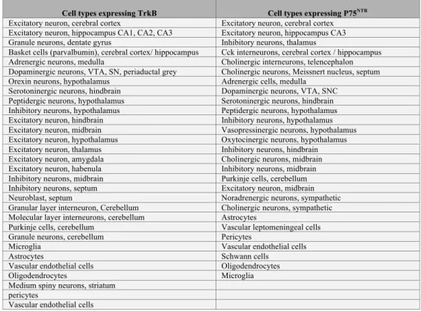
Neurotrophin ligands are expressed by a multitude of neuronal cells, for the most part excitatory (e.g. glutamatergic) and neuromodulatory (e.g. dopaminergic, serotoninergic, adrenergic…), and non-neuronal cells supporting synaptic functions (e.g. glia, vascular cells…). Most neuronal cell types in brain are able to produce some neurotrophin isoforms with the exception of cortical interneurons and striatal medium spiny neurons among others that rely upon paracrine secretion (Kohara et al., 2003). See table 1 for an inventory of single cell types expressing and cells not expressing BDNF mRNA transcripts based on based on the mouse brain atlas of single cell RNA sequencing (http://mousebrain.org/ (Zeisel et al., 2018)).
One issue is that BDNF expression is variable, depending on cues from the internal and external world (Jeanneteau and Chao, 2013). Multiple promoters drive its transcription in specific cell types and upon various stimuli (Aid et al., 2007; Cattaneo et al., 2016). Transcription of BDNF is also susceptible to epigenetic marks that are specific to individuals, possibly transmitted to progeny associated with transgenerational impact on individuals’ health (Cattaneo et al., 2016; Kundakovic et al., 2015; Roth et al., 2009; Roth and Sweatt, 2011). One consequence is that BDNF expression pattern may be unique to each individual as it reflects gender, history of living conditions and experiences (Bath et al., 2013).
Expression of the BDNF gene is a reflection of synaptic activity in selective networks of neurons and circuits. That is, BDNF expression is very responsive to neuronal activity from sensory inputs and behavioral conditioning. Therefore, it is specific for neural circuits engaged in excitation and inhibition (Hill and Martinowich, 2016). For example, stimulation of a single whisker increases Bdnf genomic expression in the cortical barrel corresponding to that whisker (Genoud et al., 2004). Other stimuli such as physical exercise, experimental seizure, olfaction, fear conditioning, food restriction, auditory and visual cues affect BDNF expression (reviewed by (Hill and Martinowich, 2016; Lu, 2003)). Mechanistically, activity-dependent expression of BDNF is mediated by promoter I and IV, which conditions the amount of BDNF available for vesicular release (Rattiner et al., 2004; Tao et al., 2002). BDNF transcripts containing the exon-I or IV are abundant in soma whereas BDNF transcripts harboring the exon-II or VI are found
carries information regarding polarized localization for dendritic translation, which is lacking in the somatic shorter isoform (An et al., 2008). As a result the many splice isoforms of BDNF, which encode the same protein, possess alternative regulatory elements necessary for the precise control of its expression in space and time (Tongiorgi et al., 2006). Yet, the vast majority of BDNF mRNA is located in the soma, barely detected in dendrites that advocates for a clear dominance for the anterograde mode of secretion over the retrograde (Will et al., 2013). The source of neurotrophins is important to determine the local effects since neurotrophins diffuse poorly (Wang et al., 1998).
III.1. Presynaptic source of BDNF
Most synaptic BDNF protein is detected in presynaptic dense core vesicles using immuno-electron microscopy with epitope-tagged BDNF expressed with its endogenous promoter in knockin mouse lines (Dieni et al., 2012). In dense core vesicles, all three ligands can be packed with other peptides like cholecystokinin and met-enkephalin (Dieni et al., 2012). This is particularly evident in the mossy fibers projecting from the dentate gyrus to CA3 region of the hippocampus. Elsewhere, BDNF levels are scarces and its protein is detectable mostly in the soma of neurons in the amygdala, cortex, hippocampus, and hypothalamus (Lambert et al., 2013; Lessmann and Brigadski, 2009; Salio et al., 2007; Sasi et al., 2017). It is interesting that BDNF is poorly detectable in the striatum, evidently not expressed in striatal cells, and yet BDNF signaling is essential to the synaptic function and plasticity of medium spiny neurons (Humbert and Saudou, 2005). Reduced BDNF levels in the striatum produce motor defects in Huntington’s disease due to a lack of anterograde transport from the cortex (Gauthier et al., 2004; Samadi et al., 2013). Presynaptic storage of BDNF ligands results from anterograde vesicular transport (Gauthier et al., 2004) as well as from local production (Lessmann and Brigadski, 2009) using axonal BDNF mRNA as template given some BDNF isoforms can be transported to the axon terminals (Jung et al., 2012). Remarkably, the highest stocks of BDNF are located at the presynapses of glutamatergic hippocampal neurons (Andreska et al., 2014). This argues strongly for presynaptic BDNF coming from excitatory synapses as a major source. However, the BDNF content is very variable
between individual presynaptic varicosities (Andreska et al., 2014), as it is perhaps recruited to some specific synaptic networks.
Whether presynaptic BDNF is produced in soma and transported or locally at the presynapse could explain how pro-BDNF can escape cleavage (Yang et al., 2009). The source of BDNF stocks could determine the combinations of ligands released in the synaptic cleft.
III.2. Postsynaptic source of BDNF
BDNF is also synthetized in dendrites from which it can be secreted to impact synaptic plasticity (Lessmann and Brigadski, 2009). The source of BDNF necessary to promote pre-synaptic terminal maturation and innervation was investigated using knockdown-rescue experiments. Complete knockdown of BDNF during embryonic development impaired innervation of trigeminal axons, which was rescued when target post-synaptic tissue was replaced with ectopic grafts expressing BDNF (Huang et al., 2007). Therefore, both retro- and post-synaptic BDNF signaling from a post-synaptic source of BDNF are central contributors of pre-synaptic differentiation of both excitatory and inhibitory neurons consistently with the classical hypothesis of target-derived neurotrophic support (Oppenheim, 1989). In dendrites, exogenous BDNF is can be recycled and stored in endosome-like vesicles. Recycling of BDNF in post-synaptic compartment involved synaptotagmin 6, which is distinct from those involved in secretion from dense core vesicles (Wong et al., 2015). Studies in cultured neurons indicate that secretory granules in dendrites and post-synaptic spines are the prime vesicular routes for retrograde secretion of BDNF (Lessmann and Brigadski, 2009).
A quantitative estimate of BDNF content in the synapse using high resolution microscopy dSTORM revealed that 90% of all BDNF synaptic immunoreactivity located in the presynapse (Andreska et al., 2014). Solely 10% overlapped in the post-synapse, again arguing that the retrograde route isn’t the principal source for synaptic release of BDNF. One way to explain this 1:9 ratio is by the reuptake of presynaptic BDNF released quanta by the post-synaptic compartment (Santi et al., 2006). However, it is striking that dendritic trafficking of BDNF mRNA is blocked by the Val66Met mutation
post-synaptic derived BDNF content is likely important for synaptic physiology related to the Val66Met polymorphism. Electrophysiological studies indicated that postsynaptic BDNF release is required for the enhancement of presynaptic glutamate release and synaptic plasticity (Harward et al., 2016; Jakawich et al., 2010; Magby et al., 2006). BDNF released optically from one postsynaptic spine can prime the neighboring spines for subsequent structural remodeling (Hedrick et al., 2016). This spreading effect of postsynaptic BDNF is different from that of the presynaptic BDNF, and suggests that BDNF can have different impact depending on its source.
III.3. Non-neuronal source of synaptic BDNF
Other source of BDNF synaptic quantum originates from the astrocytes and microglia (Chung et al., 2015). Microglia produce and secrete low levels of BDNF (Gomes et al., 2013; Trang et al., 2009). This source of BDNF is essential for the maintenance of learning-associated new dendritic spines (Parkhurst et al., 2013). The proportion of glial-derived BDNF with respect to the pre-synaptic source is considered small, but its conditional genetic deletion leads to severe deficits in behavioral performance. This argues that non-neuronal source of BDNF regulates synaptic functions.
One post-synaptic source of BDNF release from non-neuronal cells could come from the recycling of synaptic BDNF by astrocytes. For example, peri-synaptic glia recycles BDNF for LTP stabilization (Vignoli et al., 2016). Engagement of BDNF recycling by astrocytes corresponded with TrkB phosphorylation localized on adjacent neurons, a process required to sustain LTP. Additionally, astrocytes can uptake pro-BDNF via an endocytic compartment competent for pro-pro-BDNF recycling (Bergami et al., 2008). This astrocytic-derived BDNF is important for transmitter-induced secretion and suggests a specialized form of bidirectional communication between neurons and glia.
IV. What are the modes of neurotrophin synaptic release?
Contrary to the neurotrophins NGF (nerve growth factor), NT3 (neurotrophin 3) and NT4 (neurotrophin 4) that are secreted in a constitutive manner, BDNF employs both a regulated and constitutive pathways (Mowla et al., 1999). Electron microscopy indicated that BDNF is not located in the active zone of the presynapse, raising the
possibility that it might be secreted from the sides of the synapse (Lessmann and Brigadski, 2009). Secretion of neurotrophins from both pre- and post-synaptic terminals (referred to as bi-directional release) has been reported upon neuronal activation and back-propagating action potentials (Kuczewski et al., 2008; Magby et al., 2006). Pre-synaptic release of BDNF and NT3 depends on intracellular Ca2+ sensor proteins like CASP2 (Sadakata et al., 2007). Vesicular release of BDNF dense core vesicles depends on SNARE complex at least in callosal presynaptic terminals (Shimojo et al., 2015). Post-synaptic release of BDNF and NT3 depends on glutamate neurotransmission via NMDA receptor signaling but is independent of pre-synaptic neurotrophins (Kolarow et al., 2007). Additional mechanisms controlling the secretion of BDNF have been identified. Synaptotagmin-4 and -6 function as retention factors for BDNF-containing vesicles to ensures appropriate quantal release. Indeed, lack of pre-synaptic Synaptotagmin-4 increases spontaneous exocytosis of BDNF whereas loss of post-synaptic Synaptotagmin-4 increases neurotransmitter amplitude due in part to trans-synaptic trigger of neurotransmitter release by BDNF (Dean et al., 2009). Similarly, lack of Synaptotagmin-6 in postsynaptic neurons impaired activity-dependent release of endosomal BDNF from postsynaptic dendrites, which can contribute for activity-dependent synaptic modulation (Wong et al., 2015).
Trans-synaptic quantal scaling, local translation and reuptake of BDNF are consensus mechanisms for explaining the retrograde actions of BDNF (Cohen-Cory et al., 2010). The anterograde actions of BDNF in brain can be autocrine and paracrine (Harward et al., 2016; Hedrick et al., 2016). Remarkably, the Val66met polymorphism of BDNF impairs the regulated secretion by interfering with the binding of sortilin, a trans-Golgi trafficking protein (Chen et al., 2005b; Chen et al., 2006b). This would alter both the autocrine and paracrine effects of BDNF release. Consequently, homozygous carriers of the Met allele have 20% less total BDNF and poorer synaptic functions and hippocampal-dependent memory than Val carriers (Dincheva et al., 2012; Egan et al., 2003; Ninan et al., 2010).
Whether proBDNF can be secreted as a signaling molecule has been the subject of a very intense debate. Observing proBDNF directly in the extracellular milieu of mice is difficult due to the nanomolar concentrations of BDNF and the lack of antibodies that are specific and sensitive enough to identify proBDNF in such quantities. The first evidence of proBDNF secretion was obtained from a pituitary derived cell line (ATt20) infected with a rabies virus expressing recombinant human BDNF. In these conditions proBDNF as well as mature BDNF were found on the culture media of cells (Mowla et al., 1999). These conditions were useful to demonstrate that it is possible for cells to secrete proBDNF, but were very far from physiological as they rely on overexpression, and did not answer whether endogenous proBDNF could be secreted by neurons. Other cell lines including HEK293 and endothelial cell lines infected with adenovirus encoding for BDNF were used to demonstrate that proBDNF is released in the culture media and readily cleavable extracellularly when exposed to recombinant plasmin. On the contrary, the quantities of extracellular proBDNF increased after treatment of cells with the protease inhibitor aprotinin (Lee et al., 2001). Interestingly, high frequency neuronal activity was capable of inducing the secretion of proBDNF alongside with the mature BDNF on hippocampal neurons transduced with a lentivirus for the overexpression of BDNF (Nagappan et al., 2009).
Despite the demonstration of activity-dependent secretion, the yield of extracellular proBDNF remained low compared to the mature BDNF. The results suggested that (i) extracellular proBDNF is unstable, readily cleavable by activity-dependent extracellular proteases (Nagappan et al., 2009) and that (ii) extracellular proBDNF could have escaped intracellular cleavage due to the overexpression experimental approach. This controversy was supported by evidence arguing against the neuronal secretion of endogenous proBDNF (Matsumoto et al., 2008). To avoid overexpression, neuronal cultures were obtained from a knock-in mouse featuring a BDNF gene fused to a Myc tag at the N terminus. This allowed the expression of the BDNF gene under the endogenous promoters and detection of proBDNF and BDNF by very sensitive reagents against the Myc tag.
Although endogenous levels of mature BDNF and proBDNF were detectable in neuronal cell lysates, only the mature BDNF was observed in the extracellular space after
neuronal activation via NMDA receptors (Matsumoto et al., 2008). In this study, inhibitors against plasmin or matrix metalloproteinases to preserve secreted proBDNF were not included in the culture media. Also, mitotic inhibitors were not utilized to avoid glial cell growth, which could have been a source of extracellular proteases. A subsequent study addressed this issue combining a different knock-in mouse to label mature BDNF and proBDNF with a C-terminal HA tag and an antibody raised specifically against proBDNF. Endogenous levels of proBDNF were detected in the conditioned media of hippocampal neuronal cultures in the absence of glial cells, and treated with a plasmin inhibitor (Yang et al., 2009). In cultures, at least part of proBDNF secretion was activity dependent (Yang et al., 2009).
Secretion of pro-neurotrophins in vivo suggests there is a mode of escape from intracellular vesicular cleavage by proteases. Pro-BDNF is a proteolytic substrate of the protease tPA (tissue plasminogen activator), which activity-dependent synaptic release (Pang et al., 2004) offers two functional outcomes: (1) under high frequency stimulation of nerve terminals, pro-BDNF detected at active synapses should be cleaved into mature BDNF. In contrast, when low frequency stimulation is applied, pro-BDNF released into the synaptic cleft isn’t cleaved, thus producing long-term depression and synaptic destabilization (Woo et al., 2005).
Finally, cortical astrocytes can uptake and recycle proBDNF for subsequent synaptic release in an activity-dependent fashion (Bergami et al., 2008). The release from astrocytes of neuron-derived synaptically uptaken proBDNF can be rapidly re-relased (Bergami et al., 2008).
IV.2. Secretion of the mature BDNF
Several proteases have been shown to cleave proBDNF both in the intracellular and in the extracellular milieu. In some culture conditions, the latter seems to be less prominent than the intracellular cleavage. Indeed, inhibition of the main extracellular proteolytic enzymes did not affect the levels of the secreted prodomain, suggesting that most of the processing occurs within the cells (Anastasia et al., 2013). On the other hand, it has been shown that neuronal activity controls the ratio of extracellular
proBDNF/mature BDNF by regulating the secretion of extracellular proteases, like tPA (Woo et al., 2005).
It is postulated that mature BDNF are secreted only at active synapses (Tanaka et al., 2008). Indeed, pre-synaptic stimulation alone by 1-Hz spike train cannot trigger BDNF release, even though it induces a robust elevation of post-synaptic [Ca2+]i (> 10 mM) via NMDA receptors. The secretion of mature BDNF in ex vivo preparations is possible only when pre- and post-synaptic activities are synchronized (Tanaka et al., 2008). Given that such synchronous spiking activity results in short-lasting Ca2+ influx (< 2 mM) in spines, mature BDNF secretion is possible within a select [Ca2+]i range that is afforded by synchronous activity of the pre- and post-synaptic terminals.
The combination of its poor diffusion with its activity-dependent secretion suggests BDNF synaptic release follows a model of coincidence detection of pre-synaptic input activity and a post-synaptic spike. For example, dendritic spine enlargement by photolysis of caged glutamate is more efficient when paired with post-synaptic spike activity because it promotes local protein synthesis and synaptic release of mature BDNF (Tanaka et al., 2008). In this experiment, it is assumed that spike-timing plasticity induced BDNF synaptic release. Remarkably, fusion of BDNF with the pH-sensitive GFP, the pHluorin combined with ultrafast microscopy revealed secretion of BDNF post-synaptic stocks within the range of milliseconds to seconds in correlation with local photostimulated uncaging of glutamate (Harward et al., 2016; Hedrick et al., 2016).
IV.3. Secretion of the prodomain
According to the sorting and cleavage events of BDNF described before, the prodomain of BDNF is predicted to be released from neurons. A careful electron microscopy study of the adult hippocampus revealed that the prodomain is indeed present in presynaptic secretory dense-core vesicles along with the mature form of BDNF (Dieni et al., 2012). This finding suggests that the prodomain is likely to be released in the synaptic cleft, co-secreted with mature BDNF and unprocessed proBDNF. A recent report confirmed that the prodomain in isolation is detected extracellularly (Anastasia et al., 2013). The experimental approach of this report utilized cultured hippocampal neurons in conditions to reduce glia contamination, collected the conditioned media, and
detected the endogenous prodomain secreted utilizing specific monoclonal antibodies. In this study, the prodomain was secreted in an activity-dependent manner after depolarization of the cultured neurons. Incubation of the hippocampal neuron cultures with a plasmin inhibitor and/or MMP inhibitor II (which inhibits MMP1, 3, 7 and 9) to prevent extracellular cleavage of secreted proBDNF did not alter significantly the levels of the secreted prodomain in the media, in basal conditions or after depolarization (Anastasia et al., 2013).
Interestingly, both the Val66 and Met66 prodomains can be secreted after depolarization with potassium chloride; however, the levels of secreted Met66 prodomain are significantly lower as compared with the Val66 prodomain. This finding is in agreement with previous studies, which showed that the Val66Met polymorphism leads to a decrease in the trafficking of BDNF to secretory vesicles and the subsequent impairment of activity-dependent release of mature BDNF (Chen et al., 2005b; Chen et al., 2006b; Egan et al., 2003).
V. Receptors for neurotrophin ligands
Neurotrophins utilize the tropomyosin related kinase (Trk) and p75NTR as their main receptors. Other receptor systems such as sortilin family members (SorCS1, SorCS2, SorCS3) also cooperate to propagate responses. Receptors for NGF were originally defined by the binding characteristics of high and low affinity-binding sites (Sutter et al., 1979). When the p75 neurotrophin receptor (p75NTR) and TrkA receptors were identified by molecular cloning, it was assumed that the p75NTR receptor encoded the low affinity site and that TrkA (tropomyosin receptor kinase) receptor represented high affinity sites. However, this model was incorrect, since Trk receptors bind with an equilibrium binding constant of 10-9 to 10-10 M (Dechant et al., 1993; Hempstead et al., 1991; Schropel et al., 1995), which is lower in affinity than the high affinity site Kd of 10-11 M detected on sensory and sympathetic neurons. Moreover, sympathetic neurons with little expression of TrkC mRNA still possess high affinity NT3 binding sites with a Kd of 10-11 M, which were blocked by neutralizing antibodies against p75NTR (Dechant et al., 1997). Hence p75NTR display multiple affinities with different neurotrophin ligands.
Surface-plasmon resonance measurements confirmed that NGF bound TrkA with nM affinity, not pM affinity (Chao and Hempstead, 1995; Nykjaer et al., 2004). The use of the high versus low affinity nomenclature is not accurate for neurotrophins since the pro-neurotrophins can bind to p75NTR with a relatively higher affinity than mature neurotrophins (Lee et al., 2001). Contrary to p75NTR, which equally bind to all neurotrophins, the Trk receptor is more selective. When combined as co-receptor, Trk- p75NTR complexes display a higher affinity toward neurotrophins. This change in affinity is due to a relatively fast on-rate and a slow off-rate of NGF binding (Mahadeo et al., 1994).
The different affinity constants of pro- and mature neurotrophins for their receptors strongly suggest that mature neurotrophin prefers Trk whereas pro-neurotrophins prefer p75NTR (Lee et al., 2001). Therefore, the neurotrophin ligands in its proform, mature form and/or the prodomain should elicit a multitude of responses depending on the combination of cognate receptors expression in target cells. For example, striatal medium spiny neurons are much more sensitive than neocortical neurons to the knockout of BDNF for maintaining synapse number because they express different levels of Trk receptors (Baquet et al., 2004; Rauskolb et al., 2010). Cortical interneurons, which do not synthesize neurotrophins (Gorba and Wahle, 1999), rely on paracrine sources to regulate inhibitory synapse density and functional inhibition using the anterograde signaling pathway that requires TrkB (Kohara et al., 2007; Liu et al., 2007). See table 2 for TrkB and p75NTR expression in single cell types based on the mouse brain atlas of single cell RNA sequencing (http://mousebrain.org/ (Zeisel et al., 2018)).
V.1. Receptors for Pro-neurotrophins
ProNGF binds to p75NTR with an equilibrium binding constant Kd of 1 nM. Mature NGF has similar equilibrium dissociation constant for both TrkA (Kd 1nM) and p75NTR (Kd 2 nM). On the other hand, proNGF has a weaker affinity for TrkA but binds to p75NTR (Kd of 0.2 nM), suggesting proNGF could have an independent signaling mechanisms through p75NTR (Lee et al., 2001).
Sortilin was found to be essential for proNGF/p75NTR activation making this sortilin a 3rd receptor to be identified for proneurotrophins (Nykjaer et al., 2004). Sortilin binds specifically to the prodomain region of proNGF with an equilibrium binding constant of Kd= 5 nM. When sortilin and p75NTR are co-expressed they exhibit a synergetic effect on proNGF internalization rather than a simple additive effect (Nykjaer et al., 2004). Complexes of sortilin, p75NTR and proNGF were detected after crosslinking suggesting the possibility that proNGF could bind to sortilin and p75NTR at the same time (Nykjaer et al., 2004). Such a dual receptor system was later replicated for proBDNF which can bind to p75NTR utilizing its mature moiety and to sortilin by its prodomain region (Teng et al., 2010). The affinities of proBDNF for its receptors were determined using purified recombinant proBDNF and immobilized sortilin. Sortilin binds with high affinity (Kd of 0.4 nM) to the prodomain region of proBDNF whereas p75NTR affinity for proBDNF showed a Kd of 20 nM. Interestingly, ProBDNF does not bind to the TrkB receptor (Teng et al., 2005). The sortilin-p75NTR co-receptor system appears to be functionally relevant as binding of proBDNF to sortilin was necessary for p75NTR mediated cell death induced by this ligand. Abundant in the nervous system (Petersen et al., 1997), sortilin is predominantly present on intracellular membranes (Nielsen et al., 2001), which limits its capacity to transduce proBDNF signal with p75NTR. Therefore, it is important to understand how sortilin localization to the plasma membrane is regulated.
The mammalian homologue of p75NTR, NRH2 (PLAIDD or NRADD) also
interact with sortilin. This interaction reduces its lysosomal degradation, thus favoring the proportion of surface to intracellular sortilin and its association with p75NTR and proBDNF (Kim and Hempstead, 2009). It is important to note that while the first co-receptor to be identified for proneurotrophin signaling through p75NTR was sortilin, other members of the VSP10 family such as SorCS2 can also act as co-receptors (Deinhardt et al., 2011). An independent report confirmed these findings demonstrating that SorCS2 binds to the prodomain region of NGF, BDNF and NT3, as well as to p75NTR (Glerup et al., 2014). Loss-of-function of p75NTR increases the number of post-synaptic filopodia when compared to wild-type littermates whereas overexpression of p75NTR reduced the density of spines in CA1 hippocampal neurons (Zagrebelsky et al., 2005).
Importantly, the expression of p75NTR receptor is abundant during early development but is significantly reduced after birth. It is found in both the pre- and post-synaptic terminals. It is expressed in selective neuronal and many non-neuronal cells (see Table 2).
V.2. Receptors for mature neurotrophins
Mature BDNF binds with an equilibrium binding dissociation constant 10-11 M to the TrkB receptor (Rodriguez-Tebar and Barde, 1988). The binding of BDNF to TrkB is essential for synaptic strength and plasticity (Aicardi et al., 2004; Figurov et al., 1996; Gartner et al., 2006; Harward et al., 2016; Korte et al., 1995; Minichiello et al., 2002; Patterson et al., 1996). Part of this effect of BDNF is due to anterograde signaling (Zakharenko et al., 2003). But pre- and post-synaptic localization of Trk receptors indicate that neurotrophins can convey both retro- and anterograde signaling. Complete deletion of TrkB revealed a stronger synaptic phenotype than ablation of BDNF. TrkB -/- mice harbor reduced spines number (Luikart et al., 2005). Despite the reserve pool of neurotransmitter vesicles is intact, the density of neurotransmitter vesicles near the active zone and docked to the active zone is significantly decreased in TrkB -/- and TrkC -/- mice. As a result, the quantal release of neurotransmitter (glutamate or GABA) is reduced producing impaired synaptic efficacy (Genoud et al., 2004; Martinez et al., 1998). Loss-of-function of TrkB produces a more robust impairment of synapses than BDNF knockout because other TrkB ligands like NT4 and transactivation mechanisms exist (Chao, 2003; Jeanneteau et al., 2008; Lee and Chao, 2001).
The three Trk receptors are encoded by independent genes affording selectivity among neurotrophins with NGF and NT3 binding to TrkA, BDNF and NT4 binding to TrkB and NT3 binding to TrkC (Chao, 1992). Neurotrophin binding promotes homodimerization of Trk receptors, which in turn initiates intracellular phosphorylation cascades. There are several functional splice variants of Trk receptors that lack the intracellular kinase domain (Chao, 2003; Poo, 2001). These truncated receptors (TrkB.T1, TrkB.T2, TrkB.T3 and TrkB.T4) are often presented as dominant negative forms of the full-length Trk receptors (Sasi et al., 2017). The alternative view is that the truncated receptors are involved in signal transduction. Indeed, the removal of TrkB.T1
rescued the aggressive and obesity phenotypes resulting from BDNF haplo-insufficiency (Carim-Todd et al., 2009). However, specific neurotrophic signaling via the truncated TrkB receptors has been demonstrated (Carim-Todd et al., 2009; Ohira et al., 2006; Rose et al., 2003). For example, TrkB.T1 activation induces rapid intracellular Ca2+ release transients in glial cells (Rose et al., 2003).
The expression of BDNF and TrkB.T1 during development coincides well with the period of elimination of excessive axons and synaptogenesis. In the adult, neuronal TrkB.T1 is concentrated in the pre-synaptic site, whereas TrkB full-length is localized in both pre- and postsynaptic regions, suggesting a major role for pre-synaptic TrkB signaling. Overexpression of the truncated TrkB.T1 isoform increases the density of filopodia in organotypic slices (Chakravarthy et al., 2006; Hartmann et al., 2004).
V.3. Receptors for the prodomain
The BDNF prodomain binds independently to Sortilin and related Sortilin family members (SorCS) with a Kd of 0.4 nM and to p75NTR with a lower affinity (Kd of 20 nM) (Anastasia et al., 2013). Thus, preferential receptor for the BDNF prodomain is Sortilin and SorCS. But Sortilin and SorCS are not specific for the BDNF prodomain as it binds to other ligands like NGF prodomain, NT3 prodomain and neurotensin (Hempstead, 2015). Deletion of p75NTR abrogated the effect of the prodomain in several preparations (Anastasia et al., 2013; Mizui et al., 2015). This could be explained by the fact that sortilin and p75NTR heterodimerize and operate as co-receptors (Bronfman and Fainzilber, 2004; Jansen et al., 2007; Nykjaer and Willnow, 2012; Skeldal et al., 2012). P75NTR is expressed on both sides of the synapses, in presynaptic terminals and postsynaptic spines (Brito et al., 2014; Deinhardt et al., 2011; Woo et al., 2005). SorCS1 and SorCS3 are located at the synapse (Guo et al., 2016; Savas et al., 2015) and all are regulated by neuronal activity (Hermey et al., 2004). Questions remain to be answered regarding the roles of sortilin and SorCS either as co-receptor or dominant negative receptors that could regulate synaptic availability of the prodomain for P75NTR –mediated synaptic responses.
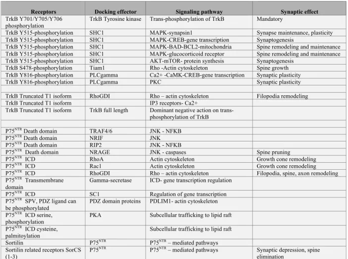
Several mechanisms have been proposed to coordinate neurotrophin receptor-to-ligand availability (see Table 3). First, incorporation of Trk receptor to the cell surface is promoted within seconds of neurotrophin exposure or neuronal activity (Du et al., 2000; Haapasalo et al., 2002). Second, only competent cells with permissive intracellular cAMP signaling increase Trk surface expression and chances to meet with the ligand (Ji et al., 2005). Third, another important mechanism for extrasynaptic Trk receptors is the translocation into lipid rafts, which may enhance responsiveness (Assaife-Lopes et al., 2010; Pereira and Chao, 2007). Local post-synaptic synthesis of BDNF and TrkB may regulate the directionality of neurotrophin signaling at the synapse (anterograde versus retrograde).
Recruitment of selective adaptor molecules discriminates signaling between the p75NTR and Trk receptors (Chao, 2003). For instance, neurotrophins activate the RhoA-JNK pathway by the p75NTR pathway that impairs the clustering of MAGUK in he postsynaptic density, elicits growth cone collapse, long-term depression and synapse elimination (Deinhardt et al., 2011; Nagerl et al., 2004; Sharma et al., 2010; Woo et al., 2005). However, Trk activates signaling pathways common to most receptor tyrosine kinases resulting from the dimerization and trans-autophosphorylation by the tyrosine kinase domains (Deinhardt and Chao, 2014; Kaplan and Miller, 2000; Reichardt, 2006).
The wave of second messengers involves PLCγ, which raises intracellular Ca2+, the Ras-Erk and PI3-kinase/Akt pathways (see table 4 for a simplified web of intracellular signaling routes). In brief, phosphorylation of TrkB-Y515 serves as docking site for the SHC1-SOS-GRB2-RAS complex that permits the activation of Pi3K/Akt-mTOR pathway for the control of protein synthesis and MAPK-CREB pathway for the control of gene transcription (Panja and Bramham, 2014; Panja et al., 2014). Phosphorylation of TrkB-Y816 serves as docking site for the PLCγ, which modulates intracellular Ca2+ level and subsequent CAMKII or PKC as effectors (Minichiello, 2009). Phosphorylation of TrkB-S478 serves as docking site for TIAM1, which activates Rac1 to promote spine growth (Lai et al., 2012).
Of all these pathways, The TrkB-Y816-PLCγ pathway seems the most important for the maintenance of long term synaptic potentiation (Minichiello et al., 2002). But the TrkB-Y515-Erk pathway is important for regulating the synaptic
responses of the glucocorticoid receptor in conditions of stress (Arango-Lievano and Jeanneteau, 2016; Arango-Lievano et al., 2015b). Very fast signaling pathway through TrkB can also mediate sodium influx that subsequently triggers fast calcium influx through voltage gated channels (Lang et al., 2007; Sasi et al., 2017).
It has been proposed that p75NTR signaling prunes silent synapses that compete with active synapses that use TrkB-mediated facilitation of synaptic plasticity and strength in a BDNF-dependent fashion (Singh et al., 2008). Therefore, mature neurotrophins may represent a synaptotrophic support whereas pro-neurotrophins may be seen as punishing signals to balance the synaptic plasticity and the resulting maintenance of competing synaptic terminals (Lu et al., 2008; Woo et al., 2005). This dual system for controlling the efficacy of the synaptic network opposes proBDNF and/or prodomain signaling via the p75NTR pathway to the mature BDNF signaling via the TrkB pathway (Giza et al., 2018; Harward et al., 2016; Tanaka et al., 2008; Yang et al., 2014). Importantly, the directionality of the signaling (anterograde versus retrograde) is not completely understood but compelling arguments suggests it could be an important determinant to modulate a synaptic engram within a specific neural network (Harward et al., 2016; Hedrick et al., 2016).
VI.1. Pre-synaptic responses
Neurotrophin ligands modulate developmental maturation of synapses by a pre-synaptic mechanism (Collin et al., 2001). Pre-pre-synaptic maturation involves the reduction of the pre-synaptic kainate receptors activity, which inhibits glutamate release (Lauri et al., 2006). Selective inhibition of TrkB receptors using a chemical-genetic approach impedes the downregulation of synaptic kainate receptors activity that controls pre-synaptic efficacy and therefore, the formation of functional synapses (Sallert et al., 2009). Consistently, the genetic ablation of BDNF is associated with delayed synaptic maturation and persistent pre-synaptic kainate receptors activity (Sallert et al., 2009).
Application of mature BDNF alters the number of synaptic vesicles, the number of docked vesicles at the active zones without affecting the reserve pool of vesicles. As a result of structural maturation, BDNF can raise the quantal release of neurotransmitters
(Tyler and Pozzo-Miller, 2001; Tyler et al., 2006). In contrast, fewer vesicles and docked vesicles are observed in the BDNF knockout mice. Therefore, BDNF might help mobilize vesicles for immediate release at existing synapse through a myosin motor mechanism (Yano et al., 2006). One of the mechanisms by which BDNF mobilizes synaptic vesicles depends on a TrkB-dependent dissociation of the cadherin-βcatenin adhesion complex (Bamji et al., 2006). Pre-synaptic structural parameters are impaired when TrkB is removed from the pre-synaptic side only or from both sides (Luikart et al., 2005). Remarkably, pre-synaptic TrkB deficiency increases the probability to contact with two or more post-synaptic densities due to the atrophy of axon terminals (Luikart et al., 2005).
Several studies have addressed the role of neurotrophin signaling in synapse stabilization. One approach is to promote destabilization of existing synapses by acute pharmacological blockade of glutamate neurotransmission (Hering and Sheng, 2001). Application of mature BDNF in the presence of APV, an inhibitor of NMDA receptors, recues the number of clusters of GFP-synaptobrevin, a marker for pre-synaptic terminals, by stabilization of existing clusters and addition of novel clusters (Hu et al., 2005). Mechanisms that translate extracellular signals into cytoskeletal rearrangements underlie morphological remodeling. The cytoskeleton is composed of actin and microtubules as well as a vast array of associated regulatory proteins. Neurotrophins may impact dynamics of filopodia, branching and synaptogenesis by local effect on cytoskeletal dynamics via cyclic nucleotides like cGMP and phosphorylation, which are used as molecular switches to regulate many of the structural proteins. For example, BDNF-induced phosphorylation of synapsin-I and Eps8 by Erk1/2 lead to actin remodeling and development of axonal terminals (Jovanovic et al., 1996; Menna et al., 2009). On the contrary, proneurotrophin causes growth cone collapse by using the p75NTR-SorCS2-rac1 pathway (Deinhardt et al., 2011). Additional experiments showed that the BDNF prodomain harboring the Met66 variation is sufficient to produce growth cone collapse by using the p75NTR-SorCS2-rac1 pathway (Anastasia et al., 2013). This means that the Met66 variation present in 25% of the general population confers a gain of function.
Structural changes in neurons often give rise to physiological consequences. Neurotrophins increase the frequency of AMPA miniature excitatory post-synaptic
currents (mEPSCs) within minutes with minimal effect on amplitude. These observations are usually attributed to a pre-synaptic change in the probability of neurotransmitter release (Tyler and Pozzo-Miller, 2001). In accordance, inactivation of pre-synaptic TrkB signaling using the truncated TrkB isoform, TrkB.T1 impaired BDNF-elicited synaptic potentiation, thus confirming a role for synaptic neurotrophin signaling (Li et al., 1998). Similar pre-synaptic effects have been observed in inhibitory neurons. For instance, BDNF increases the expression of GAD65, a GABA synthetic enzyme, as well as mIPSCs frequency without affecting amplitude (Huang et al., 1999; Ohba et al., 2005).
VI.2. Post-synaptic responses
Neurotrophins increase dendritic arbor complexity and the number of spines in several preparations (Alonso et al., 2004; McAllister et al., 1995; Sanchez et al., 2006). Knockout of BDNF does not affect spine density significantly in the hippocampus of P10-16 animals (Martinez et al., 1998). However, no addition of new spines was possible in the barrel cortex of BDNF -/- mice after sensory stimulation in contrast to the experience-dependent synaptogenesis observed in the wild-type littermates (Genoud et al., 2004). Consistently, there is an overall reduction in spine density in the hippocampus of conditional TrkB knockout mice (Luikart et al., 2005). Application of the mature BDNF or NT3 to embryonic E16 hippocampal neurons, grown in culture, can trigger the conversion of silent synapses into functional ones (Vicario-Abejon et al., 1998). Also, acute thalamocortical slices from BDNF knockout mice revealed silent synapses that are unmasked after rescuing BDNF levels back to normal (Itami et al., 2003).
Neurotrophins enhance local protein synthesis that is required for long term structural and functional synaptic plasticity (Lu et al., 2008; Tanaka et al., 2008). The post-synaptic BDNF-TrkB pathway mediates the enlargement of spine heads following synchronized pre- and synaptic spiking activities (Tanaka et al., 2008). The post-synaptic BDNF-TrkB pathway also regulates the protein content in post-post-synaptic density notably the NMDA and GABA receptor subunits (Elmariah et al., 2004; Slipczuk et al., 2009; Yamada et al., 2002). Because BDNF action is selective to active spines, it is postulated that BDNF may act as a structural tag for the selective trapping of the protein
Neurotrophins alter spine morphogenesis by changing the cytoskeleton using small GTPases to modify the dynamics of actin polymerization/depolymerization (Hedrick et al., 2016). That is, activation of Rac1/Cdc42 and inhibition of RhoA is involved in spine formation, and vice versa in spine retraction (Fu and Ip, 2007). Regulation of the small GTPase family of proteins (Rho, Rac, cdc42) by Trk and p75NTR is proposed as a mechanism to impact the dynamic structure of synapses. Indeed, the Rac1 activator, TIAM1 is directly regulated by BDNF, TrkB, PI3K and NMDA-dependent calcium levels (Miyamoto et al., 2006). Similarly, p75NTR stimulates Rac1 but dampens RhoA activity resulting in lengthening of filopodia (Gallo and Letourneau, 2004). In fact, neurotophins affects several GTPases at the same time by different receptors to impact synaptic morphology (Chen et al., 2006a; Esteban et al., 2006; Shen et al., 2006). Another signaling pathway recruited by BDNF and central to the regulation of the cellular cytoskeleton is CDK5. Several substrates of CDK5 including the WAVE proteins are known to regulate actin polymerization and dendritic spine morphology (Cheung et al., 2007; Kim et al., 2006). Post-synaptic morphology is affected by the loss of post-synaptic TrkB but not by the loss of pre-synaptic TrkB (Luikart et al., 2005). Chemical-genetic inactivation of TrkB-F616A mutant (Chen et al., 2005a) substituted for the endogenous TrkB specifically on the post-synaptic cells, is sufficient to decrease the number of dendritic spines in vivo, recapitulating the impact of chronic unpredictable mild stress and chronic corticosterone administration with no further effects when paired (Arango-Lievano et al., 2015b; Arango-Lievano et al., 2016). These observations support a cell autonomous effect of TrkB signaling on the post-synaptic architecture. Moreover, elevation of Ca2+ within spines within minutes of synchronized pre- and post-synaptic activity elicits BDNF-TrkB signaling that mediates enlargement of spine head volume (Tanaka et al., 2008). In contrast, BDNF signaling cannot shape spine head volume in absence of synchronized pre- and post-synaptic activity. Therefore, BDNF signaling orchestrates Ca2+ sensors, kinases and small GTPases to elicit structural changes of synaptic terminals.
The source of BDNF required for synaptic potentiation differed from that required for pre-synaptic differentiation. In fact, deletion of only the 3’UTR sequence of the bdnf gene, which targets BDNF mRNA to the dendrites, is sufficient to reduce spine
head volume in hippocampus, suggesting an autocrine post-synaptic pathway (An et al., 2008). This is consistent with the autocrine TrkB activation on the same spines that is crucial for structural and functional plasticity (Harward et al., 2016). Post-synaptic TrkB allows robust synaptic potentiation only when paired to a weak burst stimulation of synaptic terminal (Kovalchuk et al., 2002). However, others have reported that pre-synaptic BDNF released from CA3 neurons but not post-pre-synaptic CA1 neurons, so a paracrine pathway was essential for synaptic potentiation at these synapses (Zakharenko et al., 2003).
Neurotrophins promote stabilization of post-synaptic terminals in several preparations. Infusion of mature NGF in the septum revealed a robust potentiation of cholinergic synaptic efficacy in the septo-hippocampal neural circuit whereas infusion of NGF blocking antibodies diminished hippocampal LTP and impaired spatial memory (Conner et al., 2009). The role of BDNF in the maintenance of synapses has also been studied. For instance, blocking BDNF signaling by using anti-BDNF antibodies or overexpressing a dominant negative TrkB construct reduces mushroom spine maintenance and synaptic efficacy, accompanied by an increase in long and thin spines and filopodia (Chakravarthy et al., 2006; Sanchez et al., 2006). In agreement, the inactivation of TrkB signaling may prevent the formation of new synapses and promote the destabilization of existing spines through the post-synaptic TrkB-PI3-K pathway (Luikart et al., 2005; Luikart et al., 2008).
Contrasting with the effects of mature BDNF, the BDNF pro-peptide decreases the number of dendritic spines via a mitochondrial caspase-3 pathway (Guo et al., 2016). Prodomain of BDNF but not of NGF facilitates LFS-induced LTD, an effect that also depends on p75NTR (Mizui et al., 2015). Interestingly, the Met66 prodomain shows the opposite effect and inhibits LFS-induced LTD.. These results are consistent with a previous study describing a deficit on LFS-induced LTD in hippocampal slices of BDNFmet/met mice (Ninan et al., 2010), which suggest that the prodomain could be responsible for this effect. The LFS protocol elicits NMDA dependent LTD that relies, in part, on trafficking of AMPA receptor subunits. Mizui et al demonstrated that the Val66 prodomain facilitates LTD by promoting the surface expression of GluN2B
prodomain blocks it. These results imply a general effect of the Met66 prodomain on NMDA signaling (Mizui et al., 2015). The deficient NMDA-dependent LTP of the BDNFmet/met (Ninan et al., 2010) mice supports also this hypothesis. Unfortunately the effect on the Met66 prodomain in hippocampal LTP has not yet been reported.
This is consistent with the impact of a cleavage resistant proBDNF knockin mouse, which displayed decreased spine number, impaired LTP due to the lack of mature BDNF mediated TrkB signaling, and enhanced LTD mediated by the p75NTR pathway (Yang et al., 2014). Also, the application of proBDNF causes synaptic depression (Woo et al., 2005), which was previously associated with spine shrinkage and elimination (Nagerl et al., 2004). Removal of p75NTR by genetic methods reduces the elimination of silent pre- and post-synaptic terminals in several preparations (Cao et al., 2007; Lim et al., 2008; Singh et al., 2008; Zagrebelsky et al., 2005). Overexpression of p75NTR decreases the overall number of spines in hippocampal neurons in vivo (Zagrebelsky et al., 2005). It is assumed that the post-synaptic proBDNF-P75NTR-SorCS pathway is preferred for negative plasticity but other studies indicated that inactivation of the post-synaptic mature BDNF-TrkB pathway could do the same. In Purkinje cells, the loss of TrkB, which does not affect dendritic differentiation and synaptogenesis, impairs the developmental elimination of redundant GABAergic climbing fibers (Bosman et al., 2006). One hypothesis is that the conversion of pro-BDNF to mature BDNF could determine the nature of the response (Woo et al., 2005).
VI.3. Rapid and slow responses
Several observations of rapid synaptic neurotrophin signaling have been reported. Blocking BDNF signaling using a caged photo-activable quenching antibody revealed instructive role as synaptic potentiator within minutes (Kossel et al., 2001). Neurotrophic signaling has been shown to affect synaptic transmission within minutes by modulation of ion channel properties. Indeed, voltage-gated sodium channels, potassium channels as well as glutamate and GABA receptors have been proposed to be downstream targets of BDNF-TrkB signaling (Blum et al., 2002; Jovanovic et al., 2004; Kramar et al., 2004; Levine et al., 1998). How does rapid neurotrophic signaling translate into modifications of synapses? In vitro, high BDNF level produces repulsion
whereas low BDNF induces attraction of growth cones (Mai et al., 2009). Responses to bound BDNF gradient depend on the absolute difference rather than the relative difference in the BDNF density across the neuron. In vivo, BDNF signaling via the p75NTR results in the elimination of silent synapses whereas TrkB signaling provides a mechanism for the preservation of functional synapses (Singh et al., 2008). Moreover, fast and slow increases in BDNF concentration can differentially affect TrkB signaling (transient versus sustained) such that spine head enlargement and spine neck elongation are affected (Ji et al., 2010).
Rapid modulation of synaptic function by BDNF is attributed to both pre- and post-synaptic mechanisms. To unravel the cell autonomous mechanisms of BDNF-mediated enhancement of synaptic plasticity, TrkB signaling in the pre-synaptic sides was impaired by removal of the synaptic vesicle protein Rab3A. Knockout mice, which lack a pre-synaptic response to BDNF (miniature EPSC frequency), still display normal post-synaptic sensitivity to BDNF because Rab3a is required for the initial (<10 min) but not for the later (>10 min) phase of BDNF-enhanced transmission (Alder et al., 2005). Indeed, BDNF enhancement of postsynaptic glutamate-induced current did not differ in the mutant neurons compared to the wild type (Alder et al., 2005). One possible post-synaptic mechanism of post-synaptic potentiation by BDNF signaling is that neurotrophins induce within minutes the phosphorylation of a variety of synaptic substrates such as ion channels.
For example, NMDA and GABA receptors are phosphorylated by BDNF signaling via Erk1/2 and PKC, respectively, to regulate the probability of channel opening (Jovanovic et al., 2004; Levine and Kolb, 2000; Suen et al., 1997). In addition, BDNF induces rapid intracellular calcium transients via the Trk-PI3-K pathway and surface expression of TrpC channels (Amaral and Pozzo-Miller, 2007; Li et al., 2005). Blocking endogenous TrkB signaling decreases the frequency of spontaneous Ca2+ rises at post-synaptic sites (Lang et al., 2007). Rapid elevation of intracellular Ca2+ via Trk-PLCγ-IP3 pathway was also reported (Du and Poo, 2004). Moreover, deletion of the Y785 docking site in TrkB knock-in mice revealed the importance of the PLCγ over the ERK and PI3K pathways for the maintenance of synaptic function and the rapid
pre-signaling has been shown to be dependent on gradient of second messengers like cAMP to allow functional and structural synaptic responses (Ji et al., 2005; Mai et al., 2009).
Robust stimulation evoking long-term potentiation (lasting >180 min) elicits a large increase in BDNF secretion persisting 5-12 min beyond the stimulation period. Weaker stimulation patterns leading only to the initial phase of synaptic potentiation (about 35 min) are accompanied by a smaller increase in BDNF secretion lasting <1 min (Aicardi et al., 2004). Prolonged BDNF signaling triggered by robust synchronized pre- and post-synaptic activities allows synapse-specific potentiation and structural stabilization changes that are dependent on protein synthesis (Aicardi et al., 2004; Tanaka et al., 2008). To be specific, the newly synthesized plasticity-related proteins must be captured only at the active sites by a transient synaptic tag. TrkB signaling endosomes has been proposed to be a synaptic tag (Lu et al., 2008). Many BDNF-regulated genes are newly synthesized plasticity-related genes (Glorioso et al., 2006). The functions of several activity-regulated genes have been investigated in the context of synaptic potentiation: (i) Arc mRNA, which expression is sustained in active spines, participates in structural changes of spines associated to the consolidation of synaptic potentiation by the remodeling of actin cytoskeleton (Bramham, 2008); (ii) Homer-1 mRNA is captured only in active spines and regulates the stability of synaptic function (Okada et al., 2009; Szumlinski et al., 2006); (iii) bdnf exon IV mRNA modulates the shape and potency of neuronal networks (Barco et al., 2005; Hong et al., 2008).
Proteomic approaches have revealed that BDNF induces widespread changes in synaptic protein content and up-regulates components of the translation machinery, which requires hours (Liao et al., 2007). Several mechanisms can account for this effect. On one hand, BDNF relieves the inhibitory control of the microRNA miR-134 on synaptogenesis (Schratt et al., 2006). On the other hand, BDNF employs the post-synaptic TrkB-PI3K-mTOR pathway to regulate the local post-synaptic translation of a select group of mRNAs (GLUR1, Arc, BDNF, TrkB, Homer and CamKII…) (Schratt et al., 2004; Slipczuk et al., 2009; Wang et al., 2010). Therefore, rapid and slow signaling may serve a dual function. Promoting transcription of plasticity-dependent genes and the capture of their mRNA products where TrkB signaling endosomes persist as putative