Development and investigation of devices for ultrahigh speed gastrointestinal Optical Coherence Tomography imaging
by Kaicheng Liang
S.M. Electrical Engineering and Computer Science Massachusetts Institute of Technology, 2014
B.S. Biomedical Engineering Duke University, 2010
Submitted to the Department of Electrical Engineering and Computer Science in Partial Fulfillment of the Requirements for the Degree of
Doctor of Philosophy at the
MASSACHUSETTS INSTITUTE OF TECHNOLOGY June 2018
2018 Massachusetts Institute of Technology. All rights reserved
Signature of Author
Certified by
Accepted by
Signature redacted
Department of Electrical Engineering and Computer Science May 23, 2018
Signature redacted
Professor James G. Fujimoto Elihu Thomson Professor of Electrical Engineering and Computer Science Thesis Supervisor
Signature redacted
/
P fgor Leslie A. Kolodziejski Professor of Electrical Engineering and Computer Science Chair, Department Committee on Graduate StudentsMASSACHUSETS INSTITUTE
OF TECHNOLOGY C
U3
Development and investigation of devices for ultrahigh speed gastrointestinal Optical Coherence Tomography imaging
by Kaicheng Liang
Submitted to the Department of Electrical Engineering and Computer Science in Partial Fulfillment of the Requirements for the Degree of Doctor of Philosophy
ABSTRACT
Diseases of the gastrointestinal (GI) tract are typically diagnosed by random biopsy of tissue, which samples only a small area and often misses focal neoplasias. Existing endoscopic visualization tools including white light endoscopy, narrowband imaging and confocal laser endomicroscopy have enabled in vivo assessment to guide biopsies, but suffer from technical limitations and have demonstrated suboptimal sensitivity and specificity to neoplasia. Optical Coherence Tomography (OCT) can generate in vivo, 3-dimensional microscopic imaging. Recent efforts in ultrahigh-speed OCT systems for endoscopic applications have shown promise, but devices had limited fields of view and imprecise beam scanning mechanisms, limiting image quality and coverage.
This thesis develops a wide range of new fiber optic devices that substantially extend OCT capabilities in the GI tract, either by greatly increasing field of view for wide field mapping of entire luminal organs, or achieving high precision 2-D beam scanning with compact actuators for
in vivo microscopy. Piezoelectrically actuated fiber scanning devices enable forward viewing for
focal inspection, while micromotor actuators combined with pneumatic or piezoelectric mechanisms enclosed in tethered capsules generate side viewing over large areas. The work also advances the emerging paradigm of gastrointestinal screening without use of sedation, which promises to lower costs of screening and improve access for a broader population. Design, fabrication and benchtop evaluation of devices, as well as pre-clinical and clinical imaging protocols are reported. Results from validation studies in living swine, and human patients in collaboration with the Veterans Affairs Boston Healthcare System are discussed. The thesis work demonstrates new imaging modalities for in vivo detection and diagnosis of GI pathology that could have important applications in disease screening, surveillance, and therapeutic procedures. Thesis Supervisor: Professor James G. Fujimoto
Acknowledgments
I am forever grateful to my advisor Jim Fujimoto for his mentorship. First, he gave me the gift of being an MIT student, which I will treasure, and will always make me look smarter than I actually am. Second, he taught me how to be a rigorous and meticulous scientist, although I will still forget my scalebars from time to time. Third, he showed me how to lead, by example, without compromise, and in service to community and society. I will strive for the same (but maybe do a few things differently). And finally, he proved that scientists through their work can improve the lives of millions, and so we should dedicate our lives to trying. As I enter the next phase of my career and pursue scientific leadership, these lessons will always be on my mind. If I can be a little nicer than Jim Fujimoto while achieving a little less success than he has, then I will be content. However it is much more likely that I will end up a lot less nice and still a lot less successful.
My sincere thanks to the VA Boston Healthcare System for hosting my research. I dream to find a hospital back home with medical professionals and patients who will support cutting edge research with the same commitment, energy, and enthusiasm. Dr. Hiroshi Mashimo, fearless endoscopist, will always be my role model in his mastery of his chosen art, passion for education and mentorship, and commitment to interdisciplinary collaboration (read: saint-like toleration of bungling engineers in his clinic). The professionals of the Endoscopy unit have been gracious, patient, and incredibly supportive of our work. Having worked with veterans will always be one of the greatest privileges of my life. For each and every one of you who gulped down a tethered capsule without batting an eyelid, thank you.
Special thanks to our collaborator Vijay Jayaraman, founder of Praevium Research. Your brilliant, one-of-a-kind VCSEL has enabled my entire PhD.
How do I express my gratitude to my colleagues at MIT, who have taught me everything I know, endured my painfully ignorant questions, and inspired me to strive for excellence in all things? Hsiang-Chieh Lee PhD and Tsung-Han Tsai PhD were my first mentors, teaching me to assemble probes, polish GRIN lenses, build FC/APC connectors and countless other tasks, and continuing to dazzle me in their new careers as budding thought leaders. Chao Zhou PhD was a kind supervisor, and his suggestion of "why don't you learn how to build a probe?" set me on a path that defined my PhD. Zhao Wang PhD was an incredible collaborator - the year over which we unraveled the mysteries of the cycloid scanner was the purest agony and joy that I will probably ever experience (perhaps until I have a child). Osman Ahsen PhD worked with me longer than anybody else, and even though we often disagreed about almost everything, I was always in awe of his intelligence and commitment to his beliefs. Benjamin Potsaid PhD was the ultimate resource in OCT engineering, but more inspirational was his supreme composure in the face of adversity, whether it was seeing that I had snapped down a VCSEL yet again, or when he methodically helped me debug a system issue the night before a critical swine experiment, or when we prepared a complex grant proposal submission. Lucas Cahill and Jason Zhang were wonderful office mates. The ophthalmology team has been the perfect foil to endoscopy, and I have enjoyed our vigorous discussions while admiring your long list of publications. Special thanks to Dr. Giovanni Traverso, who helped us bring the swine model back to our lab for the first time in a very long time.
I'm grateful to A*STAR for their generous funding, which was a helpful incentive for my lab to hire me. I'm even more grateful to my parents, who have had to live half the world away from their only child for nearly a third of my life, while supporting me unconditionally from afar. My partner Michelle has stuck with me for exactly all of that time, inspired me with her own academic adventure, and continues to be a pillar of encouragement and comfort. Thank you.
Contents
1. Introduction: Clinical motivation and state of the art
7
a. Barrett's Esophagus (BE) and dysplasia
7
b. Pathogenesis and natural history of BE
8
c. Ablative therapy and challenges
10
d. Narrowband imaging (NBI) and enhanced wide field assessment
11
e. Confocal laser endomicroscopy (CLE) for high resolution
13
inspection
f. Volumetric laser endomicroscopy (VLE)
15
g. Scope of the thesis
17
2. Forward viewing endomicroscopy with piezoelectrically actuated
20
fiber scanning
a. Introduction
20
b. Materials and Methods
21
c. Results and Discussion
30
3. Ultrahigh-speed OCT capsule for enface imaging: studies in swine
36
a. Introduction
36
b. Materials and Methods
40
i. Imaging system 40
ii. Catheter design and assembly 42
iii. Scan actuation and characterization 46
iv. Animal imaging and scan protocols
49
c. Results and Discussion
51
4. Tethered capsule enface OCT of Barrett's Esophagus and dysplasia:
68
b. Materials and Methods
69
c. Results and Discussion
71
d. Bonus supplement: OCT angiography
74
5.
Cycloid scanning for wide field side viewing endomicroscopy
81
a. Introduction
81
b. Materials and Methods
84
i. Cycloid scan
85
ii. OCT system
87
iii. Capsule construction
88
iv. Scanner control and data acquisition
89
v. Image calibration and reconstruction
90
vi. Clinical imaging protocol
93
c. Results and Discussion
94
d. Supplementary Information
101
6. Tethered capsule en face and cross-sectional OCT imaging
109
in unsedated patients
a. Introduction
109
b. Materials and Methods
111
i. Imaging system and tethered capsule
111
ii. Patient recruitment and imaging procedure
112
iii. Data analysis
114
c. Results and Discussion
116
d. Bonus supplement: Prague C&M reading of capsule OCT
130
7. Conclusion and Future Work
135
Chapter 1
Introduction: Clinical motivation and state of the art
Imaging of luminal organs in vivo can have enormous impact on clinical decision making, including diagnosis, treatment planning, and real-time therapeutic guidance. In gastroenterology and the gastrointestinal (GI) tract, where advanced endoscopes have been used for decades, clinicians are no strangers to cutting edge technology for imaging and therapy. This chapter will briefly review some important clinical needs for real-time in vivo imaging, as well as a number of existing commercially available technologies that have attempted to meet these needs, with a focus on applications in the upper GI tract including the esophagus and proximal gastric cardia.
Barrett's Esophagus (BE) and dysplasia
Barrett's Esophagus (BE) is a premalignant condition that is associated with, and believed to be a consequence of, chronic (>5 years) gastroesophageal reflux disease (GERD). Acid exposure in the distal esophagus over a prolonged period can lead to metaplastic changes in the squamous epithelium, producing a columnar phenotype. BE can be observed during standard esophagogastroduodenoscopy (EGD) as having a salmon pink appearance of the mucosa, and is confirmed histologically by specialized intestinal metaplasia (IM) with presence of goblet cells, although presence of goblet cells is not required by Britain and Japan for BE diagnosis and is being debated[l]. The standard definition of BE progression is from non-dysplastic BE to low-grade/high-grade dysplasia (LGD/HGD) to esophageal adenocarcinoma (EAC), however the
EAC, which in recent decades has become the cancer with the fastest growing incidence in the Western world, although the actual incidence in the United States is still relatively low at less than
5 cases per 100,000 people[3]. Specific demographics including male gender, Caucasian race, and obesity are important risk factors for BE and EAC. Although chronic GERD is one of the most important risk factors to indicate endoscopic screening for BE, it has been noted that over 40% of EAC patients never report GERD symptoms[4]. After BE is diagnosed, patients are enrolled in regular endoscopic surveillance, during which biopsies are obtained every 2 cm of BE length in 4 quadrants, known as random biopsy or the "Seattle protocol". After a surveillance endoscopy is negative for dysplasia, risk of neoplastic progression is deemed reduced, and patients are recalled after a year and then every 3-5 years for follow up endoscopies and biopsies.
Pathogenesis and natural history of BE
The pathogenesis of BE remains unclear, and there are multiple controversies regarding the onset and progression of the disease. BE is observed manifesting as different lengths in different patients, therefore it was previously believed that BE may be occurring at the GEJ and then progressing longer over time due to increased acid exposure from a weakened lower esophageal sphincter[5]. However, multiple recent studies found that BE lengths remained relatively stable over a follow up of several years[6], suggesting that a short/long BE phenotype may be occurring relatively rapidly, and then subsequently changing little.
In general, BE develops from 'multi-potential' stem cells, which can develop into a mixture of gastric and intestinal cells. As a result, a BE segment can contain a range of mucosal phenotypes,
where gastric and intestinal epithelium coexist as a mosaic, although the former decreases in prevalence when moving from distal to proximal esophagus. As the BE evolves proximally, glands multiply by gland fission, resulting in 'patches' or clusters.
There are disagreements[7] on whether BE originates from squamous epithelium of the normal esophagus, or from the proximal gastric cardia. In the first and more traditional theory, esophageal glands and ducts of the squamous epithelium contain stem cells that are stimulated by acid reflux and encouraged to differentiate abnormally into intestinal metaplasia. Esophageal glands have been observed to contain dysplasia, adenoma, or even carcinoma in situ, further boosting this theory. In the competing theory, the columnar cells of the proximal cardia migrate up into the acid-damaged distal esophagus. This was initially dismissed because gastric-type mucosa does not contain goblet cells, unlike intestinal mucosa including intestinal metaplasia. However, it has been consistently found that regions of BE can contain a range of tissue types including gastric-type epithelium, suggesting that intestinal metaplasia is a two-step process -migration of gastric epithelium into the distal esophagus, which then undergoes intestinalization. This further implies that columnar-lined mucosa without goblet cells may simply be a precursor lesion to BE, and is thus of no less malignant potential.
Supporting this theory is the histological observation that there exists a region of 'junctional' tissue between the stomach and the esophagus that has been termed 'cardiac mucosa'[8], which appears to exist in all adult individuals but becomes longer in patients with BE. The cardiac mucosa consists of both cardiac glands and oxyntocardiac glands, the former with metaplastic potential. In patients with prior esophagectomy[9], cardiac mucosa (columnar
metaplasia) is said to develop at the neo-GEJ within the first 1-2 years, with BE only manifesting years after. These observations appear to support the theory that BE arises from the stomach.
Ablative therapy and challenges
Fortunately, there are now a multitude of endoscopic techniques for the treatment of dysplasia and early carcinoma in the esophagus that do not require surgery. Radiofrequency ablation (RFA) is now largely the standard of care, due to notable success in a large clinical trial that showed complete eradication of dysplasia in 91% of LGD patients and 81% of HGD patients[10]. Other modalities including endoscopic mucosal resection[1 1] and cryogenic ablation[ 12] are also available, which are often combined with RFA to further improve outcomes. In these treatment protocols, ablation is used to completely eradicate an entire segment of BE, which at completion is termed complete eradication/remission of intestinal metaplasia (CEIM/CRIM). However, in esophagectomy studies where large segments of esophagus were histologically analyzed[13], dysplasia was found to be highly focal, such that random biopsy would likely miss these regions. Without a confirmatory biopsy to diagnose dysplasia, therapies however effective would not be administered in a timely fashion. Therefore, there is great interest in imaging modalities that can be used during endoscopy to evaluate the esophageal mucosa for abnormalities, possibly detecting dysplasia in real time without excisional biopsy and the consequent delay for histopathology. Such a rapid protocol for dysplasia detection might motivate immediate treatment for dysplasia during the same endoscopy procedure, reducing delays to treatment and risk of loss to follow up.
While RFA has found widespread success in the eradication of dysplasia, recent large scale studies of durability at academic centers have raised serious concerns of recurrent IM and dysplasia after completion of a treatment course. In one large study of a single center where the RFA technique was first developed[14], 24% of patients developed recurrence of BE or dysplasia, at an incidence rate of 9.6% per year. The mean time to recurrence was 1.88 years after CEIM. In a larger multi-center study[15], the incidence rate of recurrence was 33% at 2 years after CEIM, and 22% of all recurrences contained dysplasia. It was observed that up to half of recurrences were found at the gastroesophageal junction (GEJ) and ~1 cm distal to the top of the gastric folds, however standard endoscopic visualization has been poor at identifying accurately the GEJ especially after ablation[ 16]. Therefore, there is an unmet need to rigorously evaluate the GEJ after RFA treatment for recurrent disease and dysplasia. The high rate of recurrence also suggests that patients will need continued endoscopic surveillance for an indefinite period, further motivating the enhancement of diagnostic yield/efficacy of endoscopic visualization.
Narrow band imaging (NBI) for enhanced wide field assessment
Since over a decade ago, there has been substantial effort by multiple research and clinical groups to improve the superficial visibility of mucosal structure and vasculature in the GI tract. A technique known as chromoendoscopy, in which the superficial mucosa is stained with a dye and then assessed with an endoscope, was found to be highly diagnostic in multiple studies[17-19], but has fallen out of favor due to perceived procedural complications and time expense. A Japanese group proposed a type of 'electronic chromoendoscopy', whereby the use of blue (415 nm) illumination and a combination of detection filters (415 nm, 445 nm, 500 nm) could enhance the
visualization of the mucosal surface[20]. This became known as 'narrow band imaging' (NBI). After initial pilot studies by clinicians in Japan, the technique began to catch on internationally after a number of high profile studies by leading groups in the US[21] and Europe[22]. NBI was found to enhance the visibility and contrast of the superficial mucosa, which contained both a mucosal and vascular pattern. NBI was marketed by Olympus as an enhanced imaging technology that was integrated into conventional endoscope systems and could be switched on/off whenever needed. Early seminal papers by Japanese groups using magnification and chromoendoscopy obtained and categorized a range of mucosal/vascular patterns observed in both upper[23] and lower[24] GI pathology, part of which became known as the Kudo pit pattern system. It became clear that NBI could visualize similar mucosal structure without the procedural difficulty of chromoendoscopy. A multitude of pattern classification systems were later developed using NBI over time[25, 26], however it became well established that NBI was extremely high performing. Early studies demonstrated up to 100% sensitivity and 99% specificity in detecting HGD in BE[21 ], and while later multi-center studies reported more conservative results, sensitivity/specificity performance for dysplasia detection hovered in the range of 80-90% when assessed by experts[26].
While NBI in the upper GI tract continues to be a leading diagnostic technique, it has notably failed to perform adequately when assessing the GEJ after RFA treatment for recurrence of BE, despite high quality visualization of the mucosal patterns. In a study of 21 patients by a leading European group, the sensitivity/specificity for identifying recurrent intestinal metaplasia were 65% and 46% respectively[27]. Another study of 198 patients found that any endoscopic finding using white light endoscopy (WLE) or NBI had only 59% sensitivity and 81% specificity to recurrence[16]. It has been suggested that high resolution imaging for the detection of goblet
cells would confirm the presence of specialized intestinal metaplasia, however ongoing controversies over histological criteria for BE also apply. Therefore, mucosal patterns and any modalities that enhance exclusively enface microstructure may not be sufficiently diagnostic for this specific yet extremely important application.
Confocal laser endomicroscopy (CLE) for high resolution inspection
WLE or enhanced endoscopy with NBI has yet to achieve optimal sensitivity/specificity to dysplasia, even in expert hands. Upon locating a region of interest using a wide field modality such as WLE/NBI, it may be desirable to examine the region with high resolution. There have been successful efforts in miniaturizing confocal microscopy into compact endoscopic devices, which is known as confocal endomicroscopy[28]. Using intravenous or topical fluorescent agents, and a blue laser for excitation, cellular imaging could be achieved. For example, when using intravenous fluorescein or topical acriflavine, an excitation wavelength of 488 nm may be used. About a decade ago, Pentax commercialized an endoscope system with an integrated confocal microscope at the distal end, in collaboration with Optiscan. Tissue could be imaged in contact with a viewing window adjacent to the endoscope optics. The distal microscope contained an electromagnetic mechanism for actuating an optical fiber in 2 axes, and a third mechanism for depth (axial) focus scanning, which led to substantial bulk but generated high quality images. This became known as endoscopic confocal laser endomicroscopy (eCLE), and quickly led to a number of compelling studies[29] such as the detection of BE-associated neoplasia with sensitivity/specificity of 93%/98% in 63 patients[30]. A competing technology also became commercially available, developed by Mauna Kea Technologies (in reference to the telescope observatories on the Hawaiian summit)
and branded as Cellvizio. The device was a small diameter catheter probe that could be passed through the 2.8 mm diameter accessory channel of a standard endoscope. Images were acquired via a 2-dimensional fiber bundle, thus no bulky scanning mechanisms were necessary. This device became known as probe-based confocal laser endomicroscope (pCLE).
Pentax/Optiscan (eCLE) Mauna Kea Cellvizio (pCLE)
Field of view 475 pm 240-600 ptm
Transverse resolution 0.7 Vtm 1-5 ptm
Axial (depth) resolution 7 pm 20-30 ptm
Imaging depth 250 pm 60-130 im
Frame rate 0.8-1.6 frames/sec 12 frames/sec
Device diameter 13 mm 2.5-2.7 mm
Table 1. Summary of specifications for commercial endomicroscopy. Adapted from [28, 29].
pCLE images were objectively lower quality than eCLE (poorer resolution and depth penetration, see Table 1) however a number of ergonomic advantages proved to be the winning recipe, including the small diameter catheter size (allowing use in any existing endoscope systems) and the rapid frame rate (minimizing motion artifacts) of pCLE. Eventually Pentax discontinued their confocal endomicroscopy product, while the Mauna Kea Cellvizio continued to be used in a number of clinical studies, although reports have complained of difficulty in obtaining stable motion-free images with adequate cellular visibility, suggesting need for substantial operator skill/experience[3 1]. The approximately half millimeter, highly limited field of view exacerbates problems with stabilizing images, particularly at the GEJ where there is substantial patient motion,
and also precludes the ability to efficiently inspect a large BE segment, such that a widefield modality is typically required in combination. In a large multicenter trial of 101 patients[32], the sensitivity/specificity of pCLE in combination with WLE/NBI for detection of BE neoplasia was 76%/84%. Notably, use of pCLE alone had poor performance (63% sensitivity, 93% specificity). A group at Rice University has worked on developing a low cost but functionally equivalent analog to the Mauna Kea system, using proflavine as a topical stain and 450 nm for the excitation laser. A number of clinical studies have been performed with this technology[33], some notably in low resource communities[34], to great success and highly promising diagnostic performance.
Volumetric laser endomicroscopy (VLE)
Optical coherence tomography (OCT) in the GI tract has been studied extensively for nearly two decades; in the year 2000 some of the first GI OCT images were reported from virtually the entire GI tract, including the esophagus, stomach, colon, duodenum and terminal ileum[35]. Early imaging speeds were infeasible for in vivo imaging, but the swept source speed revolution promised a wide range of realistic applications for OCT in gastroenterology[36, 37]. A group at Massachusetts General Hospital (MGH) developed an OCT balloon for a much larger field of view coverage than previous small-diameter catheter probes[38, 39]. This device was later commercialized by NinePoint Medical with their OCT product Volumetric Laser Endomicroscopy (VLE)[40]. VLE generates a volumetric scan over 6 cm length of the esophagus and the entire circumference, with balloon diameters 14/17/20 cm. Over a 90 second pullback scan, 1200 cross-sectional frames are acquired. The system imaging speed is 50 kHz axial scan rate, and the
rotational frame rate is 12.2 Hz[41]. The transverse and axial resolution is 40 urn (FWHM) and 7 um (tissue).
Early studies with VLE focused on understanding tissue features using precise ex vivo histological correlation[42, 43], namely visibility of layered structure, signal intensity of surface layer versus subsurface, and the presence of atypical glands/ducts. These studies were largely inspired by OCT studies nearly a decade ago that first proposed these features[44, 45]. Leggett et al.[42] showed 86% sensitivity and 88% specificity for BE dysplasia in 50 EMR specimens (34 HGD, 16 LGD + NDBE) from 27 patients, although important limitations of the study include images acquired ex vivo and one of the image readers was also responsible for developing the diagnostic criteria. Swager et al.[43] found worse performance of 83% sensitivity and 71% specificity, and used only selected cross-sectional images as well as a variable scoring system for each VLE feature arbitrarily adjusted to optimize diagnostic performance. Clinical studies by a number of leading groups investigating interobserver agreement[46] and learning curve have also been recently published.
In vivo studies of diagnostic performance will require precise histological correlation to
imaging features in order to validate diagnosis. The MGH group had previously pioneered the use of a laser to produce marks on tissue guided by imaging[47, 48]. While the concept laser marking generates an impression of precision, the production of a visible mark with continuous-wave lasers can take from one to several seconds, which is likely to be compromised by physiological motion. The latest VLE systems carry this marking capability, and clinical trials of feasibility and accuracy of laser marking to guide biopsies indicated by VLE features are ongoing.
Scope of the thesis
The primary aims of this thesis are the development of innovative, minimally invasive imaging devices with applications in studying diseases of the gastrointestinal tract, pre-clinical and clinical validation of these new technologies, and comprehensive analysis of novel imaging data to assess potential clinical utility in future diagnostic and pathogenesis studies. While these studies focus on the use of OCT to deliver ultrahigh speed 3-dimensional (3-D) optical imaging, these devices are optical scanners that can be generalized for any flying spot optical imaging or microscopy modality.
Chapter 2 is a continuation of the author's Master thesis work in developing ultrahigh speed forward imaging probes based on piezoelectric actuation of a resonant scanning fiber, and describes clinical translation and validation in patients. The technology was further miniaturized and packaged for in vivo endoscopic OCT imaging, where the probe was introduced in the accessory channel of a colonoscope. OCT angiography could be obtained by reducing the scan field to achieve oversampling. Imaging was performed in patients undergoing colonoscopy. Imaging of a range of GI mucosa was performed, including a hyperplastic polyp, for which an altered crypt pattern was observed in enface views.
Chapter 3 describes a tethered capsule device for imaging at speeds of 10-20x faster than previously reported OCT capsules. Capsules can cover large areas of luminal organs but require high imaging speeds and precision optical scanning for high quality 3-D and enface imaging. In
this work, a tethered capsule with two independent scan mechanisms - a micromotor and a pneumatic scanner was developed, imaging at 1 MHz axial scan rate for rapidly acquired enface
views. Validation results in living swine esophagus and rectum are presented.
Chapter 4 describes the successful clinical translation of high speed, en face imaging capsule technology to patients with BE. Wide field luminal imaging has the potential to detect focal areas of dysplasia. A simplified scanning capsule containing only a micromotor for precise circumferential scanning was used to image up to 24 cm length of the esophagus, in sedated patients during endoscopy. 18 patients were imaged to investigate optimal design and imaging parameters. Novel en face OCT features of dysplasia including dilated glands and irregular mucosal patterns were reported, in combination with traditional cross-sectional OCT features.
Chapter 5 describes a substantial improvement in tethered capsule precision scanning by the introduction of resonant fiber scanning. The combination of resonant scanning and micromotor scanning dramatically improved scan precision and speed from the earlier pneumatic design (Chapter 3). The device generated a novel scan trajectory known as a cycloid. Using the micromotor as the slow axis scanner, the cycloid scan could rapidly acquire 2-D en face images (or 3-D volumes) with no flyback or reset downtime. The micromotor could be slowed down to achieve oversampling for OCT angiography. This rapid 2-D scan mechanism has applications not only in OCT but any benchtop flying spot microscopy modality. Imaging was performed in the living human rectum at 3 volumes/sec over a 1 mm x 38 mm strip field of view.
Chapter 6 reports a clinical case series of 16 patients imaged with a micromotor tethered capsule without use of sedation. Volumetric and enface imaging of the esophagus and BE segment was performed over large fields of view of 4 x -10 cm in 10 seconds. Image quality metrics including tissue contact and longitudinal uniformity were assessed in relation to BE segment length and presence of sliding hiatal hernia. Image features including atypical gland clusters and irregular mucosal patterns were investigated to discover associations with patient history (pathology and treatment history).
Chapter 2
Forward viewing endomicroscopy with
piezoelectrically actuated fiber scanning
Introduction
Endoscopic optical coherence tomography (OCT) using minimally invasive catheters is an emerging imaging modality in gastroenterology[42, 49]. Early studies demonstrated side viewing catheters that scanned the OCT beam by proximal rotary and pullback actuation of a torque cable[50]. This design enabled imaging of luminal organs and continues to be the workhorse of commercially available catheter OCT systems[42]. Proximal actuation via a long catheter with multiple bends produces non-uniform rotational and/or longitudinal distortion[41, 51]. Recent catheters have used distal rotary scanning micromotors, reducing rotational distortion[52-54]. Our group developed new methods for side viewing OCT that use distal actuation for 2-dimensional scanning[55]. Distal scanning enables OCT angiography (OCTA), which uses repeated scans to detect motion contrast from blood flow to visualize microvasculature and has been used extensively in ophthalmology[56]. Our group recently demonstrated endoscopic OCTA using distal micromotor scanning[52, 57].
Forward viewing OCT catheters are of clinical interest because they generate images analogous to endoscopic views[24], and can examine small regions of interest (ROLs) at high resolution. Forward viewing devices using microelectromechanical system (MEMS) [58] or
piezoelectric actuators[59, 60] enable distal 2-D scanning, and are thus immune to catheter bending. These devices exploit mechanical resonances for scanning and are uniquely suited for high speed OCT due to their high scan speed[61, 62].
This chapter reports a forward viewing OCT piezoelectric probe and endoscopic imaging in the human gastrointestinal (GI) tract in vivo. The probe has a 3.3 mm diameter and 20 mm rigid length, and can be used into the endoscopic accessory channel, enabling use in existing endoscopic workflows. Volumetric OCT and OCTA was performed at 1 MHz axial scan (A-scan) rate to image colonic mucosa and a hyperplastic polyp in human patients. The high speed laser was a critically enabling factor in clinical translation of OCT/OCTA, particularly in endoscopy where patient motion is significant and rapid volumetric display with adequate image sampling is essential. The ability to perform high A-scan speed endoscopic forward viewing OCT has clinical potential for providing volumetric and enface microscopic tissue assessment.
Materials and Methods
This work was performed in collaboration with the VA Boston Healthcare System (Dr. Hiroshi Mashimo MD PhD and staff at the Endoscopy Unit), Thorlabs (Dr. Benjamin Potsaid) and Praevium Research (Dr. Vijaysekhar Jayaraman) for use of prototype swept source technology. Our early investigations into piezoelectric scanning were supported by Prof. Xingde Li's group at Johns Hopkins University who provided us with designs for high voltage amplification circuits and imaging probes for testing purposes.
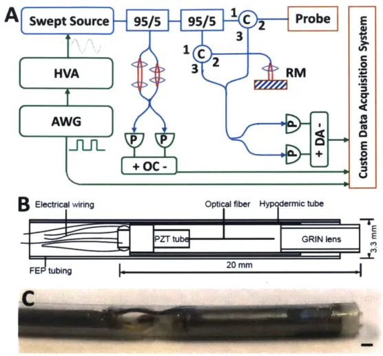
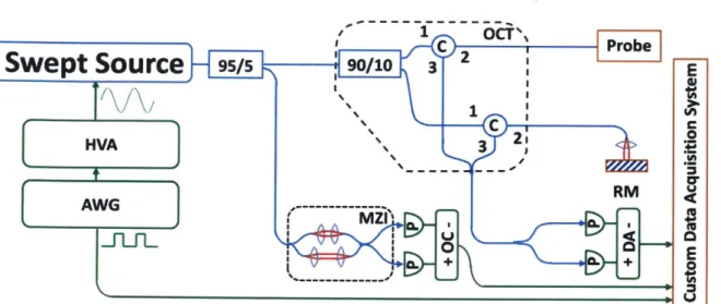
The OCT system[52] (Fig. IA) used a dual circulator interferometer with a MEMS vertical cavity surface emitting laser (VCSEL) swept source at 1310 nm (Thorlabs/Praevium)[63] running at ~500 kHz sweep rate, producing a ~1 MHz bidirectional A-scan rate. The exact VCSEL repetition rate was set to a high integer multiple of the probe resonant frequency, and the waveform generator used to drive the VCSEL was also used to clock the D/A board (National Instruments), simplifying electronic synchronization. The OCT interferometer and detector sensitivity was 103 dB at an incident power of 35 mW on the sample. The probe one-way transmission was 80%; therefore, the effective system sensitivity was ~100 dB. Losses were due to imperfect circulators/couplers (AC Photonics), use of fiber mating sleeves instead of splices, and imperfect optical surfaces of the probe lens and fiber cleave (Oxford angle cleaver). The imaging range was 2.1 mm in air and the axial resolution was 9 pm in tissue. The A/D board (AlazarTech 9370) was optically clocked up to 1.1 GHz by a Mach-Zehnder interferometer. Optical clocking with linear in k sampling enabled direct Fourier transformation, simplifying post-processing. A D/A board (National Instruments PCIe-6323) generated amplitude-modulated sinusoidal waveforms to drive a piezoelectric actuator, which were synchronized to the start of each volumetric acquisition. The phase shifted sinusoidal waveforms produced a spiral (Fig. 3B) fiber scanning trajectory.
A
Swept Source
95/5
95/
C2
Probe
E
C C 2 HVA 3RM U) AWGa P +0C-",1B
Electrical wviring Optical fiber H ermic tubePZTtt -GRIN lens E
FlE tubing20m
C
Fig. 1. (A) Ultrahigh speed OCT system. HVA: high voltage amplifier, AWG: arbitrary waveform generator, P: photodetector, C: circulator, RM: reference mirror, OC: optical clock, DA: differential amplifier. (B) Schematic and (C) photo of forward viewing probe (scale bar 1 mm).
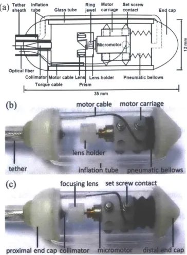
The resonant fiber scanning probe (Fig. I B-C) was similar to previous devices [5 9, 60, 64]. An optical fiber was mounted to the tip of a piezoelectric (lead zirconate titanate) tube (Boston Piezo-Optics, Bellingham MA) of 6 mm length, 1.5 mm outer diameter, 0.3 mm wall, and quartered electrodes for 2-axis actuation. <20 mm rigid length of the probe was necessary for passage through the 3.7 mm endoscopic accessory channel; therefore, piezoelectric actuator length was minimized, costing a reduction in actuating range. The fiber was aligned to a 1.8 mm diameter
deep in tissue contacting with lens). The piezoelectric actuator, optical fiber, and lens were housed in a 12G hypotube. The entire device was encapsulated in American Wire Gauge 9 fluorinated ethylene propylene tubing (Zeus) with no exposed conductive surfaces, and sealed watertight using medical epoxy.
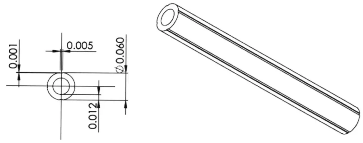
The piezoelectric tubular actuator was commercially available (e.g. Boston Piezo-Optics). At this dimension (1.5 mm OD and smaller), electrically separate electrodes on the outer surface are typically unavailable; the tubes are shipped with the outer surface uniformly metallized. The tubes were sent out to a separate vendor for laser etching of the electrodes. The depth of etch was selected based on the piezoelectric actuator vendor's specification of the electrode thickness (Fig. 2). The laser etching vendor was asked to verify electrical isolation of the electrode sectors after each etch, since there is variation in the electrode thickness and a preset etch depth may not fully separate the sectors.
0O 0.005 NO
Fig. 2. Dimensions (inches) of piezoelectric tube cross section and etch specifications.
The piezoelectric tube was hand-soldered under a magnifier to a multi-conductor cable (Cooner Wire) with 4 connections to each electrode and one earth connection to the metallic hypotube housing. Soldering to the piezoelectric tube's nickel electrode required a specialized flux
(Superior Flux) and so did soldering to the stainless steel housing (LA-CO flux). Care was taken to solder to the nickel at around 500'F (under 600'F) to prevent oxidation. The piezoelectric actuator was driven by waveforms from a 4-channel current limited 50x amplifier. This amplifier circuit was previously constructed by Tsung-Han Tsai PhD based on designs from Prof. Xingde Li's group. The output voltage after amplification was 40 V peak amplitude A.C., within the recommended limit (42 Vp) from the International Electrotechnical Commission 60601-1 medical electrical apparatus standards[65]. Electrodes on the actuator were precision laser etched (Laser Impressions, Sunnyvale CA) for symmetric actuation. It was also important for the fiber mount to be centered and symmetric in the tube, so that resonance frequency and amplitude would be similar in both axes. The fiber was centered in the tube with a ring jewel bearing (Swiss Jewel, Philadelphia PA) and fixed with heat-cured epoxy for optimal rigidity, ensuring a high (>100) resonant quality (Q) factor. The ring jewel was custom fabricated with a center hole very closely fitting the diameter of 125 ptm stripped bare fiber, and outer diameter very closely matching the inner diameter of the piezoelectric tube. The resonating fiber deflection was measured using a consumer webcam CCD sensor fitted with a microscope objective.
Deflection range I mm vs Frequency / Hz
B
Ao.9B
Axis 1 0.8- - Axis 2 0.70
E -0.6-0.5 0) 6. 5 0.3 0.2-0.1 1750 1755 1760 1765 1770 1775 Frequency / HzFig. 3. (A) Frequency response of the fiber scanner. Range refers to the scan diameter (peak to peak deflection). (B) Illustration of spiral scan showing sparse/dense sampling. (C) Enface OCT image of a nylon grid (pink arrow) of pitch 140 ptm placed over an absorbent fiber pad (orange), acquired with the probe (scale bar 100 pim).
The scanner resonance frequency was determined by fiber length, estimated using the fixed-free cantilever formula[64]:
f
=
2(1.194
216
pL
E = 72GPa and p = 2200kg / m3 are the Young's modulus and mass density of fused
silica, R = 63 x 10-6 m is the radius (for standard SMF-28 fiber) and L is the length of the fiber
cantilever, and 1.1942 is a numerically obtained constant representing the fundamental vibration
the fast circular scan. A useful rule of thumb for the resonant frequency is
f
=2.1
which can be obtained simply by plugging in the values of the constants.The piezoelectric tube has a deflection width governed by the following:
2Jid
1VL2
Ay
=
7rDh
V is the voltage, L is the length of the tube, D is the outer diameter, h is the wall thickness, and d3l is a piezoelectric constant relevant to the material, which is about 300 x 10-2i m/V for the
tube used (PZT-5H material). The fiber deflection depends in part on the fiber material, length and resonance response and is thus not exactly proportional to the tube deflection, however there is similar linear dependence with voltage. The fiber scanned a ~900 prm diameter, which was equal to the field of view (FOV) at 1:1 magnification. The 9 pm mode field diameter (I/e2) of SMF-28
gave 5 pm full-width at half maximum (FWHM) transverse resolution and ~600 resolvable spots on the outermost circle. At 1 MHz A-scan rate, each resonant period had -600 A-scans, such that the outermost circle was sampled at half-Nyquist (5 pm pitch). Each volume had 300 circular scans; the FOV radius was ~450 pm (~90 resolvable spots), therefore the slow radial axis was sampled at 3x Nyquist. For OCTA, volumes were acquired at half the scan field (-450 pm diameter), such that the outermost circle was Nyquist sampled, and the slow axis sampled at 6x Nyquist.
After probe assembly was completed, the fiber was scanned in a spiral and the trajectory assessed using a position-sensitive detector (PSD, On-Trak, Irvine CA). The phase difference and amplitudes of the two axes driving the fiber were tuned for optimal circularity[60]. The trajectory measured by the PSD was recorded and used to generate a nearest neighbor look-up table (LUT)
for converting the spiral to a 512x512 Cartesian grid. The nearest neighbor interpolation did not account for oversampling, thus multiple 'nearest' samples were not averaged. The trajectory was stable regardless of probe orientation, and re-calibration was required after 1-2 weeks. Each volume was acquired in 0.17 sec, and processed by the acquisition software (C++) for en face display before the next acquisition. The volume was acquired with the fiber actuated with increasing amplitude (outward spiral) because an inward spiral did not follow the drive waveform phase accurately. The spiral had 300 circular scans, which at ~1800 Hz frequency amounted to a volumetric acquisition time of 0.17 sec and acquisition rate of-6 volumes/sec. However, the repeat volume rate was limited by time required for the fiber to damp naturally, an additional -0.2 sec. Processing introduced a further delay resulting in ~2 volumes/sec rate. Improving processing and damping times would allow the true volumetric display rates to approach acquisition rates. Processing of spectral data was performed in MATLAB. Each OCT volume was remapped to the spiral using the LUT. Cross-sectional images analogous to traditional B-scans were extracted from the remapped volumes. OCT angiograms were post-processed and calculated by intensity decorrelation[52, 66] of the raw circular scans, which did not require registration. Remapping was performed after decorrelation. OCT volumes were used to generate OCTA, therefore the OCT and OCTA images were intrinsically co-registered at all depths.
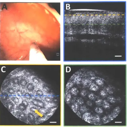
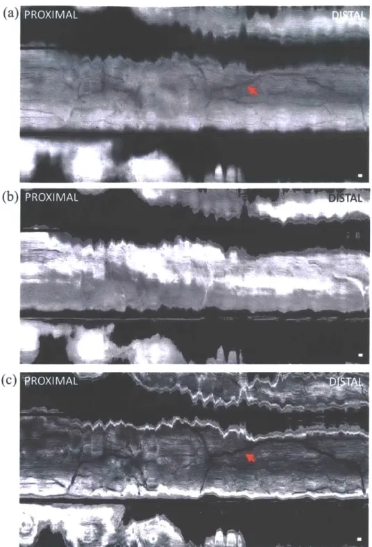
Fig. 4. (A) Endoscopic image showing probe in contact with normal colonic mucosa. (B) Cross section of OCT volume acquired. Enface OCT slices of 40 pLm depth mean intensity projection at (C) 40 ptrm depth and (D) 200 pm depth in tissue (dashed lines indicate depth) showing crypt lumens (orange arrow). Scale bars 100 pm.
Imaging was performed at the Veterans Affairs Boston Healthcare System (VABHS) with IRB approvals at the Massachusetts Institute of Technology, VABHS, and Harvard Medical School, and written informed consent obtained. Inclusion criteria was colonoscopy. The probe was introduced into the accessory channel and placed in gentle contact with ROIs in the endoscopic view to minimize compression. Multiple volumes were acquired at each location to overcome potential imaging artifacts from bulk motion.
Results and Discussion
The fiber scanner had a measured resonance of 1762 Hz (Fig. 3A). The resonance peak frequency for either axis was the same; however, the amplitude for one axis was lower at the same driving voltage. This might be related to asymmetry in fiber mounting or etching of actuator electrodes. It was possible to reduce amplitude on one axis to improve circularity; however, it was preferable to optimize FOV. Fig. 3C shows a test image of a nylon grid placed on top of an absorbent fiber pad, just before the endoscopy. The probe was held by hand and placed in contact with the grid. The image had stable reconstruction of the grid, with minor distortions due to the non-telecentricity of the field. The resolution was sufficient to visualize the absorbent fibers that were tens of microns thick.
Imaging was performed on normal colonic mucosa (Fig. 4). Cross-sectional OCT images showed vertically projecting features associated with colonic crypts. The enface plane at shallower depth showed circular/elliptical crypts, and structural features reminiscent of crypt lumens. At deeper depths, OCT shows integrated effects of light propagation/backscattering; crypt walls produced high OCT signal, while lumens and stroma produced dark vertical features, resulting in contrast inversion[54]. Imaging of a benign hyperplastic polyp (Fig. 5) showed regular crypts but slightly enlarged and more sparsely distributed, in this single illustrative example. OCTA of the polyp (Fig. 6) showed vascular patterns varying with depth and encircling the crypts.
Fig. 5. (A) Endoscopic image showing probe coming into contact with hyperplastic polyp (arrow). (B) Cross section of OCT volume from polyp. En face OCT slices of 40 jim depth projection at (C) 40 pm depth and (D) 200 pm depth showing enlarged crypts. Scale bars 100 pim.
In endoscopic and minimally invasive scenarios using catheters, 2-D distal actuation for scanning is attractive. The repeat volume rate in this study was limited because of the time for the fiber to damp before rescanning (Fig. 7). A large resonant deflection was required to maximize FOV, but the high
Q resonance produced a -0.2 sec damping time, limiting the volume rate. The
fiber resonant motion may be actively damped by driving with a waveform of the opposite quadrature, but accurate phasing is required[60]. Graphics processing units could reduce the processing delay.Forward viewing devices are an important complement to side viewing. While side viewing probes have large FOV, they are inefficient for targeting small regions flagged on the endoscopic view and requiring a closer look. Proximal scan errors exacerbate the difficulty of visualizing small regions. Forward viewing has small FOV but is suited for close-up inspection.
The resolution of scanning fiber devices depends on the number of resolvable spots, which is calculated from the fiber mode field diameter and the scan range. Constraints of the probe rigid length and driving voltages impose a limit on resolvable spots. Smaller actuators (~400 ptm diameter) have been reported[60] but are not yet commercially available. Deflection is inversely proportional to tube diameter and wall thickness[67]; smaller actuators scan larger fields for a given voltage, which will increase resolvable spot numbers and relax requirements for high
Q
factors. With improvements made to the assembly process and more experimentation with various epoxy options, it should be possible to achieve more precise control of the fiber mount rigidity and resultantQ factor. More conservative
Q factors will reduce ring-down time of the fiber resonator
and potentially improve scan reset rates.0 CT 'OT 'Il-ft,
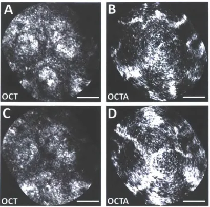
Fig. 6. Zoomed enface OCT images at (A) 150 pm and (C) 200 pm depth (40 ptm depth projection) of polyp. Intrinsically co-registered, depth-resolved OCT angiography from (B) 150 pm and (D) 200 pm depth showing vascular morphology. Scale bars 100 pm.
The resonant scan sampled at constant angular velocity, such that sampling decreased with increasing radial distance. While this resulted in non-uniform sampling, it enabled sequential circular scans to be decorrelated to generate OCTA because the spots were radially aligned. The fast resonance produced scan overlap, but the short (0.57 ms) interscan time reduced sensitivity to slow flows in small vessels. Constant linear velocity would produce uniform sampling but cannot easily generate OCTA. Although there was oversampling in the fast axis towards the center due to decreasing length of the circular scans, the latter increased uncertainty in scan-to-scan alignment; therefore, decorrelation noise did not necessarily decrease towards the center. Noise artifacts
mimicking the spiral were likely due to small synchronization errors between the laser and scanner, and small parasitic motion.
Input and measured trajectories
> >1 -1. 0 0.05 0.1 0.15 0.2 0.25 0 .3 0.35 0.4 0.45 0.5 time/ s X 0 -1 0 0.05 0.1 0.15 0.2 0.25 0.3 0.35 0.4 0.45 0.5 time/ s
Fig. 7. Illustrative figure from the author's Master's thesis [68] showing the input ramp drive to a piezoelectric actuator and the resultant resonant response from a deflecting fiber. There is a long decay time related to the fiber damping.
In GI endoscopy, the concept of 'resect and discard' i.e. assessing polyps to be benign in
situ and discarding them without histopathological analysis is gaining traction as a potentially more
cost-effective surveillance protocol, given the significant cost of histology processing[69]. The polyp in this study did not exhibit marked structural anomalies on imaging, and later showed benign histology. In this context, forward viewing OCT has promise in providing real-time tissue assessment during endoscopy.
Ultrahigh speed OCT at 1 MHz A-scan rate with a forward viewing probe generated high quality volumetric and enface images of colonic microstructure and vascularity. Future work will assess adenomas and other colonic pathology to assess the diagnostic potential of forward viewing OCT in GI surveillance.
Chapter 3
Ultrahigh speed OCT capsule for enface imaging: studies in swine
Introduction
Catheter based optical coherence tomography (OCT) enables high resolution, volumetric imaging of organ surfaces and luminal walls [50]. The rapid advancement and speed improvements of wavelength swept laser sources [70] enable in vivo three-dimensional (3D) OCT imaging at high frame rates. Increases in laser sweep rate have been accompanied by advances in optical scanning probe technologies. Early work with swept source OCT (SS-OCT) and a rotary scanning fiber optic probe with pullback demonstrated the feasibility of generating 3D gigavoxel image volumes [36, 37]. Later SS-OCT studies used novel probe designs such as a balloon catheter [38, 39, 71], MEMS scanner [72], and resonant fiber scanning [59, 61, 73]. The distally scanning micromotor OCT probe was first demonstrated a decade ago [74, 75] and more recently, multiple groups have achieved high frame rate 3D-OCT imaging over large fields using micromotor scanners [76-78]. Using ultrahigh speed MEMS tunable VCSEL laser technology and micromotor scanners, our group recently demonstrated endoscopic OCT angiography (OCTA) and en face OCT in the upper and lower gastrointestinal (GI) tract in human subjects [52, 79].
However, rotary scanning to acquire volumetric data in a luminal organ has traditionally been performed using a proximal motorized pullback for the longitudinal, slow scan direction. Proximal actuation transmits force via a long tether, such as a torque cable, which is subject to
distal end of the probe and degrades scan accuracy along the longitudinal axis. Probes with distal resonant fiber scanning have been widely investigated [59-61, 80], but the scan speed is not adjustable, image sampling is non-uniform because of the need for resonant scan patterns such as the spiral or Lissajous, and imaging is primarily in the forward direction. Two-dimensional (2D) non-resonant fiber scanners have been developed [81-84], but typically require high voltage actuation for the non-resonant axis/axes, which could limit clinical utility. A novel side-viewing probe based on the piezoelectric 'squiggle' motor achieved 2D distal side scanning using a rotating leadscrew [85], but the scan axes could not be controlled independently.
Wireless capsule endoscopy is a well-known and clinically accepted modality for comprehensive imaging of the entire gastrointestinal tract [86]. However, the wireless capsule travels rapidly down and away from the esophagus, and it is not possible to slow down or steer the device for high resolution, closer inspection of the esophageal wall. It was found that simply tying a string to a capsule endoscope was a useful and well tolerated method for diagnosing Barrett's Esophagus (BE) with good diagnostic sensitivity [87-89]. Seibel et al. developed a tethered capsule endoscope with forward viewing capability based on resonant fiber scanning which could perform white light imaging of the esophageal wall at video rates [90, 91]. The tethered capsule had the advantage that it could be swallowed by unsedated patients for screening applications, without the need for conventional endoscopy which requires sedation. More recently, the tethered capsule concept was demonstrated using SS-OCT for circumferential 3D OCT imaging [92], as well as spectrally encoded confocal microscopy [93]. These devices had a highly flexible and soft tether that was shown to be easily swallowed and well tolerated by patients [94]. In this design, the distal optics were scanned in the rotary direction via a proximally actuated torque cable and the capsule
was allowed to passively traverse the esophagus by peristalsis, then manually retracted (pullback) by the soft tether in order to scan the longitudinal extent of the esophagus. Manual pullback in order to perform the longitudinal scan is rapid and convenient in a screening context, but may not be stable or precise enough to achieve high resolution volumetric OCT for enface OCT, especially at limited frame rates supported by proximal torque cable actuation. In addition, precision longitudinal beam scanning is required for scanning microscopies such as confocal, fluorescence or multiphoton microscopy imaging. Therefore, there is a need for device technologies which can perform high speed rotary beam scanning combined with precision longitudinal scanning, maximizing image quality and reducing intraluminal device time.
Enface features in the GI tract, such as mucosal surface patterns or 'pit patterns', are known
markers for disease [24, 95]. The ability to resolve these patterns with enface OCT could improve diagnostic sensitivity. However, image quality depends on optical resolution and scan pattern repeatability. Accurate and stable beam scanning enables imaging of structural features in the en
face plane, as well as more advanced functional methods such as OCTA [51]. Previous probe
designs for circumferential luminal scanning such as balloon and capsule catheters were limited by scan instability due to proximal rotary and longitudinal actuation, and relatively poor transverse resolution because the optics have to be centered in the large diameter probe. Small diameter probes can have better transverse resolution, but cover only a limited portion of the luminal circumference, such that multiple pullbacks over different sectors of the esophagus are required to survey the entire circumference.
This chapter describes a capsule probe that incorporates 2D circumferential and longitudinal beam scanning in the distal end, thus eliminating mechanical instability from proximal torque cable actuation. The device has a 3.8 mm diameter tether that is semi-rigid to allow manual positioning, pulling back or advancing the device, depending on the mode of operation, such that peristaltic propulsion is not necessarily required. The rotary scanner is a micromotor, and the longitudinal scanner is a novel pneumatic actuator. The optical design enables focusing elements to be positioned close to the tissue surface, to improve transverse resolution. Additionally, the path length of the OCT sample arm is designed to vary with the longitudinal pneumatic scan, enabling non-uniformities in the longitudinal scan trajectory to be tracked and corrected in post-processing. The longitudinal scan can also be performed by proximal manual actuation of the entire capsule (large field coverage) via the tether, versus by distal pneumatic actuation of an internal carriage with the capsule kept stationary (small field, high stability inspection). Compared to our previously reported micromotor probe [77], the capsule probe has significantly improved field of view and 2D distal scanning capabilities. Images were acquired in the living swine esophagus, rectum, and anal canal. This study serves as an important translational step in validating the ultrahigh speed capsule OCT technology for potential clinical applications in screening and surveillance. In particular, the ability to image a wide range of field sizes, in both the upper and lower GI tract, and in sedated subjects promises to enable new applications of OCT as a next-generation GI imaging modality.
Materials and Methods
This work was performed in collaboration with Dr. Giovanni Traverso and veterinary staff at the MIT Division for Comparative Medicine, Thorlabs (Dr. Benjamin Potsaid) and Praevium Research (Dr. Vijaysekhar Jayaraman) for use of prototype swept source technology.
Imaging system
The OCT system used a dual circulator Michelson interferometer as shown in Fig. 1. The data acquisition card (AlazarTech, Quebec, Canada) had 12 bit resolution and was rated at up to 1.8 GS/s with internal clocking, but was limited to lower sampling rates when externally clocked. The acquisition card was optically clocked by a Mach-Zehnder interferometer with a variable fringe frequency of up to 1.1 GHz, which was the maximum frequency that the A/D card could perform variable clocking. The laser source was a 1300 nm MEMS-based vertical-cavity surface-emitting laser (VCSEL) [63], and was driven sinusoidally at 500 kHz by an arbitrary waveform generator and high voltage amplifier. Both forward and backward wavelength sweeps of the laser were used to achieve an effective axial scan rate of 1 MHz. The VCSEL sweep bandwidth was 115 nm, the measured axial resolution was 12 tm in air (8.5 pm in tissue), and the Nyquist imaging range was 2.3 mm in air (1.6 mm in tissue). Sensitivity roll off was negligible across the imaging range because of the long coherence length of the VCSEL and high bandwidth of the detectors. The coherence length of the VCSEL has been previously reported to be more than 100 mm in air [96]. The power emitted from the imaging capsule was 35 mW. System sensitivity was measured using an isolated reflection from a flat-cleaved fiber patch cord with 0.6% reflectivity, with
additional attenuation from a 17 dB single pass attenuator in the sample arm. The sensitivity was measured to be 105 dB, defined by the ratio of the OCT signal to the standard deviation of the magnitude of the OCT signal with the reflection blocked. The transmission through the capsule probe was 70 percent. The backcoupling efficiency of a mirror reflection into the probe was estimated to be 50 percent. The effective sensitivity of the OCT imaging engine with the capsule was -102 dB.
A custom C++ software was used to control the beam scanning and data acquisition. The OCT interferometer reference mirror was on a motorized translation stage that was triggered by the acquisition software at a preset timing delay. The translation range of the pneumatic actuator in the capsule is about 3.5 mm, which is larger than the system imaging range. Thus, the reference mirror position was scanned at a speed matched to the maximum speed of the pneumatic actuator, in order to enable imaging over the full longitudinal translation range. The system was installed on a portable cart for convenient transport to the animal housing facility for imaging experiments.
HVAA
O~r,
.M
Fig. 1. Schematic of OCT system. HVA: high voltage amplifier; AWG: arbitrary waveform generator; MZI: Mach-Zehnder interferometer; OC: optical clock; RM: reference mirror; DA: differential amplifier.
Catheter design and assembly
A schematic of the capsule device is shown in Fig. 2(a), and photographs in Figs. 2(b) and. 2(c). A collimated beam was reflected at a right angle by a prism and focused by a microlens to a focal plane outside of the glass tube enclosure. The microlens was mounted on a counterweighted lens holder on the shaft of a micromotor. The micromotor rotated the lens holder to circumferentially scan the focused beam. The micromotor was mounted on a carriage, which was centered in a glass tube by multiple contact points with non-marring set screws. The carriage was attached to a bellows that was expanded and contracted by pneumatic inflation and deflation via an inflation tube. When the bellows was actuated, the carriage slid smoothly along the glass tube, generating a precision longitudinal scan.