Methods in Molecular Biology - Chapter XX
Experimental tools to study regeneration in the sea anemone Nematostella vectensis
Aldine R. Amiel and Eric Röttinger
Université Côte d’Azur, CNRS, INSERM, Institute for Research on Cancer and Aging, Nice (IRCAN), Nice, France
Abstract
Animal regeneration is a biological process leading to the reformation of injured or lost tissues/body parts. One of the most fascinating regenerative phenomena is the so-called whole body regeneration, leading to the reformation of fully functional organisms within days after bisection. The sea anemone Nematostella vectensis is currently emerging as novel whole-body regeneration model. Here we describe the methods of inducing the regenerative process in this cnidarian as well as the fixation and staining protocols for morphological, molecular and cellular analysis.
Keywords: Whole body regeneration, bisection, fixation, permeabilization, immunohistochemistry, staining.
1. Introduction
Animal regeneration has intrigued scientists and humankind for centuries. This biological process leads to the reformation of injured or lost tissues/body and occurs at different levels from the cell, tissue, organ, structure to even the entire organism (1). Latter phenomenon is called whole body regeneration and Hydra as well as planarians are the historical models in this line of research. Since a couple of decades, the field of regeneration biology has seen the emergence of a variety of new research models, one of which is the cnidarian sea anemone Nematostella vectensis (2-5). As cnidarians are the sister group to all bilaterian animals (6,7), this marine invertebrate holds an interesting phylogenetic position and provides new insights into the evolution of this intriguing developmental trajectory. After bisection, regeneration in adult and juvenile Nematostella occurs within seven days. Developing cellular and molecular tools in order to study the dynamics of the tissues as well as cellular response/behaviour during regeneration, is crucial for our understanding of this intriguing phenomenon (5). Using 4-tentacle juveniles that are more amenable for staining and imaging approaches we have adapted/optimized protocols to highlight cellular and molecular mechanisms involved in the regeneration process in Nematostella that are based on i) small molecules, such as DAPI (4',6-Diamidine-2'-phenylindole dihydrochloride) or HOECHST (Bisbenzimide) to label nuclei, EdU (5-ethynyl-2'-deoxyuridine) to label cell proliferation/S-phase, EU (5-ethynyl-uridine) to label neo-synthesized RNA (i.e. hyper-transcription) or Phalloidin to label cell membranes (actin filaments) as well as ii) anti-bodies staining. Combined with molecular information (ex, transcriptomic time courses ((8), see chapter XX) these approaches are useful tools to determine the phenotypes of perturbation experiments (i.e. pharmacological or gene-specific knock-downs/knock-outs) (5) and predict gene network interactions.
2. Materials
Prepare and store all reagents at room temperature (unless indicated otherwise). Follow all national waste disposal regulations when discarding waste materials. 2.1 Animal care
1. Nematostella vectensis (Nv) polyps.
2. 1/3 Artificial Sea Water (1/3 ASW), pH 8.2, density 10.10, KH 7, prepared from Artificial Sea Water (ASW pH 8.2, density 10.30, KH 7) diluted to 1: 2 with Reverse Osmosis (RO) water KH7 (4,5g Sodium Bicarbonate in 20L RO water).
3. Stackable glass bowls for approx. 200ml 1/3x ASW (ex. Carolina, 250ml, #741004). 4. Incubator to maintain animals at 22˚C (ex. Pol Eco Aparatura, ST-700)
5. Artemia salina - Nauplius stages 6. Scalpes
2.2 Induction of regeneration
1. Relaxing solution - Magnesium Chloride 7,14% in 1/3 Microfiltered ASW - 0.2µm, or 1/3 MFASW): 7,14g of MgCl2 in 100ml of 1/3 ASW. Store at Room Temperature (RT)
2. Petri dishes for approximately 40ml of 1/3 ASW (ex. Greiner, #633185). 3. Plastic pipette 2ml (ex. Biosigma, #390509)
4. Scalpel handle (Swann-Morton n7) with blade (Swann-Morton n15) 5. P200 pipette (ex. Gilson P200) and tips 200µl
6. Microfiltered 1/3 Artificial Sea Water (1/3 MFASW)
2.3 Fixation, permeabilization, coating and washing Buffer for Immuno & EdU/EU -staining
1. Fixative IS: 4% Paraformaldehyde (from 32% PFA without Methanol stock solution, ex. EMS,#15714,see Note 1) in 1/3 MFSW. Can be kept at 4C for one week.
2. Ice
3. Phosphate Buffered Saline 10x (PBS 10x): for 1L solution add 2,23g NaH2PO4 (anhydrous
18.6mM); 11,94g Na2HPO4 (81,1mM); 102.2g NaCl; adjust pH to 7.4. Autoclave.
4. Tween-20 20% stock solution (autoclaved MilliQ water)
5. Tween-20 0,1% in PBS 1x (PBTw 0,1%): For 50ml add 1ml PBS 10x; 0,250ml Tween-20 20%; 48,75ml MilliQ water.
6. Triton X100 0,2% and 0,5% in PBS 1x (PBT 0,2% and PBT 0,5%, respectively) 7. Methanol – MeOH
2.4 Membrane and nuclei staining All products are stored at -20˚C
1. Alexa Fluor® 488 Phalloidin (Thermo Fisher Scientific, #A12379, see Note 1)
2. DAPI (Thermo Fisher Scientific, #D3571, see Note 1) 10,9mM stock solution or Hoechst (Thermo Fisher Scientific, #H1399, see Note 1) 10mg/ml stock solution: use at 1/5000 from stock solution in PBS1x
2.5 EdU (5-ethynyl-2'-deoxyuridine) and EU (5-ethynyl-uridine) staining
Protocol adapted from the Click-itTM kits, Thermo Fisher Scientific – EdU # C10337, EU # C10329
1. EdU solution see Note 1: dilute EdU powder in 2ml of deionized water for 10mM stock solution. Aliquot and store at -20˚C
2. EU solution (see Note 1): dilute EU powder in 373µl of deionized water for 100mM stock solution. Aliquot and store at -20˚C
3. Deionized water
4. 10x Click it reaction buffer (Click-itTM kits, stored at 4˚C)
5. CuSO4 100mM stock solution (Click-itTM kits, stored at 4˚C)
6. Alexa Fluor Azide 488 (Click-itTM kits, stored at -20˚C)
2.6 TUNEL staining
Protocol adapted from the TUNEL AP kit (Roche, #11772457001) 1. Proteinase K 0.01mg/ml in PBS1x
2. TUNEL Enzyme solution (Kit Roche, stored at -20˚C) 3. TUNEL label (Kit Roche, stored at -20˚C)
4. DNAse at 0.1U/μl for positive control
5. Fixative IS: 4% Paraformaldehyde (from 32% PFA without Methanol stock solution, ex. EMS,#15714,see Note 1) in 1/3 MFSW. Can be kept at 4C for one week.
1. Secondary fixative: 4% Paraformaldehyde (from 32% PFA without Methanol stock solution, ex. EMS,#15714,see Note 1) in PBS 1X. Can be kept at 4C for one week.
2.7 Immunostaining
1. Normal Goat Serum (NGS, Sigma #G9023) inactivated: Heat NGS stock solution at 56C for 30min; while still warm, filter the inactivated NGS through 0.22µm using a filter and syringe; aliquot in sterile tubes. Store at -20˚C.
2. Dimethylsulfoxide (DMSO, Sigma #D4540) 100% stock solution
3. Blocking buffer PA for primary antibody: NGS 10% in PBT 0,1% + DMSO 1%. Prepare fresh.
4. Blocking buffer SA for secondary antibody: NGS 10% in PBT 0,1%. Prepare fresh.
5. Primary antibody: Rabbit antiPhosphoHistone H3 antibody (ex. Abcam, #14955). Store at -20˚C.
6. Secondary antibody: Goat antiRabbit or Donkey antiRabbit Alexa 488 antibody. Store at -20˚C.
7. Glycerol 80% in PBS1x final concentration 3. Methods
3.1 Animal care
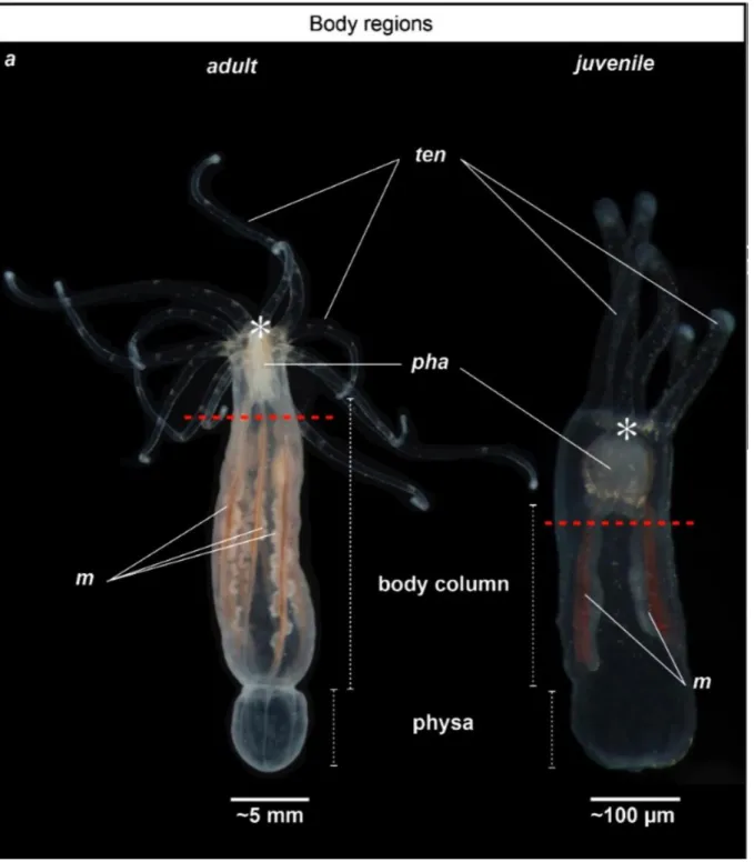
Nematostella culture, care and the induction of spawning is done according to previous publications (9-11). Adult animals (Fig. 1) are kept at 17C under dark conditions in glass bowls (~40 animal/bowl/200ml) and fed daily with freshly hatched Artemia nauplii. Change the water and clean glass bowls every two weeks. Induce spawning through a temperature (increase the temperature to 22C) and light stimulus by placing the animals on a light table for 12h. After spawning and fertilization let the embryos develop, metamorphose and raise Nematostella polyps in 1/3 ASW at 22C until they reach the desired size for regeneration experiments (e.g. 4 tentacle juveniles, ~ 6 weeks post fertilization with a smashed Artemia feeding regime, Fig. 1 (5)).
3.2 Induction of regeneration
1. Coat all plastic equipment you will use (petri dishes, tips, pipettes…) with PTw0.1% to avoid that the animals stick to the plastic and carry out the experiment at room temperature.
2. Add the Nematostellapolyps (~100 for the 4 tentacles juveniles and ~30 for the sub-adults or sexually mature adult polyps) into a petri dish in 40ml of 1/3 ASW using a plastic pipette. 3. Place the petri dish on a light table to allow the Nematostella polyps to relax for 10-15min and add 2ml of relaxing solution (7,14% MgCl2) to the petri dish. Allow the Nematostella
polyps to relax 10-15 more minutes.
4. Place the petri dish with the relaxed Nematostellaunder a binocular macroscope.
5. Use a scalpel to cut the polyp below the pharynx, perpendicularly to the oral-aboral axis of the body (Fig. 1). This microdissection results in two Nematostella parts, the isolated oral regionand the main body part.
6. Replace and rinse the MgCl2 of the used petri dish three times with 1/3 ASW.
7. Place the isolated Nematostella parts of interest in a newly coated petri dish in 40ml 1/3 ASW and let the animals regenerate at 22C until the desired step (4,5).
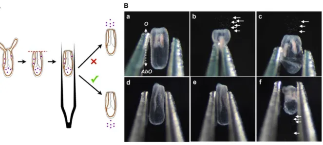
1. To study wound healing in vivo, use the compression assay developed in (5), (Fig. 2). This assay assesses the state of the opening at the amputation site following bisection on the 4-tentacles juvenile polyps. It uses nematosomes (free circulating aggregates of cnidocytes) as a marker to follow the fluid dynamics present in the gastric cavity of Nematostella”.
Use tweezers (ex. Outils Rubis SA, #3C GRIP, Stabio, Switzerland) to compress laterally, along the oral-aboral axis, the relaxed and freshly bisected or regenerating juveniles.
With an open wound, the nematosomes will be expelled at the amputation site. With a closed wound the nematosomes will either remain in the gastric cavity or leak out of the body cavity through the aboral pore.
2. To study pharynx formation, use the pharynx reformation assay develops in (5) (Fig. 3) that takes advantage of naturally occurring auto-fluorescence in the pharynx.
Mount living freshly bisected or regenerating 4-tentacles juveniles under a slide and cover slip. Use clay at the 4 corners of the cover slip to avoid smashing the fragile tissues of the bisected juvenile.
Before any observation orient the juvenile polyp with the two mesenteries laterally by using a binocular and moving/pressuring the border of the cover slips with forceps.
Place the slide under a fluorescent microscope and observe the emergence of the endogenous 488nm auto-fluorescence in the body cavity near the amputation site. The emergence of the 488nm auto-fluorescence is correlated with the emergence of pharyngeal tissues (5).
3.4 Tissue morphology, Cellular proliferation, Neo transcription and Apoptosis staining 1. Tissue morphology
1.1 Place the petri dish on an illuminated surface (ex. light table) to allow the
Nematostella polyps to relax for 10-15min and add 2ml of relaxing solution (7,14% MgCl2)
to the petri dish. Allow the Nematostella polyps to relax 10-15 more minutes.
1.2 When relaxed, collect the regenerating polyps and add them into a coated 1.5 or 2ml Eppendorf tube. Remove as much liquid as possible before fixation.
1.3 Fix relaxed polyps (controls and regenerates) by adding the fixative IS in the Eppendorf tube. Fix animals for1h at room temp (RT) or over night (ON) at 4ºC. Then, wash the fixed polyps 3 times using PBT 0.2% (see Note 2).
1.4 Dilute Alexa Fluor® 488 Phalloidin stock solution to 1/200 and DAPI or Hoechst stock solution to 1/5000 in PBTw 0.1%.
1.5 In the Eppendorf tube, containing the bisected juvenile, remove as much liquid as possible and add the solution containing Alexa Fluor® 488 Phalloidin 1/200 and DAPI or Hoechst 1/5000. Let incubate ON at 4ºC.
1.6 The next morning, rinse by performing 3 washes with PBTw 0.1%.
1.7 Remove as much liquid as possible and add 80% glycerol/PBS1x. Let settle at least half a day, as the glycerolwill partly clear the polyp tissue.
1.8 Mount the fixed and stained polyps between slide and cover slipsusing clay “feets” at its four corners.Press carefully to slightly compress the polyp and visualize properly the tissue detail under the microscope.
1.9 Use 488nm and 405nm filter under a fluorescent microscope to observe Phalloidin 488 and DAPI (or Hoechst), respectively.
2. Cellular proliferation (EdU) and Neo-transcription (EU)
2.1 Prepare 300µM EdU (to address Cell proliferation) or 1mM EU (to address Neo-transcription) working solution in 1/3 ASW (see Note 3).
2.2 Collect the polyps (controls and regenerates) and place them into a 1.5 or 2ml Eppendorf tube for the 4-tentacles juveniles or in a 24-well plate for sub-adult or sexually mature adult polyps.
2.3 Remove as much liquid as possible and add the EdU or EU solution to the polyps. Let settle and incubate in the dark (ex: wrapped foil around tube or plate) on a rocking table between 30 to 60min.
2.4 Place the Eppendorf tube or the 24-well plate containing the polyps on an illuminated surface (ex. light table) to allow the animals to relax for 10-15 minutes. Add a few drops of
relaxing solution (7,14% MgCl2) to the tube or petri dish. Allow the polyps to relax for
10-15 more minutes.
2.5 When relaxed, remove as much liquid as possible before fixation.
2.6 Fix relaxed polyps by adding the fixative IS. Fix the animals for 1h at room temp (RT) or over-night (ON) at 4ºC (if working with the 24-well plate, do not forget to place the plate under a fume hood during the fixation). Wash the fixed polyps three times using PBT 0.2% (see Note 2).
2.7 Permeabilized the animal tissue by removing as much liquid as possible and add PBT 0.5% to the fixed polyps for 20 to 30 minutes on a rocking table.
Nb. For sub-adult or sexually mature adult tissues, before developing the staining (see next steps), cut the fixed polyp in multiple identifiableparts to improve the
efficiency/accessibilityof the staining (see Note 5).
2.8 In the dark, incubate the polyps for 30 minutes in the following Click-it EdU/EU cocktail: For 100µl use 75,8ul deionized water; 10µl 10x Click-it Reaction buffer, 4µl CuSO4 (100mM stock solution); 0,25µl Alexa Fluor Azide 488; 10µl Reaction buffer
additive.
2.9 Wash 3 times in PBS1x.
2.10 At this step, the fixed and labelled polyps can also be stained with DAPI or Hoechst using the protocol described in sections 1.4 to 1.9 (Note 6). 3. Apoptosis
3.1 Collect the polyps (controls and regenerates) and add them into a 1.5 or 2ml Eppendorf tube for the 4-tentacles juveniles or in a 24-well plate for the sub-adult or sexually mature adult polyps (see Note 4).
3.2 Place the Eppendorf tube or the 24-well plate containing the bisected polyp on an illuminated surface (ex. light table) to allow the Nematostellapolyps to relax for 10-15 minutes. Add a few drops of relaxing solution (7,14% MgCl2)to the tube or petri dish.
Allow the polyps to relax for 10-15 more minutes.
3.3 When relaxed, remove as much liquid as possible before fixation.
3.4 Fix relaxedpolyps by adding the fixative IS. Fix relaxed polyps by adding the fixative IS solution. Fix the animals for 1h at room temp (RT) or over-night (ON) at 4ºC (if working with the 24-well plate, do not forget to place the plate under a fume hood during the fixation). Wash the fixed polyps 3 times using PBT 0.2% (see Note 2).
3.5 Permeabilize the animal tissue by removing as much liquid as possible and add Proteinase K 0,01mg/ml in PBS1x for 20min at RT (see Note 5).
3.6 Wash 3 times in PBS1x.
3.7 To maintain tissue integrity, fix with secondary fixative for 1h at RT. 3.8 Wash 5 times in PBS1x.
3.9 Prepare 50µl of TUNEL reaction mixture using 5µl of TUNEL-enzyme solution and 45µl of TUNEL-Label solution contained in the kit.
3.10 Remove as much liquid as possible and add 50µl of TUNEL reaction mixture to the polyps andlet incubate for 60min at RT in the dark.
3.11 Wash 3 times in PBS1x.
3.12 Observe Cell death using a 488nm filter on a fluorescent microscope. The fixed and TUNEL stained polyps can also be co-stained at this step with Alexa Fluor® 594
Phalloidin and DAPI or Hoechst using the protocol described in sections 1.4 to 1.9. 4. Immunostaining
4.1 Perform steps 3.4.1.1 to 3.4.1.3.
4.2 Permeabilize the animal tissue by removing as much liquid as possible and add PBT 0.5% to the fixed polyps for 20 to 30 min on a rocking table.
Nb. For sub-adult or sexually mature adult tissues, before developing the staining (next steps), cut the fixed polyp in multiple identifiableparts to improve the
efficiency/accessibilityof the staining (see Note 5).
4.3 Prepare blocking buffer PA for the primary antibody, remove as much liquid as possible and incubate the fixed polyps with the blocking buffer PA for 2 - 3 hours on a rocking table at RT.
4.4 Prepare the primary Ab at the desired concentration diluted in the blocking buffer + DMSO 1%. Remove as much liquid as possible from the tube with the polyps and replace with the primary antibody solution. Incubate ON at 4ºC on a rocking table.
nb. If a first purified primary Ab is tested, use it at 1/100.
4.5 Wash in PBTw 0,1% quickly two times, then, three times for 20 - 30 min each at RT. 4.6 Prepare the secondary Ab at the desired concentration diluted in the blocking buffer SA WITHOUT DMSO 1%. Remove as much liquid as possible and add the secondary Ab solutionto the polyps. Incubate ON at 4ºC on a rocking table.
nb. Usually the secondary antibody is used at 1/250 – 1/500.
4.7 Wash in PBTw 0,1% quickly two times, then, three times for 20 - 30 min each at RT. 4.8 Mount the immunostained polyps in Glycerol 80%/PBS1x under a slide and cover slip and observe the staining using the corresponding filters with a fluorescent microscope. T he fixed and immunostained polyps can also be co-stained at this step with Alexa Fluor® 594 Phalloidin and DAPI or Hoechst using the protocol described in sections 1.4 to 1.9.
4. Notes
Note 1. EdU, EU, DAPI, HOECHST, Phalloidin and Paraformaldehyde are CMR
(Carcinogenic, Mutagenic and Reprotoxic) agents. Wear gloves when handling and discard properly according to health and safety rules.
Note 2. When fixed, Nematostella polyp can be store for several weeks at 4ºC in PBS1x. Note 3. For negative EdU controls, at the step 3.4.2.8 use the developing reaction WITHOUT adding the Click-it Reaction buffer and/or Reaction buffer additive OR use 3-4 weeks starved Nematostella polyps, as their proliferative activity is strongly reduced in all tissues (3). In latter condition, use the Reaction buffer and Reaction buffer additive as mentioned in step 3.4.2.8. For positive EdU controls, use freshly fed but 2-days starved polyps, as feeding is a strong inducer of cell proliferation (3).
Note 4. For TUNEL negative control, at the step 3.4.3.9 use only the 45µl of TUNEL-Label solution contained in the kit WITHOUT adding the Enyme-TUNEL solution. For TUNEL positive control, add an additional step between step 3.4.3.8 and 3.4.3.9 by incubating the polyps with 0.1U/μl TURBO DNAse (Applied Biosystems/Ambion, Austin, TX) in 1x TURBO DNAse buffer for 10 minutes prior to the TUNEL staining (step 3.4.3.8).
Note 5. Optional - For adult tissue (thick tissues), to optimized the permeabilization and any labeling, cut the polyp in several pieces keeping a morphological characteristic in each of these pieces to orient your analysis after labeling.
Note 6: According to the manufacturer, Phalloidin staining is not compatible with the Click-iT® detection reaction.
Figure 1. General anatomy of adult and juvenile Nematostella vectensis. Photographs illustrating the adult (left) and juvenile (right) morphology of Nematostella; (a) Polyps are oriented toward the oral region to the top and aboral region to the bottom. Adult is on the left, juvenile is on the right. Red dotted lines indicate the amputation site. ten, tentacles; pha, pharynx; m, mesenteries; asterisk indicates the location of the mouth (Figure and legend reproduced from ref. 5, under the terms and conditions of the Creative Commons by Attribution (CC-BY) license).
Figure 2. Wound healing / compression assay. (A) Diagram of the compression assay during regeneration. The purple dots represent the nematosomes. The red dotted line represents the amputation site. The forceps are laterally compressing the regenerating polyp body. (B) Time series of the compression assay in an opened (Ba–Bc) or a wound-closed (Bd–Bf) polyp. The dotted double arrow in (Ba) indicates the axial orientation of the animals shown in (Ba–Bf). O, Oral; AbO, Aboral (Figure and legend reproduced from ref. 5, under the terms and conditions of the Creative Commons by Attribution (CC-BY) license).
Figure 3. (A) Image of 488+ detection in the pharynx. Fed (a,a’), starved (b–c’), regenerating 72 hpa (d–e’) Nematostella polyp juveniles. DIC optic images (a–d). Epifluorescent images (a’–d’). The red dotted line labels the amputation site under the pharynx (c–d’). The green line in (c,c’) shows the 488+ fluorescence localized in the basal part of the pharynx. The area delimitated with the dotted line in d and d' is the region where the 488+ re-emerged in the polyp at 72 hpa. (e, e’) Confocal images at 72 hpa labeled for DNA (nuclei in cyan) on the 488-negative (e) and 488-positive (e’) polyp juvenile. The area delimitated with the white dotted line in (e’) (488+ regenerating polyps) shows the pharyngeal lip/pharynx in formation that is absent from the 488-negative regenerating polyps in which only the contact between the two mesenteries is visible. pha, pharynx; m, mesentery. Scale bar in (Aa’) is 20µm and applies to (Aa-Ae, Ab’-Ae’)
(Figure and legend reproduced from ref. 5, under the terms and conditions of the Creative Commons by Attribution (CC-BY) license).
6. References
1. Bely AE, Nyberg KG. Evolution of animal regeneration: re-emergence of a field. Trends Ecol Evol (Amst). 2010 Mar;25(3):161–70.
2. Reitzel A, Burton P, Krone C, Finnerty J. Comparison of developmental trajectories in the starlet sea anemone Nematostella vectensis: embryogenesis, regeneration, and two forms of asexual fission. Invertebrate Biology. 2007;126(2):99–112.
3. Passamaneck YJ, Martindale MQ. Cell proliferation is necessary for the regeneration of oral structures in the anthozoan cnidarian Nematostella vectensis. BMC Dev Biol. BMC Developmental Biology; 2012 Dec 4;12(1):1–1.
4. Bossert PE, Dunn MP, Thomsen GH. A staging system for the regeneration of a polyp from the aboral physa of the anthozoan cnidarian Nematostella vectensis. Dev Dyn. 2013 Jul;242:1320–31.
5. Amiel AR, Johnston HT, Nedoncelle K, Warner JF, Ferreira S, Röttinger E.
Characterization of Morphological and Cellular Events Underlying Oral Regeneration in the Sea Anemone, Nematostella vectensis. Int J Mol Sci. Multidisciplinary Digital Publishing Institute; 2015 Dec 1;16(12):28449–71.
6. Zapata F, Goetz FE, Smith SA, Howison M, Siebert S, Church SH, et al. Phylogenomic Analyses Support Traditional Relationships within Cnidaria. Steele RE, editor. PLoS ONE. Public Library of Science; 2015;10(10):e0139068.
7. Chang ES, Neuhof M, Rubinstein ND, Diamant A, Philippe H, Huchon D, et al. Genomic insights into the evolutionary origin of Myxozoa within Cnidaria. Proc Natl Acad Sci USA. 2015 Dec 1;112(48):14912–7.
8. Warner JF, Guerlais V, Amiel AR, Johnston H, Nedoncelle K, Röttinger E. NvERTx: a gene expression database to compare embryogenesis and regeneration in the sea anemone Nematostella vectensis. Development (Cambridge, England). Oxford University Press for The Company of Biologists Limited; 2018 May 17;145(10):dev162867.
9. Hand C, Uhlinger KR. The Culture, Sexual and Asexual Reproduction, and Growth of the Sea Anemone Nematostella vectensis. Biol Bull. Canadian …; 1992 Apr;182(2):169–76. 10. Fritzenwanker JH, Technau U. Induction of gametogenesis in the basal cnidarian
Nematostella vectensis (Anthozoa). Dev Genes Evol. 2002 Feb 21;212(2):99–103. 11. Stefanik DJ, Friedman LE, Finnerty JR. Collecting, rearing, spawning and inducing
regeneration of the starlet sea anemone, Nematostella vectensis. Nat Protoc. 2013 Apr 11;8(5):916–23.