HAL Id: hal-01543404
https://hal.archives-ouvertes.fr/hal-01543404
Submitted on 20 Jun 2017HAL is a multi-disciplinary open access archive for the deposit and dissemination of sci-entific research documents, whether they are pub-lished or not. The documents may come from teaching and research institutions in France or abroad, or from public or private research centers.
L’archive ouverte pluridisciplinaire HAL, est destinée au dépôt et à la diffusion de documents scientifiques de niveau recherche, publiés ou non, émanant des établissements d’enseignement et de recherche français ou étrangers, des laboratoires publics ou privés.
Specific Stabilization of c-MYC and c-KIT
G-Quadruplex DNA Structures by
Indolylmethyleneindanone Scaffolds
K Diveshkumar, Saaz Sakrikar, Frédéric Rosu, S Harikrishna, Valérie
Gabelica, P.I. Pradeepkumar
To cite this version:
K Diveshkumar, Saaz Sakrikar, Frédéric Rosu, S Harikrishna, Valérie Gabelica, et al.. Spe-cific Stabilization of c-MYC and c-KIT G-Quadruplex DNA Structures by Indolylmethylenein-danone Scaffolds. Biochemistry, American Chemical Society, 2016, 55 (25), pp.3571-3585. �10.1021/acs.biochem.6b00120�. �hal-01543404�
This is an author postprint version of an article published in:
Biochemistry, 2016, 55 (25), pp 3571–3585
Publisher version can be access at http://dx.doi.org/10.1021/acs.biochem.6b00120
Specific Stabilization of c-MYC and c-KIT G
-Quadruplex DNA Structures
by Indolylmethyleneindanone Scaffolds
K. V. Diveshkumar,† Saaz Sakrikar,† Frédéric Rosu,‡,§ S. Harikrishna,†
Valérie Gabelica*,§, and P. I. Pradeepkumar*,†
†Department of Chemistry, Indian Institute of Technology Bombay, Mumbai-400076, India ‡CNRS, UMS3033/US001, Institut Européen de Chimie et Biologie, 33607 Pessac,
France
§Univ. Bordeaux, U869 ARNA Laboratory, 33600 Pessac, France
Inserm, U869 ARNA Laboratory, 33000 Bordeaux, France
*To whom correspondence should be addressed. E-mail: valerie.gabelica@inserm.fr or pradeep@chem.iitb.ac.in
Funding Sources: This work is financially supported by grants from Department of Biotechnology (DBT)-Government of India (Pilot Project Grants for Young Investigators in Cancer Biology, Grant No: 6242-P4/RGCB/PMD/DBT/PKPI/2015, to P.I.P.), IRCC-IIT Bombay (to P.I.P.), the Inserm (ATIP-Avenir Grant no. R12086GS to V.G.), the Conseil Régional Aquitaine (Grant no. 20121304005 to V.G.), and the EU (FP7-PEOPLE-2012-CIG-333611 to V.G.).
Acknowledgement: We thank Professor Jean-Louis Mergny, Professor Nand Kishore and Dr. V. Dhamodharan for fruitful discussions. Computer centre, IIT Bombay is gratefully acknowledged for providing high performance computing facilities. We are also thankful to the central facility supported by IRCC-IIT Bombay for MALDI-MS facility, and the Structural Biophysical Chemistry platform of the IECB Bordeaux for providing access to the mass spectrometers. We are also thankful to Dr. Ruchi Anand for providing access to her laboratory facilities. D.K.V. thanks Council of Scientific and Industrial Research (CSIR), India for the Ph.D. fellowship.
ABBREVIATIONS
ITC, Isothermal titration calorimetry; ESI-MS, Electrospray ionisation mass spectrometry; FDA, Food and drug administration; DMF, Dimethylformamide; ACN, Acetonitrile; AcOH, Acetic acid; MeI, Methyliodide; CD, Circular dichroism spectroscopy; EMSA, Electrophoretic mobility shift assay; PAGE, Polyacrylamide gel electrophoresis; MD, Molecular dynamics; RMSD, Root mean square deviation; InEt1, 1-methyl-1-(2-(3-((1-oxo-1H-inden-2(3H)-ylidene)methyl)-1H-indol-1-yl)ethyl)pyrrolidinium iodide; InPr1, (E)- 1-methyl-1-(3-(3-((1-oxo-1H-inden-2(3H)-ylidene)methyl)-1H-indol-1-yl)propyl)pyrolidini-umiodide; InEt2, methyl-1-(2-(3-((6-(2-(1-methylpyrrolidinium-1-yl)ethoxy)-1-oxo-1H-inden-2(3H)-ylidene)methyl)-1H-indol-1-yl)ethyl)pyrrolidinium iodide; InPr2, (E)-1-
ABSTRACT
Stabilization of G-quadruplex DNA structures by small molecules has emerged as a promising strategy for the development of anticancer drugs. Since G-quadruplex structures can adopt various topologies, attaining specific stabilization of a G-quadruplex topology to halt a particular biological process is daunting. To achieve this, we have designed and synthesized simple structural scaffolds based on indolylmethyleneindanone pharmacophore, which can specifically stabilize the parallel topology of promoter quadruplex DNAs (c-MYC, c-KIT1 and c-KIT2), when compared to various topologies of telomeric DNA and duplex DNAs. The lead ligands (InEt2 and InPr2) are water soluble and meet a number of desirable criteria for a small molecule drug. Highly specific induction and stabilization of the c-MYC and c-KITquadruplex DNAs (ΔT1/2 up to 24 °C) over telomeric and duplex DNAs (ΔT1/2 ~ 3.2
°C) by these ligands were further validated by ITC and ESI-MS experiments (Ka ~ 105-106 M−1). Low IC50 (~ 2 µM) values were emerged for these ligands from Taq DNA polymerase
stop assay with the c-MYC quadruplex forming template, whereas the telomeric DNA template showed IC50 values >120 µM. Molecular modeling and dynamics studies
demonstrated the 5'- and 3'- end stacking modes for these ligands. Overall, these results demonstrate that among the >1000 quadruplex stabilizing ligands reported so far, the indolylmethyleneindanone scaffolds stands out in terms of target specificity and structural simplicity, and therefore offers a new paradigm in topology specific G-quadruplex targeting for potential therapeutic and diagnostic applications.
INTRODUCTION
Tetrameric DNA structures formed by guanine rich sequences in the presence of monovalent metal ions are called G-quadruplexes.1,2 These structures consist of planar arrangements
called G-quartets, formed by the association of four guanine bases through the H-bonding interactions of Hoogsteen and Watson-Crick faces of the adjacent guanines. Putative G-quadruplex forming sequences have been identified in the various parts of human genome such as in telomeres,3,4 promoter regions of various oncogenes,5 introns, 6 and in the
immunoglobulin switch regions.7
G-quadruplex structures present in the telomeric and the promoter regions have emerged as attractive drug targets due to their biological relevance.5 Promoter regions of
many proto oncogenes such as c-MYC, c-KIT, BCL-2, k-RAS, VEGF, HIF-1α and PDGF-A possess G-rich sequences that have the propensity to form G-quadruplex structures.8-13 In
such regions, molecular crowding conditions due to the high concentration of macromolecules, and the dynamic forces evolved from negative superhelicity promote the formation of G-quadruplex structures from duplex DNAs.5,14 The identification of proteins
that have crucial role in unwinding the quadruplex structures further validates the existence of such G-quadruplex structures in vivo.15
Small molecules that can stabilize the quadruplex structures present in the promoter regions can effectively inhibit transcription process.5 Recently, such approaches have been
touted as promising new directions in the anticancer drug discovery.5 Major challenges in the
G-quadruplex mediated anticancer drug development are to achieve selectivity for the G-quadruplexes over the duplex DNAs, to impart specificity among the different topologies of G-quadruplexes, and moreover to engineer drug-like properties to the the stabilizing ligands.16
G-quadruplex structures can exhibit different topologies depending on the nature and length of sequence, size/length of the loops, presence of metal ions and the conformations of the glycosidic bonds.17,18 Telomeric quadruplex DNAs are highly polymorphic in nature and
exist in different topologies including antiparallel, hybrid and mixed hybrid, whereas promoter quadruplex DNAs mostly exist in parallel topology with propeller loops.19 Since all
the G-quadruplexes have identical G-quartet interfaces in their structures, achieving specificity toward a particular topology is a daunting task. However, subtle differences in the quartet surface area, size/length of the loops, and nature of the grooves of the different quadruplex topologies can be harnessed to design topology specific ligands. There are only limited reports in the literature describing those ligands, which are able to achieve topology specific stabilization of quadruplex structures. Among those, most of the ligands have shown moderate preference for one of the topologies of telomeric quadruplex DNA as revealed from few biophysical screening assays.20-24 The ligands, which show preferential stabilization
toward c-MYC quadruplex DNA over duplex DNA include furan based cyclic oligopeptides,25 ellipticine derivatives,26 piperazinylquindoline derivatives27 and
metallo-rectangles with terpyridine ligands.28 But these molecules were found to have affinity
toward telomeric quadruplex DNAs as well. Diethynyl-pyridine derivatives were reported for their selective stabilization toward various promoter quadruplexes over duplex DNAs.29
Similarly, bisaryldiketene derivatives were studied for their preferential affinity toward promoter quadruplexes over duplex DNAs and telomeric quadruplex DNAs.30 Recently, new
class of small molecules has been reported as strong inhibitor of c-MYC expression via quadruplex stabilization, which was identified using small molecule microarray.31 None of
these ligands were reported to have specificity toward parallel topology of promoter quadruplex DNAs over various topologies of telomeric and duplex DNAs. An exception to
this is the peptidomimetic ligands that have shown marginal specificity toward the c-KIT1 quadruplex structure.32
Recently, our group has reported indenopyrimidine derivatives and bisbenzimidazole carboxamide derivatives of naphthiridine and phenanthroline that can specifically stabilize c-MYC and c-KIT quadruplex DNAs having parallel topology over telomeric quadruplex and duplex DNAs.33,34 Even though these ligands were able to achieve specificity toward a
particular topology, weak stabilization property for indenopyrimidine derivatives and poor drug like properties due to the presence of multiple aromatic rings and heteroatoms for the latter one weaken the therapeutic index of these molecules. Apart from achieving high specificity toward a particular G-quadruplex topology, for clinical success, development of anticancer molecules having drug like properties is highly warranted.16 To fill this lacuna,
herein, we report a new qudruplex stabilizing ligand family having simple scaffolds based on indolylmethyleneindanone skeleton (InEt1, InEt2, InPr1 and InPr2, Figure 1). Topology specific stabilization of these ligands with c-MYC and c-KIT quadruplex DNAs having parallel topologies was unambiguously validated using various biophysical, biochemical and molecular modeling and dynamics studies.
MATERIALS AND METHODS
General Methods. Dry solvents (DMF, CHCl3, toluene) were obtained from commercial
suppliers and CH3CN, DCM were dried using calcium hydride. Thin layer chromatography
(TLC) was performed on silica gel plates pre-coated with fluorescent indicator with visualization by UV light (260 nm). Silica gel (100-200 mesh) or basic alumina was used for column chromatography. 13C NMR (100 and 125 MHz) and 1H NMR (400 and 500 MHz)
were recorded on 400 and 500 MHz instruments respectively. The chemical shifts in parts per million (ppm) were referenced to the residual signal of deuteriated solvents or TMS: TMS (0
ppm), CD3OD (3.31 ppm), and DMSO-d6 (2.5 ppm) for 1H NMR spectra; and CDCl3 (77.2
ppm), CD3OD (49.1 ppm), and DMSO-d6 (39.5 ppm) for 13C NMR spectra. Multiplicities of
1H NMR spin couplings are reported as s (singlet), d (doublet), t (triplet), q (quartet), dd
(doublet of doublets), and (q) quintet or m (multiplet and overlapping spin systems). Values for apparent coupling constants (J) are reported in Hz. High resolution mass spectra (HRMS) were obtained in positive ion electrospray ionization (ESI) mode using a Q-TOF analyser. The molecular structures of all the compounds described below are shown in Scheme 1.
Method A: General Procedure for Bromination. Aldol product (1 equiv) was dissolved in dry DMF (7 ml/mmol), and to this anhydrous K2CO3 (2 equiv) and corresponding
dibromoalkane (2 equiv) were added under nitrogen at 0 °C. After stirring at room temperature for 24 h, water was added and extracted with EtOAc. Organic layer was dried over anhydrous Na2SO4, evaporated under reduced pressure and purified by column
chromatography (12% EtOAc in pet ether) using silica gel as stationary phase to afford the brominated compounds.
Method B: General Procedure for Bromine Displacement. Brominated compound (1 equiv) was dissolved in dry ACN (6 ml/mmol) and to this pyrrolidine (3-10 equiv) was added, and was refluxed for 3-4 h. Solvent was evaporated under reduced pressure, and the crude product was purified by column chromatography (0-1% MeOH in DCM) using basic alumina as a stationary phase.
Method C: General Procedure for Methylation. Compound (1 equiv) was dissolved in dry ACN (6 ml/mmol) and to this excess methyl iodide (16-32 equiv) was added and refluxed for 12 h. Solvent was evaporated, and the solid product was washed with chloroform for removing the impurities to afford the methylated iodide salts.
Method D: General Procedure for Aldol Condensation. Aldehyde (1 equiv) and ketone (1 equiv) was dissolved in acetic acid (8 ml/mmol). To this, 4 or 5 drops of concentrated HCl was added and refluxed for 3-4 h. The reaction mixture was poured into water and extracted with EtOAc. The organic layer was dried over anhydrous Na2SO4, evaporated under reduced
pressure, and purified by column chromatography (15-20% EtOAc in pet ether) using silica gel as stationary phase to yield the condensed aldol products.
(E)-2-((1-(2-bromoethyl)-1H-indol-3-yl)methylene)-2,3-dihydro-1H-inden-1-one (2). Method A was followed using compound 1 (320 mg, 1.23 mmol), in dry DMF (6 ml), anhydrous K2CO3 (341 mg, 2.46 mmol) and 1,2-dibromoethane (0.21 ml, 2.46 mmol) to
afford compound 2 as a yellow solid (351 mg, 78%). M. p. 167-169 °C. 1H NMR (400 MHz,
CDCl3): δ 8.00 (s, 1H), 7.88 (dd, J = 14.6, 7.0 Hz, 2H), 7.47-7.56 (m, 3H), 7.37 (t, J = 7.6
Hz, 1H), 7.21-7.32 (m, 3H), 4.55 (t, J = 6.4 Hz, 2H), 3.75 (br, 2H), 3.70 (t, J = 6.4 Hz, 2H).
13C NMR (100 MHz, CDCl3): δ 193.8, 148.7, 139.1, 135.8, 134.0, 130.6, 130.0, 128.8, 127.5,
126.1, 124.9, 124.1, 123.5, 121.6, 119.6, 113.1, 109.4, 48.5, 33.2, 29.7. HRMS (ESI): Calcd for C20H17NOBr [(M+H)]+ 366.0494; found, 366.0484 (∆m- −0010 and error- −2.7 ppm).
(E)-2-((1-(3-bromopropyl)-1H-indol-3-yl)methylene)-2,3-dihydro-1H-inden-1-one (3). Method A was followed using compound 1 (305 mg, 1.18 mmol) in dry DMF (6 ml), anhydrous K2CO3 (325 mg, 2.35 mmol) and 1,3-dibromopropane (0.24 ml, 2.35 mmol) to
afford compound 3 as a yellow solid (356 mg, 79%). M. p. 165-166 °C. 1H NMR (400 MHz,
CDCl3): δ 8.06 (s, 1H), 7.91 (dd, J = 15.2, 7.36 Hz, 2H), 7.52-7.59 (m, 3H), 7.40 (t, J = 8.5
Hz, 2H), 7.26-7.32 (m, 2H), 4.42 (t, J = 6.4 Hz, 2H), 3.83 (br, 2H), 3.34 (t, J = 6.1 Hz, 2H), 2.41 (q, J = 6.1 Hz, 2H). 13C NMR (100 MHz, CDCl3): δ 193.9, 148.7, 139.2, 136.0, 134.0,
130.3, 130.2, 128.9, 127.6, 126.1, 125.2, 124.1, 123.4, 121.5, 119.6, 112.9, 109.9, 44.7, 33.3, 32.2, 30.4. HRMS (ESI): Calcd for C21H19NOBr [(M+H)]+ 381.0623; found, 381.0620 (∆m-
(E)-2-((1-(2-cyclopentylethyl)-1H-indol-3-yl)methylene)-2,3-dihydro-1H-inden-1-one (4). Method B was followed using compound 2 (84 mg, 0.23 mmol) in dry ACN (2 ml), pyrrolidine (0.06 ml, 0.69 mmol) to afford compound 4 as a yellow sticky solid (60 mg, 74%). 1H NMR (400 MHz, CDCl 3): δ 8.06 (s, 1H), 7.90 (dd, J = 12.9, 7.3 Hz, 2H), 7.60 (s, 1H), 7.49-7.57 (m, 2H), 7.22-7.39 (m, 4H), 4.31 (t, J = 7.0 Hz, 2H), 3.80 (br, 2H), 2.93 (t, J = 7.0, 2H), 2.57 (br, 4H), 1.80 (br, 4H). 13C NMR (100 MHz, CDCl 3): δ 193.9, 148.7, 139.4, 136.3, 133.9, 130.5, 129.9, 128.7, 127.5, 126.1, 125.5, 124.1, 123.2, 121.3, 119.4, 112.7, 109.9, 55.6, 54.5, 46.5, 33.3, 23.7. HRMS (ESI): Calcd for C24H25N2O [(M+H)]+ 357.1961;
found, 357.1966 (∆m- +0005 and error- +1.3 ppm).
(E)-2-((1-(3-cyclopentylpropyl)-1H-indol-3-yl)methylene)-2,3-dihydro-1H-inden-1-one (5). Method B was followed using compound 3 (175 mg, 0.46 mmol) in dry ACN (4 ml) and pyrrolidine (0.15 ml, 1.83 mmol) to afford compound 5 as a yellow sticky solid (140 mg, 82%). 1H NMR (400 MHz, CDCl3): δ 8.10 (s, 1H), 7.92 (dd, J = 12.0, 7.48 Hz, 1H),
7.52-7.61 (m, 3H), 7.38-7.42 (m, 2H), 7.24-7.31 (m, 2H), 4.30 (t, J = 6.7 Hz, 2H), 3.83 (br, 2H), 2.50 (br, 4H), 2.43 (t, J = 6.7 Hz, 2H), 2.08 (q, J = 6.7 Hz, 2H), 1.82 (q, J =3.2 Hz, 4H).
13C NMR (100 MHz, CDCl3): δ 194.0, 148.7, 139.4, 136.4, 133.9, 130.6, 129.7, 128.8, 127.5,
126.1, 125.7, 124.1, 123.1, 121.2, 119.4, 112.5, 110.1, 54.2, 52.8, 44.9, 33.4, 29.2, 23.7. HRMS (ESI): Calcd for C25H27N2O [(M+H)]+ 371.2196; found, 371.2192 (∆m- −0004 and
error- −1.2 ppm).
(E)-1-methyl-1-(2-(3-((1-oxo-1H-inden-2(3H)-ylidene)methyl)-1H-indol-1-yl)ethyl)pyrrolidinium iodide (InEt1). Method C was followed using compound 4 (30 mg, 0.08 mmol), in dry ACN (2 ml) and excess methyl iodide (1 ml, 16 mmol) to afford the final methylated iodide salt InEt1 as a yellow solid (35 mg, 89%). M. p. 251-253 °C. 1H NMR
(500 MHz, DMSO-d6): δ 8.24 (s, 1H), 7.96 (d, J = 7.74 Hz, 1H), 7.89 (s, 1H), 7.67-7.79 (m,
Hz, 2H), 4.00 (br, 2H), 3.88 (t, J = 7.2 Hz, 2H), 3.19 (s, 3H), 2.13 (br, 4H). 13C NMR (125
MHz, DMSO-d6): δ 192.6, 149.0, 138.6, 135.8, 134.5, 131.8, 130.4, 128.1, 127.8, 126.5,
124.2, 123.5, 123.3, 121.6, 118.9, 112.1, 110.9, 64.3, 61.0, 48.1, 40.7, 32.9, 21.2. HRMS (ESI): Calcd for C25H27N2O [M−I]+ 371.2118; found, 371.2122 (∆m- −0004 and error- −1.2
ppm).
(E)-1-methyl-1-(3-(3-((1-oxo-1H-inden-2(3H)-ylidene)methyl)-1H-indol-1-yl)propyl)pyrrolidinium iodide (InPr1). Method C was followed using compound 5 (35 mg, 0.09 mmol)in dry ACN (3 ml) and excess methyl iodide (1 ml, 16 mmol) to afford the final methylated iodide salt InPr1 (42 mg, 87%). M. p. 251-252 °C. 1H NMR (400 MHz,
DMSO-d6): δ 8.13 (s, 1H), 7.94 (d, J = 7.52 Hz, 1H), 7.90 (s, 1H), 7.77 (d, J = 7.52 Hz, 1H), 7.64-7.73 (m, 3H), 7.49 (d, J = 6.45 Hz, 1H), 7.30-7.36 (m, 1H), 7.26 (d, J = 7.52 Hz, 1H), 4.41 (t, J = 6.98 Hz, 2H), 4.00 (s, 2H), 2.98 (s, 3H), 2.34 (br, 2H), 2.07 (br, 4H). 13C NMR
(100 MHz, DMSO-d6): δ 192.0, 148.5, 138.2, 135.6, 133.9, 131.4, 129.4, 127.7, 127.3, 126.0,
124.1, 123.0, 122.6, 120.9, 118.3, 111.0, 110.3, 63.4, 60.2, 47.3, 32.4, 23.9, 20.7. HRMS (ESI): Calcd for C26H29N2O [M−I]+ 385.2273; found, 385.2274 (∆m- +0001 and error- +0.3
ppm).
(E)-6-(2-bromoethoxy)-2-((1-(2-bromoethyl)-1H-indol-3-yl)methylene)-2,3-dihydro-1H-inden-1-one (10). Method D was followed using compound 6 (140 mg, 0.55 mmol) and compound 8 (130 mg, 0.55 mmol) in acetic acid (4 ml) to yield compound 10 as a yellow solid (210 mg, 78%). M. p. 178-180 °C. 1H NMR (400 MHz, CDCl3): δ 8.05 (s, 1H), 7.96 (d,
J = 7.3 Hz, 1H), 7.58 (s, 1H), 7.45 (d, J = 8.2 Hz, 1H), 7.28-7.39 (m, 4H), 7.19 (dd, J = 5.8, 2.3 Hz, 1H), 4.63 (t, J = 6.7 Hz, 2H), 4.36 (t, J = 6.1 Hz, 2H), 3.81 (s, 2H), 3.73 (t, J = 6.4 Hz, 2H), 3.68 (t, J = 5.8 Hz, 2H). 13C NMR (100 MHz, CDCl3): δ 193.6, 158.1, 142.0, 140.5,
32.7, 29.8, 29.6, 29.2. HRMS (ESI): Calcd for C22H20Br2 NO2 [(M+H)] + 489.9831; found,
489.9831 (∆m- 0000 and error- 0 ppm).
(E)-6-(3-bromopropoxy)-2-((1-(3-bromopropyl)-1H-indol-3-yl)methylene)-2,3-dihydro-1H-inden-1-one (11). Method D was followed using compound 7 (105 mg, 0.39 mmol) and compound 9 (104 mg, 0.39 mmol) in acetic acid to yield compound 11 as a yellow solid (150 mg, 74%). M. p. 176-178 °C. 1H NMR (400 MHz, CDCl 3): δ 8.03 (s, 1H), 7.94 (d, J = 7.5 Hz, 1H), 7.57 (s, 1 H), 7.26-7.43 (m, 5H), 7.15 (d, J = 6.5 Hz, 1H), 4.42 (t, J = 6.0 Hz, 2H), 4.16 (t, J = 5.5 Hz, 2H), 3.76 (br, 2H), 3.62 (t, J = 6.2 Hz, 2H), 3.34 (t, J = 5.7 Hz, 2H), 2.39 (q, J = 6.0 Hz, 2H), 2.35 (t, J = 5.7 Hz, 2H). 13C NMR (100 MHz, CDCl 3): δ 193.7, 158.6, 141.6, 140.5, 136.0, 131.2, 130.2, 128.9, 126.9, 125.1, 123.4, 123.3, 121.5, 119.5, 112.9, 109.9, 106.7, 65.8, 44.7, 32.6, 32.4, 32.3, 30.4, 30.0. HRMS (ESI): Calcd for C24H23Br2NO2
[(M+H)] + 518.0149; found, 518.0152 (∆m- +0003 and error- +0.5 ppm).
(E)-6-(2-(pyrrolidin-1-yl)ethoxy)-2-((1-(2-(pyrrolidin-1-yl)ethyl)-1H-indol-3-yl)methylene)-2,3-dihydro-1H-inden-1-one (12). Method B was followed using compound 10 (50 mg, 0.1 mmol) in dry ACN (2 ml) and pyrrolidine (0.08 ml, 1 mmol) to afford compound 12 as a yellow sticky solid (40 mg, 85 %). 1H NMR (400 MHz, CDCl3): δ 8.05 (s,
1H), 7.93 (d, J = 7.5 Hz, 1H), 7.60 (s, 1H), 7.17-7.30 (m, 3H), 7.35-7.47 (m, 3H), 4.31(t, J = 7.0 Hz, 2H), 4.17 (t, J = 5.7 Hz, 2H), 3.74 (s, 2H), 2.90-2.98 (m, 4H), 2.65 (br, 4H), 2.58 (br, 4H), 1.80 (br, 8H). 13C NMR (100 MHz, CDCl3): δ 193.8, 158.8, 141.4, 140.5, 136.2, 130.8,
130.5, 128.7, 126.7, 125.4, 123.5, 123.1, 121.2, 119.4, 112.7, 109.9, 106.6, 67.5, 55.5, 55.1, 54.8, 54.5, 46.4, 32.7, 23.7, 23.6. HRMS (ESI): Calcd for C30H36N3O2 [(M+H)] + 470.2802;
found, 470.2804 (∆m- +0002 and error- +0.5 ppm).
11 (60 mg, 0.11 mmol) in dry ACN (2 ml) and pyrrolidine (0.1 ml, 1.16 mmol) to afford compound 13 as a yellow sticky solid (48 mg, 88%). 1H NMR (400 MHz, CDCl3): δ 8.03 (s,
1H), 7.90 (dd, J = 4.4, 2.0 Hz, 1H), 7.51 (s, 1H), 7.21-7.36 (m, 5H), 7.12 (dd, J = 5.5, 2.6 Hz, 1H), 4.24 (t, J = 6.4 Hz, 2H), 4.05 (t, J = 6.4 Hz, 2H), 3.67 (br, 2H), 2.63 (t, J = 7.6 Hz, 2H), 2.54 (br, 4H), 2.48 (br, 4H), 2.41 (t, J = 6.7 Hz, 2H), 1.97-2.09 (m, 4H), 1.79 (br, 8H). 13C
NMR (100 MHz, CDCl3): δ 193.8, 158.9, 141.2, 140.5, 136.3, 130.6, 130.5, 128.7, 126.7,
125.4, 123.2, 123.0, 121.1, 119.2, 112.4, 110.0, 106.6, 66.8, 54.3, 54.1, 53.1, 52.8, 44.8, 32.6, 29.1, 28.8, 23.6, 23.5. HRMS (ESI): Calcd for C32H40N3O2 [(M+H)] + 498.3115; found,
498.3117 (∆m- +0002 and error- +0.5 ppm).
(E)-1-methyl-1-(2-(3-((6-(2-(1-methylpyrrolidinium-1-yl)ethoxy)-1-oxo-1H-inden-2(3H)-ylidene)methyl)-1H-indol-1-yl)ethyl)pyrrolidinium iodide (InEt2). Method C was followed using compound 12 (30 mg, 0.06 mmol) in dry ACN (2 ml) and excess methyl iodide (2 ml, 32 mmol) to yield the final methylated iodide salt InEt2 as a yellow soild (43 mg, 90%). M. p. 262-263 °C. 1H NMR (500 MHz, DMSO-d6): δ 8.28 (s, 1H), 7.98 (d, J = 7.9 Hz, 1H), 7.90 (s, 1H), 7.77 (d, J = 8.2 Hz, 1H), 7.63 (d, J = 8.2 Hz, 1H), 7.33-7.40 (m, 3H), 7.29 (d, J = 7.78 Hz, 1H), 4.90 (t, J = 7.3 Hz, 2H), 4.59 (t, J = 4.5 Hz, 2H), 3.98 (s, 2H), 3.87-3.94 (m, 4H), 3.63 (br, 8H), 3.22 (s, 3H), 3.14 (s, 3H), 2.14 (br, 8H). 13C NMR (125 MHz, DMSO-d6): δ 192.6, 157.7, 142.5, 140.2, 136.2, 132.2, 131.4, 128.4, 127.7, 124.6, 123.6, 123.5, 121.9, 119.2, 112.3, 111.3, 107.4, 64.7, 64.6, 63.0, 62.1, 61.3, 48.5, 48.4, 48.3, 32.7, 21.5, 21.4. HRMS (ESI): Calcd for C32H41N3O2 [(M/2)] + 249.6591; found, 249.6594
(∆m- +0003 and error- +1 ppm).
(E)-1-methyl-1-(3-(3-((6-(3-(1-methylpyrrolidinium-1-yl)propoxy)-1-oxo-1H-inden-2(3H)-ylidene)methyl)-1H-indol-1-yl)propyl)pyrrolidinium iodide (InPr2). Method C was followed using compound 13 (25 mg, 0.05 mmol) in dry ACN (2 ml) and excess methyl iodide (2 ml, 32 mmol) to afford the final methylated iodide salt InPr2 as a yellow solid (32
mg, 82%). M. p. 258-260 °C. 1H NMR (500 MHz, DMSO-d6): δ 8.13 (s, 1H), 7.94 (d, J = 7.9 Hz, 1H), 7.88 (s, 1H), 7.71 (d, J = 8.2 Hz, 1H), 7.60 (d, J = 7.9 Hz, 1H), 7.23-7.35 (m, 4H), 4.42 (t, J = 7.0 Hz, 2H), 4.17 (t, J = 5.8 Hz, 2H), 3.94 (s, 2H), 3.40-3.56 (m, 12H), 3.05 (s, 3H), 2.99 (s, 3H), 2.33 (q, J = 7.6 Hz, 2H), 2.25 (q, J = 6.1 Hz, 2H), 2.08-2.12 (m, 8H). 13C NMR (125 MHz, DMSO-d6): δ 192.3, 158.0, 141.5, 139.8, 135.9, 131.7, 130.4, 128.0, 127.2, 124.4, 122.9, 122.7, 121.1, 118.6, 111.2, 110.7, 106.6, 65.2, 63.7, 63.6, 60.5, 60.4, 47.7, 47.6, 43.3, 33.5, 32.1, 24.3, 23.3, 21.1. HRMS (ESI): Calcd for C34H45N3O2 [(M/2)] + 263.6750;
found, 263.6753 (∆m- +0003 and error- +0.9 ppm).
Oligonucleotides. Oligonucleotides used for CD titration, melting, ESI-MS, and ITC experiments are listed in Table S1. Oligonucleotides except for those used for ESI-MS experiments were synthesized using a Mermade-4 DNA/RNA synthesizer and were purified by 20% PAGE using standard protocols. Integrity of all the oligonucleotides was confirmed by MALDI-TOF/TOF (Brucker autoflex speed) spectrometry. The concentration of all the oligonucleotides was measured at 260 nm in UV-Vis spectrophotometer (Perkin Elmer-Lamda Bio+) using appropriate molar extinction coefficients (ε). For the ESI-MS experiments
oligonucleotides (Table S1) were purchased from Eurogentec (Seraing, Belgium) with RP Cartridge-Gold purification and reconstituted in water as received.
CD Titration Studies. CD spectra were recorded on a Jasco J-815 CD spectrophotometer in the wavelength range of 220-320 nm using a quartz cuvette with 1.0 mm path length. The scanning speed of the instrument was set to100 nm/min and the response time was 2 sec. Baseline was measured using 50 mM Tris buffer, pH 7.2 and the strand concentration of oligonucleotide used was 12.5 µM. Each spectrum is an average of 3 measurements at 25 °C. All spectra were analyzed using Origin 8.0 software.
CD Melting Studies. For the melting studies, 10 µM strand concentration of oligonucleotide for quadruplex, and 15 µM for duplex DNAs in 10 mM lithium cacodylate (pH 7.2), required amount of monovalent salts (LiCl and KCl) and 5 molar equivalents of ligands were used. c-MYC DNA (10 µM in 1mM KCl and 99 mM LiCl), c-KIT1 DNA (10 µM DNA in 10 mM KCl and 90 mM LiCl), c-KIT2 DNA (10 µM in 1mM KCl and 99 mM LiCl),Telomeric DNA (10 µM DNA in 10 mM KCl and 90 mM LiCl), and duplex DNA (15 µM in 10 mM KCl and 90 mM LiCl) were annealed by heating at 95 °C for 5 min followed by gradual cooling to room temperature. Ligands (5 equivalents) were added to the annealed DNAs and samples were kept at 4 °C for overnight. Thermal melting was monitored at 295 nm, 263 nm and 242 nm for telomeric, promoter and duplex DNAs respectively at the heating rate of 1 °C/min. The melting temperatures were determined from sigmoidal curve fit using Boltzmann function in Origin 8.0 software.
Native Electrospray Ionization Mass Spectrometry. Electrospray ionization mass spectrometry (ESI-MS) experiments were performed on an Agilent 6560 DTIMS-Q-TOF spectrometer (Agilent Technologies, Santa Clara, CA), with the dual-ESI source operated in negative ion mode. Duplex and quadruplex solutions were prepared in 100 mM NH4OAc, pH
7.0. The drug-nucleic acids complexes were analyzed at a concentration 5 µM in 100 mM ammonium acetate. The trapping funnel was tuned to avoid energizing of the complexes (RF lower than 200 V for the ion funnel, and low extraction potentials). The data were analyzed using the Agilent MassHunter software (version B.07).
Isothermal Titration Calorimetry. Calorimetric experiments were carried out using a MicroCal iTC-200. All the DNA samples (50 µM in 100 mM KCl and 10 mM Lithium cacodylate buffer, pH 7.2) were pre-annealed by heating at 95 °C for 5 minutes and then gradual cooling to room temperature over 3-4 h. Titrations were carried out by overfilling the
DNA samples (50 µM) in the sample cell ~ 300 µL and by titrating with ligand solution (2.5 mM under similar salt and buffer conditions) over 35-40 injections. During the experiment temperature of the sample and reference cells were maintained at 25 °C. Volume for each ligand injection was 1 µL for 2 s and time interval between successive injection was 120 s. To nullify the heats of dilution, same concentration of ligand was titrated against the buffer under similar conditions, and was subtracted from the raw data prior to the curve fitting. The dilution corrected data were fitted using a sequential binding model in Origin 7 to derive the thermodynamic parameters for the DNA-ligand interactions.
5'-end-Radiolabeling of Oligonucleotides. Labelling of the primer was performed by following the previously reported protocol.35 DNA (10 pmol) was 5' end labeled by T4
polynucleotide kinase (PNK) enzyme (5 U) in 1× PNK buffer for forward reaction [50 mM Tris-HCl, pH 7.6, 10 mM MgCl2, 5 mm DTT, 0.1 mM each spermidine and 0.1 mM EDTA]
and [γ-32P]ATP (30 µCi) in a total volume of 10 µL for 1 h at 37 °C followed by deactivation
of the enzyme by heating at 70 °C for 3 minutes. The end labelled DNA was then purified using a QIAquick Nucleotide removal kit by employing protocol provided by the manufacturer.
Electrophoretic Shift Mobility Assay. Appropriate amount of labelled oligonucleotides (∼18,000 CPM) was mixed with corresponding cold oligonucleotides (5 μM in 10 mM Tris buffer, pH 7.2) and was annealed by heating at 95 °C followed by gradual cooling to room temperature over 3-4 h. Various amounts (0-10 equivalents) of ligands were incubated with the annealed DNA at 4 °C for overnight (final volume 10 µL). 1µL of 10× glycerol dye [60% glycerol (v/v), 0.1% each bromophenol blue and xylene cyanol (w/v)] was added prior to loading the reaction mixture onto the gel. Analysis was carried out in 15% native PAGE at 22 °C in which 1× TBE (89 mM of each Tris and boric acid and 2 mM of EDTA, pH ~ 8.3) was
used as running buffer and gels were autoradiographed using a phosphorimager, Storm 825. Quantification of gels was performed using ImageQuant 5.2 software.
Taq DNA Polymerase Stop Assay. This assay was performed using reported procedures.36,37
Appropriate amount of labelled primer (~ 20,000 CPM) was mixed with cold primer (50 nM) and template (100 nM) and they were annealed in an annealing buffer [5 mM Tris (pH 8), 10 mM NaCl, 0.1 mM EDTA] by heating at 95 °C for 5 min and then gradual cooling to room temperature over 3-4 h. The annealed primer-template was mixed with 1× polymerase buffer [50 mM Tris, 0.5 mM DTT, 0.1 mM EDTA, 5 mM MgCl2, 5mM KCl for c-MYC template
and 10 mM KCl for telomeric template], 1 µg/µl BSA in 5% glycerol (v/v), and 0.2 mM dNTPs. The ligands in appropriate concentration were added to the reaction mixture (10 µl total volume), and incubated for 30 min at room temperature. Finally, the primer extension reaction was initiated by adding Taq DNA polymerase enzyme (0.5 U) and incubated at 50 °C for c-MYC, and at 40 °C for telomeric DNA for 30 min. The extension reaction was stopped by adding 10 µl of 2× stop buffer (10 mM EDTA, 10 mM NaOH, 0.1% each bromophenol blue (w/v) and xylene cyanole (w/v) in formamide). Samples were analysed in 15% denaturing PAGE in which 1× TBE (89 mM of each Tris and boric acid and 2 mM of EDTA, pH ~ 8.3) was used as running buffer and gels were autoradiographed using a phosphorimager (Storm 825). Quantification of gels was performed using ImageQuant 5.2 software.
Molecular Modeling and Dynamics Studies. The coordinates of c-MYC (PDB entry: 2L7V),38 c-KIT1 (PDB entry: 2O3M)39, telomeric parallel (PDB ID: 1KF1)40, telomeric
antiparallel (PDB ID: 143D)41, and telomeric hybrid (PDB ID: 2MB3)42 G-quadruplex DNA
structures were retrieved and prepared for docking. The ligand structures were optimized using Gaussian 0943 (HF/6-31G* level). Docking was carried out using AutoDock 4.244 and
possible binding modes-intercalation, end-stacking, and groove-binding, could be revealed by docking studies. For AutoDock4.2, the Lamarckian genetic algorithm was used by following the procedure developed for G-quadruplex DNA and ligand docking.46 To facilitate the
docking to c-KIT1 DNA, the terminal 5'-nucleotide dA1 was removed from the PDB file. Subsequent to the docking studies, MD simulations were carried out using AMBER14. The procedure for MD simulations was derived from the methods reported by Haider and Neidle.46 Briefly, RESP47 charge fitted ligands was complexed with G-quadruplex DNAs.
Generalized AMBER force field (GAFF)48 was used for the ligands and force field parm99
with parmbsc0 and parmχOL4 refinement was used for the DNA.49,50 The system was then
solvated using TIP3P water molecules extended up to 10 Å in an octahedral box. The system was then neutralized by adding K+ ions. The solvated system was then subjected to
equilibration (700 ps) followed by 100 ns of MD simulation at constant temperature of 300 K (using Langevin coupling) and constant pressure at 1 atm. The non-bonded cut-off was set to 8 Å and the periodic boundary conditions were attained by PME algorithm. The coordinates were saved for each ps. Binding free energy of ligands were estimated using MM-PB/GBSA methods.51 The last 15 ns of the MD run (85-100 ns) was used for this, since, the complexes
were stabilized by this time of the simulations. RMSDs of the heavy atoms, Hoogsteen hydrogen bonding occupancies, and dihedral angles were calculated using the ptraj and cpptraj module. Trajectory analysis was carried out with UCSF Chimera (http://www.cgl.ucsf.edu/chimera) and figures were rendered using PyMOL (http://www.pymol.org).
RESULTS AND DISCUSSIONS
Ligand Design and Synthesis. Indole and indanone moieties are one of the major components in the core structures of various natural products and FDA approved drugs.52,53
rings were connected through a conjugating double bond thereby extending the delocalization. Fully conjugated indolylmethyleneindanone core perfectly match in size with the two guanine bases and thereby it can very well stack onto the G-quartet. These aromatic core structures were tuned into DNA G-quadruplex stabilizing ligands by introducing suitable side chains at appropriate positions. We have designed four indolylmethyleneindanone based molecules (InEt1, InPr1, InEt2 and InPr2; Figure 1) that fulfils the criteria for G-quadruplex stabilizing ligands (Figure 1). Minimum number of hydrogen bond acceptors and donors, low molecular weights, and lower number of aromatic rings/heteroatoms impart drug like properties to these ligands. Ethyl and propyl cationic pyrrolidine side chains in these molecules can increase the water solubility and stability of quadruplexes through their interaction with the phosphate groups present in the loops and grooves.
Synthesis of InEt1 and InPr1 was achieved from a common intermediate aldol (1) (Scheme 1), which was prepared by using reported procedures with slight modifications.54
N-alkylation of the aldol product was carried out by using 1,2-dibromoethane and 1,3-dibromopropane under basic medium to give alkyl bromides 2 with 78% and 3 with 79% yields. Bromine was displaced by using pyrrolidine to get compounds 4 in 74% and 5 in 82% yields. Finally, the amino groups were methylated by refluxing with MeI to afford the target compounds InEt1 and InPr1 in 89% and 87% yields respectively. The target molecules InEt1 and InPr1 were synthesized from the common intermediate aldol (1) with overall yields of 51% and 56% respectively.
Similarly, synthesis of InEt2 and InPr2 was achieved from alkylated intermediates 6, 7, 8 and 9, which were prepared by using reported procedures (Scheme 1).55,56 Alkylated
compounds 6, 8, and 7, 9 were coupled by employing acid mediated aldol condensation to yield the products 10 with 78% and 11 with 74% yields. Compounds 10 and 11 were refluxed
with pyrrolidine to generate the compounds 12 with 85% and 13 with 88% yields. Finally, compounds 12 and 13 were methylated by refluxing with MeI to furnish the target molecules InEt2 and InPr2 in 90% and 82% yields respectively. The ligands InEt2 and InPr2 were synthesized from compounds 6, 7, 8 and 9 with overall yields of 60% and 53% respectively.
Circular Dichroism (CD) Titration Studies and Electrophoretic Mobility Shift Assay (EMSA). CD spectroscopy is a useful technique to study the conformation of nucleic acid structures, especially G-quadruplex nucleic acids in solution.57-59 CD titration studies were
performed with telomeric and promoter quadruplex DNAs (c-MYC and c-KIT) to elucidate the ability of the ligands to induce a particular topology in the quadruplex DNAs. Telomeric DNA in the absence of added metal ions (Tris-HCl buffer, pH 7.2) exhibit a small positive peak around 295 and 251 nm, which does not correspond to any defined topology (Figure 2A and Figure S1, Supporting Information). Upon titration with 4-5 equivalents of ligands, there was no induction of any characteristic peaks for a particular topology of the telomeric quadruplex DNA (Figure 2A and Figure S1, Supporting Information). Promoter quadruplex DNAs such as c-MYC and c-KIT were reported to adopt parallel topology even in the absence of added metal ions.29 As expected, CD spectra for the c-MYC, c-KIT1, and c-KIT2 DNAs in
the absence of added metal ions showed a positive peak around 260 nm and a negative peak around 240 nm, which are characteristic peaks for the parallel topology of quadruplex DNAs.29 For the c-MYC DNA, upon titration with increasing concentration of ligands,
intensities of the characteristic peaks were dramatically increased and there was a saturation after the addition of 3-4 equivalents of the ligands (Figure 2B and Figure S2, Supporting Information).
Similarly, titration experiments for c-KIT1 DNA with all the ligands showed increase in the intensity for characteristic peaks of parallel quadruplex DNAs indicating further
induction of existing parallel topology (Figure S3, Supporting Information). c-KIT2 DNA showed very strong characteristic peaks for the parallel topology even in the absence of ligands and added metal ions.29 Upon titration with ligands, the intensities of the characteristic
peaks were retained, indicating the retention of the pre-folded parallel topology for c-KIT2 quadruplex DNA (Figure S4, Supporting Information). Overall, from the CD titration experiments, it is evident that the ligands are able to further induce or retain the existing parallel topology for c-MYC and c-KIT quadruplex DNAs and are not able to induce any particular topology of telomeric DNAs.
To further support these findings from CD titration studies, electrophoretic mobility shift assay (EMSA) was carried out with telomeric quadruplex DNAs. G-quadruplex structures are more compact in nature and migrate faster in the non-denaturing gel than non-quadruplex forms.60 Ligand-induced quadruplex formation can be detected by the faster
migration of DNAs treated with ligands as compared to the untreated DNAs.35 Telomeric
DNA was studied for the ligand-induced quadruplex formation with all the four ligands. A strong G-quadruplex inducer, 3AQN reported from our lab was used as a standard for this experiments.35 All the ligands together with the standard were incubated with the telomeric
DNA in the absence of any added metal ions in a concentration dependant manner (0-10 equivalents). Telomeric DNA treated with 3AQN (5 equivalents) migrated faster in the gel indicating the strong induction of quadruplex structure, whereas the DNAs treated with all the four ligands were retained their positions indicating the presence of non-quadruplex forms (Figure S5, Supporting Information). These results further validate the fact that the ligands are not able to induce quadruplex structures in the telomeric DNA.
CD Melting Studies. Stabilization and selectivity of ligands toward the G-quadruplex over duplex DNAs were evaluated by measuring the ligand-induced changes in the melting
temperatures at the corresponding wavelengths.61 The CD melting experiments were
performed by following reported procedures, salt and buffer concentrations were adjusted for the DNAs to melt in the range of 40-60 °C.61 Since the induction of quadruplex structure was
saturated after the addition of 5 equivalents of ligands in the CD titration spectra, the same amounts were used to evaluate ligand induced thermal stabilization.
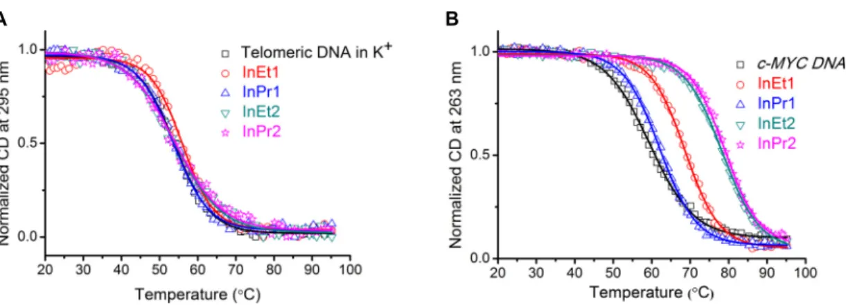
For the telomeric DNA, the experiments were carried out under 10 mM K+ conditions
by measuring the ellipticity at 295 nm; and this yielded a T1/2 of 54 °C (Figure 3A). Addition
of ligands (5 equivalents) resulted in only negligible change in the T1/2 (∆T1/2 ~ −0.2-1.4 °C,
Table 1, Figure 3A). Moreover, melting experiments were carried out for the ligand InEt2 with long telomeric DNA, which can form higher order quadruplex structures.62 Ellipticity
was monitored at 265 nm under K+ conditions. Addition of InEt2 resulted in only slight
increase in the T1/2 (∆T1/2 ~ 2.5 °C) (Figure S6, Supporting Information). For the duplex
DNA, experiments were carried out by measuring the ellipticity at 242 nm, yielding a T1/2 of
62 °C (Figure S6, Supporting Information). As expected, the addition of ligands resulted in only marginal change in the T1/2 (∆T1/2 ~ 0.7-3.2 °C, Table 1, Figure S6, Supporting
Information). These results show that the ligands are not able to stabilize telomeric quadruplex and duplex DNA structures.
CD melting experiments were monitored at 263 nm for the promoter quadruplex DNAs under 1-10 mM K+ conditions (Figure 3B and Figure S6, Supporting Information). For
the c-MYC DNA having a T1/2 of 59 °C, a moderate to high increase in T1/2 values (∆T1/2 ~
2.9-19.5 °C, Table 1) were obtained after the addition of ligands (Figure 3B). Similarly, c-KIT1 DNA yielded T1/2 of 45 °C and an impressive increase in the T1/2 values (∆T1/2 ~
4.4-24.3 °C, Table 1) were observed after the addition of ligands (Figure S6, Supporting Information). In the case of c-KIT2 DNA having T1/2 of 54 °C, a moderate increase in the T1/2
values (∆T1/2 ~ 2.6-12.4 °C, Table 1) were observed after the addition of ligands (Figure S6,
Supporting Information).
CD melting experiments revealed that irrespective of the length and the number of side chains, the ligands are not able to strongly stabilize the telomeric quadruplex (hybrid and higher order) and the duplex DNA structures. Interestingly, the ligands InEt2 and InPr2 showed high thermal stabilization, whereas InEt1 showed moderate and InPr1 showed weak stabilization with the c-MYC and the c-KIT quadruplex DNAs having parallel topologies. Out of the four ligands InEt2 and InPr2 with two methylated side chains are found to impart high stability to the c-MYC and c-KIT quadruplex DNAs than the ligands with single methylated side chains. It should be noted that the differences in side chain length (ethyl or propyl) in InEt2 and InPr2 are not reflected in the thermal stabilization properties. However, ligands with single side chain differed in the thermal stabilization and ligand with ethyl side chain (InEt1) is found to be more promising than that with the propyl side chain (InPr1).
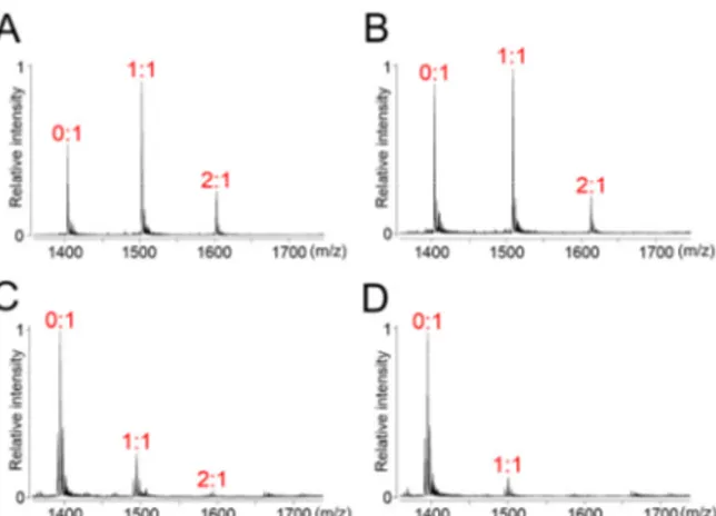
Electrospray Ionisation Mass Spectrometry Studies (ESI-MS). ESI-MS is useful to assess the non-covalent interactions between quadruplex DNAs and small molecules at low concentrations.63,64 Stoichiometries of ligand-quadruplex interactions, and hence individual
binding constants, can indeed be calculated using this technique. Experiments were carried out in a buffer containing NH4+ ions as co-existing cations with the ligands InEt2 and InPr2.
ESI-MS spectra of c-MYC quadruplex DNA (Figure 4A and 4B) in the absence of ligands showed a sharp signal around m/z 1400 with five negative charges. Upon addition of 2 equivalents of ligands, two new signals appeared in the spectra corresponding to 1:1 and 2:1 ligand-quadruplex complexes, in which the former was predominant (Figure 4A and 4B).
Binding constant values for the interaction of InEt2 with c-MYC quadruplex DNA (K1
whereas InPr2 showed moderate binding affinity (K1 ~ 105 and K2 ~ 104 M−1) (Figure 4 and
Table 2). Similarly, both the ligands, showed moderate to high stabilization (K1 and K2 ~ 105
M−1 for InEt2 and K1 ~ 106 and K2 ~ 105 M−1 for InPr2) with c-KIT1 quadruplex DNA
(Figure S7, Supporting Information and Table 2). Control experiments were performed with a tetra molecular parallel quadruplex DNA, [(dTG4T)4], and both the ligands showed moderate
to weak binding (K1 and K2 ~ 105 M−1 for InEt2 and K ~ 104 M−1 for InPr2) with 1:1 and 2:1
stoichiometries (Figure S7 and Table 2).
In order to address the topology specific binding toward c-MYC and c-KIT1 over telomeric quadruplex DNAs, similar experiments were performed with telomeric quadruplex DNA (Figure 4C and 4D). For InEt2 and InPr2 weak signals corresponding to 1:1 and 2:1 stoichiometries as compared to those for c-MYC and c-KIT quadruplex DNAs were observed. Moreover, high selectivity for the MYC (up to 56-fold) and moderate selectivity for the c-KIT1 (up to 9 fold) over telomeric quadruplex DNAs was indicated by the binding constant values (Table 2). These results are in agreement with the results from the CD melting studies and ligand InEt2 with ethyl side chains was found to be highly stabilizing and more specific toward the c-MYC and c-KIT quadruplex DNAs as compared to InPr2 with propyl side chains (Figure 4). ESI-MS analyses were also performed with four duplex sequences (DS17, DK100, DK66, and DK33; Table S1, Supporting Information) having different GC content to ensure the selectivity for quadruplex DNAs over duplex DNAs (Figure S7, Supporting Information). In the case of InEt2, as the GC content in the duplex DNAs was increased, binding affinities were found to be increasing (Table 2). But higher selectivities (up to 62 fold depending on the sequence) reflected in the binding constant values for c-MYC quadruplex over duplex DNAs validate the selectivity of ligands. In the case of c-KIT1 DNA moderate selectivity was achieved by InPr2 over duplex DNA (6-20 fold), whereas InEt2 showed poor selectivity (1.5-10 fold) with the duplex DNA depending upon the sequence of duplex DNA.
Isothermal Titration Calorimetry (ITC). ITC experiments enable to derive the thermodynamic profile of ligand - DNA interactions. We have selected InEt2 and InPr2 for the ITC studies and the c-MYC as an example from the promoter quadruplex DNAs. In the ITC experiments, both ligands showed a nonlinear isotherm pattern indicating complex multiple binding modes (Figure 5). Integrated heat data was fitted by using a sequential binding model to derive the thermodynamic profile, and the best-fit parameters are listed in Table 3. In the case of InEt2, binding of the first and the second molecule were strong enough to get a binding constant of the order of 106-105 M−1 and the binding of third molecule was
weak in nature (~ 104 M−1). Similarly, InPr2 showed a binding constant of the order of 105
-104 M−1 and a weak third binding ~ 103 M−1. As in ESI-MS, the ITC results show that one
binding site has higher affinity than the following ones. Both binding interactions are driven by a large favourable negative enthalpy change (Table 3). Among the two ligands, InEt2 was found to be having higher binding affinity with the c-MYC quadruplex DNA, which is consistent with the results from ESI-MS. Possible reason for very weak binding of the third molecule may be the formation of non-specific adducts at high ligand concentrations. To further confirm specificity of the ligands toward c-MYC quadruplex DNAs and to support the findings from CD and ESI-MS, similar experiments were conducted with telomeric quadruplex and duplex DNAs (Figure S8, Supporting Information). It was evident from the binding constant values that the ligands showed relatively weak binding (K ~ 104-103 M−1)
with telomeric quadruplex and duplex DNAs (Table S2 and S3, Supporting Information).
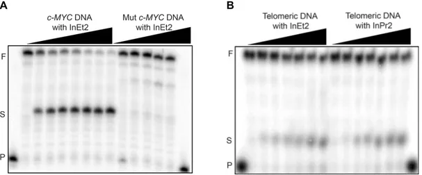
Taq DNA Polymerase Stop Assay. The Specificity of ligands to stabilize c-MYC quadruplex
DNAs was further probed with the aid of Taq DNA polymerase stop assay. Stop assay was performed with c-MYC DNA and with telomeric quadruplex DNAs. The reaction temperatures for the stop assay (50 °C for c-MYC and 40 °C for telomeric DNA) were chosen in such a way that there is no formation of stop products in the absence of ligands. At this
temperature, partially stable quadruplex structures are easily unwound by the Taq DNA polymerase enzyme.36,37 Control experiments were performed with a template containing
mutated c-MYC DNA sequences that cannot form quadruplex structure (Figure 6A and Figure S9, Supporting Information). Formation of the stop products were observed for the c-MYC DNA (minimum IC50 ~ 1.5 µM) after incubating with increasing concentration of all the
ligands (Figure 6A and Figure S10, Supporting Information). As expected, there was no formation of stop products observed with the mutated c-MYC DNA that cannot form a quadruplex structure. There was no significant amount of stop products (~ 10%) for the telomeric DNAs even at 120 µM ligand concentration (Figure 6B and Figure S9, Supporting Information).
Ligands having double side chains InEt2 and InPr2 were very efficient in stabilizing c-MYC quadruplex DNA with low IC50 values (IC50 ~ 1.5 and 2.5 µM for respectively).
Ligands with single side chains showed moderate IC50 values and the ligand with ethyl side
chain, InEt1 (IC50 ~ 10 µM) was more effective compared to the other with propyl side chain,
InPr1 (IC50 ~ 34 µM). However, formation of stop products was not prominent for mutated
c-MYC and telomeric quadruplex DNAs with all the ligands irrespective of the number and length of the side chains (Figure S9, Supporting Information).
Molecular Modeling and Dynamics Studies. Molecular modeling and dynamics studies were carried out to rationalize the topology specific binding of the ligands InEt2 and InPr2 with c-MYC and c-KIT1 G-quadruplex structures over telomeric G-quadruplex topologies. The ligand structures were geometry-optimized in Gaussian0943 at HF/6-31G* level of theory
(Figure S11, Supporting Information). These optimized structures were docked with the G-quadruplex DNA structures from the Protein Data Bank (c-MYC: PDB ID 2L7V38, c-KIT1:
143D41 and telomeric hybrid: 2MB342) using Autodock 4.2.44 Binding stoichiometry of 2:1
was revealed with both c-MYC and c-KIT1 quadruplex structures, with ligands stacking at the top (5'-end) and at the bottom (3'-end) quartets of the quadruplex DNAs. This binding stoichiometry of the ligands with both c-MYC and c-KIT1 quadruplexes is supported by the ESI-MS results. A similar 5'- and 3'- endstacking mode has also been found for indenopyrimidine ligand InPy1 with c-MYC and c-KIT1 and for quindoline to c-MYC quadruplex DNAs.33,38 These docked structures were minimized, equilibrated, and 100 ns
MD runs in AMBER1465 (using pmemd module) were carried out.
MM-PBSA analysis using the MM-PB/GBSA51 module of AMBER14 was carried
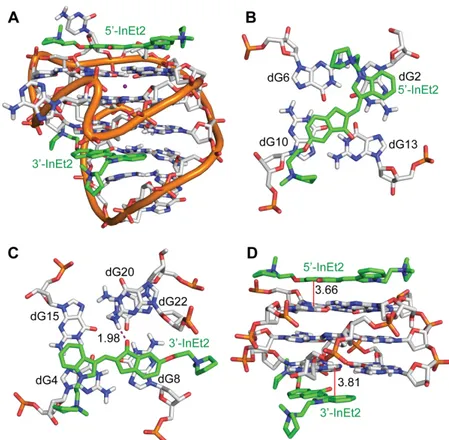
out to obtain the binding free energy values. The free energy values for the binding of InEt2 and InPr2 with c-MYC and c-KIT1 quadruplex DNAs are shown in Table 4. The 5'- and 3'- end stacking mode of InEt2 with c-MYC promoter quadruplex structure gives the most favourable binding energy (−80 kcal/mol) while InPr2, also in a 5'- and 3'- end stacking mode showed slightly reduced affinity (−66 kcal/mol) (Table S5, Supporting Information). With the c-KIT1, both InEt2 and InPr2 showed very similar binding energies (−58 and −57 kcal/mol respectively) (Table S6, Supporting Information). The specific interactions of the two ligands at the 5'- and 3'-quartet are discussed in detail below (Figures 7 and 8).
The structural stabilities of the systems through the 100 ns MD run were examined using the ptraj module of AMBER14. Root mean square deviation (RMSD) graphs and average values for the heavy atoms in the backbone, quartets, and the two ligands were also derived (Figures S12, S13 and Table S3, Supporting Information). Results indicate that the systems remained relatively stable throughout the run. This was also confirmed by the hydrogen bond occupancy for the Hoogsteen H-bonding within the quartets. The H-bonds are
present through >98.8% of the simulation time for the receptors (Table S4, Supporting Information).
The stability of these complexes results from several factors. In the complex between InEt2 and c-MYC, the 5'-InEt2 (Figure 7B) stacks its indole ring with the 5' quartet residue dG13 (average distance: 3.72 ± 0.33 Å), while its indanone ring stacks with the flanking nucleotide dG2 (3.97 ± 0.40 Å) throughout the MD run. Stacking of 3'-InEt2 with the 3' quartet (Figure 7C) was observed from both the indole and indanone rings: indole ring with dG15 (stacking distance: 3.63 ± 0.32 Å, 85% of MD run) and indanone ring with dG6, (stacking distance: 3.70 ± 0.32 Å, 77% of run). As seen in Figure 7D, 3'-InEt2 also showed a strong H-bonding interaction for the initial 44 ns between the carbonyl oxygen of the ligand and NH of dT20 of the flanking nucleotide (H-bond distance: 2.11 ± 0.49 Å). After 46 ns, a rearrangement was seen and the dT20 residue moves into a stacking position below the ligand for the rest of the run. Electrostatic interactions involving the positively charged nitrogens in the ligand side chain were found to be short-lived (5'-InEt2 N+ with ribose sugar O4' of dG4;
3'-InEt2 N+ with phosphate of dG6). The interactions of the InPr2 with the c-MYC were
found to be very similar (Figure S14, Supporting Information). For the 5'-InPr2, in addition to stacking of indole with quartet and indanone with flanking dG2, a stacking interaction of indanone with the quartet nucleotide dG17 was also seen (stacking distance: 3.70 ± 0.36 Å). In the case of 3'-InPr2, the hydrogen bond of the carbonyl oxygen of the ligand with NH2 of
dA22 persisted for 64% of the run, while for the remaining 32% of the run the oxygen atom was in H-bond with NH of dT20 (as seen with 3'-InEt2, Figure 7D ). During H-bond contact with dT20, stacking of indole ring with the quartet was disrupted. Again, the electrostatic interactions with pyrrolidine nitrogen (5'-InPr2 with phosphate of dG8, 3'-InPr2 with dT7 phosphate) were found to be short-lived. The significant difference between the binding
energy of 3'-InEt2 and 3'-InPr2 seen in Table 4 could be due to the presence of stacking interaction of 3'-InEt2 indanone ring with the quartet, which was disturbed for 3'-InPr2.
Figure 8 shows a representative structure of the MD run of InEt2 with the c-KIT1. As seen in Table 4, with the c-KIT1, both 5'-InEt2 and 3'-InEt2 have very similar binding energies and this is reflected in their stacking interactions which are also very similar. For 5'-InEt2 (Figure 8B), the indole ring stacks with the 5'-quartet terminal residue dG2 at a distance of 3.98 ± 0.51 Å, and the indanone ring shows stacking with the 5'-quartet residue dG10 (stacking distance: 3.66 ± 0.39 Å). Short-lived electrostatic interactions were seen between the pyrrolidine nitrogen and O4' of dG9 deoxyribose sugar. The indole ring of 3'-InEt2 (Figure 8C) was found to have stacking interactions with the 3'-quartet guanine dG4 (stacking distance: 3.81 ± 0.27 Å), while the indanone ring was stacking with another 3'-quartet residue dG8 (stacking distance: 3.79 ± 0.30 Å). In addition, 3'-InEt2 also showed a strong H-bond from its carbonyl oxygen to NH of dG20 (average distance: 1.98 ± 0.22Å), and electrostatic interactions with phosphate of dC9 and O4' of dA19 deoxyribose ring (Figure 8C). All these interactions were found to be stable throughout the MD simulation. InPr2 was found to have very similar interactions with c-KIT1, as shown in Figure S15, Supporting Information.
In addition, to unravel the topology specific binding of ligands with MYC and c-KIT1 G-quadruplex DNAs over telomeric quadruplex topologies, MD simulations (100 ns) of the telomeric parallel, antiparallel and hybrid topologies in complex with the InEt2 and InPr2 ligands were carried out. Docking and MD simulations results revealed that the binding stoichiometries of both the InEt2 and InPr2 ligand with telomeric DNA are 1:1 and the ligands stack on the 5'-end of the quartet (Figures S22-S24, Supporting Information). The quadruplex DNAs were quite stable during the 100 ns of MD simulations, however, both the ligands were found to be highly flexible in the complexes (Figures S16-S21, Supporting
Information). The binding energy of the ligands with telomeric DNA topologies was found to be in the range of −19 to −24 kcal/mol which is higher in comparison to the ligand-c-MYC and c-KIT1 quadruplex complexes (Table 4). From the individual binding energy components, it was observed that the ΔEMM in the c-MYC and c-KIT1 quadruplex-ligand
complexes (> −950 kcal/mol; for 1:1 binding ratio) was favourable in comparison to that for the complex formed by telomeric topologies (< −750 kcal/mol) (Table S7 and S8, Supporting Information). This is indeed reflected in the percentage life time occupancy of the stacking interactions between ligand and quartet in the promoter (>75) and telomeric topologies (<50) during the MD simulations (Table S9, Supporting Information). These unfavourable stacking interactions may be attributed to the presence of different loop structures in the antiparallel and hybrid topologies, which hinders to accommodate the pyrrolidium side chains of the ligands. In case of the parallel telomeric DNA, due to the absence of flanking nucleotides as in the c-MYC and c-KIT1 quadruplex, the ligands are flexible to move on the surface of the quartet. However, the percentage lifetime occupancy of stacking interactions was found to be <46% in the 100 ns MD simulations of ligands with parallel telomeric DNA complexes. There were no H-bonds present between the telomeric quadruplex topologies and the ligands. Also, electrostatic interactions are not observed between the positively charged side chains in the ligands and the negatively charged phosphate backbone of the DNA.
The stacking of the two aromatic groups (indole and indanone) in the ligands InEt2 and InPr2 with the G-quartet nucleobases and flanking nucleotides was found to be the main stabilizing interaction for these ligands with the c-MYC and c-KIT1 quadruplex structures. The 2:1 complexes of these ligands with c-MYC and c-KIT1 are possible because of the availability of binding sites at both the top and bottom quartets of these quadruplexes. Overall, the MD simulation results show that along with end stacking, hydrogen bonding between the carbonyl group of ligands with the flanking nucleotides, and electrostatic
interactions of the positively charged side chain play role in the specific recognition of a particular quadruplex topology. However, the stabilization can be mostly attributed to the stacking interactions of indolylmethyleneindanone core group with G-quartets as revealed the late stage MD simulation results. Similar stacking interactions were not observed between telomeric quadruplex DNA and the ligands, which attribute to their specificity toward c-MYC and c-KIT1 quadruplex structures.
CONCLUSIONS
Till date, there are >1000 small molecule ligands have been reported, which show moderate to high affinity toward G-quadruplex structures.66 Most of them harbour large aromatic core
with number of heteroatoms and as a result fall behind the typical drug-like criteria set by medicinal chemists.16 Though many of them offer target discrimination between quadruplex
and duplex structures, there are only handful of examples, which show some preferential target recognition toward a particular quadruplex topology. Since indiscriminate induction and synergic stabilization of multiple quadruplexes can lead to genomic instability,67,68 for
clinical success, search for a bona fide ligand, which specifically target a particular topology may be desirable. In this line, here we report new indolylmethyleneindanone derivatives InEt1, InEt2, InPr1 and InPr2, and confirm their specificity towards promoter quadruplex DNAs having parallel topologies using variety of biophysical and biochemical techniques. The lead compound InEt2 bearing a fully conjugated system comprised of indanone and indole moieties along with two positive side chains is able to specifically bind to the parallel topology of oncogenic promoter quadruplexes of c-MYC and c-KIT. The observed specificity is attributed to the combined effects of number of non-covalent interactions owing to the unique structural elements present in the ligands. Further structural studies are warranted to confirm this. These new unique molecular scaffolds offer opportunities to
harness their potential for therapeutic and diagnostic applications centred on promoter quadruplex structures in the genome.
ASSOCIATED CONTENT
Supporting information. CD spectra of ligands with telomeric, c-MYC, c-KIT1, c-KIT2 DNAs in the absence of added metal ions; Non-denaturing gel of telomeric and c-MYC DNAs from EMSA; CD melting curves of c-KIT1, c-KIT2 and duplex DNAs; ESI-MS mass spectra for c-KIT1, (TG4T)4 and duplex DNAs; ITC profiles of ligands with quadruplex and duplex
DNAs; PAGE of Taq DNA polymerase stop assay with c-MYC and telomeric DNAs; IC50
plots from Taq DNA polymerase stop assay; Energy optimized structure of ligands at HF/6-31G* level; Time dependent RMSD graphs of c-MYC, c-KIT1 and telomeric (parallel, antiparallel and hybrid) DNAs in complex with InEt2 and InPr2; MD snapshots of InPr2 with c-MYC and c-KIT1 quadruplex DNAs; MD snapshots of InEt2 and InPr2 with telomeric parallel, antiparallel and hybrid quadruplex DNAs; Oligonucleotides used for biophysical and biochemical studies; Thermodynamic parameters for telomeric quadruplex and duplex DNAs from ITC; Hoogsteen hydrogen bond occupancies in G-quartet during MD simulations of InEt2; Binding free energy components of c-MYC, c-KIT1 and telomeric (parallel, antiparallel and hybrid) DNAs with InEt2 and InPr2; Percentage lifetime occupancies of stacking interactions over 100 ns of MD simulations; 1H NMR & 13C NMR
spectra of compound 2, 3, 4, InEt1, InPr1, 10, 11, 12, 13, InEt2 and InPr2. This material is available free of charge via the Internet at http://pubs.acs.org/.
REFERENCES
1. Burge, S., Parkinson, G. N., Hazel, P., Todd, A. K. and Neidle, S. (2006) Quadruplex DNA: sequence, topology and structure. Nucleic Acids Res. 34, 5402-5415.
2. Collie, G. W. and Parkinson, G. N. (2011) The application of DNA and RNA G-quadruplexes to therapeutic medicines. Chem. Soc. Rev. 40, 5867-5892.
3. Meyne, J., Ratliff, R. L. and Moyzis, R. K. (1989) Conservation of the human telomere sequence (TTAGGG)n among vertebrates. Proc. Natl. Acad. Sci. U.S.A. 86, 7049-7053. 4. Wang, Y. and Patel, D. J. (1993) Solution structure of the human telomeric repeat
d[AG3(T2AG3)3] G-tetraplex. Structure 1, 263-282.
5. Balasubramanian, S., Hurley, L. H. and Neidle, S. (2011) Targeting G-quadruplexes in gene promoters: a novel anticancer strategy? Nat. Rev. Drug Disc. 10, 261-275.
6. Eddy, J. and Maizels, N. Conserved elements with potential to form polymorphic G-quadruplex structures in the first intron of human genes. (2008) Nucleic Acids Res. 36, 1321-1333.
7. Sen, D. and Gilbert, W. (1988) Formation of parallel four-stranded complexes by guanine-rich motifs in DNA and its implications for meiosis. Nature 334, 364-366.
8. Siddiqui-Jain, A., Grand, C. L., Bearss, D. J. and Hurley, L. H. (2002) Direct evidence for a G-quadruplex in a promoter region and its targeting with a small molecule to repress c-MYC transcription. Proc. Natl. Acad. Sci. U.S.A. 99, 11593-11598.
9. Rankin, S., Reszka, A. P., Huppert, J., Zloh, M., Parkinson, G. N., Todd, A. K., Ladame, S., Balasubramanian, S. and Neidle, S. (2005) Putative DNA quadruplex formation within the human c-kit oncogene. J. Am. Chem. Soc. 127, 10584-10589.
10. Dai, J., Chen, D., Jones, R. A., Hurley, L. H. and Yang, D. Z. (2006) NMR solution structure of the major G-quadruplex structure formed in the human BCL2 promoter region. Nucleic Acids Res. 34, 5133-5144.
11. Sun, D. Y., Guo, K. X., Rusche, J. J. and Hurley, L. H. (2005) Facilitation of a structural transition in the polypurine/polypyrimidine tract within the proximal promoter region of
the human VEGF gene by the presence of potassium and G-quadruplex-interactive agents. Nucleic Acids Res. 33, 6070-6080.
12. De Armond, R., Wood, S., Sun, D. Y., Hurley, L. H. and Ebbinghaus, S. W. (2005) Evidence for the presence of a guanine quadruplex forming region within a polypurine tract of the hypoxia inducible factor 1alpha promoter. Biochemistry 44, 16341-16350. 13. Qin, Y., Rezler, E. M., Gokhale, V., Sun, D. and Hurley, L. H. (2007) Characterization of
the G-quadruplexes in the duplex nuclease hypersensitive element of the PDGF-A promoter and modulation of PDGF-A promoter activity by TMPyP4. Nucleic Acids Res. 25, 7698-7713.
14. Gonz´alez, V. and Hurley, L. H. (2010) The c-MYC NHE III (1): function and regulation.
Annu. Rev. Pharmacol. Toxicol. 50, 111-129.
15. Murat, P. and Balasubramanian, S. (2014) Existence and consequences of G-quadruplex structures in DNA. Curr. Opin. Gen. Dev. 25, 22-29.
16. Neidle, S. (2016) Quadruplex Nucleic Acids as Novel Therapeutic Targets. J. Med. Chem. DOI: 10.1021/acs.jmedchem.5b01835.
17. Risitano, A. and Fox, K. R. (2004) Influence of loop size on the stability of
intramolecular DNA quadruplexes. Nucleic Acids Res. 32, 2598-2606.
18. Cevec, M. and Plavec, J. (2005) Role of loop residues and cations on the formation and stability of dimeric DNA G-quadruplexes. Biochemistry 44, 15238-15246.
19. Dai, J., Carver, M. and Yang, D. (2008) Polymorphism of human telomeric quadruplex structures. Biochimie 90, 1172-1183.
20. Hamon, F., Largy, E., Gudin-Beaurepaire, A., Rouchon-Dagois, M., Sidibe, A., Monchaud, D., Mergny, J. L., Riou, J. F., Nguyen, C. H. and Teulade-Fichou, M. P. (2011) An acyclic oligoheteroaryle that discriminates strongly between diverse G-quadruplex topologies. Angew. Chem. Int. Ed. 50, 8745-8749.