HAL Id: insu-01186040
https://hal-insu.archives-ouvertes.fr/insu-01186040
Submitted on 24 Aug 2015
HAL is a multi-disciplinary open access
archive for the deposit and dissemination of sci-entific research documents, whether they are pub-lished or not. The documents may come from teaching and research institutions in France or abroad, or from public or private research centers.
L’archive ouverte pluridisciplinaire HAL, est destinée au dépôt et à la diffusion de documents scientifiques de niveau recherche, publiés ou non, émanant des établissements d’enseignement et de recherche français ou étrangers, des laboratoires publics ou privés.
Study of the Histology of Leafy Axes and Male Cones of
Glenrosa carentonensis sp. nov. (Cenomanian Flints of
Charente-Maritime, France) Using Synchrotron
Microtomography Linked with Palaeoecology
Jean-David Moreau, Didier Néraudeau, Paul Tafforeau, Eric Dépré
To cite this version:
Jean-David Moreau, Didier Néraudeau, Paul Tafforeau, Eric Dépré. Study of the Histology of Leafy Axes and Male Cones of Glenrosa carentonensis sp. nov. (Cenomanian Flints of Charente-Maritime, France) Using Synchrotron Microtomography Linked with Palaeoecology. PLoS ONE, Public Library of Science, 2015, 10 (8), 23 p. �10.1371/journal.pone.0134515�. �insu-01186040�
RESEARCH ARTICLE
Study of the Histology of Leafy Axes and
Male Cones of
Glenrosa carentonensis sp.
nov. (Cenomanian Flints of
Charente-Maritime, France) Using Synchrotron
Microtomography Linked with Palaeoecology
Jean-David Moreau1,2*, Didier Néraudeau1, Paul Tafforeau2, Éric Dépré3
1 CNRS UMR 6118, Géosciences Rennes, Université Rennes 1, Rennes, France, 2 Beamline ID19, European Synchrotron Radiation Facility, Grenoble, France, 3 GIP-GEVES (Groupement d’Etude et de Contrôle des Variétés et Semences), Surgères, France
*jean.david.moreau@gmail.com
Abstract
We report exceptionally well-preserved plant remains ascribed to the extinct conifer Glen-rosaJ. Watson et H.L. Fisher emend. V. Srinivasan inside silica-rich nodules from the Ceno-manian of the Font-de-Benon quarry, Charente-Maritime, western France. Remains are preserved in three dimensions and mainly consist of fragmented leafy axes. Pollen cones of this conifer are for the first time reported and in some cases remain connected to leafy stems. Histology of Glenrosa has not previously been observed; here, most of internal tis-sues and cells are well-preserved and allow us to describe a new species, Glenrosa caren-tonensissp. nov., using propagation phase-contrast X-ray synchrotron microtomography, a non-destructive technique. Leafy axes consist of characteristic helically arranged leaves bearing stomatal crypts. Glenrosa carentonensis sp. nov. differs from the other described species in developing a phyllotaxy 8/21, claw-shaped leaves, a thicker cuticle, a higher number of papillae and stomata per crypt. Pollen cones consist of peltate, helically arranged microsporophylls, each of them bearing 6–7 pollen sacs. The new high resolution tomo-graphic approach tested here allows virtual palaeohistology on plants included inside a dense rock to be made. Most tissues of Glenrosa carentonensis sp. nov. are described. Lithological and palaeontological data combined with xerophytic features of Glenrosa car-entonensissp. nov. suggest that this conifer has been adapted to survive in harsh and insta-ble environments such as coastal area exposed to hot, dry conditions.
Introduction
The Cretaceous conifer Glenrosa J. Watson et H.L. Fisher emend. V. Srinivasan [1–2] shows a broad stratigraphic and geographical distribution, being reported from the Barremian to the
a11111
OPEN ACCESS
Citation: Moreau J-D, Néraudeau D, Tafforeau P, Dépré É (2015) Study of the Histology of Leafy Axes and Male Cones ofGlenrosa carentonensis sp. nov. (Cenomanian Flints of Charente-Maritime, France) Using Synchrotron Microtomography Linked with Palaeoecology. PLoS ONE 10(8): e0134515. doi:10.1371/journal.pone.0134515
Editor: William Oki Wong, Institute of Botany, CHINA Received: March 4, 2015
Accepted: July 7, 2015 Published: August 19, 2015
Copyright: © 2015 Moreau et al. This is an open access article distributed under the terms of the
Creative Commons Attribution License, which permits unrestricted use, distribution, and reproduction in any medium, provided the original author and source are credited.
Data Availability Statement: All relevant data are within the paper, and high quality plates used for this study are available online on the open access database for palaeontology of the ESRF:http://paleo. esrf.eu.
Funding: This work was supported by funding from Géosciences Rennes, CNRS-UMR6118, to J-DM, DN; and the European Synchrotron Radiation Facility, to J-DM, PT. The funders had no role in study design, data collection and analysis, decision to publish, or preparation of the manuscript.
Cenomanian of America, Asia and Europe [1–8]. Glenrosa is characterized by an unusual sto-matal arrangement inside crypts. Stosto-matal crypts consist of ampulla-shaped pits that are sunken in the mesophyll and contain stomatal apparatuses. The genus was first erected to include two species from the lower Cretaceous of Texas, Glenrosa texensis and Glenrosa pagio-phylloides [1], that were previously assigned to Brachyphyllum texense and Sequoia pagiophyl-loides by Fontaine [9]. Glenrosa texensis was described from the Glen Rose Formation and the Trent’s Reach locality, which are late Aptian–earliest Albian and Barremian–earliest Aptian in age respectively. Glenrosa pagiophylloides was only reported from the first [1]. Four other spe-cies were later distinguished based on phyllotaxy, leaf morphology, and cuticular features: Glenrosa hopewellensis and Glenrosa virginiensis from the middle Albian Patapsco Formation of Virginia [2], Glenrosa nanjingensis from the late Early Cretaceous Gecun Formation of China [3], and Glenrosa falcata from the upper Barremian of La Huérguina Formation in Spain [7]. All previous Glenrosa species were based only on highly compacted and isolated cuti-cle remains, and internal histology of leaf tissues has not been described. Likewise, attached reproductive structures have not been found, although isolated cone scales and microsporo-phylls without in situ pollen sacs have tentatively been assigned to Glenrosa [2]. Initially, Glen-rosa was tentatively assigned to the Cheirolepidiaceae [1]. However, Srinivasan [2] and Zhou [3] noted that the stomatal arrangement differs considerably from members of Cheirolepidia-ceae. Based on isolated reproductive structures, Srinivasan [2] tentatively compared Glenrosa with Cupressaceae. Affinities of the genus remain unresolved.
In western France, fragmented cuticles of leafy axes ascribed to Glenrosa sp. have been pre-viously reported from upper Albian and Cenomanian deposits of many localities of Charente-Maritime and Charente (Figs1and2; [6,8]). Exceptionally preserved Glenrosa specimens have been recently recovered from this area inside Cenomanian flint nodules that preserve the cuti-cle and the histology of vegetative structures, as well as attached pollen-producing cones. In the present paper, we describe a new species, Glenrosa carentonensis sp. nov. Given the preserva-tion of this exceppreserva-tional new material in tough flint, we used a non-destructive imaging tech-nique useful for the observation of inner and hidden structures: the propagation phase-contrast X-ray synchrotron microtomography (PPC-SRμCT). The plant-bearing nodules being large and dense, this work required to test new tomographic protocols combining high energy and multiscale approaches. We discuss the interest and the limits of the synchrotron microto-mography, and compare G. carentonensis sp. nov. with other Glenrosa species. The only known other Cretaceous conifer bearing stomatal crypts is Sedites rabenhorstii (Geinitz) Kunz-mann from the upper Turonian of the Bohemian Cretaceous Basin [10]. Given the rarity of sto-matal crypts in conifers [11], we also discuss their potential palaeoecological significant in Glenrosa.
Geological Setting
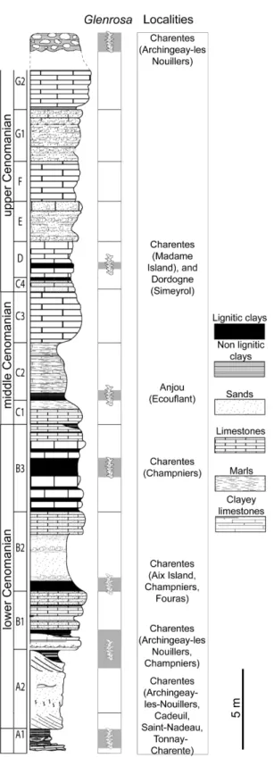
In Charente and Charente-Maritime, the upper Albian–Cenomanian series are divided into seven units, A to G (Fig 2; [12]). In the Font-de-Benon quarry, the beds are exposed from sub-unit A1 to subsub-unit B1 (Figs1and2; [13–14]). Uppermost Albian (A1) consists of sandy beds with cross-bedded stratifications and clay lenses. A1 mainly yielded wood and amber contain-ing abundant and diverse arthropods inclusions [15–17]. The lowermost Cenomanian (A2) shows alternations of sand and clay beds, and yielded abundant and diverse plant meso-and macrofossils bearing cuticles [5,6,18]. B1 consists of sands yielding abundant marine fauna such as molluscs, echinoderms, foraminifers (Orbitolina) and vertebrates [19]. Locally, at the top of the quarry, deposits are overlapped by 1-to-2 metres horizontal beds of diachronic flint. Fossiliferous and siliceous nodules used for this study have been collected in this latter facies.
Histology and Ecology of the Conifer Glenrosa carentonensis sp. nov.
Competing Interests: The authors have declared that no competing interests exist.
Nodules are irregular in shape and 10-50-cm wide. Flint nodules yielded marine invertebrates such as echinoids (c. Catopygus carinatus Goldfuss, Mecaster sp. Pomel), foraminifers (c. Orbi-tolina conica d’Archiac), and unidentified sponge spicules [14]. Marine invertebrates co-occurred with terrestrial plants. These latter are represented by conifers such as Brachyphyllum Brongn. emend. Harris, Frenelopsis (Schenk) emend. J. Watson, Geinitzia Endl., and Glenrosa J. Watson et H.L. Fisher emend. V. Srinivasan [13–14]. Based on the co-occurrence of Orbito-lina conica and Catopygus carinatus, a Cenomanian age was assigned to the flints.
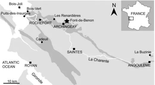
Albian–Cenomanian deposits yielding plant fossils have been reported from several locali-ties of western France such as Charente-Maritime [5], Dordogne [20], and Maine-et-Loire [21] (Figs1and2). Glenrosa have been reported from the uppermost Albian (A1) of Cadeuil [22], Font-de-Benon (Archingeay- Les Nouillers; [15]) and Les Renardières (Tonnay-Charente; [12]); the lowermost Cenomanian (A2 and B1) of Font-de-Benon (Archingeay-Les Nouillers; [6]) and La Buzinie (Champniers); the upper lower Cenomanian (B2 and B3) of La Buzinie (Champniers; B.G. pers. obs.), Bois-Joli (Aix Island; [8]), Bois-Vert (Fouras; [5]); and the upper Cenomanian limestones (D) of Puits-des-Insurgés (Madame Island; D.N. pers obs.).
Material and Methods
The plant-bearing flints were collected from the Cenomanian of the Font-de-Benon quarry between the villages of Archingeay and Les Nouillers, Charente-Maritime, western France (Fig 1; GPS: 45°56’15.5”N 0°41’40.3W). The quarry is no longer accessible, as it has been partially
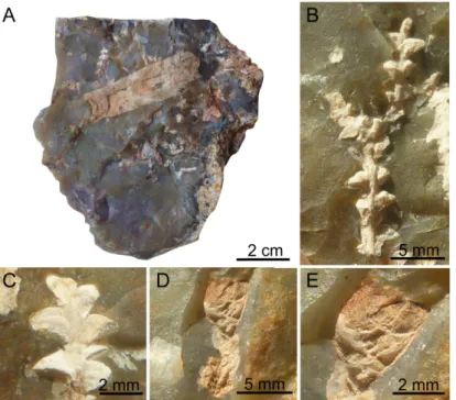
filled in. All necessary permits were obtained for this study, which complied with all relevant regulations, and sampling access was provided by the MARCHAND company (Archingeay, France), the owner of the quarry. The studied flint nodules were irregular in shape and did not exceed 15 cm, although only fragments were studied. Some nodule fragments showed plant fossils on breaken surfaces in addition to those contained inside (Fig 3A–3E). However, they contain hidden plant inclusions inside matrix. Plant remains are up to 5 cm long, and consist of vegetative structures and some pollen cones, occasionally in connection. Specimens of the genus Glenrosa, as determined by diagnostic helical phyllotaxy and the presence of stomatal
Fig 1. Geological map of Charente and Charente-Maritime. Black dots indicate Albian–Cenomanian localities yielding Glenrosa J. Watson et H.L. Fisher emend. V. Srinivasan and the black star indicates the Font-de-Benon quarry.
doi:10.1371/journal.pone.0134515.g001
Fig 2. Stratigraphic section from the Albian–Cenomanian in western France with indication of the beds yieldingGlenrosa J. Watson et H.L. Fisher emend. V. Srinivasan.
doi:10.1371/journal.pone.0134515.g002
crypts, are the main component of the flora preserved in the flint nodules. It represents the first example of silicified leafy stems and male cones of Glenrosa discovered to date. Three pollen cones have been discovered, two of them remain connected with leafy axes. Plants remains are preserved as silica permineralizations, with three dimensions of external and internal micro-structures. The flint nodules are housed in the collection of Géosciences Rennes, CNRS UMR 6118, Université Rennes 1 (Rennes, France). Seven of them have been investigated
(SIL_ARC_1 to SIL_ARC_7). Fourteen leafy stems and the three pollen cones have been investigated.
Multiscale propagation phase contrast synchrotron tomography
Multiscale approach. The specimens were scanned at beamlines BM05 and ID19, at the European Synchrotron Radiation Facility (ESRF), Grenoble, France. The dense flint nodules were examined using propagation phase-contrast X-ray synchrotron microtomography (PPC-SRμCT). For studying arthropod inclusions in low density and absorption opaque amber, Lak et al. [23] developed an approach combining medium resolution microradiography and high resolution PPC-SRμCT. In contrast, the high density and high absorption of siliceous nodules required the use of higher energies and made impossible the localisation of inclusions by using radiographic approach only. Low resolution scans of the whole broken-open nodules were performed with 28.9μm, 12.8 μm, and 11.9 μm of voxel sides depending on the specimen dimension. Virtual 2D sections and 3D renderings were reconstructed for the whole rock speci-mens. Thereby, low resolution scans of opaque flint nodules allow fossil plant inclusions to be located. High resolution scans of small areas were performed for targeted plants to describe the morphology and histology (Figs4–13). Local scans of plant fossils were performed with 3.5μm, 0.7 μm, and 0.6 μm of voxel side on selected specimens. Due to the large size and the
Fig 3. Plant fossils embedded inside flint nodules. (A) Broken flint nodule with diverse conifer inclusions, SIL_ARC_2. (B) Leafy twigs of Glenrosa carentonensis sp. nov., SIL_ARC_3_1. (C) Leafy twigs of Glenrosa carentonensissp. nov., SIL_ARC_3_2. (D) Leafy axis of Glenrosa carentonensis sp. nov. bearing distally a microsporangiate cone on its distal tip, SIL_ARC_2_1. (E) Detail of the same cone, SIL_ARC_2_1. doi:10.1371/journal.pone.0134515.g003
high absorption of the original flint pieces, the highest resolution used in this paper (0.28μm voxel side) required smaller sample. Thus, one sample (c. 18 mm long and 7 mm wide) was iso-lated from the siliceous matrix, without any damage of the fossil, and using a diamond disk mounted on a high precision cut-off saw T200, Pressi (Jean Wirtz, Germany).
Acquisition parameters and data processing. The distance between the sample stage and the source is 55 m and 145 m, at BM05 and ID19, respectively. ID19 is characterized by a higher coherence of the beam than BM05 and by more flux at higher energy, allowing to work on larger specimens at higher resolution than BM05. In total, seven different setups have been used (Table 1). We used a pink-beam configuration [24]. For more information on the acquisi-tion protocol see the article published by Moreau et al. [13]. The scans were reconstructed using a back-projection algorithm implemented in the PyHST software (High Speed Tomogra-phy in python version, ESRF). It was coupled with single distance phase retrieval process modi-fied from Paganin’s algorithm [25] to incorporate an unsharp mask applied to the retrieved phase maps [24]. Data were finally converted into a 16 bit tiff stack of slices corrected for resid-ual ring artefacts and cropped.
3D segmentation. The three dimensional reconstructions of specimens were achieved with the software VG Studio Max 2.2 (Volume Graphics, Heidelberg, Germany). The virtual dissection and segmentation were made using 3D region growing and manual selections.
Results: Systematic Palaeontology
Order—Coniferales Family—Incertae sedis
Genus—Glenrosa Watson et Fisher, 1984 emend. Srinivasan, 1992
Species—Glenrosa carentonensis sp. nov. Moreau, Néraudeau, Tafforeau et Dépré Etymology. Derived from the latin name (Carentonia) of the river Charente.
Fig 4. PPC-SRμCT, virtual sections of Glenrosa carentonensis sp. nov. reconstructed using a single distance phase retrieval process. (A) Longitudinal section of leafy axis, SIL_ARC_3_1. (B) Transversal section of leafy axis, SIL_ARC_3_1. (C) Longitudinal section of twig and cone, SIL_ARC_2_1. Voxel side, (A–B) = 11.8 μm, (C) = 28.9 μm.
doi:10.1371/journal.pone.0134515.g004
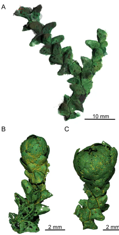
Fig 5. PPC-SRμCT, 3D renderings of Glenrosa carentonensis sp. nov. (A) Helically arranged leafy axis, SIL_ARC_3_1. (B–C) Leafy axes bearing a microsporangiate male cone, SIL_ARC_2_2 and SIL_ARC_2_1, respectively. Voxel side = 11.9μm.
doi:10.1371/journal.pone.0134515.g005
Holotype. SIL_ARC_2_1. Here designated. Figs3D–3E;4C;5C;7A–7C;10A, 10C–10H, 10J
and 10K
Paratypes. SIL_ARC_2_2, SIL_ARC_2_3, SIL_ARC_3_1, and SIL_ARC_7_1. Here desig-nated. Figs3B and 3C;4A and 4B;5A and 5B;6A–6D;8A–8L;9A and 9B;10B, 10I and 10L;
11A–11H;12A–12D;13A–13G
Type locality. Font-de-Benon quarry, Archingeay-Les Nouillers, Charente-Maritime, west-ern France.
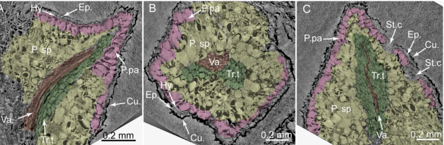
Fig 7. PPC-SRμCT, virtual histological sections of a Glenrosa carentonensis sp. nov. leaf. (A) Sagittal section. (B) Transversal section. (C) Tangential section. Cu., cuticle; Ep., Epidermis; Hy., hypodermis; P.pa, palisade parenchyma; P.sp, spongy parenchyma; St.c, stomatal crypts; Tr.t, transfusion tracheids; Va., vascular bundle. Voxel side = 0.6μm. SIL_ARC_2_1.
doi:10.1371/journal.pone.0134515.g007
Fig 6. 3D renderings of aGlenrosa carentonensis sp. nov. leaf with stomatal crypts. (A) Top view of adaxial surface. (B) Bottom view of abaxial surface. (C) Right view. (D) Oblique top-left view. Cr.a., apertures of stomatal crypts; Le.a., apex of leaf. Voxel side = 11.9μm. SIL_ARC_3_1.
doi:10.1371/journal.pone.0134515.g006
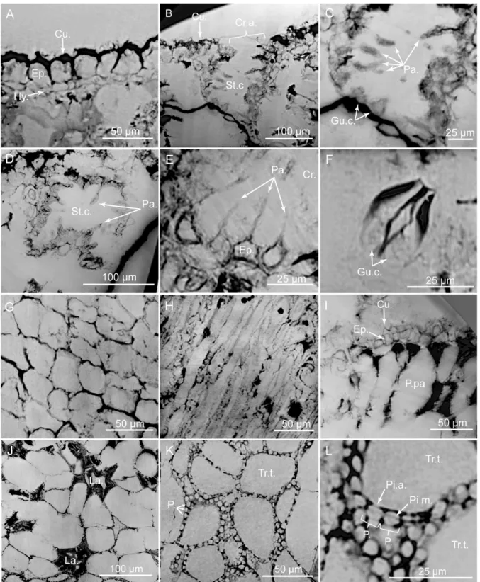
Fig 8. PPC-SRμCT, virtual histological sections of Glenrosa carentonensis sp. nov. leaf. (A) Uppermost adaxial surface with cuticle, epidermis and hypodermis in transversal section. (B) Stomatal crypt in transversal section. (C) Detail of B. (D) Stomatal crypt in tangential section. (E) Hair-like projections of epidermal cells inside a crypt. (F) Guard cells of stomatal apparatus. (G) Epidermal cells in tangential section. (H) Hypodermal cells in tangential section. (I) Elongated palisade parenchyma cells in transversal section. (J) Spongy parenchyma with lacunae in tangential section. (K) Transfusion tracheids with numerous pits. (L) Detail of transfusion tracheid pits. Cr.a., apertures of stomatal crypts; Cu., cuticle; Ep., epidermis; Gu.c., guard cells; Hy., hypodermis; La., lacunae; P., pits; P.pa, palisade parenchyma; Pa., papillae; Pi.a, pit aperture; Pi.m., putative pit membrane; St.c., stomatal crypts; Tr.t., transfusion tracheids. Voxel side = 0.28μm. SIL_ARC_7_1.
doi:10.1371/journal.pone.0134515.g008
Type horizon. Cenomanian flints exposed at the top of the stratigraphic section in the Font-de-Benon quarry.
Diagnosis
Shoots bearing helically arranged leaves. Leaves, claw-shaped, and thick. Leaf apices obtuse to sub-acute. Leaves keeled with a thick cuticle. Leaves amphistomatic with abundant stomatal crypts. Stomatal crypts, 8–18 per leaf, and randomly distributed. Crypts as wide as deep, with circular to oval apertures, and partially occluded by interdigitate, elongated papillae. Papillae up to 48 per crypt. Stomata, 7–12 per crypt, and basally packed. Epidermis thinner on adaxial surface, and with polygonal ordinary cells. Hypodermis thin, and mainly developed on the adaxial surface. Palisade parenchyma with one (rarely two) layer(s) of elongated cells. Spongy
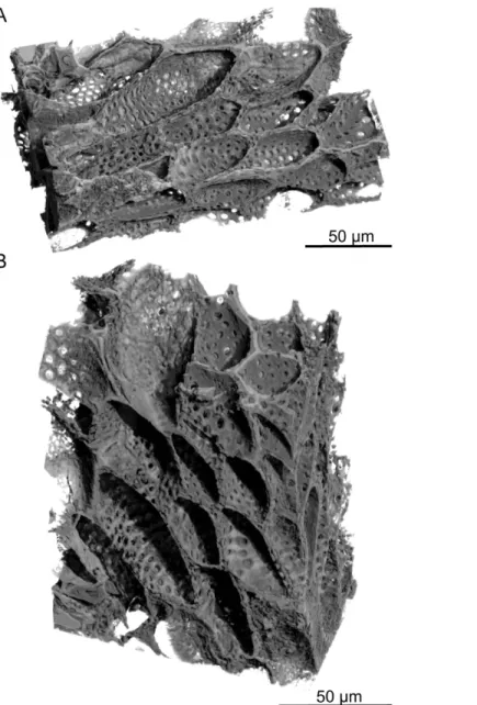
Fig 9. PPC-SRμCT, 3D renderings of transfusion tracheids in leaf of Glenrosa carentonensis sp. nov. (A–B) longitudinal section showing transfusion tracheids with spherical pits. Voxel side = 0.28 μm.
SIL_ARC_7_1.
doi:10.1371/journal.pone.0134515.g009
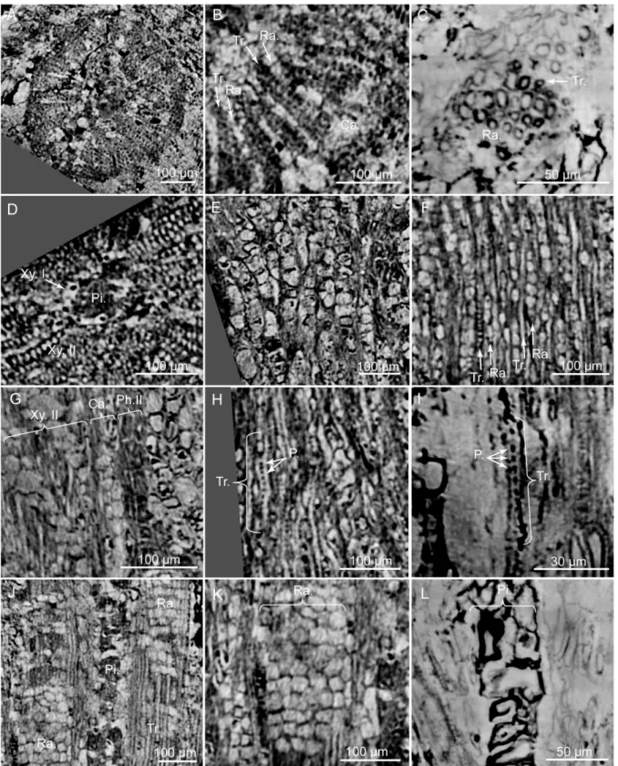
Fig 10. PPC-SRμCT, virtual sections of Glenrosa carentonensis sp. nov. wood. (A) General view of wood in cross section. (B) Detail of wood with tracheids and rays in cross section. (C) Detail of tracheids in cross section. (D) Detail of secondary xylem, primary xylem and pith in cross section. (E) Cambium in tangential section. (F) Uniseriate rays in tangential section. (G) Secondary phloem, cambium and secondary xylem in radial section. (H) Tracheids and pits in radial section. (I) Detail of a tracheid and pits in radial section. (J) Tracheids and rays in radial section. (K) Rays in radial section. (L) Pith in radial section. Ca., cambium; P., pits; Ph.II, secondary phloem; Pi., pith; Ra., rays; Tr., tracheids; Xy.I, primary xylem; Xy.II, secondary xylem. Voxel side, (A, C, D, E, F, G, H, J, K) = 0.63μm; (B, I, L) = 0.28 μm. (A, C, D, E, F, G, H, J, K) SIL_ARC_2_1; (B, I, L) SIL_ARC_7_1.
doi:10.1371/journal.pone.0134515.g010
parenchyma well-developed on abaxial and adaxial sides with lacunae and large, rounded cells. Transfusion tracheids, abundantly pitted, well-developed and limited to the abaxial side of leaves. Single vascular bundle per leaf. Continuous vascular cylinder in the stem. Tracheids elongated, narrow and bearing regularly arranged pits. Rays abundant, mainly uniseriate, 3–20 cells high. Pith with large parenchyma cells. Pollen cones spherical to ovoid, slightly longer than wide. Microsporophylls imbricate, helically arranged, peltate, and up to 40 in number per cone. Microsporophyll stalks elongated, exmedially attached to the head and triangular in transversal plane. Microsporophyll heads flat to convex, and rhomboidal. Pollen sacs 6–7 per microsporophyll.
Description
Gross morphology of leafy axes. Shoots are up to twice branched. Branches arise at 30– 70°. The longest specimen is 50 mm long. Shoots are up to 7.0 mm in diameter. Leaves are
Fig 11. PPC-SRμCT, virtual histological sections of microsporangiate cone with microsporophylls bearing pollen sacs. (A) Cone in longitudinal section. (B) Cone in transversal section. (C) Detail of A showing large head insertion and pollen sacs. (D) Pollen sacs of a microsporophyll in transversal section. (E) In situ pollen grains. (F) Transversal section of microsporophylls showing epidermis and parenchyma-like cells. (G) Longitudinal section of like cells. (H) Putative gland structure. Co., cone axis; Ep., epidermis; Mi.h., microsporophyll head; Mi.s., microsporophyll stalk; P. parenchyma-like tissue; Po., pollen sacs; Po.g., pollen grains. Voxel side = 0.6μm. SIL_ARC_2_3.
doi:10.1371/journal.pone.0134515.g011
helically arranged (Figs3B and 3C;4A–4C;5A–5C). Leaves arise at up to 140 degrees (phyllo-taxy 8/21). Leaves are triangular in abaxial and adaxial views, rhomboidal in transversal sec-tion, claw-shaped in sagittal secsec-tion, and slightly keeled on both sides (Fig 6A–6D). They are 1.5–3.5 mm long and 1.0–1.5 mm wide. The leaf margin is entire. The apex of leaves is obtuse
Fig 12. PPC-SRμCT, 3D renderings of a Glenrosa carentonensis sp. nov. microsporophyll with six pollen sacs. (A) Inner surface in front view. (B) Right view. (C) Outer surface in front view. (D) Bottom view. Mi.h., microsporophyll head; Mi.s., microsporophyll stalk; Po., pollen sacs. Voxel side = 3.5μm. SIL_ARC_2_3.
doi:10.1371/journal.pone.0134515.g012
to sub-acute. Outer surface of leaves shows the outlines of epidermal cells (Figs7A–7Cand
8A).
Histology of leafy axes. Adaxial cuticles of leaves are slightly thicker than abaxial cuticles, being 5–18 μm and 3–14 μm thick, respectively. Both surfaces bear stomatal crypts (Fig 6A– 6D). Each leaf bears 8–18 spherical to ovoid stomatal crypts (c. 4–7 per mm2). Abaxial surface shows a higher number of stomatal crypts than adaxial surface. Crypts are generally as wide as deep, or slightly deeper than wide. Stomatal crypts are 80–200 μm in diameter, and 80–220 μm deep (Figs7Cand8B–8D). Stomatal crypts seem to be randomly distributed (Fig 6A–6D). Crypt apertures are circular to oval, and measure 60–120 μm long and 40–80 μm wide. Crypt apertures of the abaxial and the adaxial surfaces have similar sizes. The crypt aperture is par-tially reduced by interdigitate papillae, being 20–60 μm long and up to 15 μm wide (Fig 8B– 8E). Papillae are 20–48 in number per crypt, mainly developed inside the crypts. Papillae show
acute apices. Stomata are located at the bottom of crypt, being 7–12 per crypt. Stomata are up to 40μm long and up to 30 μm wide (Fig 8F). Adaxial epidermis is slightly thinner than abaxial epidermis, being 6–25 μm and 13–35 μm thick, respectively (Figs7A–7Cand8A). Ordinary epidermal cells are square, rectangular, or polygonal, being 20–90 μm long and 15–30 μm wide. The anticlinal walls of ordinary epidermal cells are straight to slightly sinuous, and are up to 5μm thick (Fig 8G). Leaf hypodermis mainly occurs on the adaxial surface, being partially
Fig 13. PPC-SRμCT, 3D renderings of Glenrosa carentonensis sp. nov. pollen grains. (A) Group of pollen grains contained in a pollen sac. (B–G) Two pollen grains; B–E, equatorial view; F–G, polar view. Voxel side = 0.66μm. SIL_ARC_2_3.
doi:10.1371/journal.pone.0134515.g013
or totally absent on the abaxial surface (Fig 7A). Hypodermis consists of a single layer of elon-gate cells, up to 150μm long, up to 30 μm wide, and 10–20 μm high (Fig 8A and 8H). The anti-clinal walls of the hypodermal cells are thin, less than 3μm thick. Palisade parenchyma usually consists of one layer (rarely two) of elongated cells (Figs7A–7C,8A and 8I). Palisade paren-chyma occurs on both sides but is mainly developed on the adaxial side (Fig 7A–7C). It devel-ops along the entire leaf length. Cells are 40–100 μm long and 30–70 μm wide. Anticlinal walls are curve to straight, and are less than 3μm thick. Spongy parenchyma is well-developed both on abaxial and adaxial sides (Fig 7A–7C). Spongy parenchyma is thinner toward to the leaf apex and absent on this latter. Spongy parenchyma consists of up to six layers of large and rounded cells. Cells are separated by large intercellular spaces, forming lacunae (Fig 8J). Cells vary in size, being 20–130 μm in diameter. Lacunae are up to 90 μm wide (Fig 8J). The anticli-nal walls are curve and are less than 3μm thick. Transfusion tissue occurs on the abaxial side, and is up to 150μm thick (Fig 7A and 7B). It consists of up to seven layers of transfusion tra-cheids without transfusion parenchyma (Figs8K, 8L,9A and 9B). Transfusion tracheids are slightly elongate and ovoid to polygonal (Fig 9A and 9B). Transfusion tracheids are 30–100 μm
long and 10–90 μm wide. Walls are straight to slightly convex, and are 1–4 μm thick. They show numerous pits (Figs8K, 8L,9A and 9B). Pit chambers are spherical and up to 6μm in diameter. Pit apertures are circular and 2–3 μm in diameter. Putative pit membranes are observed in the central area of the pit chamber (Fig 8L). A single vascular bundle ends before the leaf apex (Fig 7A and 7C). Vascular bundle forms an angle of up to 45° with the stem. Vas-cular bundle is up to 1.8 mm long, up to 250μm wide (Fig 7B). Vascular bundle is basally embedded in transfusion tissue.
In the stem, the vascular cylinder is continuous, secondary phloem, cambium and secondary xylem forming rings (Fig 10A). Secondary phloem is 30–100 μm thick (Fig 10G). Putative sieve cells are 20–60 μm long, 20–30 μm in diameter, and form longitudinal rows (Fig 10G). Walls of putative sieve cells are less than 3μm thick. Cambium is up to 70 μm wide (Fig 10B and 10G). Cambium consists of up to four layers of square to polygonal cells, 20–50 μm wide (Fig 10E). Secondary xylem is up to 250μm wide (Fig 10A and 10B). No growth rings and no distinction between early wood and late wood is evident. Tracheids are 160–270 μm long and 5–15 μm in
Table 1. Acquisition parameters for each of the seven configurations used at beamlines BM05 and ID19.
Voxel side (μm) 29.88 12.73 11.88 3.47 0.66 0.63 0.28
Energy (keV) 122 100 131 70 70 110 26.5
Filters (mm) Al, 23.0; Cu, 6.0 Al, 17.0; Cu, 6.0 Al, 2.0; Cu, 4.0; Mo, 0.5 Al, 2.0; W, 0.3 Cu, 0.10; Mo, 0.15; Au, 0.05 Al, 5.60; Cu, 1.40; Mo, 0.14 Al, 0.7
Refractive lenses - - - 80 Be lenses 10 Be lenses
Radius of curvature lenses (mm) - - - 0.5 0.5 Propagation distance (cm) 240 480 400 120 23 30 1.3 Camera FReLoN 2K CCD FReLoN 2K CCD FReLoN 2K CCD FReLoN 2K CCD
FReLoN 2K CCD FReLoN 2K CCD FReLoN 2K CCD Scintillator LuAG 750 LuAG 750 LuAG 750 LuAG 750 LuAG 25 GGG24 GGG10
Insertion device - - W150 W150 - W150 U13
Gap (mm) - - 35 45 - 40 11.5
Number of projections 4998 4998 3000 2500 6400 8000 6000
Half-acquisition mode yes yes Yes no yes Yes yes
Exposure time (s) 0.1 0.10 0.05 0.30 1.20 2.4 0.7
Beamline BM05 BM05 ID19 ID19 BM05 ID19 ID19
doi:10.1371/journal.pone.0134515.t001
diameter. Radial walls of tracheids show pits, less than 3μm in diameter (Fig 10H and 10I). Pits seem to be regularly distributed along the tracheids but we cannot ascertain that pits are uniseriate. Tracheid walls are up to 4μm thick. Rays are abundant, mainly uniseriate, rarely biseriate, 3–20 cells of 50–300 μm high (Fig 10F, 10J and 10K). Each cell is polygonal, measures 15–40 μm long, 7–20 μm wide and 15–30 μm high. Walls of ray cells are up to 4 μm thick. No resin canals are present. Primary xylem is not clearly distinguished but seems to be up to 20μm thick (Fig 10D). Pith forms a slightly flattened central cylinder up to 130μm in diameter (Fig 10D, 10J and 10L). Parenchyma cells of pith are large, rounded to ellipsoidal, and up to 90μm in diameter. Walls of pith cells are convex, and less than 3 μm thick.
Gross morphology of male cones. Pollen cones are borne apically (Figs3D and 3E;4C;
5B and 5C). Cones are spherical to ovoid. They are 2.8–5.0 mm long and 2.3–4.1 mm wide. Cone axes are up to 4 mm long, up to 0.9 mm wide, and bear up to 40 imbricate, helically arranged, peltate microsporophylls. Microsporophyll stalks are regularly distributed along the cone axis (Fig 11A and 11C). The Microsporophyll stalk is exmedially attached to the head (Fig 12A and 12B). They are 0.4–0.8 mm long, less than 0.2 mm wide, and triangular in the trans-versal plan. The microsporophyll heads are flattened to convex, rhomboidal in shape, and show acuminate or acute apice (Figs11A–11Cand12A–12D). They are 1–2 mm long, 0.5–1.9 mm wide, up to 0.25 mm thick. Head margin is entire. 6–7 pollen sacs are tightened in the lower part of the head, forming a cluster on the adaxial surface, near the stalk (Figs11C, 11D
and12A–12D). The clusters of pollen sacs are up to 0.5 mm wide, and protected by the head margin that folds upward (Fig 12A–12D). Each pollen sac is up to 0.30 mm wide (Fig 11D– 11E). Pollen sacs contain preserved pollen grains (Figs11D, 11Eand13A–13G).
Histology of male cones. The microsporophyll stalks show some cells, 20–30 μm wide. They form parallel cell rows, 20–30 μm wide. The current tomographic configuration used for high resolution imaging on the complete flint pieces of several centimetres does not allow pre-cise histological survey of these tissues. Abaxial and adaxial cuticles of microsporophyll heads seem to be less than 10μm thick. We do not distinguished stomatal crypts or stomatal appara-tuses on the abaxial and adaxial surfaces of microsporophyll heads. Unlike, the adaxial surface bears 6–9, 40–100 μm wide, and guttiform putative gland structures (Fig 11H). They are located in the upper part of the heads. These structures are limited by a single layer of small cells, up to 10μm thick. Each putative gland structure forms a projection outwards, 10–40 μm long and 20–30 μm wide (Fig 11H). This projection shows a circular aperture, 5–20 μm in
diameter. Epidermis consists of one layer of cells, 10–25 μm thick (Fig 11F). A parenchyma-like tissue is 40–250 μm thick and shows one to three layers of rounded to elongated cells, 40– 150μm long and 10–40 μm wide (Fig 11F and 11G). The anticlinal walls of parenchyma-like tissue are straight and 1.0–3.0 μm thick. Tissues of the cone axes are difficult to distinguish. Locally, some cells form parallel rows, 20–30 μm wide. Cells are 30–110 μm long and 20– 30μm wide.
Pollen grains. Pollen grains cannot be determined. They are slightly flattened (Fig 13A– 13G). Pollen grains are 17.3–20.3 μm long and 10–16 μm wide. No sacci are present.
Discussion
Interests and limits of the PPC-SR
μCT
During the last decade, propagation phase-contrast X-ray synchrotron microtomography (PPC-SRμCT) has proved to be an efficient, non-destructive technique for the study of inner structures of fossil specimens (e.g. [26]). In the field of palaeobotany, until recently, synchro-tron microtomography studies mainly dealt with small plant specimens removed from the sed-imentary matrix [27–40]. In some cases, small blocks of rock embedded in a suitable resin and
containing plant inclusions have been cut in order to obtain high resolution data [41]. Here, the technical developments combining high energy and multiscale analyses allowed us, for the first time, to non-destructively access to small plant inclusions inside large and dense silica-rich nodules. This new approach enables the gross morphology and the histological details to be described without breaking or slicing plant specimens. This non-destructive technique can be preferentially applied for unique or rare specimens. The 3D preservation of Glenrosa caren-tonensis sp. nov. from Charente-Maritime is a perfect example of the usefulness of the
PPC-SRμCT. Previously, Glenrosa was only examined based on isolated cuticles and under LM and SEM [1–7,12]. Here, the combination of unique siliceous permineralization and
PPC-SRμCT gives for the first time detailed information about the histology of twigs, leaves and male cones as well as pollen sacs. Although the resolution used in the current tomographic configurations and probable preservation bias do not allow all details of pollen grains and his-tology to be observed (e.g., arrangement of subsidiary cells of stomatal apparatuses, arrange-ment of tissues in the vascular bundle of leaves, tissues of cones), the submicron scans enable the production of an infinite number of virtual palaeohistological sections and the characteri-zation of most of tissues and cells.
Size of samples being a factor limiting the resolution of the tomographic data, more infor-mation would be obtained with invasive approaches such as micro-sampling of leafy stems and cones. The large size of the flint nodules precluded the use of nanotomography as previously described by Moreau et al. [39]. However, for the present study, we decided to stay non-destructive, considering that the rapid technical progress in synchrotron imaging may make possible such very high resolution imaging on large specimens at high energy in the coming years.
Comparison with other Glenrosa species
Although the seven Glenrosa species show helical leaf arrangement, the phyllotaxy varied greatly and only G. carentonensis sp. nov. shows an 8/21 phyllotaxy (Table 2). Leaves of all Glenrosa species are similar in size, usually 1.0–3.5 mm in length, and 0.5–2.5 mm in width (Table 2), although they vary considerably in shape (Fig 14). G. carentonensis sp. nov. leaves are most similar in shape to G. pagiophylloides [1]. G. carentonensis sp. nov. leaves lack the hook-like shape of G. falcata [7], they are proportionally narrower than the scale-like leaves of G. texensis [2], although they are proportionally wider than those of G. hopewellensis [2]. The entire leave margins of G. carentonensis sp. nov. differ with the fimbriate margins of G. texensis [2], and G. nanjingensis [3].
The cuticle of G. carentonensis sp. nov. is slightly thicker than those of G. falcata, G. nanjin-gensis, and G. pagiophylloides [2,3,7]. All Glenrosa species show amphistomatic leaves, except G. nanjingensis. G. carentonensis sp. nov. shows a number of stomatal crypts per leaf similar to that of G. nanjingensis [3], but higher than that of G. falcata [7]. Crypt density per mm2is close to that of G. pagiophylloides (c. up to 7 per mm2; [1]) and G. texensis (c. up to 8 per mm2; [2]). The stomatal crypt dimensions are quite close among the different Glenrosa species except G. pagiophylloides that shows smaller ones ([1];Table 2). The number of stomata per crypt is higher than those of all other species (c. up to 8 stomata per crypt for G. falcata and G. texensis [1,7]). Although the total number of papillae per crypt is somewhat difficult to estimate in Glenrosa species preserved only as cuticle remains. G. carentonensis sp. nov. appears to show a greater number of papillae (Table 2). The microsporophylls of G. carentonensis sp. nov. are quite similar in size and in shape to isolated scales assigned to Glenrosa by Srinivasan [2], but they do not produce bisaccate grains. The interpretation of the putative gland structures observed on microsporophylls of G. carentonensis sp. nov. is not obvious. We do not exclude
Table 2. Comparison of morphological and histological features inGlenrosa species. Taxa Glenrosa falcata Glenrosa pagiophylloides Glenrosa texensis Glenrosa nanjingensis Glenrosa virginiensis Glenrosa hopewellensis Glenrosa texensis Glenrosa carentonensis Age upper Barremian late Aptian to earliest Albian late Barremian to earliest Albian late Early Cretaceous
middle Albian middle Albian middle Albian Cenomanian
Localities Spain Texas Texas China Virginia Virginia Virginia France
Phyllotaxy 1+3 - 1+2 8+13 - - - 8/21
Leaf shape Horn to hook-shaped Falcate Scale-like, adpressed Scale-like, adpressed Scale-like, adpressed basally
Falcate Scale-like Claw-shaped
Leaf length (mm) 1–3 3 3 1.1–3.0 3.0–3.4 2.0–2.5 1.10–2.75 1.5–3.5 Leaf width (mm) 0.7–1.5 2 2.5 0.85–2.00 1.6–1.8 0.5–1.0 0.85–2.50 1.0–1.5 Cuticle thickness (μm) 3–10 Up to 10 Thick 3.0–6.5 (Ad. f.); 4.5–10.0 (Ab.f.) Rather thick and leathery Moderately thick Moderately thick 5–18 (Ad.f.); 3–14 (Ab.f.) Epidermal cells length (μm) 15–30 28 38 25–45 (Ad.f.); 40–60 (Ab.f.) 18–60 20–100 22–100 20–90 Epidermal cells width (μm) 7–19 23 16 15–30 (Ad.f.); 30–40 (Ab.f.) 12–14 10–30 9–22 15–30 Anticlinal wall thickness (μm) 3–7 5 5 1.5–2.0 (Ad. f.); 0.5–1.0 (Ab.f.) - - - up to 5 Stomatal crypt distribution
Amphistomatic Amphistomatic Amphistomatic Hypostomatic Amphistomatic Amphistomatic Amphistomatic Amphistomatic
Stomatal crypt number per leaf 5–10 Up to 7 per mm2 Up to 8 per mm2 6–17 - - - 8–18 (4–7 per mm2) Stomatal chamber length (μm) 70–190 78 112 (Ab.f.); 90 (Ad.f.) 100–120 80–200 67–130 130–160 80–220 long Stomatal chamber width (μm) 40–100 - 69 (Ab.f.); 66 (Ad.f.) 65–75 55–110 50–110 70–80 80–200 wide Stomata per crypt 2–8 up to 4 4–8 2–3 2–6 1–6 4–6 7–12 Stomata dimensions 40–70 * 30– 60 - 75* 50 55* 38 45–70 * 35–40 47–75 * 45– 60 39–63 * 30– 45 40* 30 (without subsidiary cells) Papillae number
More than 8 Some More than 12 More than 10 - a few - 20–48
Sources [7] [1] [1] [3] [2] [2] [2] This study Ab.f, abaxial surface; Ad.f, adaxial surface.
doi:10.1371/journal.pone.0134515.t002
probable resinous glands. Finally, G. carentonensis sp. nov. mainly differs from other species in phyllotaxy, shape of leaves, thickness of cuticle, number of papillae and stomata per crypt.
Palaeoecophysiological implications and palaeoenvironment of
Glenrosa carentonensis
sp. nov.
Among conifers, the only other taxon with stomatal crypts is Sedites rabenhorstii (Geinitz) Kunzmann from the upper Turonian of the Bohemian Cretaceous Basin [10]. Among extant angiosperms, invagination of leaf epidermis to house stomata is more widespread. This is often considered as an adaption to dry climates, although stomatal crypts are also present in plants in wet environments as well [42–45]. For example, Banksia, Blossfeldia, and Nerium produce stomatal crypts and inhabit a range of environments. Only very pronounced or deep encryp-tions are associated with significant drought [46], which suggests that the deep stomatal crypts of Glenrosa may have similarly functioned as an adaptation to dry climate conditions.
The role of papillae in these crypts has also been debated with respect to environment. They have long been interpreted as reducing water-loss [45], although a numerical model suggests they may have only a minor influence on water-saving function of the stomatal crypts [47]. Rather than limit the water-loss, papillae are a barrier to invasion by pathogens and prevent blockage of crypt by dusts or liquid water [45,48]. Indeed, the excess of water in crypts may highly decrease the CO2diffusion [49], and dusts may prevent stomata closure, increasing the
water-loss. Thus, numerous and well-developed papillae of G. carentonensis sp. nov, may sug-gest that this conifer was probably not only adapted to withstand desiccant conditions but also adapted to tolerate open and weakly protected habits that should be occasionally instable or disturbed.
Studies in living angiosperms showed that the location of stomata within the stomatal crypts influences transpiration [47]. Roth-Nebelsick et al. [47] explained that stomata placed at the
Fig 14. Comparison of the leafy axes morphology for the differentGlenrosa species. (A) Glenrosa carentonensissp. nov. (B) Glenrosa pagiophylloides. (C) Glenrosa virginiensis. (D) Glenrosa hopewellensis. (E) Glenrosa texensis. (F) Glenrosa nanjingensis. (G) Glenrosa falcata. Redrawn from [1–3,7].
doi:10.1371/journal.pone.0134515.g014
bottom of crypts are less exposed to extreme temperatures than at the leaf surface. We can sus-pect that the bottom position inside the crypts and the small size of stomata of G. carentonensis sp. nov., probably contributed to offset the water-loss, particularly in dynamic conditions such as fluctuating insolation.
The role of hypodermis can be to decrease the ultraviolet penetration in the leaves, allowing the thermal load to be reduced [43,50]. The presence of an hypodermis under the epidermis of the adaxial surface of G. carentonensis sp. nov. may be a benefit to tolerate habitat with extreme sunlight exposition and hot temperatures.
In many localities of Charente-Maritime, lithological and palaeontological data support that the Cenomanian Glenrosa-bearing beds has been deposited in coastal environment highly influenced by marine inputs and partially or occasionally open to the sea (Figs1and2; [5]). Based on highly fragmented and mixed fossil remains from both marine and terrestrial envi-ronments in the flints, storm events cannot be excluded. The combination of xerophytic fea-tures such as fleshy shoots, small and thick leaves, thick cuticle, deep invagination of stomatal crypts in the mesophyll, small stomatal apparatuses basally packed in the crypts, numerous and well-developed interdigitate papillae, and well developed hypodermis, supports that G. car-entonensis sp. nov. may have been a plant particularly adapted to withstand intense sunlight, and coastal environments exposed to desiccant conditions coupled with salty sea wind, and mechanical damages induced by recurrent storms. However, although the crypts of G. carento-nensis sp. nov. were clearly involved in the control of transpiration, based on numerical models, Hassiotou et al. [45] demonstrated that in extant plants (e.g. Banksia) the role of crypts can be also to facilitate the diffusion of CO2to the sites of assimilation. They explained that this is
par-ticularly specified for thick leaves. We cannot exclude that stomatal crypts of G. carentonensis sp. nov. had a multi-function combining the protection of the stomata, the control of the tran-spiration and the regulation processes of the CO2.
The abundance of G. carentonensis sp. nov. inside the flints of Charente-Maritime suggests that this species was one of the main component of coastal conifer-dominated forests, being so well-adapted to survive in this harsh and instable environments. The other conifers that co-occurred with G. carentonensis sp. nov. inside the flints also support this hypothesis. Brachy-phyllum Brongn emend. Harris and Frenelopsis (Schenk) emend. J. Watson also show xero-morphic features generally present among plants inhabiting terrestrial environments highly influenced by marine or haline inputs [6,8,11,12,51].
Acknowledgments
This project was supported by the beamlines ID19 and BM05 at the European Synchrotron Radiation Facility. We thank the Academic Editor, Dr. William Oki Wong, and the three anon-ymous reviewers who provided useful comments and suggestions. We thank Anne Bonnin, Vincent Fernandez and Tamzin Lafford for their help during tomographic experiments at BM05. And we thank Bernard Gomez and Véronique Daviero-Gomez for help and discussions during the redaction of the first drafts of the manuscript.
Author Contributions
Conceived and designed the experiments: PT DM DN. Performed the experiments: PT DM. Analyzed the data: DM DN PT ED. Contributed reagents/materials/analysis tools: J-DM DN PT ED. Wrote the paper: J-J-DM DN PT ED.
References
1. Watson J, Fisher HL. A new conifer genus from the Lower Cretaceous Glen Rose Formation, Texas. Palaeontology. 1984; 77: 719–727.
2. Srinivasan V. Two new species of the conifer Glenrosa from the Lower Cretaceous of North America. Rev Palaeobot Palynol. 1992; 72: 245–255.
3. Zhou Z, Thévenard F, Barale G, Guignard G. A xeromorphic conifer from the Cretaceous of East China. Palaeontology. 2000; 43: 561–572.
4. Gomez B, Martín-Closas C, Méon H, Thévenard F, Barale G. Plant taphonomy and palaeoecology in the lacustrine delta of Uña (Upper Barremian, Iberian Ranges, Spain). Palaeogeogr Palaeoclimatol Palaeoecol. 2001; 170: 133–148.
5. Gomez B, Daviero-Gomez V, Perrichot V, Thévenard F, Coiffard C, Philippe M, et al. Assemblages flor-istiques de l'Albien-Cénomanien de Charente-Maritime (SO France). Ann Paléontol. 2004; 90: 147– 159.
6. Gomez B, Coiffard C, Dépré E, Daviero-Gomez V, Néraudeau D. Diversity and histology of a plant litter bed from the Cenomanian of Archingeay-Les Nouillers (SW France). CR Palevol. 2008; 7: 135–144. 7. Gomez B, Ewin TAM, Daviero-Gomez V. The conifer Glenrosa falcata sp. nov. from the Lower
Creta-ceous of Spain and its palaeoecology. Rev Palaeobot Palynol. 2012; 172: 21–32.
8. Néraudeau D, Vullo R, Gomez B, Girard V, Lak M, Videt B, et al. Amber, plant and vertebrate fossils from Lower Cenomanian paralic facies of Aix Island (Charente-Maritime, SW France). Geodiversitas. 2009; 31: 13–27.
9. Fontaine WM. Notes on some fossil plants from the Trinity Division of the Comanche Series of Texas. Proc US Nat Mus. 1893; 16: 261–282.
10. Kunzmann L. Geinitzia reichenbachii (Geinitz, 1842) Hollick and Jeffrey, 1909 and Sedites rabenhorstii Geinitz, 1842 (Pinopsida; Late Cretaceous) reconsidered and redescribed. Rev Palaeobot Palynol. 2010; 159: 123–140.
11. Barale G. Les stratégies adaptatives de quelques végétaux du Jurassique/Crétacé inférieur de la Prov-ince européenne. Geobios. 1997; 21: 17–22.
12. Néraudeau D, Vullo R, Gomez B, Perrichot V, Videt B. Stratigraphie et Paléontologie (plantes, verté-brés) de la série paralique Albien terminal–Cénomanien basal de Tonnay-Charente (Charente-Mari-time, France). CR Palevol. 2005; 4: 79–93.
13. Moreau J-D, Néraudeau D, Gomez B, Tafforeau P, Dépré E. Plant inclusions from the Cenomanian flints of Archingeay—Les Nouillers, western France. Lethaia. 2014a; 47: 313–322.
14. Moreau J-D, Néraudeau D, Gomez B, Tafforeau P, Dépré E. Inclusions of conifers, echinoids, foramini-fers and sponges in flints from the Cenomanian of Charente-Maritime (France): contribution of synchro-tron microtomography. CR Palevol. 2014b; 13: 455–461.
15. Néraudeau D, Perrichot V, Dejax J, Masure E, Nel A, Philippe M, et al. Un nouveau gisementà ambre insectifère et à végétaux (Albien terminal probable): Archingeay (Charente-Maritime, France). Geobios. 2002; 35: 233–240.
16. Nel A, Perrault G, Perrichot V, Néraudeau D. The oldest ant in the Lower Cretaceous amber of Cha-rente-Maritime (SW France) (Insecta: Hymenoptera: Formicidae). Geol Acta. 2004; 2: 23–29. 17. Perrichot V, Néraudeau D, Tafforeau P. Charentese amber. In: Penney D (Ed.), Biodiversity of fossils in
amber from the major world deposits, Siri Scientific Press; 2010. pp. 192–207.
18. Coiffard C, Gomez B, Thiébaut M, Kvaček J, Thévenard F, Néraudeau D. Intramarginal veined Laura-ceae leaves from the Albian-Cenomanian of Charente-Maritime (western France). Palaeontology. 2009; 52: 323–336.
19. Vullo R, Néraudeau D, Videt B. Un faciès de type falun dans le Cénomanien basal de Charente-Mari-time. Annal Paléontol. 2003; 89: 171–189.
20. Saint Martin JP, Saint Martin S, Néraudeau D. L’ambre associé aux lignites cénomaniens du Sarladais (Dordogne, SO France). Ann Paléontol. 2013; 99: 289–300.
21. Néraudeau D, Redois F, Ballèvre M, Duplessis B, Girard V, Gomez B, et al. L’ambre cénomanien d’An-jou: stratigraphie et paléontologie des carrières du Brouillard et de Hucheloup (Ecouflant, Maine-et-Loire). Ann Paléontol. 2013; 99: 361–374.
22. Néraudeau D, Perrichot V, Colin J-P, Girard V, Gomez B, Guillocheau F, et al. A new amber deposit from the Cretaceous (uppermost Albian-lowermost Cenomanian) of southwestern France. Cretac Res. 2008; 29: 925–929.
23. Lak M, Néraudeau D, Nel A, Cloetens P, Perrichot V, Tafforeau P. Phase contrast X-ray synchrotron imaging: opening access to fossil inclusions in opaque amber. Microsc Microanal. 2008; 14: 251–259. doi:10.1017/S1431927608080264PMID:18312722
24. Sanchez S, Ahlberg PE, Trinajstic K, Mirone A, Tafforeau P. Three dimensional synchrotron virtual paleohistology: A new insight into the world of fossil bone microstructures. Microsc and Microanal. 2012; 18: 1095–1105.
25. Paganin D, Mayo SC, Gureyev TE, Miller PR, Wilkins SW. Simultaneous phase and amplitude extrac-tion from a single defocused image of a homogeneous object. J Microsc. 2002; 206: 33–40. PMID:
12000561
26. Tafforeau P, Boistel R, Boller E, Bravin A, Brunet M, Chaimanee Y, et al. Applications of X-ray synchro-tron microtomography for non-destructive 3D studies of paleontological specimens. Appl Phys A-Mater. 2006; 83: 195–202.
27. Feist M, Liu J, Tafforeau P. New insights into Paleozoic charophyte morphology and phylogeny. Amer J Bot. 2005; 92: 1152–1160.
28. Friis EM, Crane PR, Pedersen KR, Bengtson S, Donoghue PCJ, Grimm GW, et al. Phase-contrast X-ray microtomography links Cretaceous seeds with Gnetales and Bennettitales. Nature. 2007; 450: 549–552. PMID:18033296
29. Friis EM, Pedersen KR, von Balthazar M, Grimm GW, Crane PR. Monetianthus mirus gen. et sp. nov., a nymphaealean flower from the Early Cretaceous of Portugal. Int J Pl Sci. 2009; 17: 1086–1101. 30. Friis EM, Pedersen KR, Crane PR. Cretaceous diversification of angiosperms in the western part of the
Iberian Peninsula. Rev Palaeobot Palynol. 2010; 162: 341–361.
31. Friis EM, Crane PR, Pedersen KR. The Early Flowers and Angiosperm Evolution. Cambridge, UK: Cambridge University Press; 2011.
32. Friis EM, Pedersen KR, Endress PK. Floral structure of extant Quintinia (Paracryphiales, campanulids) compared with the Late Cretaceous Silvianthemum and Bertilanthus. Int J Pl Sci. 2013; 174: 647–664. 33. Friis EM, Marone F, Pedersen KR, Crane PR, Stampanoni M. Three-dimensional visualization of fossil flowers, fruits, seeds, and other plant remains using synchrotron radiation X-ray tomographic micros-copy (SRXTM): new insights into Cretaceous plant diversity. J Paleontol. 2014; 88: 684–701. 34. von Balthazar M, Pedersen KR, Crane PR, Stampanoni M, Friis EM. Potomacanthus lobatus gen. et
sp. nov., a new flower of probable Lauraceae from the Early Cretaceous (early to middle Albian) of Eastern North America. Amer J Bot. 2007; 94: 2041–2053.
35. Scott AC, Galtier J, Gostling NJ, Smith SY, Collinson ME, et al. Scanning electron microscopy and syn-chrotron radiation X-Ray tomographic microscopy of 330 million year old charcoalified seed fern fertile organs. Microsc Microanal. 2009; 150: 166–173.
36. Friis EM, Pedersen KR. Canrightia resinifera gen. et sp. nov., a new extinct angiosperm with Retimono-colpites-type pollen from the Early Cretaceous of Portugal: missing link in the eumagnoliid tree? Grana. 2011; 50: 3–29.
37. Heřmanová Z, Kvaček J, Friis EM. Budvaricarpus serialis Knobloch & Mai, an unusual new member of the Normapolles complex from the Late Cretaceous of the Czech Republic. Int J Pl Sci. 2011; 172: 285–293.
38. Schönenberger J, von Balthazar M, Takahashi M, Xiao X, Crane PR, Herendeen P. Glandulocalyx upa-toiensis, a fossil flower of Ericales (Actinidiaceae/Clethraceae) from the Late Cretaceous (Santonian) of Georgia, USA. Ann Bot. 2012; 109: 921–936. doi:10.1093/aob/mcs009PMID:22442339
39. Moreau J-D, Cloetens P, Gomez B, Daviero-Gomez V, Néraudeau D, Lafford TA, et al. Multiscale 3D virtual dissections of 100-million-year-old flowers using X-Ray synchrotron micro- and nanotomogra-phy. Microsc and Microanal. 2014c; 20: 305–312.
40. Steart DC, Spencer AR, Garwood RJ, Hilton J, Munt MC, Needham J, et al. X-ray Synchrotron Microto-mography of a silicified Jurassic Cheirolepidiaceae (Conifer) cone: histology and morphology of Para-raucaria collinsonae sp. nov. PeerJ. 2014; 2, e624. doi:10.7717/peerj.624PMID:25374776
41. Strullu‐Derrien C, Kenrick P, Tafforeau P, Cochard H, Bonnemain JL, Le Hérissé A, et al. The earliest wood and its hydraulic properties documented in c. 407‐million‐year‐old fossils using synchrotron microtomography. Bot Journ Linn Soc. 2014; 175: 423–437.
42. Lösch R, Tenhunen JD, Pereira JS, Lange OL. Diurnal courses of stomatal resistance and transpiration of wild and cultivated Mediterranean perennials at the end of the summer dry season in Portugal. Flora. 1982; 172: 138–160.
43. Larcher W. Physiological Plant Ecology, Ed 4. Cambridge, UK: Cambridge University Press; 2003. 44. Roth-Nebelsick A. Computer-based studies of diffusion through stomata of different architecture. Ann
Bot. 2007; 100: 23–32. PMID:17483152
45. Hassiotou F, Ludwig M, Renton M, Veneklaas EJ, Evans JR. Influence of leaf dry mass per area, CO2 and irradiance on mesophyll conductance in sclerophylls. J Exp Bot. 2009; 60: 2303–2314. doi:10. 1093/jxb/erp021PMID:19286919
46. Jordan GJ, Weston PH, Carpenter RJ, Dillon RA, Brodribb TJ. The evolutionary relations of sunken, covered, and encrypted stomata to dry habitats in Proteaceae. Am J Bot. 2008; 95: 521–530 doi:10. 3732/ajb.2007333PMID:21632378
47. Roth-Nebelsick A, Hassiotou F, Veneklaas EJ. Stomatal crypts have small effects on transpiration: a numerical model analysis. Plant Physiol. 2009; 151: 2018–2027. doi:10.1104/pp.109.146969PMID:
19864375
48. Levin DA. The role of trichomes in plant defense. Q Rev Biol. 1973; 48: 3–15
49. Brewer CA, Smith WK. Leaf surface wetness and gas-exchange in the pond lily Nuphar polysepalum (Nymphaeaceae). Am J Bot. 1995; 82: 1271–1277.
50. Darling MS. Epidermis and hypodermis of the saguaro cactus (Cereus giganteus): anatomy and spec-tral properties. Am J Bot. 1989; 76: 1698–1706.
51. Thévenard F, Gomez B, Daviero-Gomez V. Xeromorphic adaptations of some Mesozoic gymno-sperms. A review with palaeoclimatological implications. CR Palevol. 2005; 4: 67–77.