S-layer protein-AuNP systems for the colorimetric detection of metal and
metalloid ions in water
J. Jung
a,⁎,1, M. Lakatos
b,1, S. Bengs
c, S. Matys
d, J. Ra
ff
e, A. Blüher
f,⁎, G. Cuniberti
aaInstitute for Materials Science and Max Bergmann Center of Biomaterials, TU Dresden, 01062, Dresden, Germany
bPhotonic Nanosystems, Department of Physics, University Fribourg, 1700, Fribourg, Switzerland
cDepartment of Nuclear Medicine, Cardiovascular Gender Medicine, University Hospital Zurich, Switzerland
dHelmholtz-Institute Freiberg for Resource Technology, Chemnitzer Str. 40, 09599, Freiberg, Germany
eHelmholtz-Zentrum Dresden Rossendorf, Institute of Resource Ecology, Bautzner Landstraße 400, 01328, Dresden, Germany
fSchool of Engineering Science, TU Dresden, 01062, Dresden, Germany
A R T I C L E I N F O
Keywords: S- layer Gold nanoparticles Colorimetric assay Rare earth elements
A B S T R A C T
Bacterial surface layer proteins (S-layer) possess unique binding properties for metal ions. By combining the binding capability of S-layer proteins with the optical properties of gold nanoparticles (AuNP), namely plas-monic resonance, a colorimetric detection system for metal and metalloid ions in water was developed. Eight S-layer proteins from different bacteria species were used for the functionalization of AuNP. The thus developed biohybrid systems, AuNP functionalized with S-layer proteins, were tested with different metal salt solutions, e.g. Indium(III)-chloride, Yttrium(III)-chloride or Nickel(II)-chloride, to determine their selective and sensitive binding to ionic analytes. All tested S-layer proteins displayed unique binding affinities for the different metal ions. For each S-layer and metal ion combination markedly different reaction patterns and differences in con-centration range and absorption spectra were detected by UV/vis spectroscopy. In this way, the selective de-tection of tested metal ions was achieved by differentiated analysis of a colorimetric screening assay of these biohybrid systems. A highly selective and sensitive detection of yttrium ions down to a concentration of 1.67 × 10⁻⁵mol/l was achieved with S-layer protein SslA functionalized AuNP. The presented biohybrid sys-tems can thus be used as a sensitive and fast sensor system for metal and metalloid ions in aqueous syssys-tems.
1. Introduction
Most surface layer protein (S-layer) bearing bacteria survive in ex-treme environmental conditions, such as high temperature, strong acidic surroundings, high salt concentrations, or high pressure. Therefore it is assumed the S-layer proteins primarily serves as pro-tection against the environment. Those microorganisms have developed various mechanisms for this purpose, including dealing with toxic metals by binding them to their S-layer [1–4]. S-layers form the out-ermost structures of these bacteria and archaea. They consist of sub-units of identical (glyco-) proteins that can self-assemble into highly ordered crystalline structures of varying symmetries with pores of 2–8 nm and a thickness of 5–15 nm [5,6]. Studies revealed unique and characteristic affinities for different metal and metalloid ions for S-layer species [7–10]. These natural binding properties, originated by the evolutionary development, can be used for a wide variety of nano-technological applications [11–15], e.g. for the development of water
treatment systems or for the recovery of valuable metals from water [16].
Moreover, the metal-binding properties of S-layer proteins can be used in a broad range of sensing applications. One example of such a sensing platform is the arsenic sensing in water using colloidal AuNP functionalized with the S-layer protein of Lysinibacillus sphaericus JG-A12 [17]. Contaminant (bio) monitoring and assessment of valuable elements or pollutants in an aqueous environment has become in-creasingly important for both environmental protection and commer-cial applications. Notably, the ability to detect rare-earth elements has growing significance over the past few years, also due to their wide-spread incorporation in electronic devices [18,19].
Metallic compounds enter the aqueous systems of the environment by means of mining and industrial production processes, as well as by natural weathering processes. This entry of pollution has become an increasing problem within the recent years and affected the living conditions of organisms, as well as human health [20–26]. The
⁎Corresponding authors.
E-mail addresses:[email protected](J. Jung),[email protected](A. Blüher).
1Equally contributed.
http://doc.rero.ch
Published in "Colloids and Surfaces B: Biointerfaces 183(): 110284, 2019"
which should be cited to refer to this work.
detection of heavy metals in water using biological markers poses certain challenges: Variations in species composition, conditions at sampling sites, differences in seasonal sampling, and age of organism, as well as different metal levels in different parts of the organism, make the interpretation of results difficult [27]. This underlines the im-portance of the development of fast and precise detection methods for such elements.
Established systems for the identification of these metal and me-talloid ions can be divided into two groups. Thefirst group is highly sensitive and can detect a broad range of analytes and includes e.g. high performance liquid chromatography, inductively coupled plasma atomic emission spectroscopy, and inductively coupled plasma time-of-flight mass spectrometry [28–31]. The drawbacks of such methods are: they are very expensive, time consuming, and often require special sample preparation. The second group involves the use of specialized test kits. This approach utilizes characteristic chemical reactions, and results can be analyzed by UV/Vis spectroscopy, fluorescence mea-surements, or even visually [32]. Although they provide fast results, these tests are limited by design to detect only one specific analyte based on a particular reaction type. The development of novel bio-based materials for the detection of metal and metalloid ions offers new screening tools, which will overcome these limitations. In particular, biohybrid materials show great promise due to a range of advantageous characteristics associated with the combination of inorganic and bio-logical-derived materials [33]. The aim of our investigation was to as-sess the behavior of different S-layer-functionalized AuNP systems with a variety of ions or ionic complexes of noble and rare-earth metals, in order to test their suitability in sensor applications in an aqueous en-vironment.
2. Materials and methods
All solutions were prepared with ultrapure water (MembraPure, electrical conductivity≤ 0,055μS).
2.1. AuNP synthesis and characterization
Spherical AuNP with 20–30 nm in diameter were synthesized using the adapted Turkevich-Method [34,35]. The volume of 2 ml triso-diumcitrate trihydrate solution (C₆H₅Na₃O₇∙2H₂O, 3.4 × 10⁻²mol/l)
was added to a continuously stirring solution of 100 ml chloroauric acid (HAuCl4∙3H2O; 0.5 × 10⁻³mol/l) at 100 °C. Within a few minutes the
color changed from slight yellow to dark blue, before turning burgundy, indicating the formation of a colloidal solution of spherical AuNP. To complete the reaction, heating and stirring was continued for one hour. To prevent particle agglomeration by evaporation of the solvent, the amount of water (100 ml) was always kept constant during the whole process, by manually adding the proper amount.
The absorption of the colloidal AuNP solutions was measured with a Varian Cary 100 UV/Vis spectrophotometer (Varian Inc., Canterbury, Australia). Scanning electron microscopy (SEM, LEO 982 Gemini, LEO Elektronenmikroskopie GmbH, Oberkochen, Germany) and transmis-sion electron microscopy (TEM, Zeiss Libra 200, Zeiss Oberkochen, Germany) were used to determine the size and shape of the AuNP. 2.2. S-layer protein extraction and AuNP functionalization
The extraction of the eight different S-layer proteins was carried out as described by Blüher et al. 2015 with slight modifications [36]. The bacteria species Lysinibacillus fusiformis DSMZ 2898, Lysinibacillus sphaericus JG-A12 (SlfB), Lysinibacillus sphaericus JG-B53 (Slp1), Lysi-nibacillus sphaericus JG-B62, LysiLysi-nibacillus sphaericus NCTC 9602 (SlfA) and Sporosarcina ureae ATCC 13881 (SslA) were grown for 17–20 h under aerobic conditions in liquid medium (5 g/l BactoTMPeptone, Becton, Dickinson and Company, Sparks, USA, and 3 g/l meat extract, Merck, Germany) at 30 °C. Two bacteria species are thermophilic,
Geobacillus stearothermophilus ATCC 12980 (SbsA) and Thermo-anaerobacterium thermosulfurigenes EM1. Therefore, they were grown at a higher temperature of 60 °C. Additionally T. thermosulfurigenes EM1 was grown under anaerobic conditions [37,38].
S-layer oligomeric solutions were obtained by disassembling the crystalline S-layer fragments (tubes or sheets) in suspension with 6 mol/ l guanidine hydrochloride, followed by centrifugation at 14,000g for 30 min at 4 °C and a subsequent dialysis against 0.5 × 10⁻³mol/l TRIS buffer. The oligomer solutions were stored at 4 °C.
The biofunctionalization of AuNP with S-layer proteins was done by adsorption processes. In afirst step, the concentration of required S-layer proteins to stabilize the AuNP was assessed by mixing a series of 50μl S-layer proteins of decreasing concentration (from 1–0.001 mg/ ml) with a constant amount of 100μl AuNP (OD523= 1; absorbance
maximum of 1 at 523 nm). For protein adsorption to the gold surfaces, the mixtures were incubated for 10 min at RT. After 10 min 10μl of a 10% sodium chloride solution (NaCl) were added, to check the stability of the formed biohybrid nanoparticles. By this so called salt test [39], insufficient stabilized nanoparticles tend to agglomerate, indicated by a color change of the AuNP solution from red to blue or purple within 5–10 min after adding NaCl. For the following analytical experiments, in principle the lowest required S-layer concentration, which still pro-duces a stable biohybrid AuNP solution, was used. No differences in reaction time for the adsorption of different S-layer proteins with the AuNP were observed (see Supplementary information, Fig. SI1).
In the second step, the volume of the AuNP solution with the esti-mated S-layer protein concentration from step one for the functionali-zation was scaled up. Therefore, 10 ml of an AuNP solution (20–30 nm size, OD523= 1) were incubated with the calculated appropriate
con-centration of S-layer protein for at least 6 h. To achieve a full saturation of the nanoparticle surface with the protein, very careful shaking was applied. To eliminate unbound protein from the biofunctionalized AuNP, the suspension was centrifuged 2–3 times at 4 °C for 30 min at 3000g. The supernatant was carefully removed and the pellet re-suspended in 0.1% polyvinyl alcohol (PVA) until afinal OD525 = 1 was
achieved and the nanoparticles received an enhanced stability by ad-dition of this short polymer. The subsequent biofunctionalized AuNP solutions shows a red color and were characterized by UV/Vis spec-troscopy and TEM imaging (the latter using WSXM software) [40]. 2.3. Metal salt detection by colorimetric screening with S-layer-AuNP systems
In an experiment, 100 μl of S-layer functionalized AuNP (OD525= 1) were mixed with 50μl of a metal salt solution of Yttrium
(III)-chloride (YCl3), Copper(II)-sulfate (CuSO4), Indium(III)-chloride
(InCl3), Potassium tetrachloroplatinate(II) (K2PtCl4), Gold(III)-chloride
(HAuCl4), Holmium(III)-nitrate (Ho(NO3)3), Samarium(III)-nitrate (Sm
(NO3)3), Gallium(III)-nitrate (Ga(NO3)3), Copper(II)-nitrate (Cu
(NO3)2), Sodium-hydrogen-arsenate(V) (Na2HAsO4) or
Nickel(II)-chloride (NiCl2). This was repeated for a series of dilutions in a
con-centration range of 0.08–0 mol/l of the analyte. To define the baseline values of the experimental series for the specific biohybrid-systems and analytes, the same experiments were carried out with non-functiona-lized AuNP, which were stabinon-functiona-lized by 0.1% PVA (OD525= 1).
Analyte detection was achieved by monitoring the color change of the solution, which occurred within thefirst 5 min. These color changes are based on the agglomeration of the functionalized AuNP and were quantified by UV/Vis absorption spectra analysis of the solutions with a Tecan Infinite 200 Pro plate reader (Tecan Group AG, Männedorf, Switzerland).
2.4. Comparison of protein sequences using the universal protein database (UniProt)
To investigate the genetic similarity of the S-layer proteins, the
UniProt database was employed [41–43]. In order to compare two protein sequences, the sequence data in FASTA format were analyzed with a NCBI Standard basic local alignment search tool (BLAST). BLAST is an algorithm used to determine the similarity between primary bio-logical sequence information, such as amino-acid sequences of proteins [44,45]. A high similarity in the genetic sequence indicates a high structural similarity. Due to the fact that three protein structures are either not available in the database or still unknown in their sequences, onlyfive S-layer proteins were considered.
The following UniProt data were used:
L. sphaericus JG-A12 - Q5K102 (Q5K102_LYSSH), L. sphaericus JG-B53 - M4N8T6 (M4N8T6_LYSSH), L. sphaericus NCTC 9602 - Q5K104 (Q5K104_LYSSH), S. ureae ATCC 13881 - Q3T908 (Q3T908_SPOUR), G. stearothermophilus 12980 - P35825 (SLAP_GEOSE). 3. Results and discussion
3.1. Characterization of S-layer functionalized AuNP
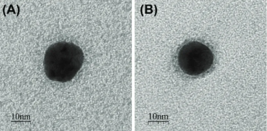
The synthesized S-layer protein functionalized AuNP were char-acterized by TEM (Fig. 1) and UV/Vis spectroscopy (Fig. 2).Fig. 1
shows two examples of AuNP before and after functionalization with S-layer proteins. Particles without protein functionalization are typically characterized by a sharp border (Fig. 1(A)). In contrast, functionalized AuNP are surrounded by a homogenous corona of dark grey, re-presenting the shell of appropriate S-layer proteins (Fig. 1(B), see also Supplementary information Fig. SI3), which are visualized by negative staining of the TEM samples with 2% uranyl acetate. According to the TEM analysis of the dried samples, the general thickness of all protein shells was 1–2 nm. Based on DLS measurements, however, a larger size of the native state of the protein shell in solution, which are covering the AuNP surface, can be assumed [17] (see also Supplementary in-formation Fig. SI2). The difference in the visible size of the AuNP-bio-hybrids between TEM and DLS can be attributed to the different mea-surement principles. DLS represents the hydrodynamic diameter of the particles in solution, which is bigger than in the dried TEM samples.
Due to the fact that the plasmon resonance strongly depends on the dielectric environment, UV/Vis spectroscopy can be used to measure the protein adsorption of the biohybrid. As depicted in Fig. 2, the maximum of absorbance of the citrate capped AuNP, slightly shifted to a higher wavelength in the presence of S-layer proteins on the AuNP-surface. Typically, this shift was in a range of 4–5 nm. To ensure that no incorrect measurements of the biohybrid systems taken place, in-adequate colloidal solutions that already started particle agglomeration indicated by peak broadening, shoulder, or additional peaks at higher wavelengths, where not used.
Altogether, the protein corona visualized by TEM, the peak shift at
specific UV–vis spectra and the stability of the functionalized AuNP against ionic-destabilization (describe in Materials and Methods sec-tion), prove the successful functionalization of the AuNP with the S-layer proteins. All tested S-S-layer proteins exhibited suitable properties for the biofunctionalization of AuNP under optimized conditions of stable pH and a suitable protein concentration.
3.2. Interactions of eight biohybrid S-layer-AuNP systems with different analytes
The unique potential of the eight biohybrid S-layer-AuNP systems to detect different metals and metalloid ions in an aqueous system was investigated by colorimetric assays. Accordingly, eight S-layer proteins from different bacteria species were used for the bio-functionalization of the AuNP.
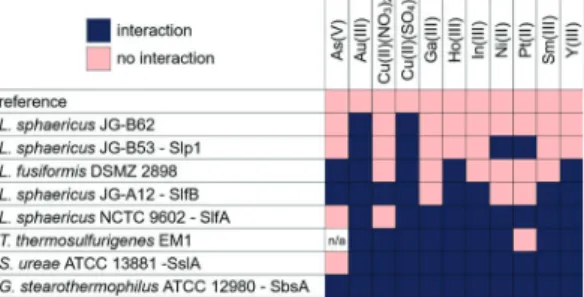
Table 1 depicts an overview of the experimental matrix for each biohybrid system with one of the eleven assessed analytes. Blue colored columns visualize combinations of S-layer functionalized AuNP with a clear interaction with an analyte, e.g. a color change from red to blue. The red colored columns illustrate no reaction of the biohybrid with the corresponding analyte. From this representation, it can be seen only whether a significant reaction with the analyte is indicated (blue), but not that each of these reactions looks different, e.g. different con-centration ranges, stronger/weaker peak shift, or a different reaction pattern. This will be specified in the next sections with taking an in-depth look at the different reactions.
All used S-layer proteins within this study were reactive at least with two of the examined analytes. Because of the numerous functional
Fig. 1. TEM image of (A) a non-functionalized AuNP, and (B) a functionalized AuNP with Slp1 protein from L. sphaericus JG-B53.
Fig. 2. UV/Vis spectra of citrate capped AuNP in water and AuNP functiona-lized with S-layer protein from L. sphaericus JG-B53.
groups at the protein surface of the S-layers, such as NH2, NH, OH, CO,
COOH, SH and PO4, this is no surprise, and resulted in a variety of
interactions depending on the unique binding properties of the S-layer proteins. If those observed interactions between the multivalent posi-tively charged analytes and the S-layers would have based on an un-specific electrostatic interaction, all of the results should finally show the same reaction. However, this was not the case. Therefore, the re-sults inTable 1supported our hypothesis of a specific interaction be-tween the S-layers AuNP biohybrid system and analytes.
As expected, none of the utilized S-layer biohybrids responded identically for all analytes, as well as all biohybrid systems displayed different interaction behaviors depending on their S-layer and used analyte. Accordingly, if an S-layer-AuNP hybrid showed an interaction difference with at least two analytes, we examined and compared those differences to each other. For some analytes, several proteins displayed typical reactions, e.g. for Au(III), Cu(II)SO4, Ho(III), Y(III) (seeTable 1).
Interestingly, all of our S-layer AuNP systems reacted with Au(III) and Cu(II)SO4, despite their different amino acid sequences or structures, as
assessed with UniProt. Therefore, it can be assumed that all of these S-layers must have at least one identical binding sites for these two analytes.
While Au(III) and Cu(II)SO4were detected by all eight tested
S-layer-AuNP systems, the other analytes demonstrated reduced binding efficiency to at least three different S-layer-AuNP systems. It is also interesting to note that the two featuring different copper solutions, Cu (II)SO4and Cu(II)(NO3)2, were detected differently by four of the eight
sensory active solutions.
Notable, the S-layer-AuNP system functionalized with the protein SbsA of G. stearothermophilus ATCC 12980 displayed reactions with all tested analyte solutions.
Interestingly, all biohybrid systems with the used four S-layer types of L. sphaericus species displayed different reaction patterns to the analytes. Here, we expected similarities in the reaction pattern due to their phylogenetic proximity. While the biohybrid system with the SlfA protein of L. sphaericus NCTC 9602 reacted with nine analytes, the one with L. sphaericus JG-B62 displayed a reaction with only two analytes. Within the tested S-layer-AuNP system, the one with protein SslA of S. ureae ATCC 13881 interacted with all chosen analytes, except As(V). The biohybrid system with the S-layer of T. thermosulfurigenes EM1 interacted with all analytes, except Pt(II). Andfinally, the S-layer of L. fusiformis DSMZ 2898 in the biohybrid system showed a reaction with As(V), Au(III), Cu(II)SO4, Ho(III) and Y(III).
Due to the variety of the observed interaction patterns of the tested analytes with the S-layer biohybrid systems, the following two sections are addressed to further demonstrate the selectivity of the method. First, by the reaction of various S-layer proteins with only one specific analyte, and second for different reaction pattern of one S-layer bio-hybrid system with different analytes.
3.3. Detection of Y(III) by using four S-layer-AuNP systems
Fig. 3gives a summary of the results for the colorimetric detection of Y(III) by four different S-layer biohybrids. AuNP stabilized with 0.1% PVA are used for reference.
The S-layer-AuNP system functionalized with Slp1 of L. sphaericus JG-B53 displayed no reactions at all tested Y(III) concentrations. The remaining three biohybrid systems showed different reaction patterns in different concentration ranges of Y(III). The S-layer-AuNP system functionalized with slfA protein showed a color change from red to blue, beginning at a concentration of 8.33 × 10⁻⁴ mol/l up to 8.33 × 10⁻² mol/l Y(III). The SslA-functionalized biohybrid AuNP system displayed a reaction with Y(III) in a range between 1.67 × 10⁻⁵
mol/l and 3.33 × 10⁻⁴mol/l. In the last case of AuNP functionalized with the S-layer protein of T. thermosulfurigenes EM1, the color changed from red to blue from 3.33 × 10⁻⁴mol/l up to 8.33 × 10⁻²mol/l. The
accompanying specific reactions of the different S-layer AuNP systems with defined concentration ranges of an analyte depict the possibility to selectively distinguish an analyte by its color / absorbance shift. Utilizing a dilution series of the sample, the presence and also the concentration of a single metal ion can be determined by comparing the results with known reaction patterns as shown in Fig. 3. Due to the visible color change during the reaction, and therefore the detection of an analyte within some minutes, the method can easily be transferred to field measurements.
However, the biohybrid AuNP system with SslA protein showed a weak visual response that was perceivable by the eye. This outcome represents good example of the limitations of the system, thus heigh-tening the need for additional evaluation methods, such as UV/Vis measurements, to get more detailed information about the sample and concentration range. Accordingly,Fig. 4displays the UV/Vis spectra for the above-mentioned conditions of SslA AuNP system interacting with Y (III) within an estimated concentration range between 1.67 × 10⁻⁶ mol/l. till ×10-2mol/l. The reference without Y(III) ions showed an absorption peak at 525 nm, whereas no peak shift was visible at a concentration of 1.67 × 10⁻⁶mol/l Y(III). With increasing concentra-tion of Y(III), the shift of the absorpconcentra-tion peak to higher wavelengths became more pronounced, up to the strongest shift to 562 nm at a concentration of 1.67 × 10⁻⁴mol/l Y(III).
As previously mentioned, the shift of the plasmon resonance oc-curred from the agglomeration of functionalized AuNP while inter-acting with the analyte (see Fig. SI4 in Supplementary information). At higher concentration, the peak shifts back to smaller wavelengths with a maximum at 534 nm. This smaller shift results from a saturation of the binding sites of the S-layer by the increased Y(III) ion concentration. The saturation leads to less cross-linkage, because there are less free binding sites available, and therefore results in the formation of minor Table 1
Schematic representation of the colorimetric reactions for eight different S-layer protein functionalized AuNP and the tested analytes. Blue signifies an interaction with the respective analyte, while the light red squares represent no detectable reaction. For T. thermosulfurigenes EM1 the value with As(V) is not available (n.a.). (For interpretation of the references to colour in this Table legend, the reader is referred to the web version of this article.).
Fig. 3. Visible reaction pattern of the four S-layer-AuNP systems with varying concentrations of Y(III). Stable and weak reacting AuNP display red colors, while agglomerated and reactive AuNP are depicted in shades of blue (For in-terpretation of the references to colour in thisfigure legend, the reader is re-ferred to the web version of this article).
agglomerates.
In order to obtain a clear presentation of the data and to enable a better comparability of the different reaction patterns of the tested S-layer-AuNP systems, the results of the UV/Vis measurements can be reduced to the absorbance at 575 nm.
Fig. 5illustrates those relative changes of the extinction at 575 nm for different Y(III) concentration. The extinction of the reference was subtracted from the measured value at the same wavelength, resulting in a percentage based demonstration of the different S-layer AuNP systems according to the different Y(III) concentration (see Equation 1 in Supplementary Information). The resulting curves now can be plotted against the YCl3concentration, compared more easily and the
difference in reaction ranges and reaction patterns can be extracted. The biohybrid AuNP system functionalized with Slp1 of L. sphaericus JG-B53 showed no reaction at all tested Y(III) concentrations. In con-trast, the biohybrid AuNP system functionalized with SlfA showed an increase in the relative extinction at 575 nm, beginning with 8.33 × 10⁻⁴ mol/l up to 8.33 × 10⁻² mol/l, with a maximum of
25.92 ± 5.87% at 1.67 × 10⁻³ mol/l Y(III). The SslA-AuNP system
displayed a strong change of the extinction in a range between 1.67 × 10⁻⁵mol/l and 3.33 × 10⁻⁴mol/l with the maximum extinc-tion change of 118.12 ± 4.82% at 3.33 × 10⁻⁴mol/l. Finally, for the S-layer protein of T. thermosulfurigenes EM1 the relative extinction change ranged from 3.33 × 10⁻⁴mol/l up to 8.33 × 10⁻²mol/l, with the strongest signal of 121.81 ± 7.58% at 3.33 × 10⁻⁴ mol/l.
Additionally, different reaction patterns of the S-layer-AuNP systems were shown and can be distinguished at an extinction at 575 nm.
Visually, the reactions of the S-layer-AuNP systems functionalized with SlfA or with the S-layer of T. thermosulfurigenes EM1 were in a similar concentration range, which can be simplified by using the ex-tinction at 575 nm. The simple specific differentiation therein, which of the two S-layer proteins has interacted in particular with the Y(III) di-lution series, can be distinguished. According to those experiments, the example SslA-AuNP system showed the lowest detection limit for Y(III) at 1.67 × 10⁻⁵mol/l for all the used experiments within this study.
Here, we were able to describe the selectivity of several S-layer functionalized AuNP hybrids, for one specific analyte.
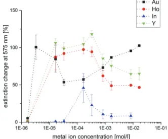
3.4. Reactivity of the biohybrid AuNP system functionalized with SslA protein with Au(III), Ho(III), In(III) and Y(III)
Next, the selectivity of one S-layer-AuNP system for several analytes will be defined. Therefore, the previously described SsIA functionalized AuNP system, and the analytes Au(III), Ho(III), In(III), as well as Y(III) were used. When examining the interactions of this one S-layer-AuNP system with the various analytes, the color shift visually was partly similar from red to blue. As discussed previously, the results of UV/Vis measurements allow more nuancedfindings, than the detection with the eyes. Therefore, the values of the calculations from the relative change of extinction at 575 nm were plotted against the corresponding metal or metalloid ion concentration (Fig. 6). The graphs show different
reaction patterns depending on the type of metal or metalloid ion (see also Fig. SI6 in Supplementary information).
For the biohybrid AuNP system functionalized with SslA, the lowest detection limit of 3.33 × 10⁻⁶mol/l was measured for Au(III) with the strongest extinction change with 100.63 ± 16.33%. By increasing the analyte concentration, the extinction initially decreased to about half with 53.68 ± 4.12%, prior of a new rising up to 102.72 ± 0.11% at a concentration of 1.67 × 10⁻²mol/l. No extinction changes was
asses-sable at the lowest measured Au(III) concentration of 1.67 × 10⁻⁶mol/ l (5.21 ± 1.22%).
For Ho(III) and Y(III), the SslA functionalized AuNP system showed similar reaction patterns. The detection limits were measured between 1.67 × 10⁻⁶mol/l and 1.67 × 10⁻⁵mol/l, respectively, with the
stron-gest changes of extinction between 3.33 × 10⁻⁵mol/l and 1.67 × 10
-4mol/l. At higher concentrations, the signal decreased to 50–60 %. The
similarity in the reaction patterns for Ho(III) and Y(III) indicated an analogous binding mechanism of SslA protein with both elements. Fig. 4. UV/Vis spectra of SslA AuNP system with protein from S. ureae ATCC
13881 treated with different concentrations of Y(III).
Fig. 5. Relative extinction change at 575 nm of four different bio-hybrid AuNP systems in interaction with YCl3.
Fig. 6. Relative extinction change at 575 nm of bio-hybrid AuNP system func-tionalized with SslA protein from S. ureae ATCC 13881 in interaction with different analytes in various concentrations indicated in the legend.
Here, the available reactive groups of the S-layer surface might interact with the analytes by forming novel binding sites, which seems to be similar for Ho(III) and Y(III) as evidenced by their comparable reaction patterns.
In general, the differences in the interaction pattern can be used to specifiy the present metal ions of a sample. Further, as shown inFig. 6, the specific extinction curves (see Fig. SI6 in Supplementary informa-tion), together with a dilution series of the sample, might determine the unknown concentration of a sample with known metal ions. By testing a sample with multiple S-layer-AuNP systems, a distinction between multiple ions in solution via colorimetric assay would be possible.
However, this approach to develop a screening assay has to be in-vestigated in further experiments, especially with regard to cross-re-activity in the presence of different analytes. These findings could be a starting point for the development of a fast and simple screening assay for different analytes. However, it has to be stated, that the system is influenced by complex parameters and not yet optimized. Since the system is not buffered, the influence of different pH values has to be investigated further in detail. Moreover, the effect of different anions as well as the respective present ionic species in general has to be eluci-dated.
3.5. Sequence comparison of S-layer proteins SlfB, Slp1, SlfA, SslA and SbsA
The variability in the reaction of the S-layer proteins to the appro-priate analytes demonstrated that S-layer represent interesting ex-ploration objects. It is of great interest to correlate these results pre-cisely to their biological phylogeny and sequence. Pollmann et al. [9] described a high similarity between the S-layer protein SlfB (L. sphaericus JG-A12) and SlfA (L. sphaericus NCTC 9602), with 98% se-quence identity and a difference of only 3 amino acids / 13 nucleotides [46,47]. Similarly, Lederer et al. [48] compared Slp1 (L. sphaericus JG-B53) to SlfB with a 67% sequence identity, as well as 65% for Slp1 to SlfA sequence identity.
Differences between the literature and our results can be explained by the different used software for sequence comparison, as well as the used version.Despite this fact that the protein BLAST results showed differing values for the sequence identity, the highest sequence identity values (79%) were evaluated for both SlfB and SlfA (Table 2). Ad-ditionally, the three S-layer proteins SlfA, SlfB and Slp1 from the same species L. sphaericus demonstrated high sequence identities between SlfB and SlfA, and for SlfB and Slp1.
It should also be noted that even though SlfA and SlfB display high sequence identity, they reacted very differently with metal and me-talloid ions. Therefore, the hypothesis of similar amino acid sequences leading to comparable binding properties for those proteins might be rejectable. The fact that based on the amino acid sequence, it is not possible to define the binding pattern of the S-layer proteins confirms that their specific binding affinities must be experimental determined more intensively. In the experimental design, additional influencing factors, such as the pH value, must be taken into account. This in turn affects the net charge of the proteins and the chemical speciation of the analytes. Studies have already shown differences in the growth condi-tions of the bacteria can lead to post-translational modifications of
S-layer proteins [49]. These post-translational protein modifications
might also influence binding properties. Recently, Lederer et al. (2013) recently described various environmental factors that could impact the binding capacity of S-layer proteins [48]. In particular, the authors noted that cells can present different S-layer variants with specific abilities to cope with fast-changing environmental conditions. 4. Conclusions
The herein described results represent one of the most extensive studies for the colorimetric detection of different analytes with S-layer protein-functionalized AuNP. Eleven different analytes, including the rare-earth elements Ho(III), Sm(III), and Y(III), as well as noble metal complexes such as Au(II), and Pt(II); as well as the toxic element As(V), were analyzed by our established colorimetric biohybrid assays. Especially for the rare-earth elements, to-date there are only a limited number of analytic methods available.
In our experimental setting, the typical colorimetric reaction was confirmed visually by a color shift from red to blue. The detection threshold of our method was improved by UV/Vis measurements and a wavelength dependent correlation method. For the major example de-scribed in our study, a highly specific detection of Y(III) up to a con-centration of 1.67 × 10⁻⁵ mol/l was achieved for the SslA-AuNP system.
Furthermore, our data verified a unique reaction pattern for each of the verified S-layer-AuNP hybrid systems with all tested analytes, fa-cilitating the specific detection of metal and metalloid ions in an aqu-eous solution. First attempts to correlate the binding mechanisms of the layer and the analytes to the specific sequence identity data of the S-layers, indicated that the interaction capability not only depends on the amino acid sequence, but also on essential environmental or epigenetic factors.
The colorimetric biohybrid system described herein can be estab-lished as a rapid and easy screening assay for the tested rare-earth elements as well as expanded to other metal ions.
Declaration of Competing Interest
There are no conflicts of interest to declare. Acknowledgements
We thank Beate Katzschner for supporting the preparation of S-layer proteins, and Phil Goldberg for providing an external point of view and in-depth discussion. This research has been funded by the Bundesministerium für Bildung und Forschung (BMBF, 03WKCL03D) and European Social Fund (ESF, 100284305).
Appendix A. Supplementary data
References
[1] S. Schultze-Lam, T.J. Beveridge, Nucleation of celestite and strontianite on a cya-nobacterial S-layer, Appl. Environ. Microbiol. 60 (1994) 447–453.
[2] V.R. Phoenix, R.W. Renaut, B. Jones, F.G. Ferris, Bacterial S-layer preservation and rare arsenic–antimony–sulphide bioimmobilization in siliceous sediments from Champagne Pool hot spring, Waiotapu, New Zealand, J. Geol. Soc. Lond. 162
(2005) 323–331,https://doi.org/10.1144/0016-764903-058.
[3] M.C. Allievi, S. Florencia, P.A. Mariano, P.M. Mercedes, S.M. Ruzal, S.R. Carmen, Metal biosorption by surface-layer proteins from Bacillus species, J. Microbiol.
Biotechnol. 21 (2011) 147–153https://doi.org/10.4014/jmb.1009.09046.
[4] E. Gerbino, P. Carasi, P. Mobili, M.A. Serradell, A. Gómez-Zavaglia, Role of S-layer
proteins in bacteria, World J. Microbiol. Biotechnol. 31 (2015) 1877–1887,https://
doi.org/10.1007/s11274-015-1952-9.
[5] U.B. Sleytr, T.J. Beveridge, Bacterial S-layers, Trends Microbiol. 7 (1999) 253–260,
https://doi.org/10.1016/S0966-842X(99)01513-9.
Table 2
Sequence identity data from protein BLAST offive S-layer proteins. Green in-dicates high sequence identity, and red low sequence identity. (For inter-pretation of the references to colour in this Table legend, the reader is referred to the web version of this article.).
[6] M. Sára, U.B. Sleytr, S-layer proteins, J. Bacteriol. 182 (2000) 859–868,https://doi. org/10.1128/JB.182.4.859-868.2000.
[7] S. Selenska-Pobell, P. Panak, V. Miteva, I. Boudakov, G. Bernhard, H. Nitsche, Selective accumulation of heavy metals by three indigenous {Bacillus} strains, {B}-cereus, {B}-megaterium and {B}-sphaericus, from drain waters of a uranium waste pile, FEMS Microbiol. Ecol. 29 (1999) 59–67 //000080187400006.
[8] J. Raff, U. Soltmann, S. Matys, S. Selenska-Pobell, H. Böttcher, W. Pompe, Biosorption of uranium and copper by biocers, Chem. Mater. 15 (2003) 240–244,
https://doi.org/10.1021/cm021213l.
[9] K. Pollmann, J. Raff, M. Merroun, K. Fahmy, S. Selenska-Pobell, Metal binding by bacteria from uranium mining waste piles and its technological applications,
Biotechnol. Adv. 24 (2006) 58–68,https://doi.org/10.1016/j.biotechadv.2005.06.
002.
[10] M. Suhr, N. Unger, K.E. Viacava, T.J. Günther, J. Raff, K. Pollmann, Investigation of metal sorption behavior of Slp1 from Lysinibacillus sphaericus JG-B53: a combined
study using QCM-D, ICP-MS and AFM, Biometals 27 (2014) 1337–1349,https://
doi.org/10.1007/s10534-014-9794-8.
[11] U.B. Sleytr, P. Messner, D. Pum, M. Sara, Crystalline bacterial cell surface layers: general principles and application potential, Mol. Microbiol. 10 (1993) 911–916,
https://doi.org/10.1111/j.1365-2672.1993.tb04339.x.
[12] U.B. Sleytr, H. Bayley, M. Sára, A. Breitwieser, S. Küpcü, C. Mader, S. Weigert, F.M. Unger, P. Messner, B. Jahn-Schmid, B. Schuster, D. Pum, K. Douglas, N.A. Clark, J.T. Moore, T.A. Winningham, S. Levy, I. Frithsen, J. Pankovc, P. Beale, H.P. Gillis, D.A. Choutov, K.P. Martin, VI. Applications of S-layers, FEMS Microbiol.
Rev. 20 (1997) 151–175,https://doi.org/10.1016/S0168-6445(97)00044-2.
[13] W. Shenton, D. Pum, U.B. Sleytr, S. Mann, Synthesis of cadmium sulphide super-lattices using self-assembled bacterial S-layers, Nature 389 (1997) 585–587,
https://doi.org/10.1038/39287.
[14] U.B. Sleytr, C. Huber, N. Ilk, D. Pum, B. Schuster, E.M. Egelseer, S-layers as a tool kit for nanobiotechnological applications, FEMS Microbiol. Lett. 267 (2007) 131–144,
https://doi.org/10.1111/j.1574-6968.2006.00573.x.
[15] U.B. Sleytr, B. Schuster, E.M. Egelseer, D. Pum, S-layers: principles and applications,
FEMS Microbiol. Rev. 38 (2014) 823–864,https://doi.org/10.1111/1574-6976.
12063.
[16] K. Pollmann, S. Kutschke, S. Matys, S. Kostudis, S. Hopfe, J. Raff, Novel bio-technological approaches for the recovery of metals from primary and secondary
resources, Minerals 6 (2016) 54,https://doi.org/10.3390/min6020054.
[17] M. Lakatos, S. Matys, J. Raff, W. Pompe, Colorimetric as (V) detection based on
S-layer functionalized gold nanoparticles, Talanta 144 (2015) 241–246,https://doi.
org/10.1016/j.talanta.2015.05.082.
[18] M. Zhuang, J. Zhao, S. Li, D. Liu, K. Wang, P. Xiao, L. Yu, Y. Jiang, J. Song, J. Zhou, L. Wang, Z. Chu, M. Zhuang, J. Zhao, S. Li, D. Liu, K. Wang, P. Xiao, L. Yu, Y. Jiang, J. Song, J. Zhou, L. Wang, Z. Chu, M. Zhuang, J. Zhao, S. Li, D. Liu, K. Wang, P. Xiao, L. Yu, Y. Jiang, J. Song, J. Zhou, L. Wang, Z. Chu, Concentrations and health risk assessment of rare earth elements in vegetables from mining area in
Shandong, China, Chemosphere 168 (2017) 578–582,https://doi.org/10.1016/j.
chemosphere.2016.11.023.
[19] K.M. Goodenough, F. Wall, D. Merriman, The rare earth elements: demand, global resources, and challenges for resourcing future generations, Nat. Resour. Res. 27
(2018) 201–216,https://doi.org/10.1007/s11053-017-9336-5.
[20] J.O. Duruibe, M.O. Ogwuegbu, J.N. Egwurugwu, Heavy metal pollution and human
biotoxic effects, Int. J. Phys. Sci. 2 (2007) 112–118,https://doi.org/10.1016/j.
proenv.2011.09.146.
[21] L.M. Jakubek, S. Marangoudakis, J. Raingo, X. Liu, D. Lipscombe, R.H. Hurt, The inhibition of neuronal calcium ion channels by trace levels of yttrium released from
carbon nanotubes, Biomaterials 30 (2009) 6351–6357,https://doi.org/10.1016/j.
biomaterials.2009.08.009.
[22] P.C. Nagajyoti, K.D. Lee, T.V.M. Sreekanth, Heavy metals, occurrence and toxicity
for plants: a review, Environ. Chem. Lett. 8 (2010) 199–216,https://doi.org/10.
1007/s10311-010-0297-8.
[23] Z. Li, Z. Ma, T.J. van der Kuijp, Z. Yuan, L. Huang, A review of soil heavy metal pollution from mines in China: pollution and health risk assessment, Sci. Total
Environ. 468–469 (2014) 843–853,https://doi.org/10.1016/j.scitotenv.2013.08.
090.
[24] B. Wei, L. Yang, A review of heavy metal contaminations in urban soils, urban road
dusts and agricultural soils from China, Microchem. J. 94 (2010) 99–107,https://
doi.org/10.1016/j.microc.2009.09.014.
[25] S. Audry, J. Schäfer, G. Blanc, J.M. Jouanneau, Fifty-year sedimentary record of heavy metal pollution (Cd, Zn, Cu, Pb) in the Lot River reservoirs (France), Environ.
Pollut. 132 (2004) 413–426,https://doi.org/10.1016/j.envpol.2004.05.025.
[26] F. Sulotto, C. Romano, A. Berra, G.C. Botta, G.F. Rubino, E. Sabbioni, R. Pietra, Rare
earth pneumoconiosis: a new case, Am. J. Ind. Med. 9 (1986) 567–575,https://doi.
org/10.1002/ajim.4700090609.
[27] D.L. Tomlinson, J.G. Wilson, C.R. Harris, D.W. Jeffrey, Problems in the assessment
of heavy-metal levels in estuaries and the formation of a pollution index,
Helgoländer Meeresunters. 33 (1980) 566–575,https://doi.org/10.1007/
BF02414780.
[28] J.F. Boissonneau, M.J. Repellin, A. Eglem, Elecrochemical detection of rare earths and yttrium after chromatographic separation by high performance liquid chro-matography, Analusis 8 (1980) 230–237.
[29] K. Yoshida, H. Haraguchi, Determination of rare earth elements by liquid chro-matography/inductively coupled plasma atomic emission, Anal. Chem. 56 (1984)
2580–2585,https://doi.org/10.1021/ac00277a069.
[30] P.L. Smedley, The geochemistry of rare earth elements in groundwater from the Carnmenellis area, southwest England, Geochim. Cosmochim. Acta 55 (1991)
2767–2779,https://doi.org/10.1016/0016-7037(91)90443-9.
[31] K. Benkhedda, H. Goenaga Infante, E. Ivanova, F.C. Adams, Determination of sub-parts-per-trillion levels of rare earth elements in natural waters by inductively coupled plasma time-of-flight mass spectrometry after flow injection on-line sorp-tion preconcentrasorp-tion in a knotted reactor, J. Anal. At. Spectrom. 16 (2001)
995–1001,https://doi.org/10.1039/b101343m.
[32] D.W. Hicklin, W.M. Willingham, Evaluation of Commercially Available Cyanide Test Kits Against Various Matrices, (2016).
[33] E. Ruiz-Hitzky, M. Darder, P. Aranda, K. Ariga, Advances in biomimetic and
na-nostructured biohybrid materials, Adv. Mater. 22 (2010) 323–336,https://doi.org/
10.1002/adma.200901134.
[34] J. Turkevich, P.C. Stevenson, J. Hillier, A study of the nucleation and growth processes I N the synthesis of colloidal gold, Discuss. Faraday Soc. 11 (1951) 55–75. [35] G. Frens, Controlled nucleation for the regulation of the particle size in
mono-disperse gold suspensions, Nat. Phys. Sci. 241 (1973).
[36] A. Blüher, K. Ostermann, P. Jäckel, A. Clemens, B. Katzschner, G. Rödel, M. Mertig,
Extraction and long-term storage of S-layer proteins andflagella from Lysinibacillus
sphaericus NCTC 9602: building blocks for nanotechnology, Eng. Life Sci. 15 (2015)
410–415,https://doi.org/10.1002/elsc.201400182.
[37] E. Brechtel, M. Matuschek, A. Hellberg, E.M. Egelseer, R. Schmid, H. Bahl, Cell wall of thermoanaerobacterium thermosulfurigenes EM1: isolation of its components and attachment of the xylanase XynA, Arch. Microbiol. 171 (1999) 159–165,
https://doi.org/10.1007/s002030050694.
[38] D. Fritze, Taxonomy of the genus Bacillus and related genera: the aerobic
en-dospore-forming bacteria, Phytopathology 94 (2004) 1245–1248,https://doi.org/
10.1094/PHYTO.2004.94.11.1245.
[39] W.D. Geoghegan, G.A. Ackerman, Adsorption of horseradish ovomucoid and anti-immunoglobulin to colloidal for the indirect detection of concanavalin a, wheat germ anti- G on cell surfaces at the electron microscopic level: a new method, theory and application, J. Histochem. Cytochem. 25 (1977) 1187–1200. [40] I. Horcas, R. Fernández, J.M. Gómez-Rodríguez, J. Colchero, J. Gómez-Herrero,
A.M. Baro, WSXM: a software for scanning probe microscopy and a tool for
nano-technology, Rev. Sci. Instrum. 78 (2007),https://doi.org/10.1063/1.2432410.
[41] U. Hinz, From protein sequences to 3D-structures and beyond: the example of the
UniProt Knowledgebase, Cell. Mol. Life Sci. 67 (2010) 1049–1064,https://doi.org/
10.1007/s00018-009-0229-6.
[42] The UniProt Consortium, UniProt Database.http://www.uniprot.org(Accessed 1
September 2017).
[43] UniProt Consortium, UniProt: a hub for protein information, Nucleic Acids Res. 43
(2015) D204–D212,https://doi.org/10.1093/nar/gku989.
[44] National Center for Biotechnology Information, U.S. National Library of Medicine,
NCBI Protein Basic Local Alignment Search Tool– BLAST.https://blast.ncbi.nlm.
nih.gov/Blast.cgi?PROGRAM=blastp&PAGE_TYPE=BlastSearch&LINK_LOC= blasthome(Accessed 1 September 2017).
[45] S.F. Altschul, W. Gish, W. Miller, E.W. Myers, D.J. Lipman, Basic local alignment
search tool, J. Mol. Biol. 215 (1990) 403–410,
https://doi.org/10.1016/S0022-2836(05)80360-2.
[46] K. Pollmann, J. Raff, M. Schnorpfeil, G. Radeva, S. Selenska-Pobell, Novel surface layer protein genes in Bacillus sphaericus associated with unusual insertion
ele-ments, Microbiology 151 (2005) 2961–2973,https://doi.org/10.1099/mic.0.
28201-0.
[47] K. Pollmann, S. Matys, Construction of an S-layer protein exhibiting modified self-assembling properties and enhanced metal binding capacities, Appl. Microbiol.
Biotechnol. 75 (2007) 1079–1085,https://doi.org/10.1007/s00253-007-0937-5.
[48] F.L. Lederer, U. Weinert, T.J. Günther, J. Raff, S. Weiß, K. Pollmann, Identification of multiple putative S-layer genes partly expressed by Lysinibacillus sphaericus
JG-B53, Microbiology (United Kingdom) 159 (2013) 1097–1108,https://doi.org/10.
1099/mic.0.065763-0.
[49] Z. Guan, S. Naparstek, D. Calo, J. Eichler, Protein glycosylation as an adaptive re-sponse in archaea: growth at different salt concentrations leads to alterations in Haloferax volcanii S-layer glycoprotein N-glycosylation, Environ. Microbiol. 14
(2012) 743–753,https://doi.org/10.1111/j.1462-2920.2011.02625.x.