1
Multifunctional Nano-Biointerfaces: Cytocompatible Antimicrobial
Nanocarriers from Stabilizer-free Cubosomes
Mahsa Zabara1, Berna Senturk1, Mark Gontsarik1, Qun Ren1, Markus Rottmar1, Katharina Maniura-Weber1, Raffaele Mezzenga2, Sreenath Bolisetty2, Stefan Salentinig1,3*
1
Laboratory for Biointerfaces, Department Materials meet Life, Empa Swiss Federal Laboratories for Materials Science and Technology, Lerchenfeldstrasse 5, 9014 St. Gallen, Switzerland
2
Laboratory for Food & Soft Materials Science, Institute of Food, Nutrition & Health, ETH Zurich, Schmelzbergstrasse 9, LFO, 8092, Zürich, Switzerland
3
Department of Chemistry, University of Fribourg, Chemin du Musée 9, 1700 Fribourg, Switzerland
2
Figure S1. (a) Experimental SAXS patterns (symbols) and the fit calculated with the indirect Fourier
transformation method (red curve) for the stabilizer-free GMO/LL-37 self-assemblies at a weight ratio of 50/50. (b) The corresponding pair distance distribution function, p(r). Note that the maximum dimension in p(r) at p(r) = 0 does not reflect the overall vesicle dimensions. As the maximum dimensions of the vesicles are above the resolution limit of our SAXS set-up the p(r) was mathematically forced to 0 at 100 nm.
a
b
0 200 400 600 800 1000 -0.0005 0.0000 0.0005 0.0010 0.0015 0.0020 0.0025 r [Å] p(r) [a.u.] 0.01 0.1Intensity[a.u.]
q[Å-1] q-23
Figure S2. Dynamic light scattering (DLS) measurements of stabilizer-free GMO cubosomes directly and 21 days after preparation. The DLS correlation functions of the particles represent rather monomodal particles with only a single relaxation time. The shift of the decay in the correlation function to longer relaxation times results from the increase in the particle size after 21 days; the slight change in the slope is from the relatively small changes in PDI after 21 days.
4
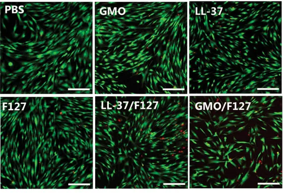
Figure S3. Cytotoxicity of HDF cells treated with the control groups. The Live/Dead assay shows live
cells in green (calcein-AM staining) and dead cells in red (ethidium homodimer staining).
Figure S4. Uptake of FAM-labelled LL-37 from different formulations into HDF cells. The cells were
treated with (a) 37, (b) GMO/37, (c) 37/F127 and (d) GMO/37/F127. The FAM-labelled LL-37 is shown in green, cell nuclei and actin filaments were stained with DAPI (blue) and phalloidin (red). Scale bar; 20 μm.
5
Strains GMO/LL-37 GMO/LL-37/F127
Gram positive Staphylococcus aureus DSMZ 20231 No antibacterial activity (up to 250μg/ml LL-37) No antibacterial activity (up to 250μg/ml of LL-37) Staphylococcus epidermis ATCC 4961 No antibacterial activity (up to 250μg/ml LL-37) No antibacterial activity (up to 250μg/ml of LL-37) Bacillus Subtilis ATCC 6633 OD0.108 No antibacterial activity (up to 64μg/ml LL-37) No antibacterial activity (up to 64μg/ml of LL-37) Gram negative Escherichia coli DSMZ 30083 Antibacterial effect MIC ≤ 40 μg/ml Antibacterial effect MIC ≤ 80 μg/ml Pseudomonas aeruginosa CIP A22 DSMZ 25123 Antibacterial effect MIC ≤ 32 μg/ml Antibacterial effect MIC ≤ 64 μg/ml