jA72
Beam Characterization for Accelerator-Based Boron Neutron Capture Therapy Using the 9Be(d,n) Nuclear Reaction
by
Susan Marie White B.S. Nuclear Engineering (1996)
Purdue University
Submitted to the Department of Nuclear Engineering in Partial Fulfillment of the Requirements for the Degree of
Master of Science in Nuclear Engineering at the
Massachusetts Institute of Technology June 1998
© 1998 Massachusetts Institute of Technology All rights reserved
Signature of Author ...
Department of Nuclear Engineering May 8, 1998
Certified by ... ..
J
//i Jacquelyn C. Yanch Associate ro fessor of Nuclear Engineering and Whitaker College of Health Sciences and Technology Thesis Supervisor R ead by ...Ruth PShefer Newton Scientific, Inc. Thesis Reader A ccepted by ... . . . ...
Lawrence M. Lidsky Chairman, Department Committee on Graduate Students
Beam Characterization for Accelerator-Based Boron Neutron Capture Therapy Using the 'Be(d,n) Nuclear Reaction
by
Susan Marie White
Submitted to the Department of Nuclear Engineering on May 8, 1998 in partial fulfillment of the requirements for the Degree of Master of Science in
Nuclear Engineering ABSTRACT
Use of the 9Be(d,n) nuclear reaction for accelerator-based boron neutron capture therapies (AB-BNCT) was investigated. The moderated neutron spectra produced at several deuteron bombarding energies were evaluated in terms of dose rates and dosimetric profiles in a
water-filled brain phantom using an existing heavy water moderator and lead reflector assembly. Dosimetry results were obtained using the dual ionization chamber technique coupled with bare
and cadmium-covered gold foils. Data have been taken with deuteron beams of 1.3 MeV to 1.8 MeV. As deuteron energy was increased, the tumor dose rate correspondingly improved due to the neutron yield increase. However, the data suggest that the advantage depth decreased, and the ratio of the fast neutron dose rate to the thermal neutron dose rate at a depth of I cm increased, although error bars are significant. All deuteron energies investigated produced a beam that, once moderated, appears viable for AB-BNCT. No conclusion was drawn about the best energy in terms of a high tumor dose rate, a significant advantage depth, and a low fast to
thermal neutron dose rate ratio. Treatment times assuming 20 Gy to a tumor located 4 cm deep using a 4 mA accelerator ranged from 18 -59 minutes, assuming a tumor boron concentration of 40 ppm and RBE values of 1.0 for photons, 3.2 for neutrons, and 3.8 for boron in tumor tissue. The average advantage depth was 6.4 ± 0.7 cm, so these moderated beams could be used to treat tumors near the brain centerline. The 9Be(d,n) nuclear reaction is exothermic, and is accessible to inexpensive, small particle accelerators.
Thesis Supervisor: Jacquelyn C. Yanch
Title: Associate Professor of Nuclear Engineering and Whitaker College of Health Sciences and Technology
TABLE OF CONTENTS A B STRA CT ... ... 2 TABLE OF CONTENTS ... 3 LIST OF FIGURES ... 4 LIST OF TABLES ... 6 1. INTRODUCTION ... 7
1.1 BORON NEUTRON CAPTURE THERAPY ... 7
1.2 ACCELERATOR-BASED BNCT ... 9
1.3 AB-BNCT USING THE 9Be(d.n) REACTION ... 10
2. DOSIMETRY METHODS ... 12
2.1 EXPERIMENTAL SETUP ... 12
2.2 MIXED FIELD DOSIMETRY ... 17
2.2.1 Dual Ionization Chamber Technique ... 17
2.2.1.1 Theory ... ... 17
2.2.1.2 M ethod ... ... ... 21
2.2.2 Cadmium-Difference Method ... 23
2.2.2.1 Theory ... 23
2.2.2.2 M ethod ... 25
2.2.3 Kerma Factor Method ... 27
2.2.3.1 Theory ... 27
2.2.4 Tumor and Healthy Tissue Dose Rate Calculations ... 28
2.3 SECONDARY ELECTRON EFFECT ON CURRENT MEASUREMENT ... 28
2.4 MONTE CARLO CALCULATION ... 30
2.5 FIGURES OF MERIT ... ... 32
3. RESULTS ... 33
3.1 EXPERIMENTAL RESULTS ... 33
3.2 SIMULATION RESULTS ... 39
4. DISCUSSION AND CONCLUSIONS ... 45
4.1 DEUTERON ENERGY ... 45
4.2 EFFECT OF CHANGING BORON COMPOUND ... 46
4.3 COMPARISON OF SIMULATED AND EXPERIMENTAL RESULTS ... 52
4.4 RECOMMENDATIONS FOR FUTURE WORK ... 60
LIST OF FIGURES
Figure 2.1: Moderator/Reflector Assembly used in experimental work ... 13
Figure 2.2: Ellipsoidal Head Phantom used in experimental work ... 15
Figure 2.3: Schematic of Ellipsoidal Head Phantom used in experimental work ... 16
Figure 2.4: Secondary Electron Effect on Current Measurement ... 29
Figure 2.5: 2D Slice of MCNP modeled experimental setup ... 31
Figure 3.1: Experimental Dosimetry Results for 1.3 MeV deuterons on a beryllium target ... 34
Figure 3.2: Experimental Dosimetry Results for 1.5 MeV deuterons on a beryllium target ... 35
Figure 3.3: Experimental Dosimetry Results for 1.6 MeV deuterons on a beryllium target ... 36
Figure 3.4: Experimental Dosimetry Results for 1.7 MeV deuterons on a beryllium target ... 37
Figure 3.5: Experimental Dosimetry Results for 1.8 MeV deuterons on a beryllium target ... 38
Figure 3.6: Figure of Merit 1 - Tumor Dose Rate @ 4 cm as a function of Deuteron Energy ... 40
Figure 3.7: Figure of Merit 2 - Advantage Depth as a function of Deuteron Energy ... 41
Figure 3.8: Figure of Merit 3 - Ratio of Fast Neutron to Total Tumor Dose Rates @ 1 cm as a function of Deuteron Energy ... 42
Figure 3.9: Simulated Dosimetry Results for 1.5 MeV deuterons on a beryllium target ... 43
Figure 4.1: Figure of Merit 1 - Tumor Dose Rate @ 4 cm as a function of Deuteron Energy for different B-10 Concentrations ... 49
Figure 4.2: Figure of Merit 2 - Advantage Depth as a function of Deuteron Energy for different B-10 Concentrations ... 50
Figure 4.3: Figure of Merit 3 - Ratio of Fast Neutron to Total Tumor Dose Rates @ 1 cm as a function of Deuteron Energy for different B-10 Concentrations ... 51
Figure 4.4: Comparison of 1.5 MeV d-Be Experimental and Simulated Results - Dose Rate due to Thermal Neutrons ... 53
Figure 4.5: Comparison of 1.5 MeV d-Be Experimental and Simulated Results - Dose Rate due to 40 ppm B-10 ... ... 54
Figure 4.6: Comparison of 1.5 MeV d-Be Experimental and Simulated Results - Dose Rate due to Fast Neutrons ... 55
Figure 4.7: Comparison of 1.5 MeV d-Be Experimental and Simulated Results - Dose Rate due to Photons ... 56
Figure 4.8: Comparison of 1.5 MeV d-Be Experimental and Simulated Harder Spectrum Results --Dose Rate due to Fast Neutrons ... 58 Figure 4.9: Comparison of 1.5 MeV d-Be Experimental and Simulated Softer Spectrum Results --Dose
LIST OF TABLES
Table 2.1: Errors in Fast Neutron and Photon Doses ... 23
Table 2.2: Errors in Thermal Flux Measurement ... 26
Table 4.1: Figure of Merit results ... 45
Table 4.2: Various "'B concentrations in tumor and healthy tissue ... 47
1. INTRODUCTION
Accelerator-based boron neutron capture therapy (AB-BNCT) is currently being studied as an alternative to reactor-based BNCT at MIT's Laboratory for Accelerator Beam Applications (LABA) [Yanch et al 1992]. A tandem accelerator designed by Newton Scientific, Inc. was recently constructed and installed at LABA [Shefer et al 1994]. Previous work has indicated that the Be(d,n) reaction may be promising for use in AB-BNCT [Yanch et al 1997]. These early studies investigated the use of deuterons at 2.6 MeV. This beam showed potential but would not be clinically usable due to the large fast neutron component. A softer neutron spectrum can be produced using lower deuteron energies. Therefore, the work described here considered deuterons of lower energies. Deuteron energies from 1.3 MeV to 1.8 MeV were examined. Beam design has been performed to determine what energy deuterons produce the most useful therapy beam.
1.1 BORON NEUTRON CAPTURE THERAPY
Boron neutron capture therapy (BNCT) is an experimental therapy that is being investigated as a means of killing tumor cells. This experimental technique was first attempted in the 1950's, but results were discouraging due to unanticipated late effects [Slatkin 1991]. Much research has been done since those early trials, to improve both neutron delivery and tumor-specific boronated compounds, and
clinical trials are underway currently in the United States [Zamenhof et al 1997, Capala et al 1997]. BNCT consists of bombarding a tumor with thermal neutrons. The thermal neutrons are either scattered or captured by the different elements found in tissue. Thermal neutron elastic scattering results in reducing the neutron's energy, whereas thermal neutron capture results in loss of the neutron and the emergence of an energetic particle. Tissue consists primarily of hydrogen, oxygen, carbon, and nitrogen. Macroscopic cross sections indicate that the most likely events are thermal neutron scattering by the tissue elements, or capture by hydrogen and capture by nitrogen.
Thermal neutron scattering by tissue elements results in a transfer of energy from the neutron to the recoil nuclei. Energy transferred is on the order of tenths of an eV or less, and does not significantly contribute to patient dose. Capture of a thermal neutron by hydrogen produces a deuteron and a 2.22 MeV photon. These high energy photons contribute to patient dose over a long range as the photons lose energy through collisions with atomic electrons. Ninety percent of these 2.22 MeV photons will deposit all of their energy within 50 cm of tissue, so the dose from photons is not a local phenomenon. The
14N(n,p)14C capture reaction creates an energetic recoil proton that also contributes to dose. Thermal neutron capture by nitrogen has a Q value of 0.626 MeV, and this energy is delivered locally to the soft
tissue, due to the short ranges of protons and heavy nuclei in tissue [Turner 1995]. The 0.626 MeV will be split so that the recoil proton has 0.584 MeV and the carbon nucleus has 0.042 MeV, due to
conservation of energy and momentum [Turner 1995]. The range of a 0.584 MeV proton in soft tissue is 1x103 cm, and the range of a 0.042 MeV carbon nucleus is less than 1x106 cm [Turner 1995]. Thus the energies from the recoil proton and recoil carbon nucleus following thermal neutron capture by nitrogen are deposited within 100 pm of the interaction and can be considered locally deposited in soft tissue.
In BNCT, a boronated compound is given to the patient so that boron is present in tissue, along with hydrogen, nitrogen, carbon, and oxygen. "oB has a high capture cross section of 3840 b for thermal neutrons [Turner 1995]. The reaction that occurs is either:
'OB + 'n 7Li* + 4a (96%) Q=2.31 MeV or (1.1) 0B + 'n 7 7Li + 4
a0 (4%) Q=2.79 MeV (1.2)
The lithium and alpha particles are energetic particles that share the Q value of the reaction. The lithium nucleus will have either 0.84 MeV (96%) or 1.01 MeV (4%), and the alpha particle will have 1.47 MeV (96%) or 1.78 MeV (4%). However, they are both heavy particles having an extremely short range in the irradiated tissue, and will deposit all of their energy locally. The range of the alpha particle in tissue is approximately 4x104 cm, and that of the lithium nucleus is approximately 2x104 cm [Turner 1995]. The excited lithium nucleus will decay by emitting a prompt 0.48 MeV gamma ray, which also contributes to the dose [Turner 1995].
Prior to the irradiation, the patient is given the boronated compound, either intravascularly or orally [Busse et al 1997]. The tumor is preferentially loaded with the boron, while the healthy tissue
does not take up as much boron as the tumor cells do. Ratios of boron in tumor tissue to boron in healthy tissue are approximately 3.5 to 1 in current clinical trials using the compound boronated phenyalaline (BPA) [Kiger 1997]. Therefore, if the tumor has three-and-a-half times as much boron as healthy tissue, the tumor will be preferentially irradiated and the healthy tissue will be spared. The different
mechanisms that cause preferential uptake of the boronated compounds by tumor tissue are not understood.
A major research component of BNCT is the development of boronated compounds that will be taken up by the tumor cells, but not the healthy cells. Another is the development of useful therapy beams. The tumor should be irradiated with thermal, or low energy, neutrons to take advantage of the 3840 b oc of i'B at En = 0.0253 eV. If the tumor is located on the skin surface, then a thermal beam can
be used directly. However, if the tumor is located at some depth below the skin surface, then an
epithermal, or intermediate energy, beam is needed. The neutrons lose energy by elastic scattering with
the tissue nuclei, and may become thermalized by the time they reach the tumor. For this case of a tumor beneath the skin surface, the healthy skin tissue will be irradiated by the most energetic neutrons, while the tumor tissue will be irradiated by the lower energy neutrons.
The advantage depth is used to evaluate the therapeutic potential of a given beam and is defined as the depth at which the tumor dose equals the maximum healthy dose [Clement et al 1990]. If the
tumor is found at a depth greater than the advantage depth, then some part of the healthy tissue is being irradiated to a greater extent than the tumor tissue, and there is no preferential tumor killing. The advantage depth is also dependent upon the boron concentrations in both tumor and tissue, and therefore upon the boronated compound used.
A useful therapy beam consists of epithermal neutrons of approximately 4 eV to 40 keV [Yanch et al 1991]. However, all neutrons are born fast, so the therapy beam is generated by moderating a
source of higher energy neutrons to the desired energy. Moderation is accomplished by passing the neutron beam through a material with a high os, and the neutrons lose energy by elastic scattering to produce a beam with a large epithermal component. Energy loss due to elastic scattering is a statistical process, such that a monoenergetic neutron beam becomes a beam of variable energy neutrons following moderation. The extent of moderating material is chosen such that therapy energy neutrons are
maximized, and the number of fast and thermal non-therapeutic neutrons is reduced.
In developing useful therapy beams simulation is generally the first step, followed by
experimental dosimetry [Kota et al 1997]. Monte Carlo codes such as MCNP (A General Monte Carlo
N-Particle Transport Code) [Briesmeister 1997] are very effective in simulating the dose to tissue for a set of initial conditions, provided that the neutron source spectrum is known. The therapy beam is simulated by passing the source neutron spectrum through the moderation material. Experimental dosimetry in a water-filled tissue-equivalent phantom is used to confirm the simulation results.
1.2 ACCELERATOR-BASED BNCT
BNCT using an accelerator-based neutron source, as opposed to a reactor-based neutron source, is gaining widespread attention [Klinkowstein et al 1997, Gahbauer et al 1997, Chu et al 1997,
Teichmann and Crawford 1997]. Accelerator-based BNCT (AB-BNCT) will require several milliamperes of particle current, and particle energies of 1 to 4 MeV to compete with reactor-based BNCT [Shefer et al 1994]. The primary neutron-producing reactions being considered are 7Li(p,n), 9Be(d,n) and 9Be(p,n). These reactions have high neutron yields for fairly low energy incident particles. Total yields of 1012 n/sec-mA can be achieved from 2.3 MeV protons on lithium, from 2.1 MeV
deuterons on beryllium, and from 4 MeV protons on beryllium [Burrill 1964].
Different accelerator types are being considered for AB-BNCT. These include electrostatic, radio frequency quadrupole (RFQ), and electrostatic quadrupole (ESQ) designs. Tandem electrostatic accelerators appear to be well suited for AB-BNCT. They allow continuous tuning of the particle beam current and energy over large ranges. The continuous current on target reduces the peak thermal load, as opposed to the pulsed current delivery method of the RFQ. Tandem accelerators are very electrically efficient, need very little cooling, and can operate at higher acceleration gradients than an RFQ or ESQ [Shefer et al 1994]. The accelerator at LABA used for the experimental work described here is a 4 mA
tandem electrostatic accelerator designed and installed by Newton Scientific, Inc. [Klinkowstein et al 1997]
1.3 AB-BNCT USING THE 9Be(d,n) REACTION
The two neutron-producing targets most often studied for AB-BNCT are lithium and beryllium [Yue et al 1997]. High currents will be needed on target to generate particle fluxes capable of competing with reactor-based BNCT. However, lithium is not suitable for high currents. Its melting point is only 181 C, and it has a thermal conductivity of only 85 W/m C. Thus, cooling lithium to remain below its melting point is difficult. On the other hand, beryllium is well suited for high currents. Its melting point is 1287 C, and it has a thermal conductivity of 200 W/m OC [Lide and Frederikse 1994].
Heat removal from a beryllium target has been considered in some detail at LABA. A submerged jet impingement cooling device was designed and tested for cooling a beryllium target. Experimental heat removal in excess of 5 kW/cm2 was possible over a 15.5 cm2 target area [Blackburn et al 1997].
The two neutron-producing reactions using a beryllium target that appear viable for AB-BNCT are 'Be(p,n) and 'Be(d,n). 9Be(p,n) is an endothermic reaction, with a threshold of 2.059 MeV, whereas 'Be(d,n) is an exothermic reaction with a low threshold of -300 keV. Much lower deuteron energies can be used to produce neutrons, as compared with the high proton energy threshold, so a smaller, less expensive accelerator could be used. In addition, neutron yields are higher for the (d,n) reaction as compared to the (p,n) reaction for same energy bombarding particles, due to their respective exothermic and endothermic natures. A total yield of 1012 n/sec-mA can be achieved from 4 MeV protons or from only 2.1 MeV deuterons on beryllium [Burrill 1964].
The Laboratory for Accelerator Beam Applications (LABA) at MIT is currently studying AB-BNCT using the 9Be(d,n) reaction. If the reaction could be utilized at low deuteron energies to produce a
useful therapy beam for AB-BNCT, it would be accessible to small, inexpensive, low energy accelerators, as well as to higher energy accelerators such as the tandem electrostatic accelerator at LABA. The work presented here examines the tradeoff between treatment time (neutron yield) and advantage depth (neutron energy spectrum) in an attempt to determine the most useful deuteron energy using the 'Be(d,n) reaction with LABA's accelerator and experimental setup. Dosimetry methods are outlined in chapter 2. Experimental and simulation results are given in chapter 3 and are discussed in chapter 4.
2. DOSIMETRY METHODS
Boron neutron capture therapy is an experimental cancer treatment modality that involves
irradiating patients with an epithermal neutron beam. Previous work has shown that the optimum therapy energy range for BNCT is an epithermal beam of approximately 4 eV to 40 keV. [Yanch et al 1991] Characterization of the dosimetry of the epithermal neutron beams is needed to give guidance as to what beam to use. A brief discussion of neutron energy categorization follows. Thermal, or low energy, neutrons are defined as having energies up to 0.4 eV, with an average energy of 0.025 eV at room temperature [Turner 1995]. The upper end of 0.4 eV is the so-called cadmium-cutoff energy. Cadmium has a high absorption cross section for neutrons up to 0.4 eV, and then the cross section rapidly drops. The microscopic cross section for neutrons below 0.4 eV is 21,000 barns [Turner 1995]. Epithermal, or intermediate energy, neutrons have energies greater than 0.4 eV, up to approximately 10 to 40 keV. Fast, or high energy neutrons, have energies greater than the upper bound chosen for the epithermal energy range. The energy cutoff between epithermal and fast neutrons varies for different applications.
Beam dosimetry is complicated because the irradiation field is not a pure neutron field, and the neutrons are not monoenergetic. The irradiation field consists of fast, epithermal, and thermal neutrons. Photons are present as well due to decay of excited states following inelastic neutron scattering and to (n,y) reactions. The origination of these particles is described in the following section. To determine the therapeutic quality of this mixed beam, the dose due to the neutrons must be separated from the dose due to photons. In addition, dose is delivered to the patient through the 'OB(n,a) reaction, the desired result of BNCT, and the dose due to this oB(n,a) reaction must also be determined. This mixed field dosimetry can be accomplished by simulation or experimental methods. Simulations have been performed using monte carlo codes such as MCNP [Briesmeister 1997]. Experimental dosimetry measurements have been performed using the dual ionization chamber technique for mixed field dosimetry coupled with bare and cadmium-covered gold foils [Rogus et al 1994, Raaijmakers et al 1995], and will be described in section 2.2.
2.1 EXPERIMENTAL SETUP
Experimental beam characterization of the 9Be(d,n) reaction utilizing different incident deuteron energies has been carried out as part of this work. The accelerator used in these experiments is a high current tandem accelerator built by Newton Scientific, Inc. that can accelerate protons or deuterons up to 4.1 MeV [Klinkowstein et al 1997]. The moderator/reflector assembly used (Figure 2.1) consists of a 24 cm diameter, 27 cm long DO0 moderator surrounded by an 18 cm thick lead reflector [Yanch et al 1992].
Figure 2.1: Moderator/Reflector Assembly used in experimental work. Moderator tank is empty in this picture. The head
phantom can be seen in the far right of the picture.
:;:fL-.
L
The accelerated deuteron beam impinges upon the beryllium target, at the end of the beam tube 1 cm within the D20 moderator, and produces energetic neutrons. These neutrons are moderated by the heavy water, through elastic scattering reactions with deuterium and oxygen. This elastic scattering of the energetic neutrons produces a neutron beam consisting of neutrons of different energies.
The moderator is designed so that most of the neutrons will leave the moderator with epithermal energies. However, some contamination with fast and thermal neutrons always remains. This
moderator/reflector assembly was designed for the 7Li(p,n) reaction at 2.5 MeV [Yanch et al 1992], and included removable lead inserts to allow the moderator length to be varied. The moderator length has been increased from the optimum for 2.5 MeV 7Li(p,n) by removing all lead inserts, to account for the harder 9Be(d,n) spectrum. Large angle scattering of the neutrons by the lead reflects many of the neutrons back into the beam. The neutrons leave the moderator and may then interact within the brain phantom. The ellipsoidal head phantom (Figures 2.2 and 2.3) has major outer axes of 13.6, 19.6, and
16.6 cm, to simulate a standard-sized human brain [Harling et al 1995], and is on-loan from the MIT Reactor BNCT Group. Fast neutrons interact predominantly by elastic scattering with 'H, producing recoil protons that deposit their energy locally. This recoil proton energy is the source of the fast neutron dose to the patient. The thermal neutrons from the beam are primarily captured by the 'H or 14N.
Capture of thermal neutrons by 1H causes a 2.2 MeV prompt gamma to be released. These prompt gammas are the major source of photon dose to the patient. Source photons are also present in the mixed beam from the decay of excited 0B states following the 9Be(d,n)10B reaction, and from the decay of excited aluminum nuclei following neutron capture by the aluminum beam tube. Thermal neutron capture by 14N results in the emission of an energetic recoil proton as described in Chapter 1. The epithermal neutrons are moderated by elastic scattering, resulting in a thermalized neutron beam at the tumor location.
The neutron yield for the 9Be(d,n) reaction decreases with decreasing deuteron energy, from 8x10⁸ n/sec-[LA at 2 MeV to lx108 n/sec- iA at 1 MeV [Burrill 1964]. Lowering of the neutron yield will result in longer treatment times for a constant moderator/reflector geometry. However, the neutron spectrum gets softer with decreasing deuteron energy, consisting of neutrons with a lower average energy. Neutrons of thermal energies are needed to interact with the boron in the patient. The lower percentage of fast neutrons will reduce the patient dose due to fast neutrons. Thus an optimization process must be undertaken in AB-BNCT to find the tradeoff between the lower neutron yield and the
softer spectrum that occurs when reducing the incident deuteron energy. This optimization is discussed further in Chapter 4. Less moderation is also an option when reducing deuteron energy, but was not
Plastic end cap
Ellipsoidal shell
of acrylic
Tube with
ionization
chamber
-- Acrylic base
- Guide tube
-
Rubber O-rings
Hole in tube to
preclude pressurizing
the flushing gas
Figure 2.3: Schematic of Ellipsoidal Head Phantom used in
experimental work [Rogus 1994]
considered in this work.
2.2 MIXED FIELD DOSIMETRY
Mixed field dosimetry is used experimentally to evaluate dose profiles resulting from various neutron beams in a water-filled phantom. This method consists of using the dual ionization chamber method to separate the neutron and photon dose components, and the cadmium difference method to provide a measurement of the thermal flux [Rogus et al 1994]. Doses are calculated by using flux to dose conversion factors. Finally, dose rate vs. depth curves can be generated. Measurements were taken at the surface and at depths of 1, 2, 3, 4, 6, 8, and 10 cm along the brain phantom centerline. This mixed field dosimetry method has been studied extensively by the BNCT group at the MIT Reactor [Rogus et al
1994], and is used as the dosimetry method for the reactor-based BNCT human clinical trials currently being undertaken at MIT [Harling et al 1995], and in Petten, The Netherlands [Raaijmakers et al 1995].
2.2.1 Dual Ionization Chamber Technique
The dual ionization chamber technique consists of using one ion chamber that is nearly equally sensitive to both neutrons and photons, and one ion chamber that is only sensitive to either neutrons or photons, and has a very low sensitivity to the other component. Measurements are taken using both ion chambers, and the neutron contribution can then be separated from the photon contribution by
subtraction, as discussed in section 2.2.1.1.
Photons interact with atomic electrons, so photon detection requires a material with an equivalent Z value as the material of interest. Tissue equivalent ion chambers for detecting photons are most often composed of graphite, and flushed with CO2 gas, because carbon is a low Z material similar to tissue. On the other hand, neutrons interact with the nucleus and the nuclear forces present, so when detecting neutrons equivalent means a material of equivalent nuclear composition as the material of interest. Tissue equivalent ion chambers for detecting neutrons are constructed of material containing the same elements as soft tissue (hydrogen, oxygen, nitrogen, and carbon).
2.2.1.1 Theory
Radiation produces ionizations and excitations of atoms and molecules. Ionization chambers are used to detect the secondary particles that the incoming radiation creates in the ion chamber walls and in the ion chamber gas. With a positive voltage applied to the outside of the ion chamber, the positive ions are repelled to the electrode. This signal is sent along a triax cable to the electrometer to be read. A
Keithley 617 electrometer used in these experiments records the total charge collected by the ion chamber over a length of time. The ionization current is the charge collected divided by the collection time.
The dual ionization chamber technique is used for mixed field dosimetry because it enables the user to experimentally separate the dose due to photons and the dose due to neutrons. This dose component separation is done by making separate measurements of the current responses of the two different ion chambers. The responses can be used to calculate the doses by:
QTE =A ED +BTED, (2.1)
QCG=ACGD +BCGDn (2.2)
where Q = the experimental current response of the ion chamber, A = the known response of the ion chamber per unit of photon dose, B = the known response of the ion chamber per unit of neutron dose, and D = the experimental dose due to photons or neutrons.
The ion chambers are calibrated to photon dose (parameter A) at an AAPM Accredited
Dosimetry Calibration Laboratory using a 60Co exposure calibration where the free-air exposure rate X is a known quantity [Attix 1986]. The absorbed dose in Gray at the center of an equilibrium sphere of a 0.52 g/cm2 radius of tissue from an exposure rate X (C/kg) at the same location is given by [Attix 1986]:
D = A eqX( W)a r en tissue (2.3)
e p
where
f
= the transient charged particle equilibrium constant 1.003, Aq = the photon attenuation in penetrating to the center of the tissue sphere s 0.988, (W/e).r = the average energy needed to create anion pair in air 34 J/C, and (en/P),, n
ssue = the mass energy-absorption coefficient for tissue divided by the mass energy-absorption coefficient for air = 1.102 [Attix 1986]. Substituting these values allows the calculation of the absorbed dose from a known exposure rate to be:
where the dose is in Gy and the exposure rate is in C/kg. D, is then a known quantity for the 6Co calibration beam. The ionization chambers are then placed in the 6Co beam and their responses are measured. If the charge collected by the ionization chamber in the exposure calibration is Q, then:
ATE - (2.5)
DY
c yCG
ACG - (2.6)
where ATE and AcG are in C/Gy. The calibrations are performed with the ionization chambers open to air, and the results are multiplied by air-to-gas conversion factors because the experiments are run with the ion chambers flushed with gas. The air-to-gas conversion factors for the ion chambers used in this work have been determined by the MITR Group to be 1.19 for the tissue equivalent chamber and 1.57 for the carbon graphite chamber [Rogus 1994]. Theoretical values calculated using Bragg-Gray theory are 1.17 for the tissue equivalent chamber and 1.57 for the carbon graphite chamber [Pearson et al 1980].
The response of the ion chambers per unit of neutron dose (parameter B) is calculated from the neutron to gamma sensitivity, or B/A ratio, once A is known. A formula for the B/A ratio for the tissue equivalent ionization chamber has been developed by Attix [1986], assuming that charged particle equilibrium is attained and that the Bragg-Gray relation is valid:
B TE en tissue (W/e)g (Sy/p)TE
()TE = (Fn)tissue( TE (2.7)
A P (W /e)g (S,/p) TE
where (Fn),ssueT = the neutron kerma factor of Fn for TE plastic divided by the neutron kerma factor of F. for brain tissue, ( en/p)E " s sue = the mass energy-absorption coefficient for tissue divided by the mass energy-absorption coefficient for tissue equivalent gas, (W,/e)g = the average energy needed by species i to create an ion pair in tissue equivalent gas, and (Si/p)gT = the mass collision stopping power of TE
plastic divided by the mass collision stopping power of TE gas for species i's secondary particles.
Equation 2.7 can be further simplified by noting that the ratio (,en/p)Tssue is equal to 1 + 1% [Rogus et al
-4~""1
-~-1994], and that the ratios (S,/p),T are nearly unity [Attix 1986]. Equation 2.7 then reduces to:
B TE (W /e)g
(-)TE (Fn)sssue (2.8)
Neutron kerma factors are tabulated in the literature [Caswell and Coyne 1980], as are the W values [Goodman and Coyne 1980, Leonard and Boring 1973]. The value for (B/A)T used in this work was 0.92, which was obtained by averaging the (B/A), values over the range 0.1 - 10 MeV [Rogus et al
1994].
A parallel analysis could be used to determine the (B/A)CG value [Attix 1986], by simply replacing the TE chamber values with CG chamber values, and the gas g is no longer tissue equivalent gas but is CO2 instead. However, this method would not be as accurate as determining the (B/A)cG value experimentally using the narrow beam lead filtration method, due to the extremely low neutron
sensitivity [Attix 1986]. Neutron sensitivities of carbon graphite chambers flushed with carbon dioxide have been reported in the literature [ICRU 1977, Waterman et al 1979], and are smooth functions of
energy from 0-10 MeV [Rogus et al 1994]. A spectral analysis study was performed by Ashtari [1982] that determined a value of 0.044 for (B/A)CG for the MITR thermal beam. This value is currently used by the MITR BNCT group [Rogus et al 1994], and was also used in this work. A spectral analysis study was not performed to determine the precise value of (B/A)CG for these d-Be beams because previous work has shown that varying the value from 0.01-0.07 changed the fast neutron and photon dose rates by less than 5% [Howard 1997].
The doses can now be solved for as:
A
TEQCG -ACGQTE D= (2.9) SATEB CG-ACGBTE BTEQCG-BCGQTE D = (2.10) BTEACG-BCGATEionization chambers to thermal neutrons must be subtracted. The thermal neutron response of each chamber was previously experimentally determined [Ashtari 1982]. The corrected current response of each chamber is then the experimental current response minus the thermal flux times the response of that chamber to thermal neutrons. Thus
IcG = QCG f CG( (2.11)
IE = QTE - fOE, #(2.12)
where I = the corrected current response of the ion chamber, f, = the thermal neutron response of the chamber, and
)
= the thermal neutron flux. The doses are then calculated as in Eqs. 2.1 and 2.2, replacing Q with I.2.2.1.2 Method
This work used a tissue equivalent ionization chamber and a carbon graphite ionization chamber, both made by Far West Technology, Inc. The tissue equivalent chamber (IC-18) is constructed of A-150 tissue equivalent plastic, has an outer diameter of 9.59 mm, and a wall thickness of 2.51 mm. The carbon graphite chamber (IC-18G) is made of high purity graphite, has an outer diameter of 7.87 mm, and a wall thickness of 1.65 mm. The tissue equivalent chamber is flushed with tissue equivalent gas (64.4% CH4,
32.4% CO2, and 3.2% N2), and is nearly equally sensitive to neutrons and photons. Sensitivity of the
tissue equivalent chamber to neutrons is 92% of the sensitivity to photons. The carbon graphite chamber is flushed with 99.9% pure CO2 gas and is sensitive to photons, but not neutrons. Sensitivity of the carbon graphite chamber to neutrons is only 4% of the sensitivity to photons. These sensitivities were determined by calibration from a 60Co source at an AAPM accredited laboratory [Rogus et al 1994].
The ionization chamber is placed into the phantom in the desired radial position. A high voltage cable is connected to the ionization chamber's HV terminal, and to a power supply set to +250 V. This is the voltage recommended by the manufacturer, and it is in the power supply's plateau region. A triax cable is connected to the center electrode, and carries the signal to the electrometer. Gas is flushed through the ion chamber by means of a thin tube. The gas flow rate is set at 20 cm3/min as this is the flow rate used for calibration. The ion chamber must be flushed with the gas for at least a quarter of an hour before the charge collection is stable enough to proceed, and the electrometer must be warmed up for at least an hour.
First the dark current must be determined. Dark current is the electrometer reading when no charge is actually being collected by the ion chamber. The dark current must be subtracted from the
experimental data so that the data consist of the charge due solely to neutron and photon irradiation, and not to background current. Typical dark current values are 0.06 to 0.12 pC/min. Experimental results are normalized to the integrated charge collected on the beryllium target, as measured by a wire screwed onto the target. Target temperature is monitored by a thermocouple to insure melting will not occur.
The experiment begins with the ion chamber in the location deepest into the brain phantom (10 cm). Irradiation at that position lasts for several minutes and data are taken every minute by recording the charge collected by the electrometer and the collection time. Values ranged from 0.8 to 6.4 pC/min depending upon the deuteron energy and the depth at which the ion chamber was located. The charge collected by the electrometer increased as the deuteron energy was increased, reflecting the larger neutron yield with increasing deuteron energy. The charge collected also increased as the depth into the brain phantom was reduced, due to the decreased moderation, leakage, and absorption. The ion chamber is then pushed in the phantom to a smaller depth and the procedure is repeated until data have been collected at all seven depths. At each depth, it was confirmed that the dark current was less than 10% of the total signal. The procedure is then repeated with the other ion chamber. Total acquisition lasts approximately an hour and a half per ionization chamber running at a deuteron beam current of 30 [IA on target.
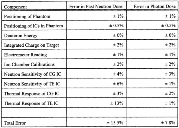
The errors associated with the dual ionization chamber technique have been quantified by the MITR group [Rogus et al 1994], and are shown in Table 2.1. Errors due to the positioning of the
phantom and of the ion chambers in the phantom were calculated knowing that the phantom was always positioned within 2 mm of the previous irradiation position, and that the ion chambers were always positioned within 1 mm of their intended depth. The accelerator energy stability is such that the error in knowing the deuteron energy is 0.01%, which produces no error in determining the dose rates.
Determination of the integrated charge on target due to the presence of secondary electrons (section 2.3) is within 2%, which adds an error of 2% to the dose rate determination. Dose rate errors due to the electrometer, ion chamber calibrations, and sensitivities of the chambers were quantified by Rogus et al [1994]. The overall error associated with calculating the fast neutron dose is ± 15.5%, and the error in
Component Error in Fast Neutron Dose Error in Photon Dose
Positioning of Phantom ± 1% ± 1%
Positioning of ICs in Phantom ± 0.5% + 0.5%
Deuteron Energy + 0% + 0%
Integrated Charge on Target ± 2% ± 2%
Electrometer Reading ± 1% + 1%
Ion Chamber Calibrations ± 2% + 2%
Neutron Sensitivity of CG IC ± 4% ± 3%
Neutron Sensitivity of TE IC ± 6% ± 1%
Thermal Response of CG IC ± 3% ± 2%
Thermal Response of TE IC ± 13% ± 1%
Total Error - 15.5% ± 7.8%
Table 2.1: Errors in Fast Neutron and Photon Doses
2.2.2 Cadmium-Difference Method
Bare and cadmium-covered gold foils are used to experimentally measure the thermal neutron flux via the cadmium difference method. Bare 197Au foils are activated by fast and thermal neutrons. Cadmium has a high cross section of 2 1,000 b for thermal neutrons [Turner 1995], so the cadmium-covered foils are only activated by energetic neutrons greater than 0.4 eV. The activated Au foils then decay by emitting a 411 keV gamma ray.
197Au + in - 198Au* (2.13)
198Au* -1 98Au + y(411 keV) t.,=2.695 d (2.14) Following irradiation, the foils are counted with a germanium detector, and the thermal neutron flux can be determined from these values.
2.2.2.1 Theory
Activation foils are used to measure the average neutron flux over the irradiation period. The foils give a measure of the number of interactions that occurred during the entire irradiation process, and
the average current on target is used as the normalization parameter. Thus measurements using activation foils allow calculation of an average neutron flux per mA of target current.
If we let N be the number of '9 8Au nuclei, X be its decay constant, and R be the production rate of
198Au, then:
dN
- R-,N (2.15)
dt
If the neutron flux is constant over the irradiation, then R is also a constant. Assuming there are no '98Au nuclei at time zero, this equation can be integrated to give:
N(t) =-
R (1 -e
(2.16)
Activity is defined as A=XN so:
A(t)=R(1 -e -t) (2.17)
As time goes to infinity, the activity will approach R asymptotically. R is thus the saturated activity, A.. The foil irradiation lasts for a time to, after which time the foil activity will be:
AO=A (1-e ') (2.18)
Counting of the foil follows the irradiation. Once the irradiation is finished, the activity will decrease as the nuclei begin to decay. If the foil is counted at a time tj and is counted until a time t2, both measured from the beginning of the irradiation, and E is the overall efficiency, then the net counts will be:
t2
C= eA oe (tdt= e (e - -e ) (2.19)
The overall efficiency includes the detector efficiency, the abundance of the 411 keV decay gamma, and the self-absorption of the 411 keV gamma by the gold foil. Rearranging this equation to solve for Ao and substituting into Eq. 2.11 allows solving for A. as:
AC
A -e - -. t°(e - t -Xt2 (2.20)
A saturated activity is calculated for both the bare foil and the Cd-covered foil at each position [Knoll 1989]. Each saturated activity is normalized by the average current on target, which is the integrated charge divided by the irradiation time. The thermal neutron flux can then be calculated from:
SMW( - FCd (2.21)
A v( M bare F Cd
A~o
mbare mCdwhere MW is the molecular weight of 197Au, a is its microscopic thermal absorption cross section, A, is Avogadro's number, m is the foil mass, and Fcd is a correction factor, dependent upon cadmium
thickness, that accounts for those neutrons absorbed by cadmium that are of nonthermal energies [Rogus
et al 1994]. A value of 1.02 has been suggested for 0.020" cadmium covers by R. Fairchild of
Brookhaven National Laboratory [Rogus et al 1994].
2.2.2.2 Method
Gold foils are cut from a piece of thin gold foil so that they are approximately 0.25 cm
2. The
foils are then precisely weighed to within 10 gg. Bare foils are taped to a polyethylene rod at depths of 1, 2, 3, 4, 6, 8, and 10 cm. This rod is then inserted into a butyrate tube that is then filled with water. This water-filled tube is then completely inserted into brain phantom port along the central axis. Another foil is taped onto the outside of the phantom to provide the surface measurement. The foils are then irradiated and the integrated charge on target is recorded for normalization purposes. The irradiation start time and end time are recorded. The target temperature and dose rate at the console are monitored during the irradiation.
Another set of foils is prepared. These are covered with a 0.020" layer of cadmium, and then taped onto the polyethylene rod. These Cd-covered foils must be placed at least 2 cm apart to eliminate any reduction of foil activation due to the proximity of other cadmium covers [Choi 1991]. Foils are placed at depths of 2, 4, 6, 8, and 10 cm on one rod, and at 1 and 3 cm on another. The rest of the
procedure is identical to the bare foil irradiation, with the surface foil being irradiated at the same time as the first Cd-covered rod.
Following the irradiation, each foil is counted by a germanium detector. The counting start time, and counting elapsed time are recorded, as are the counts collected by the detector for the 411 keV peak. All foils are counted to an error of less than 8% with most foils being less than 5%. Finally, a standard source is counted with the same geometry as the gold foils, to determine the detector efficiency.
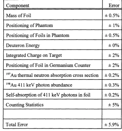
The errors associated with the cadmium difference method of determining the thermal neutron flux have been quantified by the MITR group [Rogus et al 1994], and are shown in Table 2.2. The foils were weighed within 0.01 mg, which produced an error in the dose rate of 0.5%. Errors in thermal flux calculation due to the positioning of the phantom and the positioning of the detectors in the phantom, to the deuteron energy stability, and to the integrated charge on target were discussed in section 2.2.1.2. Errors in the thermal flux calculation due to the 19 7Au thermal neutron absorption cross section, to 198Au
411 keV photon abundance, and to self-absorption of 411 keV photons in foil were quantified by Rogus
et al [1994]. Counting statistics were ± 5% which contributed ± 5% error to the thermal flux calculation. The overall error in calculating the thermal neutron flux is ± 5.9% in this work.
Table 2.2: Errors in Thermal Flux Measurement
Component Error
Mass of Foil - 0.5%
Positioning of Phantom ± 1%
Positioning of Foils in Phantom - 0.5%
Deuteron Energy ± 0%
Integrated Charge on Target ± 2%
Positioning of Foil in Germanium Counter ± 2% 197Au thermal neutron absorption cross section ± 0.2%
'
98Au 411 keV photon abundance ± 0.3% Self-absorption of 411 keV photons in foil ± 0.2%
Counting Statistics ± 5%
Total Error ± 5.9%
2.2.3 Kerma Factor Method
Calculation of the dose due to thermal neutrons, and the dose due to the boron concentration is carried out using the kerma factor method. Once the thermal neutron flux is known, from the bare and cadmium-covered gold foils, the doses can be determined by multiplying by a kerma factor.
Kerma is the initial kinetic energy released per unit mass. Kerma is equal to dose under the condition of charged particle equilibrium if losses due to bremsstrahlung are negligible. Charged particle equilibrium exists when the ionization energy lost from a volume when electrons escape is exactly compensated by the ionization energy gained by other electrons entering the volume [Turner 1995]. For charged particle equilibrium:
D = K = 4)Fn (2.22)
where D is the absorbed dose (cGy/min), K is the kerma (cGy/min),
4 is the thermal flux (n/cm
2-sec), and F, is the kerma factor (cGy-cm2/n). Kerma factors are calculated from:Fn= 1.602*10-8- 'E (2.23)
m
where a is the microscopic cross section (cm2), Nt is the number of target atoms, m is the mass (g), and E is the kinetic energy transferred per interaction (MeV). Kerma factors for ioB and 14N are tabulated in the literature [Zamenhofet al 1975, Caswell and Coyne 1980].
2.2.3.1 Theory
Thermal neutrons deliver dose to tissue predominantly via the following reactions:
'n + 1H -+2H + y (2.24)
'n + 14N -_14C + lp (2.25)
Thermal neutron capture by hydrogen produces a prompt gamma of energy 2.22 MeV. The ionizations due to these prompt gammas are measured by the ionization chambers. The recoil deuteron does not have enough energy to ionize, so it delivers no dose. Thermal neutron capture by nitrogen has a Q value of 0.626 MeV, and this energy is delivered locally to the soft tissue where the interaction occurs, due to the short ranges of protons and heavy nuclei in tissue as described in Section 1.1 [Turner 1995]. The dose absorbed by the tissue due to this 14N(n,p) reaction is calculated using the kerma factor method. Thermal neutron capture by boron also contributes to dose to tissue, and was described in Section 1.1. Again, the kerma factor method is used to calculate the dose to tissue from this 'oB(n,a) interaction.
2.2.4 Tumor and Healthy Tissue Dose Rate Calculations
The previous sections have described the determination of the dose rates in tissue due to fast and thermal neutrons, photons, and due to the loB(n,a) reaction. The dose rates to tumor tissue and healthy tissue are calculated from these dose rate components, based upon their RBE values and the
concentration of i'B in tissue. The dose rates are calculated as:
Dtssue = Dfast*RBEfast + Dther*RBEtheral + Dphoton*RBEphoton+DB 0o*RBEB-10*ConcB-1o (2.26) where Dssue is the dose rate to either tumor or healthy tissue, Dfs, is the dose rate due to fast neutrons, RBEt is 3.2, Dthe, is the dose rate due to thermal neutrons, RBEthem is 3.2, Dphoton is the dose rate due to photons, RBEphoton is 1.0, DB-10 is the dose rate due to 1 ppm of 'OB in the tissue, RBEB.10 is 3.8, and ConcB1 0 is 40 ppm in tumor tissue and 11.4 ppm in healthy tissue. The above RBE values and 'OB concentrations are based upon the MIT clinical trials using the boronated compound BPA [Kiger 1997, Zamenhof et al 1997].
2.3 SECONDARY ELECTRON EFFECT ON CURRENT MEASUREMENT
In order to determine the accuracy of the dose rates measured in these experiments, the accuracy of the target current measurement was checked. Secondary electrons emitted from the beryllium target following deuteron interactions may escape from the target, if the length of the target is not sufficiently long compared with the target diameter. The loss of these negative particles from the current
measurement effectively increases the current on target reading. Since the dosimetry results are normalized to the current on target, it is important to know the total target current due to the interacting particles and also to these lost secondary electrons. The length to diameter ratio of the beryllium target is 0.5. Previous work on a similar target indicated that the loss of secondary electrons could artificially inflate the current reading by roughly 30% [Song 1997]. That experiment was repeated on the beryllium target to determine its secondary electron effect.
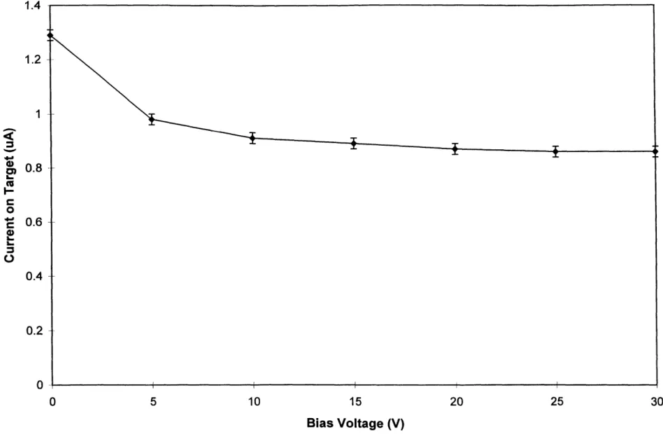
A positive bias voltage relative to the beamline was applied to the target after emptying the moderator tank of D20 and ensuring that the target itself was dry. Current on target was measured
against increasing bias voltage, and results are shown in Figure 2.4. The current saturates at the value representing the current on target due to both the primary particles and to all of the secondary particles, once the bias voltage is large enough to attract all of the secondary electrons.
Initial target current was 1.29 ± 0.02 jIA and the saturated value was 0.86 + 0.02 IA. Thus the loss of secondary electrons inflates the current measurement by 50 ± 6%. This effect was taken into account in the mixed field dosimetry experiments described below.
1.4
1.21
1 I--L. =r o 0.8 0.6 0.4 0.20
0
5
10
15
20
25
Bias Voltage (V)
Figure 2.4: Secondary Electron Effect on Current Measurement2.4 MONTE CARLO CALCULATION
Beam dosimetry has been modeled using the code MCNP, a general Monte Carlo N-Particle transport code [Briesmeister 1997]. MCNP is a computer code that transports neutrons, photons, and
electrons, beginning with a source spectrum entered by the user. The geometry is also entered by the user, and consists of using Boolean algebra to define three-dimensional bounded "cells" from
user-defined surfaces. The user-user-defined surfaces can consist of planes, spheres, cylinders, cones, ellipses, and other geometric shapes. Boolean algebra is used to form bounded three-dimensional volumes by
intersections, unions, and complements of surfaces. MCNP can therefore be used to model very complex geometries.
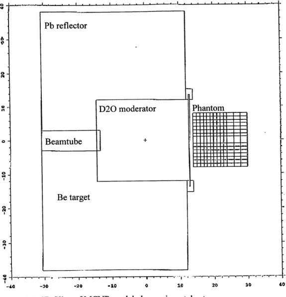
The geometry of the moderator/reflector assembly used experimentally in this work (Figure 2.5) was previously modeled at LABA [Yanch et al 1992], and was used in this simulation. MCNP includes
in depth cross sectional data from ENDF/B V that allow accurate modeling of neutron scattering and absorption interactions [Briesmeister 1997]. The ellipsoidal brain phantom was approximated by modeling a right-circular cylinder. This approximation is valid for simulating doses along the phantom's central axis [Yanch et al 1992]. The simulation assumed the right-cylinder was filled with water. Water is used experimentally in the brain phantom because it is chemically similar to soft tissue which is 63% hydrogen and 26% oxygen [Turner 1995].
Spectral data for the 9Be(d,n) reaction are available in the literature for deuterons of 2.6 MeV and higher at zero degrees [Meadows 1991], but no spectral data exist for deuterons of lower energies
impinging upon a beryllium target. Recent work has been performed at the Schonland Research Center of the University of Witwatersrand on measuring the neutron source spectra from the 9Be(d,n) reaction at different neutron energies. Data were provided by Hamm [1997] for the neutron source spectrum resulting from the 'Be(d,n) reaction using deuterons of 1.5 MeV.
MCNP can calculate flux, across surfaces or averaged over cells, and energy deposition over cells. Tallies used in this work were the f2:n or neutron surface flux, and the f4:n or track length estimate of neutron flux in a cell [Briesmeister 1997]. Photon tallies (f2:p and f4:p) were also performed. These tallies were modified by dose energy (de) and dose function (df) cards to determine the energy dependent dose per source particle. The de and df cards allow the user to enter flux-to-dose conversion factors as a function of energy [Briesmeister 1997]. Kerma factors used in this work for oB and '4N are tabulated in the literature [Zamenhof et al 1975, Caswell and Coyne 1980]. The track length estimate of flux is then modified by these flux-to-dose conversion factors to determine the energy dependent dose per source particle. This procedure was performed to determine the simulated dose rate due to fast and thermal
-40 -36 -20 -10 0
Phantom
II4 -24w t
.4 20 10 40
Figure
2.5:
2D Slice of MCNP modeled experimental setup
_I _. ,._,~~~_...~ ..I . I ~ I ~ ~ I i i ~ r i ~
.. . . . . . . . . I''
?-neutrons, photons, and to the "1B concentration in tissue per source particle. A rough estimate of the neutron yield (section 4.3) was used to calculate the dose rate components per mA of accelerator current. The dose rates to tumor and healthy tissue were then calculated as described in section 2.2.4. Results are reported in Chapter 3.
2.5 FIGURES OF MERIT
Three criteria were chosen to evaluate the experimental and simulated dosimetry results. These were the advantage depth, the tumor dose rate at a depth of four centimeters, and the ratio of the fast neutron dose rate to the total tumor dose rate at a depth of one centimeter. A large advantage depth, large tumor dose rate, and small ratio of fast to thermal dose rates are desirable.
The advantage depth has been extensively used in beam design research for BNCT and is the depth at which the tumor receives the maximum healthy tissue dose [Clement et al 1990]. A therapeutic
beam should have a high advantage depth. Any tumor at a depth less than or equal to the advantage depth will receive more dose than any volume of healthy tissue. Therefore, a large advantage depth means that the treatment would be beneficial for deep-seated tumors, as well as for tumors found close to the skin surface.
The tumor dose rate should also be as high as possible, to reduce treatment time. The tumor dose rate is calculated as described in section 2.2.4 and is given in units of RBE-cGy/min-mA, or dose per unit time per unit current. The higher the dose rate, the less time needed to reach the necessary dose to tumor.
The final criterion is the ratio of the fast to tumor dose rate at a depth of 1 cm. This criterion is important because it is an indicator of the beam quality. The fast neutron dose component drops rapidly with depth. A high fast neutron component will contribute significantly to the healthy tissue dose, and only marginally to the tumor dose, because the tumor dose is dominated by the dose due to boron. The healthy tissue dose near the surface is dominated by the fast neutron component. A low ratio is therefore desirable to protect the healthy tissue. This ratio and the advantage depth go hand-in-hand. As the ratio increases, the advantage depth decreases, and vice versa.
Analysis of these figures of merit for each of the different beams studied allows conclusions to be drawn about which deuteron energy should be chosen. See Chapter 3 for these results.
3. RESULTS
The 'Be(d,n) reaction is under investigation at LABA for possible use in accelerator-based BNCT. MCNP simulations and experimental data acquisition have been previously performed using 2.6 MeV deuterons [Yanch et al 1997]. The initial experiment using 2.6 MeV deuterons on a beryllium
target determined that the fast neutron dose component was unacceptably high; therefore, the deuteron energy should be reduced to produce a softer spectrum. However, reducing the deuteron energy will also result in a reduction in the neutron yield. The purpose of the work described here was to determine the deuteron energy that optimizes the tradeoff between advantage depth and dose rate to tumor.
Complete experiments were performed using 1.3, 1.5, 1.6, 1.7, and 1.8 MeV deuterons impinging upon the beryllium target, using the mixed field dosimetry method described in Chapter 2. Spectral data for the 9Be(d,n) reaction for deuteron energies lower than 2.6 MeV are not currently found in the
literature. Unpublished spectral data of 1.5 MeV deuterons on a beryllium target were provided by Hamm [1997], and a monte carlo simulation was performed.
3.1 EXPERIMENTAL RESULTS
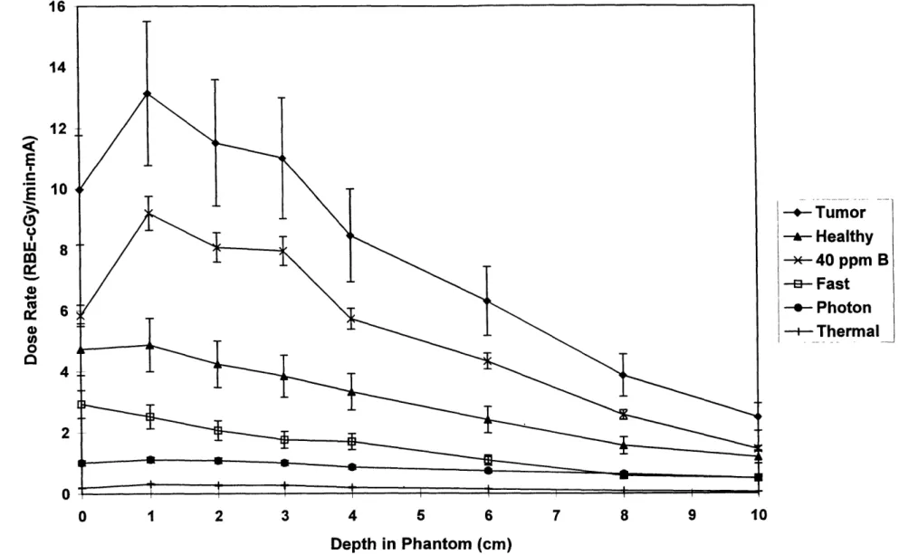
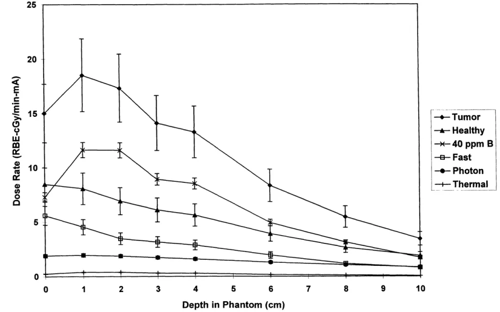
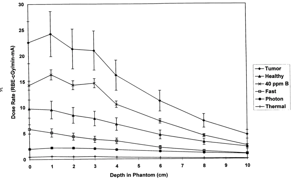
Mixed field dosimetry was used to characterize five different neutron beams produced by the 'Be(d,n) reaction using different deuteron energies. The deuteron energies considered were 1.3, 1.5, 1.6, 1.7, and 1.8 MeV. Measurements were taken at depths of 1, 2, 3, 4, 6, 8, and 10 cm along the axial centerline of the brain phantom, and results are shown in Figures 3.1-3.5. Error bars reflect total experimental error as described in chapter 2.
As can be seen in Figures 3.1-3.5, the major component of the tumor dose rate is the dose rate due to the 40 parts per million (ppm) 'oB concentration in tumor tissue. Thus, by simply increasing the 'oB concentration in the tumor tissue, the tumor dose rate could be significantly increased. On the other hand, the major component of the healthy tissue dose rate is the dose rate due to the fast neutron
component. To reduce the healthy tissue dose rate, the fast neutron dose rate should be decreased, if possible.
The results shown assume boron concentrations of 40 ppm in tumor tissue, and 11.4 ppm in healthy tissue. These concentrations are based on those obtained in ongoing BNCT clinical trials at MIT using the compound boronated phenyalaline (BPA) [Zamenhof et al 1997]. The relative biological effectiveness (RBE) values chosen were also based upon the MIT clinical trials, assuming no
fractionation. The RBE values used were 3.8 for boron dose in tumor, 1.35 for boron dose in healthy tissue, 3.2 for fast and thermal neutron doses, and 1.0 for photon doses [Kiger 1997].
-
Tumor
-A- Healthy-* 40 ppm B
---
Fast
-e-
Photon
-+-
Thermal
1
2
3
4
5
6
7
8
9
Depth in Phantom (cm)
Figure 3.1: Experimental Dosimetry Results for 1.3 MeV deuterons on a beryllium targetC
E
ca(0 o 0 12 10 8 6 4Tumor
-A
Healthy
---
40 ppm B
-E-Fast
-e-
Photon
--
Thermal
1 2 3 4 5 6 7 8 9Depth in Phantom (cm)
Figure 3.2: Experimental Dosimetry Results for 1.5 MeV deuterons on a beryllium target4E
<€
E
..d0
(
U) U)I 0a
--
Tumor
--
Healthy
---
40 ppm B
-a-
Fast
-e-
Photon
-+-
Thermal
Figure 3.3: Experimental Dosimetry Results for 1.6 MeV deuterons on a beryllium target
2
25
2015
10
*
-E
eON
00
0
1
2
3
4
5
6
7
8
9
1C
Depth in Phantom (cm)
I
mL
40
35
30
-
25
-
Tumor
o
---
Healthy
u
20
m
2
--
40 ppm B
-s-
Fast
cc
S15
-e-
Photon
-+-
Thermal
o10
5
0
1
2
3
4
5
6
7
8
9
10
Depth in Phantom (cm)
Figure 3.4: Experimental Dosimetry Results for 1.7 MeV deuterons on a beryllium target-+- Tumor
-I-Healthy
---
40 ppm
I
-e- Fast
-e- Photon
-+-
Thermal
0
1
2
3
4
5
6
7
8
9
Depth in Phantom (cm)
Figure 3.5: Experimental Dosimetry Results for 1.8 MeV deuterons on a beryllium target60
50
40 3020
E
C,E
0m
A 0, 0 100
The results were evaluated according to the criteria outlined in Section 2.2, and are shown in Figures 3.6-3.8. As the deuteron energy is decreased, the tumor dose rate at a depth of 4 cm
correspondingly decreases. This decrease in tumor dose rate with decreased deuteron energy is the expected result, as the neutron yield monotonically decreases with decreasing energy [Burrill 1964]. A high tumor dose rate is preferable, as the treatment time would be reduced. As an example, the total
tumor dose rate at 4 cm for the 1.6 MeV d-Be reaction is 16 RBE-cGy/min-mA or 64 RBE-cGy/min for a 4 mA accelerator. This result compares well with the approximately 25 RBE-cGy/min total tumor dose rate at 4 cm for the MITR beam used in the BNCT clinical trials [Rogus et al 1994].
Alternatively, as the deuteron energy is decreased from 1.8 MeV, the data suggest that the advantage depth increases, although the error bars are significant. A large advantage depth is desired to spare the healthy tissue. As an example, the advantage depth for the 1.6 MeV d-Be reaction is 6.5 cm. The corresponding value for the MIT clinical trials is approximately 7 cm [Rogus et al 1994]. Finally, as the deuteron energy is decreased, the data suggest that the ratio of the fast neutron dose rate to the tumor dose rate at a depth of 1 cm decreases. Again the error bars are significant and it is difficult to be certain that the ratio is decreasing and is not constant in this deuteron energy range. A low ratio is advantageous due to skin sparing. The 1.6 MeV d-Be beam has a ratio of 21% and the MIT BNCT clinical trial beam has a ratio of approximately 11% [Rogus et al 1994].
3.2 SIMULATION RESULTS
Monte carlo simulations were performed using the code MCNP, as described in Chapter 2. Neutron source spectra from the 9Be(d,n) reaction for deuterons of less than 2.6 MeV are not currently available in the literature. However, neutron source spectra for the 9Be(d,n) reaction from low energy deuterons are currently being measured at the University of Witwatersrand (UW) [Guzek et al 1997]. The neutron source spectrum resulting from the 9Be(d,n) reaction with 1.5 MeV deuterons was provided by the Schonland Research Center [Hamm 1997] so that a comparison could be made between
experimental and simulated results. The MCNP simulation results are shown in Figure 3.9. Error bars shown indicate the statistical error only. Measurement errors are undoubtably also present in the UW spectral measurement, but are not reflected in these graphs. Simulation dose components are compared with the experimental dose components in Chapter 4.
Figure 3.9 shows that the major component of the healthy tissue dose rate is the fast neutron dose rate component. The experimental results (Figure 3.2) also showed that the fast neutron dose rate was the major component of the healthy tissue dose rate. The simulated tumor dose rate is equally composed


![Figure 2.3: Schematic of Ellipsoidal Head Phantom used in experimental work [Rogus 1994]](https://thumb-eu.123doks.com/thumbv2/123doknet/14148113.471466/16.918.211.808.136.986/figure-schematic-ellipsoidal-head-phantom-used-experimental-rogus.webp)