Cellular/Molecular
Spinal
and ␦ Opioids Inhibit Both Thermal and
Mechanical Pain in Rats
Audrey Normandin,
1Philippe Luccarini,
4Jean-Louis Molat,
4Louis Gendron,
1,2,3and Radhouane Dallel
41De´partement de physiologie et biophysique, Faculte´ de me´decine et des sciences de la sante´,2Institut de pharmacologie de Sherbrooke, and3Centre de
recherche clinique E´tienne-Le Bel, Universite´ de Sherbrooke, Sherbrooke, QC J1H 5N4, Canada, and4Clermont Universite´, Universite´ d’Auvergne,
NEURO-DOL, BP 10448, F-63000, CLERMONT-FERRAND Inserm, U1107, F-63001 Clermont-Ferrand, France
The expression and contribution of
(MOPR) and ␦ opioid receptors (DOPR) in polymodal nociceptors have been recently challenged.
Indeed, MOPR and DOPR were shown to be expressed in distinct subpopulation of nociceptors where they inhibit pain induced by
noxious heat and mechanical stimuli, respectively. In the present study, we used electrophysiological measurements to assess the effect
of spinal MOPR and DOPR activation on heat-induced and mechanically induced diffuse noxious inhibitory controls (DNICs). We
recorded from wide dynamic range neurons in the spinal trigeminal nucleus of anesthetized rats. Trains of 105 electrical shocks were
delivered to the excitatory cutaneous receptive field. DNICs were triggered either by immersion of the hindpaw in 49°C water or
applica-tion of 300 g of mechanical pressure. To study the involvement of peptidergic primary afferents in the activaapplica-tion of DNIC by noxious heat
and mechanical stimulations, substance P release was measured in the spinal cord by visualizing neurokinin type 1 receptor
internaliza-tion. We found that the activation of spinal MOPR and DOPR similarly attenuates the DNIC and neurokinin type 1 receptor internalization
induced either by heat or mechanical stimuli. Our results therefore reveal that the activation of spinal MOPR and DOPR relieves both
heat-induced and mechanically induced pain with similar potency and suggest that these receptors are expressed on polymodal,
sub-stance P-expressing neurons.
Introduction
In peripheral tissues, noxious mechanical, thermal, and chemical
stimuli are detected by nociceptors and converted to action
po-tentials by lightly myelinated A␦ and nonmyelinated C-fibers
(
Julius and Basbaum, 2001
;
Stein and Lang, 2009
). Unlike other
sensory receptors, most nociceptors are thought to be polymodal
(
Perl, 1996
;
Cain et al., 2001
) (i.e., sensitive to both heat and
mechanical noxious stimuli) (
Julius and Basbaum, 2001
). Until
recently, discrimination between different pain modalities was
therefore considered to happen primarily at spinal and/or
su-praspinal sites (
Perl, 2007
). Indeed, recent findings revealed that
noxious stimuli of various natures specifically activate different
neuronal pathways and, consequently, that the distinction
be-tween pain modalities would instead occur at the level of primary
afferents (
Abrahamsen et al., 2008
;
Cavanaugh et al., 2009
;
Scher-rer et al., 2009
). In particular, this model suggests that heat and
mechanical sensitivity are processed by distinct subpopulations
of primary afferent fibers. Indeed, in mice, a subpopulation of
lightly myelinated A␦ fibers (
Koltzenburg et al., 1997
;
Cain et al.,
2001
) and nonpeptidergic unmyelinated IB4-positive C-fibers
(
Cavanaugh et al., 2009
;
Scherrer et al., 2009
) were shown to be
specific mechanosensitive nociceptors. By contrast, peptidergic
IB4-negative C-fibers expressing substance P and calcitonin
gene-related peptide preferentially mediate heat sensitivity (
Ca-vanaugh et al., 2009
;
Scherrer et al., 2009
).
Adding to the controversy,
Scherrer et al. (2009)
had recently
reported that, in mice, the
␦ opioid receptors (DOPRs) were
absent from peptidergic nociceptors expressing
opioid
recep-tors (MOPRs). In support of a role of primary afferents in the
discrimination of pain modalities, they further showed that
acti-vation of spinal DOPR preferentially inhibits mechanical pain,
whereas activation of spinal MOPR preferentially inhibits
ther-mal pain (
Scherrer et al., 2009
). However, numerous studies have
shown that the selective activation of MOPR with [D-Ala
2,
N-Me-Phe
4, Gly
5-ol]-enkephalin (DAMGO) injected at the level
of the spinal cord can efficiently alleviate both heat-induced
(
Porreca et al., 1984
;
Malmberg and Yaksh, 1992
;
Nagasaka and
Yaksh, 1995
;
Kondo et al., 2005
;
Scherrer et al., 2009
;
van Rijn et
al., 2012
) and mechanically induced pain (
Nichols et al., 1995
;
Sluka et al., 2002
;
Kondo et al., 2005
;
Chen and Pan, 2006
;
van
Received April 17, 2013; revised June 4, 2013; accepted June 7, 2013.Author contributions: A.N., P.L., J.-L.M., L.G., and R.D. designed research; A.N., P.L., and J.-L.M. performed research; A.N., P.L., J.-L.M., L.G., and R.D. analyzed data; A.N., P.L., J.-L.M., L.G., and R.D. wrote the paper.
This work was supported by the Natural Sciences and Engineering Research Council of Canada and the Canadian Institutes of Health Research grants to L.G. and a Laboratory exchange program opportunity grant from the Fonds de Recherche Que´bec-Sante´ (FRQ-S)-funded Quebec Pain Research Network to L.G. and R.D. L.G. is the recipient of a FRQ-S Junior 2 salary support. A.N. was the recipient of the Frederick Banting and Charles Best Canada Graduate Scholarships-Master’s Award awarded by the Canadian Institutes of Health Research and a Master scholarship from the FRQ-S. This work was also supported by the Institut National de la Sante´ et de la Recherche Me´dicale, Universite´ d’Auvergne-Clermont 1 (France) and Re´gion Auvergne. We thank He´le`ne Beaudry for technical assistance and Robert Dumaine and Philippe Sarret (Universite´ de Sherbrooke) for granting access to the Olympus confocal micro-scope funded by the Canadian Foundation for Innovation.
The authors declare no competing financial interests.
Correspondence should be addressed to either of the following: Dr. Louis Gendron, De´partement de physiologie et biophysique, Universite´ de Sherbrooke, Faculte´ de me´decine et des sciences de la sante´, 3001 12th Avenue North, Sherbrooke, QC J1H 5N4, Canada, E-mail: Louis.Gendron@USherbrooke.ca; or Pr. Radhouane Dallel, INSERM UMR 1107, Neuro-Dol, Douleur Trige´minale et Migraine, Faculte´ de Chirurgie Dentaire, 11 boulevard Charles de Gaulle, 63000 Clermont-Ferrand, France, E-mail: Radhouane.dallel@udamail.fr.
DOI:10.1523/JNEUROSCI.1631-13.2013
Rijn et al., 2012
). Similarly, the activation of spinal DOPR was
shown to relieve heat (
Stewart and Hammond, 1994
;
Tseng et al.,
1997
;
Qiu et al., 2000
;
Cahill et al., 2001
,
2003
;
Morinville et al.,
2003
;
Gendron et al., 2007a
,
b
;
Beaudry et al., 2009
;
Overland et
al., 2009
;
Dubois and Gendron, 2010
) and mechanical pain (
Mi-askowski et al., 1990
,
1991
;
Sutters et al., 1990
;
Holdridge and
Cahill, 2007
;
Scherrer et al., 2009
;
Otis et al., 2011
). Therefore, a
more thorough investigation of the role of spinal MOPR and
DOPR and their ability to inhibit various pain modalities is
required.
In the present study, we investigated whether spinal MOPR
and DOPR differentially regulate thermal and mechanical pain
by assessing the effects of intrathecal DAMGO and deltorphin II
on both diffuse noxious inhibitory controls (DNICs) and
spi-nal neurokinin type 1 (NK
1) receptor internalization triggered
by thermal and mechanical noxious stimulations of the rat
hindpaws.
Materials and Methods
Animals
Studies in rats. Adult male Sprague Dawley rats weighting 250 –300 g (from
Charles River Laboratories) were maintained on a 12 h light/dark cycle. Laboratory food and water were available ad libitum. Experiments were ap-proved by the animal care committees at the Universite´ d’Auvergne-Clermont 1 (Comite´ d’E´thique en Matie`re d’Expe´rimentation Animale Auvergne CE 26-12) and the Universite´ de Sherbrooke (protocol 242-10). All procedures were in compliance with the policies and directives of the Canadian Council on Animal Care and guidelines from both the Interna-tional Association for the Study of Pain and the Directives of the European Parliament and the Council on the protection of animals used for scientific purposes (Dir.2010/63/UE).
Studies in mice. Adult male C57BL/6 mice (20 –25 g: Charles River
Laboratories) were housed in groups of four and maintained on a 12 h light/dark cycle. Laboratory food and water were available ad libitum. Experiments were approved by the animal care committee at the Univer-site´ de Sherbrooke (protocol 242-10). All procedures were in compliance with the policies and directives of the Canadian Council on Animal Care and guidelines from the International Association for the Study of Pain. The experiments were designed to minimize the number of animals used and their suffering.
Drugs
DAMGO-enkephalin acetate salt (Tocris Bioscience, batch 22; and Sigma-Aldrich, lot BCBB7749), a MOPR-selective agonist, and deltor-phin II (Dlt II) (American Peptide, lot M08048T1; and Sigma-Aldrich, lot 079K8741), a DOPR-selective agonist, were dissolved in saline solu-tion (0.9% NaCl). The drugs were injected intrathecally either by lumbar catheterization for electrophysiological studies (5g of DAMGO in a volume of 10l; corresponding to 9.7 nmol and 8 g of Dlt II in a volume of 10l; corresponding to 10.2 nmol) or lumbar puncture for NK1 receptor internalization studies (5g of DAMGO in a volume of 30 l and 10g of Dlt II in a volume of 30 l; corresponding to 12.7 nmol). To ascertain drug specificity and selectivity, respectively, for MOPR and DOPR, in the electrophysiological studies, naloxone 0.4 mg/kg intrave-nously, a nonselective opioid antagonist, and naltrindole 4 mg/kg intra-venously, a DOPR-selective antagonist, were injected via the jugular vein. Selectivity of DAMGO and Dlt II in the NK1receptor internalization studies was demonstrated in a previous study (Beaudry et al., 2011).
Electrophysiological recordings
Animal preparation
As previously described (Lapirot et al., 2011), the rats were anesthetized with 2% halothane in a NO2/O2mixture (2:1). After intramuscular in-jection of 100g atropine sulfate, the trachea was cannulated. The ca-rotid artery and external jugular vein were catheterized. The animals were then paralyzed by intravenous perfusions of vecuronium bromide (2.4 mg/h) and artificially ventilated with volume-controlled pumps
(54 –55 strokes/min). The levels of O2, N2O, and end-tidal CO2(3.5– 4.5%) were monitored by an anesthetic gas analyzer (Dra¨ger Vamos) during the entire experimental period. These parameters were digitally displayed and under the control of alarms. The arterial catheter was attached to a calibrated pressure transducer (UFI) connected to an am-plifier (Stoelting) for continuous monitoring of the mean arterial blood pressure (90 –110 mmHg). The analog output from the blood pressure amplifier was connected to a computer data sampling system (Cam-bridge Electronics Design 1401 computer interface). The heart rate was continuously monitored, and cutaneous vascularization was periodically checked by observing the color of the paw extremities and the rapidity by which they regained normal color after pressure application. The colo-rectal temperature was kept constant at 38⫾ 0.5°C with a feedback-controlled heating blanket. The surgical level of anesthesia was verified by a stable mean arterial blood pressure and a constant heart rate during noxious stimulation.
The animals were placed in a stereotaxic frame with the head fixed in a ventroflexed position (incisor bar dropped 5 mm under the standard position) by means of an adapted metallic bar. For spinal trigeminal subnucleus oralis (Sp5O) recordings, a craniotomy was performed on the right side at the level of the occipitoparietal suture, and the dura mater was removed.
For the lumbar catheterization, a 2 cm longitudinal skin incision was made directly over the L5–L6 lumbar vertebrae. The subcutaneous soft tissue was eliminated, and the skin incision was drawn to the dorsal midline. The paravertebral muscles were separated to expose the L5 and L6 vertebrae. A curved Friedman-Pearson rongeur (F.S.T.) was used to break the spinal crest of the L6 vertebrae and then expose the dura. We cut a rat intrathecal polyurethane short catheter (Alzet, Durect) into 4.5-cm-long 28G tubing. A Teflon-coated, stainless steel stylet guided the catheter into the subarachnoid space from the lumbar spinal column (L5–L6) to the thoracic spinal column (T12–T13). DAMGO (5g) or Dlt II (8g) was injected via this catheter over a period of 2 s. Appropri-ate placement of the catheter was verified by dissecting the animals at the end of the experiment. After the surgery, the level of halothane was reduced to 0.6 – 0.7% and maintained during the entire recording period.
Recordings
Unitary extracellular recordings were made from the right Sp5O with glass micropipettes (7–10 M⍀) filled with a mixture of 5% NaCl and pontamine sky blue. The brainstem was explored 2.4 –3.0 mm laterally to the midline and between the frontal planes AP⫺1.1 and AP ⫺2.6 mm from the interaural line (Paxinos and Watson, 1997). Single-unit activi-ties were amplified and displayed on oscilloscopes. The activiactivi-ties went into a window discriminator connected to a CED 1401plus interface (Cambridge Electronic Design) and a PC computer (Spike 2.06 soft-ware), which allowed sampling and analysis of the spontaneous and evoked neuronal activity. Wide-dynamic range (WDR) neurons were recognized based on their responses to mechanical non-noxious (brush-ing with a soft brush) and noxious (pinch with forceps) stimulations of their receptive fields as previously described (Dallel et al., 1999). Specif-ically, each neuron that responded in a graded manner with increasing firing rates to the stimulus range from non-noxious to noxious intensity was classified as a WDR cell. Once a neuron was identified, electrical square-wave stimuli (2 ms duration) were applied through a pair of stainless steel needle electrodes subcutaneously placed into the center of the receptive field; the thresholds for eliciting A- and C-fiber-evoked responses were determined. In poststimulus time histograms (PSTHs), A- and C-fiber-evoked responses were distinguished by their latencies, but only C-fiber-evoked responses were considered in the detailed anal-ysis. Previous research showed that burst discharges at latencies⬎30 ms are elicited by C-fibers (Hu, 1990;Dallel et al., 1999). Therefore, all spikes occurring between 30 and 200 ms after a stimulus were considered to be C-fiber-evoked.
Experimental design
Sequences of 105 electrical stimuli (0.66 Hz) were applied to the receptive field at 3 times the threshold for C-fiber activation every 10 min. DNICs
were triggered by alternatively immersing one hindpaw into a 49°C water bath or applying a mechanical pressure of 300 g to the other hindpaw with calibrated forceps between the 36th and 60th stimuli (i.e., for 37.5 s) (seeFig. 1A). The pressure of 300 g was determined from pilot
electro-physiological experiments, which showed that this was the minimal pres-sure required to induce a reduction of at least 40% of C-fiber-evoked action potentials. This pressure is also in the range of the pressures reached in the Randall-Selitto’s test in which 400 g (Chen and Pan, 2006;
Sibilia et al., 2012) or higher pressures (Miaskowski et al., 1991; Muth-uraman and Singh, 2011) are commonly used as a cutoff. Only one hindpaw at a time received a noxious stimulation, and the order of ap-plication of the noxious stimulation was alternated for each animal. For example, for one animal, we began with the thermal stimulus and then proceeded with the mechanical stimulus. For another animal, we instead began with mechanical stimulation, followed with thermal stimulation and so forth. Thus, when all the tested animals were considered, the effects of 5g DAMGO or 8 g Dlt II on both heat-induced and me-chanically induced DNICs were tested 10, 20, 30, and 40 min after the administration of the opioid. To ascertain the drug specificity in the observed effects, naloxone (0.4 mg/kg intravenously), a nonselective opi-oid receptor antagonist, was injected 50 min after the administration of DAMGO in animals. For the Dlt II-injected rats, naltrindole (4 mg/kg intravenously), a DOPR-selective antagonist, was used.
The analyses were performed as previously described (Lapirot et al., 2011). Briefly, in each sequence, the PSTHs determined from the 21st to 35th responses were used for studying the unconditioned response (be-fore noxious stimulation; control period), and those derived from re-sponses 36th to 105th were used for studying the DNICs. To assess the inhibitory effect of noxious heat or a mechanical stimulus, only the 46th– 60th responses were considered because responses induced between the 36th– 45th noxious stimuli correspond to the time required (5–10 s) to reach the maximal inhibitory effects. In each sequence, PSTHs derived from the 46th– 60th (DNIC effect) were normalized to PSTHs derived from the 21st to 35th responses (unconditioned response; control pe-riod). The inhibitory effect of the DNIC was computed as the percentage decrease in the number of C-fiber-evoked action potentials.
DAMGO (5g in 10 l) or Dlt II (8 g in 10 l) was intrathecally injected after two stable control sequences were recorded (i.e., variation in unconditioned C-fiber response⬍10%). Each rat received only one injection of DAMGO or Dlt II.
NK
1receptor internalization
Animal perfusion
Studies in rats. The animals were deeply anesthetized in an induction cage
with 5% isoflurane–95% medical air mixture (the anesthetic used in Sherbrooke) and maintained during the entire procedure with 2% of the same anesthetic distributed via a mask. In those experiments, DAMGO (5g in 30 l), Dlt II (10 g in 30 l), or saline was administered via direct lumbar puncture using a technique with a high success rate that was previously described by others (Fairbanks, 2003) and extensively used in our laboratory (Beaudry et al., 2009,2011;Otis et al., 2011). Briefly, a 30-gauge 0.5-inch needle mounted on a Luer tip Hamilton syringe (VWR) was inserted into the L5–L6 intervertebral space of the anesthetized animal (at the level of the cauda equina), and the injection was performed over a period of 2 s. Appropriate placement of the needle was confirmed by observing a brief twitch of the tail. DAMGO, Dlt II, or saline was intrathecally injected 10 min before application of either heat stimulation at 49°C or a mechanical pressure of 300 g for 38 s on the right hindpaw. As previously described (Beaudry et al., 2011), 10 min after the noxious stimulation (i.e., 20 min after DAMGO, Dlt II, or saline injec-tion), the animals were perfused through the aortic arch with 500 ml of 4% PFA and 0.18% picric acid in 0.1Mphosphate buffer with pH 7.4 at 4°C. The spinal cord was then removed, postfixed in the same solution for 1 h, and cryoprotected for 48 h in a solution of 30% sucrose in 0.2M phosphate buffer. The lumbar segment of the spinal cord was snap frozen in isopentane at⫺45°C and stored at ⫺80°C until sectioning. The lum-bar segments of the spinal cord were sliced in transverse sections (30m thick) on a frozen microtome, and immunolabeling of NK1receptors was performed on free-floating sections. Briefly, the slices were incubated for
30 min at room temperature in 1% sodium borohydride diluted in PBS to quench aldehyde-induced fluorescence present in tissues and thor-oughly rinsed. The sections were incubated overnight at 4°C in a rabbit anti-NK1receptor polyclonal antibody (directed against the C-terminal of NK1receptors of rat origin, amino acids 393– 407; Sigma-Aldrich) diluted 1:5000 in a solution containing 3% normal goat serum and 0.3% Triton X-100 in PBS. The slices were washed in PBS and then incubated with a fluorescent goat anti-rabbit secondary antibody (Alexa-488, Invit-rogen) diluted 1:1000 in PBS for 1 h at room temperature.
Studies in mice. Mice were deeply anesthetized as described for rats.
They then received a noxious heat (immersion in a 49°C water bath) or mechanical (application of a 200 g pressure) stimulation for 38 s on their right hindpaw. Ten minutes after mice had received the noxious stimu-lation, they were intracardially perfused with 30 ml of 4% PFA and 0.18% picric acid in 0.1Mphosphate buffer with pH 7.4 at 4°C. The spinal cord was then removed, postfixed in the same solution for 2 h, and cryopro-tected for 48 h in a solution of 30% sucrose in 0.2Mphosphate buffer. The lumbar segment (L4 –L6) of the spinal cord was snap frozen in isopen-tane at⫺45°C and stored at ⫺80°C until sectioning. The dorsal part of the L4 –L6 spinal cord segment was cut on a cryostat into 8 –10 serial 20-m-thick horizontal sections to examine superficial laminae I–II. Horizontal sections were directly mounted on HistoBond adhesive mi-croscope slides (VWR), and immunohistochemistry was performed di-rectly on the slides. Briefly, the slices were thawed at room temperature before rehydration for 10 min in PBS. They were then incubated for 30 min at room temperature in 0.1Mglycine diluted in PBS to quench aldehyde-induced fluorescence present in tissues and rinsed. The sec-tions were incubated overnight at 4°C, in a humid container, with the anti-NK1receptor polyclonal antibody (1:5000) solution described above. The slices were washed in PBS and then incubated with the fluorescent secondary antibody (1:1000) solution for 1 h at room temperature.
The proportion of spinal cord neurons exhibiting internalized NK1 receptors was quantified by a previously described method (Abbadie et al., 1997;Marvizon et al., 2003;Kondo et al., 2005;Nazarian et al., 2008;
Beaudry et al., 2011). Briefly, we used an epifluorescence microscope (Leica DM4000B; Leica Microsystem) to count NK1receptor-like immu-noreactive cell bodies. Neurons with and without NK1receptor internal-ization located in the superficial lamina I were counted, and the percentage of NK1receptor immunoreactive cells with internalization was calculated. The number of neurons with NK1receptor internaliza-tion was determined by averaging the counts made in 3–5 rats per con-dition (14 –18 sections per animal) or 4 or 5 mice per concon-dition (3– 6 sections per animal). During the counting procedure, the observer was unaware of the treatment received by the animals. A neuron was consid-ered having internalized NK1receptors if the membrane was barely la-beled and if⬎50 vesicle-like fluorescent puncta were observed in the cell body. The results were expressed as the mean⫾ SEM.
For the purpose of illustrating our observations, representative confo-cal images were collected with the Olympus Fluoview 1000 (FV1000) laser-scanning confocal microscope fitted with an U Plan S-Apo 60⫻ (1.35 NA) oil-immersion objective.
Calculations and statistical analysis
The calculations were performed with Excel 2007 (Microsoft), graphs were created with SigmaPlot 11.0, and statistical analyses were performed with Prism GraphPad 5.0. The data are expressed as the mean⫾ SEM. p values are presented in the figure legends.
Results
Characteristics of trigeminal WDR neurons
Seventeen WDR neurons were recorded within the Sp5O to test
the effect of DAMGO (n
⫽ 8) and Dlt II (n ⫽ 9) on noxious heat
and mechanical stimulations. None of the neurons exhibited
spontaneous activity. All of them had ipsilateral receptive fields
that included the intraoral or perioral region. They were sensitive
to both innocuous and noxious mechanical stimuli and
re-sponded by increasing their firing rates as the stimulus intensity
increased into the noxious range. When
2-ms-long percutaneous electrical stimuli
were applied to the center of the receptive
fields of the neurons, responses
attribut-able to peripheral activation of A- and
C-fibers could be observed (data not
shown). The longest latency responses
(89
⫾ 3.7 ms, n ⫽ 17) were
C-fiber-evoked. Indeed, the computed
conduc-tion velocity (
⬃0.5 m/s) was in the range
of those previously reported for C-fibers
(
Hu, 1990
;
Dallel et al., 1999
;
Coste et al.,
2008a
,
b
;
Lapirot et al., 2011
). In addition,
long latency responses could only be
evoked by high intensity stimulations
(mean threshold, 10.3
⫾ 0.8 mA, n ⫽ 17),
and they exhibited windup.
Figure 1
A
il-lustrates the experimental design used for
the electrophysiological assays. As shown
in
Figure 1
(examples), immersion of the
hindpaw in a 49°C water bath (
Fig. 1
B) or
application of a 300 g pressure (
Fig. 1
C)
between the 36th and 60th electrical
stim-ulus induced a strong inhibition of
C-fiber-evoked firing of Sp5O WDR
neu-rons. This inhibition reflects the
activa-tion of DNICs.
The effects of intrathecal DAMGO and
Dlt II injection on the DNIC induced by
a noxious heat stimulus
All tested neurons were under the
influ-ence of the DNICs. The C-fiber-evoked
responses of all those neurons were
strongly depressed by the application of noxious heat stimulation
(49°C) to one of the hindpaws (
Fig. 2
A, E, examples).
Regarding the experiments with DAMGO (n
⫽ 8),
quantita-tive analysis showed that noxious heat stimulation induced a
mean inhibition of 89
⫾ 4% before the injection of the drug. The
intrathecal injection of DAMGO (5
g) alone (i.e., without the
heterotopic conditioning stimulus) has no effect on the
C-fiber-evoked responses of trigeminal WDR neurons (
Fig. 2
B; see also
Fig. 4
B, between the first and the 36th electrical stimuli), but the
injection strongly reduced the inhibitory effect induced by
nox-ious heat stimulation of the hindpaw (
Fig. 2
B). Ten to 20 and
30 – 40 minutes after the administration of DAMGO, the
inhibi-tions of C-fiber-evoked responses during heat application were
33
⫾ 7% and 32 ⫾ 8%, respectively (
Fig. 2
D). Indeed, spinally
administered DAMGO induced reductions of 62
⫾ 8% and 65 ⫾
9% of the DNICs ( p
⬍ 0.0001, F ⫽ 20.30, one-way ANOVA
for repeated measures followed by Bonferroni’s
multiple-comparison test). The specificity of the effects of intrathecally
administered DAMGO upon the DNIC was tested by
adminis-tering the opiate antagonist naloxone. For all the tested neurons,
systemic administration of naloxone (intravenous injection, 0.4
mg/kg) antagonized the effect of DAMGO, indicating that it was
opioid receptor-mediated (
Fig. 2
C,D). Immersion of one of the
hindpaw of the rats in the water bath reduced by 87
⫾ 5% the
C-fiber-evoked action potentials of Sp5O WDR neurons. As for
DAMGO, the intrathecal injection of Dlt II (8
g) alone (i.e.,
without the heterotopic conditioning stimulus) has no effect on
the C-fiber-evoked responses of trigeminal WDR neurons (
Fig.
2
F; see also
Fig. 4
F, between the first and the 36th electrical
stim-uli), but the injection strongly reduced the inhibitory effect
in-duced by noxious heat stimulation of one hindpaw (
Fig. 2
F ). Ten
to 20 and 30 – 40 minutes after the administration of Dlt II, the
inhibitions of C-fiber-evoked responses during heat application
were 36
⫾ 8% and 55 ⫾ 8%, respectively (
Fig. 2
H ). Indeed,
spinally administered Dlt II induced reductions of 56
⫾ 11% and
38
⫾ 8% of the DNICs (p ⬍ 0.0001, F ⫽ 13.81, one-way ANOVA
for repeated measures followed by Bonferroni’s
multiple-comparison test). The selectivity of the effects of intrathecally
administered Dlt II upon the heat-induced DNIC was tested by
administering the DOPR-selective antagonist naltrindole. For all
the tested neurons, systemic administration of naltrindole
(intra-venous injection, 4 mg/kg) antagonized the effect of Dlt II,
indi-cating that it was DOPR-mediated (
Fig. 2
G,H ).
In an interesting way, the effects of spinally administered
DAMGO and Dlt II (at equimolar doses), when compared
to-gether, show no significant difference in their ability to reduce the
activation of heat-induced DNICs ( p
⫽ 0.1451, F ⫽ 2.36,
two-way ANOVA for repeated measures followed by Bonferroni’s
multiple-comparison test).
The effects of intrathecal DAMGO and Dlt II injection on
heat-induced NK
1receptor internalization
A well-known phenomenon is that, after the activation of
pepti-dergic primary afferent fibers by noxious stimulation, substance
P is released in the superficial laminae of the spinal cord, where it
binds to and activates NK
1receptors. Upon activation, NK
1re-ceptors are rapidly internalized (
Mantyh et al., 1995
;
Abbadie et
al., 1997
;
Marvizon et al., 2003
;
Nazarian et al., 2008
;
Beaudry et
Figure 1. Schematic representation of the electrophysiological experimental design. A, Sequences of 105 percutaneous elec-trical stimulations (0.66 Hz at 3 times the C-fiber threshold intensity) were applied to the excitatory receptive field. The DNICs were triggered by alternately immersing one hindpaw into a 49°C water bath (B) or applying a 300 g mechanical pressure (C) on the other hindpaw between the 36th and 60th electrical stimuli (corresponding to 37.5 s). DAMGO (5gin10l)orDltII(8gin10 l) was injected to the lumbar spinal cord via an intrathecal catheter. B, C, Histograms showing the C-fiber-evoked responses of trigeminal WDR neurons to 105 successive electrical stimulations. B, Effect of the noxious heat stimulus on the C-fiber-evoked response of a trigeminal WDR neuron. C, Effect of the noxious mechanical stimulus on the C-fiber-evoked response of a trigeminal WDR neuron.al., 2011
). NK
1receptor internalization can therefore be used as
an indication of the activation of peptidergic primary afferent
fibers.
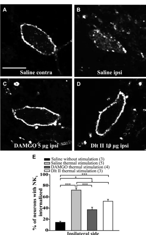
In the ipsilateral side of control rats, immunostaining of
NK
1receptors in the rat spinal cord revealed intense
immu-nofluorescent labeling located mainly at the cell surface of
laminae I of the lumbar spinal cord (
Fig. 3
E; see
Fig. 5
E, first
column; 15
⫾ 2% of lamina I neurons have internalized NK
1receptors). In animals pretreated with intrathecal saline,
nox-ious heat stimulation (immersion of the right hindpaw in 49°C
water bath during 38 s) induced NK
1receptor internalization
in the majority of the lamina I neurons of the ipsilateral
lum-bar spinal cord (
Fig. 3
B, E; 72
⫾ 4% of laminae I neurons have
internalized NK
1receptors). In contrast, only discrete NK
1receptor internalization was observed in the contralateral side
of the spinal cord (
Fig. 3
A). Intrathecal DAMGO (
Fig. 3
C) and
Dlt II (
Fig. 3
D) administered 10 min before noxious heat
stim-ulation significantly decreased the proportion of neurons with
internalized NK
1receptors to 37
⫾ 4% and 52 ⫾ 3%,
respec-tively (
Fig. 3
E). Those results correspond to a reduction of
⬃49% for DAMGO and 28% for Dlt II, of laminae I neurons
with internalized NK
1receptors. We found no significant
dif-ference between the effects of DAMGO and Dlt II on their
ability to inhibit heat-induced NK
1receptor internalization
(
Fig. 3
E) ( p
⬍ 0.0001, F ⫽ 4.03, one-way ANOVA followed by
Bonferroni’s multiple-comparison test).
The effects of intrathecal DAMGO and Dlt II injection on the
DNIC induced by a noxious mechanical stimulus
C-fiber-evoked responses were strongly depressed by the
appli-cation of noxious mechanical (300 g) stimulation to the hindpaw
(
Fig. 4
A, E, examples). For assays where DAMGO was tested,
quantitative analysis showed a mean inhibition of 56
⫾ 8% of
C-fiber-evoked firing of Sp5O neurons before the injection of the
MOPR-selective agonist. After intrathecal injection of DAMGO
(5
g), the DNICs triggered by noxious mechanical stimulation
of the hindpaw were strongly reduced (
Fig. 4
B).
Figure 4
D
illus-trates the results of the quantitative analysis. Ten to 20 min and
30 – 40 min after DAMGO injection, mechanically induced
inhi-bition of C-fiber-evoked responses was 23
⫾ 9% and 29 ⫾ 7%,
respectively. The DNICs were therefore reduced by 64
⫾ 13%
and 53
⫾ 7% (p ⬍ 0.0001, F ⫽ 22.90, one-way ANOVA for
re-peated measures followed by Bonferroni’s multiple-comparison
test). The specificity of the effects of intrathecally administered
DAMGO upon the DNIC was tested by administering the opiate
antagonist naloxone. For all the neurons tested, systemic
admin-istration of naloxone (intravenous injection, 0.4 mg/kg)
antago-nized the reduction of the DNIC induced by DAMGO (
Fig.
4
C,D).
In animals where Dlt II was tested (n
⫽ 9), application of a
300 g pressure to the hindpaw of the rats before the
administra-tion of the DOPR-selective agonist reduced by 57
⫾ 7% the
C-fiber-evoked action potentials of Sp5O WDR neurons. The
Figure 2. Intrathecal DAMGO and Dlt II both inhibit the DNIC induced by a noxious heat stimulus. A–C, E–G, Histograms showing representative C-fiber-evoked responses of two trigeminal WDR neurons to 105 successive electrical stimulations recorded before (A,E) and 10 min after intrathecal (i.t.) administration of DAMGO 5g(B)or20minafterDltII8g(F).Betweenthe36thand60th stimulation, one hindpaw was immersed into a 49°C water bath. The DNICs triggered by heat noxious stimulation of the hindpaw are reduced after both DAMGO (B) and Dlt II (F ) injection. The intravenous (i.v.) administration of naloxone (C) and naltrindole (G) reversed the effect of DAMGO and Dlt II, respectively. D, H, Graphic representation showing the mean (n⫽ 8 for D, and n ⫽ 9 for H ) percentage of inhibition of C-fiber-evoked action potentials before and 10 –20 min and 30 – 40 min after the intrathecal injection of the opioids. The data are individually normalized to those before administration of the opioids. The selective MOPR and DOPR agonists significantly reduced the percentage of inhibition of C-fiber-evoked action potentials either 10 –20 or 30 – 40 min after their administration. Naloxone significantly reversed those opioidergic-induced effects, and naltrindole significantly reversed the DOPR-mediated effect 10 –20 min after Dlt II administration. **p⬍0.01(one-wayANOVAforrepeatedmeasureswithBonferroni’sposthoctest).***p ⬍ 0.001 (one-way ANOVA for repeated measures with Bonferroni’s post hoc test). Error bars indicate the SEM.
intrathecal injection of Dlt II strongly
re-duced the inhibitory effects evoked by
noxious mechanical stimulation of the
hindpaw (
Fig. 4
F ). Ten to 20 and 30 – 40
minutes after the administration of Dlt II,
the inhibitions of C-fiber-evoked responses
during the 300 g pressure application were
28
⫾ 7% and 46 ⫾ 11%, respectively (
Fig.
4
H). In brief, spinally administered Dlt II
induced reductions of 51
⫾ 14% and 25 ⫾
13% of the DNICs. Indeed, the effect of
Dlt II is only significant 10 –20 min after
its administration ( p
⬍ 0.0002, F ⫽ 10.00,
one-way ANOVA for repeated measures
followed by Bonferroni’s
multiple-compa-rison test). The selectivity of the effects of
intrathecally administered Dlt II upon the
heat-induced DNICs was tested by
admin-istering the DOPR-selective antagonist
nal-trindole. For all the tested neurons, systemic
administration of naltrindole (intravenous
injection 4 mg/kg) antagonized the effect
of Dlt II, indicating that it was
DOPR-mediated (
Fig. 4
G,H ).
As for thermal stimulation, intrathecal
DAMGO and Dlt II have comparable
in-hibitory effects on mechanically induced
DNICs (p
⫽ 0.1654, F ⫽ 2.13, two-way
ANOVA for repeated measures followed by
Bonferroni’s multiple-comparison test).
Interestingly, when the effects of
intra-thecal injection of DAMGO on the DNICs
triggered by either noxious heat or
mechan-ical stimulation were compared, no
signifi-cant difference was found ( p
⫽ 0.643,
F
⫽ 0.2244, two-way ANOVA for repeated
measures followed by Bonferroni’s
mul-tiple-comparison test). Similarly, Dlt II
was found to inhibit DNICs triggered by
either noxious heat or mechanical
stimu-lation with comparable potencies ( p
⫽
0.499, F
⫽ 0.48, two-way ANOVA for
repeated measures followed by Bonferroni’s
multiple-comparison test).
The effects of intrathecal DAMGO and
Dlt II injection on mechanically
induced NK
1receptor internalization
As seen for noxious heat stimulation,
appli-cation of a 300 g pressure induced a strong
NK
1receptor internalization (
Fig. 5
B; 66
⫾
5% of ipsilateral lamina I neurons). Only
discrete NK
1receptor internalization was
observed in the contralateral side of the
lumbar spinal cord (
Fig. 5
A). In animals
pretreated with intrathecal DAMGO (
Fig.
5
C) or Dlt II (
Fig. 5
D), a significant decrease
in the proportion of neurons showing NK
1receptor internalization was observed (
Fig.
5
E; 33
⫾ 7% and 38 ⫾ 4%, respectively, for
DAMGO and Dlt II, of ipsilateral laminae I
neurons have internalized NK
1receptors).
This result corresponds to a reduction of
Figure 3. Intrathecal DAMGO and Dlt II reduce heat-induced NK1receptor internalization. Internalization of NK1receptors was induced by immersing the right hindpaw of male Sprague Dawley rats in a 49°C water bath for 38 s, and lamina I NK1 receptor-immunoreactive neurons were observed by immunofluorescence. The noxious heat stimulation was applied 10 min after intra-thecal injection of saline (A,B), DAMGO 5g (C), or Dlt II 10 g (D). Confocal images of neurons on the contralateral (A) and ipsilateral (B–D) sides of the spinal cord are shown. On the contralateral side of the saline-injected animals, immunolabeling of NK1 receptors appeared to be at the cell surface (A). However, on the ipsilateral side of the same animals, the noxious heat stimulation induced a significant increase in NK1receptor internalization, as evidenced by the intensely labeled intracellular vesicle-like structures (B). When DAMGO (C) or Dlt II (D) was injected, a significant reduction in NK1receptor internalization was observed on the ipsilateral side compared with the same side in saline-injected rats. The animals that had received a saline injection but no noxious stimulation had low basal proportions of neurons with internalized NK1receptors. This result is illustrated in the graphic representation showing the percentage of neurons with NK1receptor internalization induced by heat stimulation for ipsilateral side of the lumbar spinal cord (E). *p⬍ 0.05 (one-way ANOVA with Bonferroni’s post hoc test). ***p ⬍ 0.001 (one-way ANOVA with Bonferroni’s post hoc test). The numbers in parentheses represent the number of animals per group. Error bars indicate the SEM. Scale bar: A, 30m.
⬃50% and 42% of laminae I neurons with internalized NK
1recep-tors. In addition, there is no significant difference between the effects
of DAMGO and Dlt II on inhibition of mechanically induced NK
1receptor internalization (
Fig. 5
E; p
⫽ 0.0002, F ⫽ 13.82, one-way
ANOVA followed by Bonferroni’s multiple-comparison test).
Heat-induced and mechanically induced NK
1receptor
internalization in mice
We then studied the effects of noxious heat and mechanical
stim-uli on the internalization of NK
1receptors in mice. We observed
the localization of NK
1receptors after either immersion of the
right hindpaw of mice in a 49°C waterbath or application of a
200 g pressure with calibrated forceps for 38 s. As shown in
Figure
6
, in control mice the NK
1receptor labeling was mostly found at
the cell surface (
Fig. 6
A, D; 14
⫾ 1% of lamina I neurons had
internalized NK
1receptors). Immersion of the hindpaw in the
49°C waterbath induced a strong proportion of internalized NK
1receptors (
Fig. 6
B, D; 71
⫾ 2% of neurons). Application of the
mechanical pressure also induced a significant internalization of
NK
1receptors (
Fig. 6
C,D; 53
⫾ 4% of lamina I neurons have
internalized NK
1receptors).
Discussion
In the present study, we investigated the role of spinal MOPRs
and DOPRs in heat and mechanical pain regulation using
elec-trophysiological and immunohistochemical approaches. By
measuring the firing of WDR neurons of the spinal trigeminal
subnucleus oralis (Sp5O), we demonstrated that intrathecal
ad-ministration of either DAMGO or Dlt II, respectively,
MOPR-and DOPR-selective agonists, was able to inhibit the activation of
DNICs triggered by heat and mechanical noxious stimuli. We
further observed that both spinal MOPR and DOPR activation
blocks substance P release and inhibits NK
1receptor
internaliza-tion induced by thermal and mechanical stimuli.
The existence of polymodal nociceptors implies that
discrim-ination of pain modalities occurs at the higher spinal cord level
and in the brain (
Melzack and Wall, 1962
;
Perl, 2007
). This
dogma was, however, recently challenged. Indeed, using
pharma-cological ablation of mouse sensory neurons expressing the
so-dium channel Na
v1.8 or genetic ablation of Mrgprd-expressing
sensory neurons, others revealed that specific pain modalities are
encoded by distinct subpopulations of nociceptors (
Abrahamsen
et al., 2008
;
Cavanaugh et al., 2009
). These findings were further
supported by the fact that, in mice, nonpeptidergic IB4-positive
DRG neurons were found to specifically mediate the responses to
mechanical pain, whereas a subpopulation of IB4-negative
neu-rons mediates thermal pain (
Scherrer et al., 2009
).
In primary afferents, MOPR are mainly expressed by small
IB4-negative peptidergic neurons (
Ji et al., 1995
;
Minami et al.,
1995
;
Zhang et al., 1998b
;
Wang and Wessendorf, 2001
;
Rau et al.,
2005
), whereas DOPR was found to be expressed in large- and
small-diameter neurons (
Mansour et al., 1994
;
Wang and
Wes-sendorf, 2001
). Although the presence of DOPR in substance
Figure 4. Intrathecal DAMGO and Dlt II both inhibit the DNIC triggered by a noxious mechanical stimulus. A–C, E–G, Histograms showing representative C-fiber-evoked responses of two trigeminal WDR neurons to 105 successive electrical stimulations recorded before (A,E) and 30 min after intrathecal (i.t.) administration of DAMGO 5g (B) or 10 min after Dlt II 8 g (F). Between the 36th and 60th stimulation, a 300 g mechanical pressure was applied on one hindpaw of the animal with calibrated forceps. The DNICs triggered by mechanical noxious stimulation of the hindpaw are reduced after both DAMGO (B) and Dlt II (F ) injection. The intravenous (i.v.) administration of naloxone (C) and naltrindole (G) reversed the effect of DAMGO and Dlt II, respectively. D, H, Graphic representation showing the mean (n⫽ 8 for D, and n ⫽ 9 for H) percentage of inhibition of C-fiber-evoked action potentials before and 10–20 min and 30–40 min after the intrathecal injection of the opioids. The data are individually normalized to those before administration of the drugs. The selective-MOPR agonist significantly reduced the percentage of inhibition of C-fiber-evoked action potentials either 10 –20 or 30 – 40 min after its administration. Naloxone significantly reversed those opioidergic-induced effects (D). The selective-DOPR agonist significantly reduced the percentage of inhibition of C-fiber-evoked action potentials only 10 –20 min after its administration and naltrindole significantly reversed this DOPR-mediated effect (D). *p⬍ 0.05 (one-way ANOVA for repeated measures with Bonferroni’s post hoc test). **p⬍ 0.01 (one-way ANOVA for repeated measures with Bonferroni’s post hoc test). ***p ⬍ 0.001 (one-way ANOVA for repeated measures with Bonferroni’s post hoc test). Error bars indicate the SEM.
P- and calcitonin gene-related
peptide-containing neurons has been shown (
Ji et
al., 1995
;
Zhang et al., 1998a
;
Riedl et al.,
2009
;
Wang et al., 2010
), enhanced green
fluorescent protein-tagged DOPR is
con-fined to nonpeptidergic fibers in knockin
mice (
Scherrer et al., 2009
). In agreement
with the hypothesis that specific pain
mo-dalities are encoded by distinct
subpopula-tions of nociceptors, it was observed that
spinal DAMGO specifically inhibited
heat-induced pain whereas activation of spinal
DOPR specifically attenuated mechanical
stimuli-induced behaviors (
Scherrer et al.,
2009
).
In the current study, we measured the
effects of heat and mechanical noxious
stimuli applied to the hindpaws of rats on
the firing of trigeminal WDR neurons and
confirmed previous studies showing the
existence of DNIC acting on these
trigem-inal neurons (
Le Bars, 2002
;
Lapirot et al.,
2011
). As opposed to stimulus-induced
reflex or withdrawal responses studied in
most behavioral tests, DNICs can only be
triggered by noxious stimuli (
Le Bars et
al., 1979
) and therefore represent an
effi-cient and unbiased strategy to evaluate the
analgesic properties of drugs applied
in-trathecally. Here, we demonstrated that
activation of both MOPR and DOPR at
the spinal level resulted in a profound
de-crease in the effects of the DNIC on the
firing of WDR neurons. Indeed,
intrathe-cal DAMGO and Dlt II were able to
simi-larly reduce the effects of the DNIC
induced by heat or mechanical noxious
stimulation of the hindpaw. The fact that
intrathecal administration of DAMGO or
Dlt II alone had no effect on
C-fiber-evoked responses of trigeminal WDR
neurons in the absence of heterotopic
conditioning stimuli suggests that the
re-duction of DNIC-induced analgesia does
not result from direct action of MOPR or
DOPR agonists in the trigeminal nucleus.
The efficacy of MOPR agonists to
in-hibit mechanical pain in rodents has also
been shown by others (
Nakazawa et al.,
1985
;
Chaki et al., 1988
;
Nichols et al.,
1995
;
Kondo et al., 2005
;
Back et al., 2006
;
Iwai et al., 2012
;
Lee et al., 2012
).
Simi-larly, a number of studies demonstrated
that intrathecal DOPR agonists efficiently
alleviate noxious heat-induced behaviors
in rats and in mice (
Stewart and
Ham-mond, 1994
;
Tseng et al., 1997
;
Qiu et al.,
2000
;
Cahill et al., 2001
,
2003
;
Morinville
et al., 2003
;
Gendron et al., 2007a
,
b
;
Be-audry et al., 2009
,
2011
;
Overland et al., 2009
;
Dubois and
Gend-ron, 2010
). Lessons from knock-out mice also provided
evidence supporting a role for MOPR and DOPR in the
mod-ulation of diverse pain modalities. Indeed, both MOPR and
DOPR knock-out mice were found to have increased sensitivity
to thermal (
Sora et al., 1997
;
Matthes et al., 1998
;
Qiu et al.,
2000
;
Martin et al., 2003
) and mechanical pain (
Martin et al.,
2003
).
Figure 5. Intrathecal DAMGO and Dlt II reduce mechanical stimulus-induced NK1receptor internalization. Internalization of NK1 recep-tors was induced by applying a 300 g mechanical pressure on the right hindpaw with calibrated forceps for 38 s. Lamina I NK1 receptor-immunoreactiveneuronswereobservedbyimmunofluorescence.Thenoxiousmechanicalstimulationwasapplied10minafterintrathecal injection of saline (A,B), DAMGO 5g(C),orDltII10g(D).Confocalimagesshowedthatapplicationofthemechanicalpressuredidnot affect the cell-surface localization of NK1receptors in the contralateral side of the lumbar spinal cord of saline-injected rats (A). In contrast, applicationofthesamestimulusinducedastronginternalizationofNK1receptorsintheipsilateralsideforthesameanimal,asshownbythe intracellular localization of the immunolabeling (B). The injection of DAMGO or Dlt II both significantly inhibited mechanically induced NK1 receptorinternalization(E).Forcomparisonpurposes,datafromthesaline-injectedgroupwithoutnoxiousstimulationpresentedinFig.3E
are reported in panel E. *p⬍0.05(one-wayANOVAwithBonferroni’sposthoctest).**p⬍0.01(one-wayANOVAwithBonferroni’spost
hoc test). ***p⬍ 0.001 (one-way ANOVA with Bonferroni’s post hoc test). Values in parentheses represent the number of animals per
In accordance with the polymodal nature of nociceptors,
peripheral thermal (
Zachariou and Goldstein, 1996
;
Abbadie
et al., 1997
;
Allen et al., 1997
;
King et al., 2005
), mechanical
(
McCarson and Goldstein, 1991
;
Zachariou and Goldstein,
1996
;
Abbadie et al., 1997
;
King et al., 2005
;
Kondo et al.,
2005
), and chemical (
Marvizon et al., 2003
;
Nazarian et al.,
2008
;
Beaudry et al., 2011
) noxious stimulations were shown
to evoke the release of substance P in the superficial layers of
the rat spinal cord. Interestingly, the blockade of NK
1recep-tors expressed by spinoparabrachial neurons was previously
shown to inhibit DNICs triggered by intraplantar formalin (
La-pirot et al., 2009
). To determine whether DAMGO and Dlt II can
inhibit the release of substance P induced by heat and mechanical
noxious stimulation of the hindpaw in rats, we measured NK
1receptor internalization in the superficial laminae of the lumbar
spinal cord. As shown previously (
Kondo et al., 2005
;
Zhang et al.,
2010
), we observed that DAMGO blocked the NK
1receptor
in-ternalization induced by noxious mechanical stimulation. We
also showed, for the first time, that intrathecal DAMGO
de-creased the NK
1receptor internalization evoked by noxious
heat stimulation of the paw. Similarly, intrathecal Dlt II
blocked NK
1receptor internalization induced by heat and
me-chanical noxious stimuli. The fact that
intra-thecally injected Dlt II has an effect on NK
1receptor internalization confirms previous
reports showing that a subpopulation of
substance P-containing nociceptors
ex-presses DOPRs (
Ji et al., 1995
;
Zhang et al.,
1998a
;
Riedl et al., 2009
;
Wang et al., 2010
;
Beaudry et al., 2011
). Our results also reveal
that peptidergic primary afferents are
in-volved in DNICs triggered by noxious heat
and mechanical stimulations and that these
fibers are regulated by both MOPRs and
DOPRs.
One may argue that the apparent
dis-crepancies between our observations and
those made by others could be explained
by interspecies differences. Indeed, as
pre-viously mentioned, a dichotomy between
C-fiber subpopulations and pain
modali-ties was observed in mice (
Abrahamsen et
al., 2008
;
Cavanaugh et al., 2009
;
Scherrer
et al., 2009
). Thus, transient receptor
po-tential vanilloid 1, an important but not
unique transducer of thermal nociception
(
Caterina et al., 2000
;
Davis et al., 2000
;
Woodbury et al., 2004
), is almost
exclu-sively found in heat-specific peptidergic
sensory neurons in mice (
Zwick et al.,
2002
;
Woodbury et al., 2004
;
Lawson et
al., 2008
;
Cavanaugh et al., 2011
) while
it is found in both peptidergic and
non-peptidergic C-fibers in rats (
Gold et al.,
1996
;
Tominaga et al., 1998
;
Guo et al.,
1999
;
Michael and Priestley, 1999
;
Woodbury et al., 2004
;
Yu et al., 2008
).
Others hypothesized that the observed
dichotomy between C-fiber populations
and nociceptive modalities; thus, the effects
of intrathecal MOPR and DOPR agonists
may not apply to rats or higher-order
pri-mates (
Saeed and Ribeiro-da-Silva, 2012
;
Taylor et al., 2012
). For instance, we observed here that both noxious
thermal and mechanical stimuli can induce NK
1receptor
internal-ization in both rats and mice, confirming that peptidergic primary
afferents participate in thermal and mechanical nociception in both
species.
Alternatively, it is possible that the mechanical stimuli used
by
Scherrer et al. (2009
), namely, von Frey filaments, were not
noxious and therefore that paw withdrawals were the
conse-quence of stimulation of non-nociceptive A
␦ and/or A-fibers
shown to express DOPR. von Frey filaments are indeed
com-monly used to measure allodynia, that is, pain induced by a
non-noxious stimulus (e.g., light touch), in injured or
sensi-tized subjects. As a noxious mechanical stimulus, we rather
used a 200 g (mice) or 300 g (rats) pressure applied for 38 s to
the hindpaw with calibrated forceps and show that it
effi-ciently recruits mechanical nociceptors, induces the release of
substance P by primary afferents and, most importantly,
acti-vates DNICs, which are only triggered by noxious stimuli.
In conclusion, our results reveal that the activation of
ei-ther MOPR or DOPR can equally relieve both pain modalities
(i.e., thermal and mechanical), possibly via direct action on
peptidergic C-fibers. Admittedly, although we found that
spi-Figure 6. Heat-induced and mechanically induced NK1receptor internalization in mice. In mice, internalization of NK1receptors was induced by immersion of the right hindpaw for 38 s in a 49°C water bath or by applying (with calibrated forceps) a 200 g mechanical pressure on the right hindpaw, also for 38 s. Animals were perfused without receiving any noxious stimulation (A), 10 min after receiving the noxious thermal (B) or the noxious mechanical (C) stimulation. Lamina I NK1receptor-immunoreactive neurons were observed by immunofluorescence. Confocal images showed that nonstimulated animals present a cell-surface localization of NK1receptor labeling (A). Application of the thermal stimulus (B) or the mechanical pressure (C) both induces strong internalization of NK1receptors in the ipsilateral side of the lumbar segment of the spinal cord (D). **p⬍ 0.01 (one-way ANOVA with Bonferroni’s post hoc test). ***p⬍ 0.001 (one-way ANOVA with Bonferroni’s post hoc test). Values in parentheses represent the number of animals per group. Error bars indicate the SEM. Scale bar: A, 30m.
nal DAMGO and Dlt II similarly inhibited the DNIC triggered
by heat and mechanical noxious stimuli applied to the
hind-paws, other MOPR and DOPR agonists may not be equally
potent at inhibiting all pain modalities. As an example,
neu-ropathic pain is often found to be less sensitive than
inflam-matory pain to MOPR agonists (
Arner and Meyerson, 1988
;
Obara et al., 2009
). Despite the fact that we cannot exclude
that spinal DAMGO and Dlt II could inhibit multiple
sub-populations of selective heat-sensitive and mechanosensitive
nociceptors, the current study revealed that peptidergic
C-fibers are involved in heat and mechanical pain processing
in rodents, a hypothesis recently challenged by others (
Ca-vanaugh et al., 2009
;
Scherrer et al., 2009
).
References
Abbadie C, Trafton J, Liu H, Mantyh PW, Basbaum AI (1997) Inflamma-tion increases the distribuInflamma-tion of dorsal horn neurons that internalize the neurokinin-1 receptor in response to noxious and non-noxious stimula-tion. J Neurosci 17:8049 – 8060.Medline
Abrahamsen B, Zhao J, Asante CO, Cendan CM, Marsh S, Martinez-Barbera JP, Nassar MA, Dickenson AH, Wood JN (2008) The cell and molecular basis of mechanical, cold, and inflammatory pain. Science 321:702–705.CrossRef Medline
Allen BJ, Rogers SD, Ghilardi JR, Menning PM, Kuskowski MA, Basbaum AI, Simone DA, Mantyh PW (1997) Noxious cutaneous thermal stimuli in-duce a graded release of endogenous substance P in the spinal cord: im-aging peptide action in vivo. J Neurosci 17:5921–5927.Medline
Arne´r S, Meyerson BA (1988) Lack of analgesic effect of opioids on neuro-pathic and idioneuro-pathic forms of pain. Pain 33:11–23.CrossRef Medline
Back SK, Lee J, Hong SK, Na HS (2006) Loss of spinal-opioid receptor is associated with mechanical allodynia in a rat model of peripheral neurop-athy. Pain 123:117–126.CrossRef Medline
Beaudry H, Proteau-Gagne´ A, Li S, Dory Y, Chavkin C, Gendron L (2009) Differential noxious and motor tolerance of chronic␦ opioid receptor agonists in rodents. Neuroscience 161:381–391.CrossRef Medline
Beaudry H, Dubois D, Gendron L (2011) Activation of spinal - and ␦-opioid receptors potently inhibits substance P release induced by pe-ripheral noxious stimuli. J Neurosci 31:13068 –13077.CrossRef Medline
Cahill CM, Morinville A, Lee MC, Vincent JP, Collier B, Beaudet A (2001) Prolonged morphine treatment targets␦ opioid receptors to neuronal plasma membranes and enhances␦-mediated antinociception. J Neurosci 21:7598 –7607.Medline
Cahill CM, Morinville A, Hoffert C, O’Donnell D, Beaudet A (2003) Up-regulation and trafficking of␦ opioid receptor in a model of chronic inflammation: implications for pain control. Pain 101:199 –208.CrossRef Medline
Cain DM, Khasabov SG, Simone DA (2001) Response properties of mecha-noreceptors and nociceptors in mouse glabrous skin: an in vivo study. J Neurophysiol 85:1561–1574.Medline
Caterina MJ, Leffler A, Malmberg AB, Martin WJ, Trafton J, Petersen-Zeitz KR, Koltzenburg M, Basbaum AI, Julius D (2000) Impaired nociception and pain sensation in mice lacking the capsaicin receptor. Science 288: 306 –313.CrossRef Medline
Cavanaugh DJ, Lee H, Lo L, Shields SD, Zylka MJ, Basbaum AI, Anderson DJ (2009) Distinct subsets of unmyelinated primary sensory fibers mediate behavioral responses to noxious thermal and mechanical stimuli. Proc Natl Acad Sci U S A 106:9075–9080.CrossRef Medline
Cavanaugh DJ, Chesler AT, Bra´z JM, Shah NM, Julius D, Basbaum AI (2011) Restriction of transient receptor potential vanilloid-1 to the peptidergic subset of primary afferent neurons follows its develop-mental downregulation in nonpeptidergic neurons. J Neurosci 31: 10119 –10127.CrossRef Medline
Chaki K, Sakurada S, Sakurada T, Sato T, Kawamura S, Kisara K, Watanabe H, Suzuki K (1988) Comparison of the antinociceptive effects of new [D-Arg2]-dermorphin tetrapeptide analogs and morphine in mice. Pharma-col Biochem Behav 31:439 – 444.CrossRef Medline
Chen SR, Pan HL (2006) Loss of TRPV1-expressing sensory neurons re-duces spinal opioid receptors but paradoxically potentiates opioid an-algesia. J Neurophysiol 95:3086 –3096.CrossRef Medline
Coste J, Voisin DL, Luccarini P, Dallel R (2008a) A role for wind-up in
trigeminal sensory processing: intensity coding of nociceptive stimuli in the rat. Cephalalgia 28:631– 639.CrossRef Medline
Coste J, Voisin DL, Miraucourt LS, Dallel R, Luccarini P (2008b) Dorsal horn NK1-expressing neurons control windup of downstream trigeminal nociceptive neurons. Pain 137:340 –351.CrossRef Medline
Dallel R, Duale C, Luccarini P, Molat JL (1999) Stimulus-function, wind-up and modulation by diffuse noxious inhibitory controls of responses of convergent neurons of the spinal trigeminal nucleus oralis. Eur J Neurosci 11:31– 40.CrossRef Medline
Davis JB, Gray J, Gunthorpe MJ, Hatcher JP, Davey PT, Overend P, Harries MH, Latcham J, Clapham C, Atkinson K, Hughes SA, Rance K, Grau E, Harper AJ, Pugh PL, Rogers DC, Bingham S, Randall A, Sheardown SA (2000) Vanilloid receptor-1 is essential for inflammatory thermal hyper-algesia. Nature 405:183–187.CrossRef Medline
Dubois D, Gendron L (2010) ␦ opioid receptor-mediated analgesia is not altered in preprotachykinin A knockout mice. Eur J Neurosci 32:1921– 1929.CrossRef Medline
Fairbanks CA (2003) Spinal delivery of analgesics in experimental models of pain and analgesia. Adv Drug Deliv Rev 55:1007–1041.CrossRef Medline
Gendron L, Pintar JE, Chavkin C (2007a) Essential role of opioid receptor in the regulation of␦ opioid receptor-mediated antihyperalgesia. Neuro-science 150:807– 817.CrossRef Medline
Gendron L, Esdaile MJ, Mennicken F, Pan H, O’Donnell D, Vincent JP, Devi LA, Cahill CM, Stroh T, Beaudet A (2007b) Morphine priming in rats with chronic inflammation reveals a dichotomy between anti-hyperalgesic and antinociceptive properties of deltorphin. Neurosci-ence 144:263–274.CrossRef Medline
Gold MS, Dastmalchi S, Levine JD (1996) Co-expression of nociceptor properties in dorsal root ganglion neurons from the adult rat in vitro. Neuroscience 71:265–275.CrossRef Medline
Guo A, Vulchanova L, Wang J, Li X, Elde R (1999) Immunocytochemical localization of the vanilloid receptor 1 (VR1): relationship to neuropep-tides, the P2X3 purinoceptor and IB4 binding sites. Eur J Neurosci 11: 946 –958.CrossRef Medline
Holdridge SV, Cahill CM (2007) Spinal administration of a␦ opioid recep-tor agonist attenuates hyperalgesia and allodynia in a rat model of neuro-pathic pain. Eur J Pain 11:685– 693.CrossRef Medline
Hu JW (1990) Response properties of nociceptive and non-nociceptive neurons in the rat’s trigeminal subnucleus caudalis (medullary dorsal horn) related to cutaneous and deep craniofacial afferent stimulation and modulation by diffuse noxious inhibitory controls. Pain 41:331– 345.CrossRef Medline
Iwai S, Kiguchi N, Kobayashi Y, Fukazawa Y, Saika F, Ueno K, Yamamoto C, Kishioka S (2012) Inhibition of morphine tolerance is mediated by painful stimuli via central mechanisms. Drug Discov Ther 6:31–37.
CrossRef Medline
Ji RR, Zhang Q, Law PY, Low HH, Elde R, Ho¨kfelt T (1995) Expression of -, ␦-, and -opioid receptor-like immunoreactivities in rat dorsal root ganglia after carrageenan-induced inflammation. J Neurosci 15:8156 – 8166.Medline
Julius D, Basbaum AI (2001) Molecular mechanisms of nociception. Nature 413:203–210.CrossRef Medline
King T, Gardell LR, Wang R, Vardanyan A, Ossipov MH, Malan TP Jr, Vanderah TW, Hunt SP, Hruby VJ, Lai J, Porreca F (2005) Role of NK-1 neurotransmission in opioid-induced hyperalgesia. Pain 116: 276 –288.CrossRef Medline
Koltzenburg M, Stucky CL, Lewin GR (1997) Receptive properties of mouse sensory neurons innervating hairy skin. J Neurophysiol 78:1841–1850.
Medline
Kondo I, Marvizon JC, Song B, Salgado F, Codeluppi S, Hua XY, Yaksh TL (2005) Inhibition by spinal- and ␦-opioid agonists of afferent-evoked substance P release. J Neurosci 25:3651–3660.CrossRef Medline
Lapirot O, Chebbi R, Monconduit L, Artola A, Dallel R, Luccarini P (2009) NK1 receptor-expressing spinoparabrachial neurons trigger diffuse nox-ious inhibitory controls through lateral parabrachial activation in the male rat. Pain 142:245–254.CrossRef Medline
Lapirot O, Melin C, Modolo A, Nicolas C, Messaoudi Y, Monconduit L, Artola A, Luccarini P, Dallel R (2011) Tonic and phasic descending do-paminergic controls of nociceptive transmission in the medullary dorsal horn. Pain 152:1821–1831.CrossRef Medline
Lawson JJ, McIlwrath SL, Woodbury CJ, Davis BM, Koerber HR (2008) TRPV1 unlike TRPV2 is restricted to a subset of mechanically insensitive