HAL Id: tel-03215815
https://tel.archives-ouvertes.fr/tel-03215815
Submitted on 3 May 2021HAL is a multi-disciplinary open access
archive for the deposit and dissemination of sci-entific research documents, whether they are pub-lished or not. The documents may come from teaching and research institutions in France or abroad, or from public or private research centers.
L’archive ouverte pluridisciplinaire HAL, est destinée au dépôt et à la diffusion de documents scientifiques de niveau recherche, publiés ou non, émanant des établissements d’enseignement et de recherche français ou étrangers, des laboratoires publics ou privés.
colistin) to improve their penetration through mucus
and pulmonary bacterial biofilms
Rana Zaidan Awad
To cite this version:
Rana Zaidan Awad. Formulations of cationic antibiotics (tobramycin and colistin) to improve their penetration through mucus and pulmonary bacterial biofilms. Human health and pathology. Univer-sité de Poitiers, 2020. English. �NNT : 2020POIT1805�. �tel-03215815�
THESE
Pour l’obtention du Grade de
DOCTEUR DE L’UNIVERSITE DE POITIERS
(Faculté Médecine et Pharmacie)
(Diplôme National - Arrêté du 25 mai 2016)
Ecole Doctorale « Sciences Biologiques & Santé »
Secteur de Recherche : Pharmacologie et Sciences du médicament
Présentée par :
Rana AWAD
************************
Formulations d’antibiotiques cationiques (tobramycine et colistine)
afin d’améliorer leur pénétration dans le mucus
et les biofilms bactériens pulmonaires
************************
Directeurs de Thèse :
Madame le Professeur Sandrine MARCHAND
Monsieur le Docteur Mohamad NASSER
Monsieur le Docteur Fréderic TEWES
************************
Soutenue le 17 décembre 2020
devant la Commission d’Examen
************************
JURY
Professeur Emmanuelle Dé
Université de Rouen
Rapporteur
Docteur Nicolas Anton
Université de Strasbourg
Rapporteur
Professeur Christophe Burucoa
Université de Poitiers Examinateur
Docteur Layale Safa
Université Libanaise
Examinateur
Docteur Fréderic Tewes
Université de Poitiers Directeur de Thèse
Professeur Sandrine Marchand
Université de Poitiers Directrice de Thèse
The research work presented in this thesis was performed under the supervision of
Professor Sandrine MARCHAND and of Doctor Frederic TEWES from the INSERM
U1070 Pharmacology of antimicrobial
agent’s laboratory, Faculty of Medicine and
Pharmacy, University of Poitiers, France. This work was in co-direction with Doctor
Mohamad NASSER from the Platform for Research and Analysis in Environmental
Science (PRASE) laboratory, Doctorate school of Science & Technology, Lebanese
University, Beyrouth-Lebanon.
To my special friend
You told me to never stop no matter what!
If you were still with us, you would have been proud of my success
For that, I owe it all to you…
ACKNOWLEDGMENT
Undertaking this PhD has been a truly life-changing experience for me and it would not have been possible to do without the support and guidance that I received from many people.
Foremost, I would like to express my deepest thanks and appreciation to the director of INSERM U1070, Pr. William COUET, for accepting me in this lab. Thank you for your constructive advises, for your efforts and for the environment you provided for me and other students to proceed with our work.
I would like to express my sincere gratitude to my director Pr. Sandrine MARCHAND, I am very grateful to you for your indispensable scientific help, patient guidance, enthusiastic, encouragement and useful criticism of this thesis. I would also like to thank you for your advice and assistance in keeping my progress on schedule. It is an honor to have you as the director of my thesis.
My very special gratitude goes out to my supervisor Dr. Frederic TEWES without whom his project would never have been achievable. I appreciate your contributions of ideas and time to make my Ph.D. experience productive and enthusiastic. I am sincerely honored by the irreplaceable and valuable critiques you gave me which actually made me the one I am today. Thank you for being patient and always keeping me in check throughout these three years. I could not have imagined having a better advisor and mentor for my Ph.D. study.
My sincere appreciation goes to my co-director at the Lebanese University, Dr Mohamad NASSER. I am grateful to your stimulating and encouraging advices. Thank you for transmitting your faith and support throughout these three challenging years.
I would like to thank the committee members: Pr Emmanuel DÉ, Dr Nicolas ANTON, Pr Christophe BURUCOA, Dr Layale SAFA, Dr Frederic TEWES and Pr Sandrine MARCHAND. Thank you for your valuable time to read and evaluate my dissertation. I am honored to have you in my defense.
My sincere gratitude goes towards Pr Emmanuel DÉ from Université de Rouen and Dr Nicolas ANTON from Université de Strasbourg, for accepting to be the evaluators for my Ph.D thesis. My grateful thank to Pr Christophe BURUCOA and Dr Layale SAFA for accepting to be the part of jury members as an examiner.
My special thanks go out to the organization of “AL-Massar International College & University Institute”, which financially supported me throughout my thesis. I am especially grateful also for the financial support I received from the “University of Poitiers” which allowed me to pursue my thesis following the post-economics crisis assessment in Lebanon.
I would like to thank all the members of the lab for their warm welcome and their good humor that made it possible for us to live those three years as the most enjoyable years of thesis.
To Dr Julien BUYCK, thank you for the great support and precious advices I received from you for enriching my microbiology knowledge.
My sincere gratitude goes to Dr Nicolas GREGOIRE, Pr Blandine RAMMAERT, Dr Julien BRILLAULT for their precious advises.
I would like to thank Helene, Christophe and Julian, who provided me many advices in the analytical part.
Special thanks to Théo, for his technical assistance during this thesis.
To my colleagues in office, Hari my savior friend, Shachi my supporter friend, Grace my helpful friend, Luc Mr informatics, thank you for maintaining a good and rich talkative environment in our room.
To all the students I have met during this thesis: Jennifer, Kévin, Vincent, Alexia, Barbara, Betty, Romain C., Romain A., Etienne, Chantal, Bruna, Karin, Zahyra, Hanyssa, Quentin, Jean-Philippe, François, Lucien, Aline, Noémie, Eduardo, Mathilde, thank you for all the good times we've had together.
I thank my fellow lab mates in INSERM 1070: Muriel, Agnès, Laure, Isabelle, Celia, Jérémie, Emma, Miroslava, for the stimulating discussions and for all the fun we had in the last few years.
I am indebted and indeed grateful to my family without whom nothing would have been special. To my parents, thank you for your continuous support and love. You always pushed me forward and never stopped believing in me. To my siblings, Rim, Rabih, Mohamad and Ali... Thank you for being always there for me, listening and supporting me with all you have got.
I am indeed thankful to my huge family in France, especially to, Sarah, Abir, Bissane, Zaynab, Hassan, Sami, Hend, Kassem, Rana, Achraf, Maya, Youssef, Hussein, Hawraa… Some of you are indeed special and dear to me, I thank you all for your continuous support which I will always need and appreciate.
I would like to thank specially my friends in Lebanon; Jinan AWAD and Fatimah Kheireddine, with whom I spent a precious and memorable time.
I wish to express my most sincere praise and gratitude to the Lord, the grace giver of my life and success.
To Lebanon, my home country that I love, I could only taste the happiness of belonging to you. I dedicate all this work to you, because you are the holy ground that I honor dearly.
ABSTRACT
Pseudomonas aeruginosa (PA) and Acinetobacter baumannii (A. baumannii) are responsible for chronic lung infections often associated with the development of biofilms, i.e. bacterial aggregates trapped in a self-produced matrix of anionic polymers such as alginate or DNA. Within these biofilms, bacteria are protected from cationic antibiotics (ATB) such as tobramycin (TOB) and colistin (COL), in part through their interaction with matrix polymers, which reduce their diffusion. The main objectives of this thesis were first to develop a lung biofilm model that mimics those found in chronic lung infections of patients with cystic fibrosis (CF), one of the main lung diseases for which biofilms develop. Then use this model to evaluate a new ATB delivery system made of lipid nanoparticles developed to treat lung biofilms.
In CF patients, PA forms biofilms consisting of aggregates (5-100 μm) enclosed in their pulmonary mucus. An in vitro lung biofilm model composed of polymers present in vivo (alginate, mucins, DNA) and consisting of PA trapped in alginate beads has been developed and evaluated using COL and TOB. This model was refined by evaluating the effect of the size of the beads and the growth medium in which they were dispersed. Changing the size of the beads from 60 to 1200 µm and using a dispersion medium simulating the composition of mucus further decreased the effectiveness of TOB in this model. For high TOB concentrations, small-colony variants (SCV) of PA, also found in the CF patient, were observed in the model, suggesting a good mimicry of the conditions in vivo. In this model, COL efficacy was less impacted than that of TOB, and prevented the appearance of SCV. Pegylated lipid nanoparticles loaded with COL and containing a lipophilic adjuvant to COL such as Farnesol (FAR) have been developed to increase its effectiveness against biofilms. Encapsulation of COL improved its efficiency by two in the lung biofilm model PA developed, but also restored the sensitivity to COL of a resistant A. baumannii and improved its efficiency in an abiotic adherent biofilm model with A. baumannii.
Keywords: Pseudomonas aeruginosa, Acinetobacter baumannii, tobramycin, colistin, lung
RÉSUMÉ
Pseudomonas aeruginosa (PA) et Acinetobacter baumannii (A. baumannii) sont responsables d'infections pulmonaires chroniques souvent associées à la formation de biofilms, i.e. des agrégats bactériens piégés dans une matrice riche en polymères anioniques tels que l'alginate ou l’ADN.
Au sein de ces biofilms, la diffusion des antibiotiques cationiques (ATB), tels que la tobramycine (TOB) et la colistine (COL), est en partie réduite due aux interactions avec les polymères de la matrice. Les principaux objectifs de cette thèse ont été dans un premier temps de développer un modèle de biofilm pulmonaire mimant ceux retrouvés dans les infections pulmonaires chroniques des patients atteints de mucoviscidose (CF), l'une des principales maladies pulmonaires dans laquelle ces biofilms se développent. Un nouveau système d’administration d'ATB, composé de nanoparticules lipidiques chargées de COL, a été par la suite développé et testé grâce à ce modèle de biofilms pulmonaires.
Chez les patients atteints de CF, PA forme des biofilms constitués d'agrégats (5-100 μm) incorporés dans leur mucus pulmonaire. Un modèle de biofilm pulmonaire in vitro composé des polymères présents in vivo (alginate, mucines, ADN) et constitué de PA piégés dans des billes d'alginate, a été développé et évalué à l'aide de la COL et de la TOB. Ce modèle a été optimisé en évaluant l'effet de la taille des billes et du milieu de croissance dans lequel elles ont été dispersées. La modification de la taille des billes de 60 à 1200 µm et l'utilisation d'un milieu de dispersion simulant la composition du mucus pulmonaire ont permis de réduire davantage l'efficacité de la TOB dans ce modèle. Pour des concentrations élevées en TOB, des variants de PA formant de très petite colonies (SCV), également trouvés chez les patients atteints de CF, ont été observés dans le modèle, ce qui suggère une bonne imitation des conditions in vivo. Dans ce modèle, l'efficacité de la COL a été moins affectée que celle de la TOB, et aucune SCV a été observé. Des nanoparticules lipidiques PEGylées chargées de COL et contenant un adjuvant lipophile à la COL tel que le Farnésol ont été développées pour augmenter son efficacité contre les biofilms. L'encapsulation de la COL a amélioré son efficacité par un facteur deux dans le modèle de biofilm pulmonaire à PA précédemment développée. Ces nanoparticules ont permis également de restaurer la sensibilité à la COL d'une souche résistant d’A. baumannii et ont amélioré son efficacité dans un modèle de biofilm d’A. baumannii formé sur une surface abiotique.
Mots clés : Pseudomonas aeruginosa, Acinetobacter baumannii, tobramycine, colistine,
TABLE OF CONTENTS
ABSTRACT ... i
RÉSUMÉ ... ii
LIST OF COMMUNICATION ... v
LIST OF ABBREVIATIONS ... vii
LIST OF FIGURES ... x
LIST OF TABLES ... xi
LITERATURE REVIEW ... 1
I. Introduction ... 2
II. Pulmonary biofilm ... 4
a. Lung biofilms and associated pathogens ... 4
1) Mucus dispersed biofilms ... 4
2) Biofilms adhering to abiotic surfaces ... 7
b. Bacterial biofilm regulations ... 9
1) Bis-(3′-5′) cyclic diguanosine monophosphare ...10
2) Quorum sensing ...10
c. Biofilm tolerance to ATBs ...12
1) Limitation of the ATBs transport through the biofilm ...12
i. Extracellular DNA ...13
ii. Polysaccharides (Alginate,Psl,Pel) ...14
2) Host mucus can limit the transport the transport of ATBs ...15
d. Investigated therapies specific to biofilms ...16
1) Agents decreasing intracellular c-di-GMP concentration ...16
i. Nitric oxide ...16
ii. Change in nutrient concentrations ...17
iii. Antibiofilm peptides ...18
iv. Interaction with quorum sensing ...18
2) Agents that directly target the matrix ...20
III. How to studying and evaluation of biofilms? ...21
a. In vitro biofilms models ...21
1) Static biofilm model ...24
2) Dynamic biofilm model ...25
b. In vivo models ...28
c. Parameters measured with biofilm models ...31
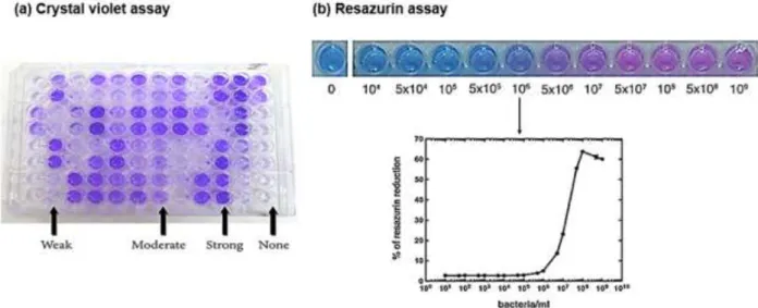
i. Quantitative methods ...32
ii. Qualitative biofilm characterization methods...34
2) In vivo methods ...36
IV. Drug Delivery system to target lung biofilms ...37
a. Nanotechnologies or nanoencapsulations for DDS ...38
1) Lipid-based nanoparticles ...40
i. Liposomes ...40
ii. Lipid nanoparticles (SLN and NLC) ...44
2) Polymeric Nanoparticles ...47
i. Poly(lactic-co-glycolic acid) ...47
ii. Chitosan ...48
iii. Dendrimers ...48
3) Inorganic nanoparticles ...50
b. Others strategies used for antibiotic delivery ...56
1) Bacteriophages ...56 2) PEGylation of antibiotics ...58 EXPERIMENTAL WORK ...56 ARTICLE 1 ...58 ARTICLE 2 ...69 ARTICLE 3 ...98
GENERAL DISCUSSION& PERSPECTIVES ... 135
LIST OF COMMUNICATIONS
Articles:
1. Bruna Gaelzer Silva Torres, Rana Awad, Sandrine Marchand, William Couet,
Frederic Tewes. In vitro evaluation of Pseudomonas aeruginosa chronic lung
infection models: Are agar and calcium-alginate beads interchangeable? Eur.
J. Pharm. Biopharm. 143, 35‑43 (2019). DOI: 10.1016/j.ejpb.2019.08.006
1. Rana Awad, Sandrine Marchand, William Couet, Mohamad Nasser, Frederic
Tewes. Refinement of an in vitro lung biofilm model of Pseudomonas
aeruginosa to evaluate the efficacy of cationic antibiotics. (In manuscript)
2. Rana Awad, Sandrine Marchand, William Couet, Mohamad Nasser, Frederic
Tewes. Antibacterial and antibiofilm activity of colistin-loaded nanoparticles
against adherent and non-adherent lung biofilms. (In manuscript)
International conférences :
1. Rana Awad, Sandrine Marchand, William Couet, Mohamad Nasser, Frederic
Tewes. Modèle in-vitro de biofilm pulmonaire à Pseudomonas aeruginosa.
Congrès de Ricai, Paris 2019. (Poster P-116)
2. Rana Awad, Sandrine Marchand, William Couet, Mohamad Nasser, Frederic
Tewes. Invitro model of Pseudomonas aeruginosa pulmonary biofilm to
evaluate the efficacy of cationic antibiotics. European Congress of Clinical
Microbiology and Infectious Diseases, Paris, 2020. (Poster P1687)
3. Rana Awad, Sandrine Marchand, William Couet, Mohamad Nasser, Frederic
Tewes Antibacterial and antibiofilm activity of Colistin-loaded nanoparticles
against Acinetobacter baumannii. European Congress of Clinical Microbiology
and Infectious Diseases, Paris, 2020. (Poster P339)
4. Rana Awad, Sandrine Marchand, William Couet, Mohamad Nasser, Frederic
Tewes Antibacterial and antibiofilm activity of Colistin-loaded nanoparticles
against Acinetobacter baumannii. Société Française de Pharmacologie et de
Thérapeutique, Lille 2020. (Poster P-116)
5. Rana Awad, Sandrine Marchand, William Couet, Mohamad Nasser, Frederic
Tewes. In vitro model of Pseudomonas aeruginosa pulmonary biofilm to
evaluate the efficacy of cationic antibiotics. 21ème Colloque français des jeunes
chercheurs. Paris, 2020. (Poster)
6. Rana Awad. Limitation de traitement des infections respiratoires chroniques
chez les patients atteints de mucoviscidose. 21ème Colloque français des
jeunes chercheurs. Paris, 2020. (oral presentation)
LIST OF ABBREVIATIONS
CF
:
Cystic fibrosis
ATB
:
Antibiotic
DDS
:
Drug delivery system
TOB
:
Tobramycin
COL
:
Colistin
PA
:
Pseudomonas aeruginosa
A. baumannii
:
Acinetobacter baumannii
ETT
:
Endotracheal tube
VAP
:
Ventilator-associated pneumonia
COPD
:
Chronic obstructive pulmonary disease
EPS
:
Extracellular polymeric substances
eDNA
:
extracellular DNA
SCVs
:
Small colony variants
PMNs
:
Polymorphonuclear leukocytes
ROS
:
Reactive oxygen species
NETs
:
Extracellular neutrophil traps
QS
:
Quorum sensing
c-di-GMP
:
Bis-(3′-5′) cyclic diguanosine monophosphate
PDEs
:
Phosphodiesterases
AHLs
:
Acyl homoserine lactones
3-oxo-C12-HSL
:
N-(3-Oxododecanoyl)-L-homoserine lactone
C4-HSL
:
N-butyryl-L-homoserine lactone
T3SS
:
Type 3 secretion systems
MIC
:
Minimal inhibitory concentration
LPS
:
Lipopolysaccharides
CIP
:
Ciprofloxacin
NO
:
Nitric oxide
QSI
:
Quorum sensing inhibitors
DNase
:
Deoxyribonucleases
rhDNase
:
Recombinant human DNase
AlgL
:
Alginase or alginate lyase
CDC
:
Center for disease control
MBC
:
Minimal bactericidal concentration
EUCAST
:
European Committee on Antimicrobial Susceptibility TestingCLSI
:
Clinical and Laboratory Standards Institute
MBEC
:
Minimal biofilm eradication concentration
MBIC
:
Minimal biofilm inhibitory concentration
BBC
:
Biofilm bactericidal concentration
BPC
:
Biofilm prevention concentration
CFU
:
Colony-forming unit
CLSM
:
Confocal laser scanning microscopy
CV
:
Crystal violet
GFP
:
Green fluorescent protein
PI
:
Propidium iodide
FISH
:
Fluorescence in situ hybridization
PNA
:
Peptide nucleic acid
LNA
:
Locked nucleic acids
PCR
:
Polymerase chain reaction
LNP
:
Lipid nanoparticle
CMS
:
Colistimethate sodium
NPs
:
Nanoparticles
LIPs
:
Liposomes
PMB
:
Polymyxin B
SUV
:
Small unilamellar vesicles
LUV
:
Large unilamellar vesicles
LMV
:
Large multilamellar vesicles
DPPC
:
Dipalmitoylphosphatidylcholine
DSPC
:
Distearoylphosphatidylcholine
PEG
:
Polyethylene glycol
Bi
:
Bismuth-ethanedithiol
SLN
:
Solid lipid nanoparticles
NLC
:
Nanostructured lipid carriers
EE
:
Encapsulation efficiency
FDA
:
Food and drug administration
EMA
:
European Medicines AgencyPAMAM
:
Polyamidoamine
Au
:
Gold
Ag
:
Silver
Cu
:
Copper
MHB
:
B
ouillon Muller-HintonASM
:
M
ucus pulmonaire artificielATP
:
Adenosine triphosphate
CMI
:
Concentration minimale inhibitrice
ZP
:
Zeta potential (ζ-potential)
FA
:
Fatty acid
DA
:
Decanoic acid
DA
:
Lauric acid
LIST OF FIGURES
Figure 1. Schematic representation of different steps of formation of mucus dispersed
biofilms ... 6
Figure 2. Schematic representation of different steps of formation of the biofilm
adherent to abiotic surface ... 8
Figure 3. Scheme presented the characteristics of PA transition from planktonic to
biofilm lifestyle, corresponding to PA microevolution during the CF airways infection
... 11
Figure 4... 13
Figure 5. Scheme representative for static biofilm models linked to lung infections 24
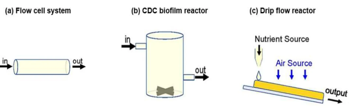
Figure 6. Scheme represent the principe of the medium flow through dynamic biofilm
models. ... 26
Figure 7. Schematic drawing of flow cell system ... 26
Figure 8. Schematic diagram of Center for Disease Control (CDC). ... 27
Figure 9. Schematic diagram of Drip flow reactor. ... 27
Figure 10. Schematic representation of the quantification of bacterial colonies (CFU)
from a biofilm. ... 32
Figure 11. Schematic representation of indirect measurements methods. ... 34
Figure 12. Schematic representation of microscopic methods... 36
Figure 13. The chemical structure of TOB and COL. ... 37
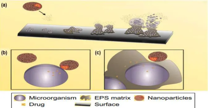
Figure 14. Schematic representation of interaction of nanoparticles in DDS with
different biofilm stages. ... 38
Figure 15. Schematic representation of NP as a drug carrier ... 39
Figure 16. Schematic representation of different types of LIP. ... 40
Figure 17. Different types of NPs used as drug delivery system. ... 52
Figure 18. Scheme of Research work ... 57
LIST OF TABLES
Table 1. In vitro biofilm models linked to gram-negative chronic lung infections ... 22
Table 2. In vivo models linked to PA chronic lung infections ... 29
Table 3. Advantages and disadvantages of the NPs formulations ... 53
LITERATURE
REVIEW
I.
Introduction
Bacteria are responsible for both acute invasive infections and chronic localized infections. Differences between these infections are often attributable to the mode of growth of the bacterium. Bacteria growing as isolated cells or planktonically are mainly associated to acute infections. For most of the chronic bacterial infections, bacteria grow in slime‑enclosed aggregates known as biofilms which are recognized as a significant cause of antimicrobial tolerance (Bjarnsholt 2013; Boisvert et al. 2016; Chirgwin et al. 2019; Costerton 1999). These biofilm infections, such as pneumonia in cystic fibrosis (CF) patients, chronic wounds, chronic otitis media, implant‑ and catheter‑associated infections, affect millions of people and increase their risk of death (González et al. 2018).In these biofilms, bacteria are protected from the environment and the concentration of antibiotics (ATBs) needed to eradicate a biofilm are up to a 1000-fold higher than the concentration needed to kill planktonic cells. Therefore, biofilm infections are not easily responsive to existing antimicrobial treatment (Høiby et al. 2010a). Reduced susceptibility in biofilms is a consequence of complex physical and biological properties with multiple factors such as resistance and tolerance against antimicrobial agents through metabolic dormancy or molecular persistence programs (Bjarnsholt 2013). Besides, mismanagement of ATBs treatment used to fight biofilm accelerates the emergence of bacterial resistance to the conventional ATBs (Høiby et al. 2015; Ridenhour et al. 2017) as well as the lack of the discovery of new antimicrobial agents drove to develop another strategies to cure the biofilm infections. In this context, the therapies currently used to combat bacterial infections must be improved in order to be able to treat biofilms, for example by using new administration strategies (Forier et al. 2014; Martin et al. 2014; Smith 2005).
For lung biofilms, promising approaches currently focus on pulmonary drug delivery systems (DDS) which, when inhaled, can improve the targeting of ATBs to lung biofilm (Ciofu et al. 2015; Forier et al. 2014). This new approach consists of inhaling ATBs encapsulated in a particulate DDS such as nanoparticles (NPs) or liposomes to improve its penetration into biofilms and to control its release into the biofilm, in order to increase its local concentration and minimize its toxicity (Sajid et al. 2014). Several excellent reviews discuss how bacteria develop biofilms and their protective mechanisms against ATBs and specific therapeutic strategies developed to combat biofilms (Bjarnsholt et al. 2010 ; Høiby et al. 2010a ; Høiby et al. 2010b ; Ciofu et al. 2015 ; Høiby et al. 2015 ; Klinger-Strobel et al. 2015 ; Koo et al. 2017 ; Fleming and Rumbaugh 2017). The main objective of this thesis was to evaluate a new DDS of lipid nanoparticles (LNP) to increase diffusion of cationic ATBs (Tobramycin (TOB) and Colistin (COL)) into lung biofilms. Our first step was to develop a Pseudomonas aeruginosa
(PA) lung biofilm model that mimics those found in chronic lung infections of CF patients and to validate this model by studying efficacy of cationic ATBs against PA biofilms and Acinetobacter baumannii (A. baumannii) biofilms.
In the general part of this work, biofilms structure will be developed as well as their mechanisms of regulation and tolerance towards ATBs. Secondly, in vitro and in vivo biofilm models found in literature will be presented before ending in the last part on drug delivery systems to target lung biofilms.
II.
Pulmonary biofilm
a. Lung biofilms and associated pathogens
Depending on the bacterial species and the surrounding environment, bacteria produce various structured biofilms with different characteristics. In the lung, there are two main types of biofilm associated with chronic infections, where the biofilm is either produced in the mucus (Bjarnsholt et al. 2009a; Høiby et al. 2010b; Sønderholm et al. 2017a) or adhered to the wall of the endotracheal tubes (ETTs) (Boisvert et al. 2016). The first type of biofilm is characterized by unattached aggregates dispersed in the airway mucus. It is mainly observed for chronic pulmonary infections with PA associated with CF, but also with bronchiectasis (Boisvert et al. 2016; Macia et al. 2014; Marsh et al. 2015). The second type is found in patients with ventilator-associated pneumonia (VAP) and can be formed by various species often forming polyspecies biofilms. Multidrug-resistant pathogens from ESKAPE group (acronym corresponding to the names of six bacterial pathogens commonly found to carry antimicrobial resistance genes (Enterococcus faecium, Staphylococcus aureus, Klebsiella pneumoniae, A. baumannii, PA, Enterobacter spp) play a dominant role in VAP etiology, and these organisms are frequently identified in ETTs biofilms (Bauer et al. 2002; Diaconu et al. 2018).
1) Mucus dispersed biofilms
Bacterial biofilms developed in mucus are non-attached biofilms that are not associated with a foreign body (Høiby et al. 2015). Their formation is accompanied by a reduction in the motility and expression of virulence genes of bacteria. In the airways of CF patients, abnormal mucus present in excess, decreases bacterial clearance and provides a nutrient-rich environment promoting the biofilm establishment (Balázs and Mall 2019; La Rosa et al. 2019; McDaniel et al. 2015). PA is the most common pathogen that persists as a mono-species biofilm suspended in the mucus of these patients, resulting in ineradicable chronic lung infections (Bjarnsholt et al. 2009a; Boisvert et al. 2016; Sousa and Pereira 2014). Therefore, in CF patients, the persistence of PA in the airways mucus as a biofilm is strongly correlated with the decline in lung function and a high rate of patient morbidity and mortality (Sousa and Pereira 2014). In chronic obstructive pulmonary disease (COPD), another chronic inflammatory lung disease in which mucus is produced in excess, no direct observation of PA biofilm formation was reported (Blasi et al. 2016; Hassett . 2014; Martínez-Solano et al. 2008). Still, the ATB resistance patterns in COPD patients are similar to those observed for CF chronic infections and isolates from lungs of COPD patients were shown to produce more in vitro
biofilm than isolate collected from the blood of the same patients, suggesting the possible presence of biofilm in these patients (Blasi et al. 2016; Rodrigo‑Troyano et al. 2016).
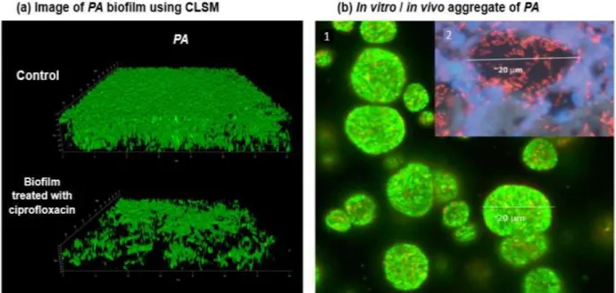
In CF patient’s airways, PA mature biofilms are characterized by small structured bacterial aggregates embedded in a self-produced viscous matrix of extracellular polymeric substances (EPS) composed of polysaccharides (mainly alginate, Psl, and Pel), extracellular DNA (eDNA), proteins such as CdrA, and lipids (Bjarnsholt et al. 2009a; Maunders and Welch 2017) (Figure 1). These aggregates which are enclosed in a second polymer matrix composed of the patient's mucus have a size ranging from 5 to 100 µm (Figure 1) (Bjarnsholt et al. 2013 ; Bjarnsholt et al. 2009; Sønderholm et al. 2018; Sønderholm et al. 2017). The polysaccharide composition of EPS changes during the biofilm life cycle. In the early stages, two exopolysaccharides named Psl and Pel are produced and serve to initiate the formation of biofilm. Psl is a neutral polysaccharide consisting of a pentasaccharide repeat containing glucose, mannose, and rhamnose. This polysaccharide is essential for the adherence to mucin surface and epithelial cell airway and promotes cell-cell interactions (Irie et al. 2017; Ryder et al. 2007). Pel is a cationic polysaccharide made of acetylgalactosamine and N-acetylglucosamine, involved in interactions between bacteria in the biofilm and in the adhesion of bacteria to mucins (Colvin et al. 2012; Irie et al. 2017; Jennings et al. 2015; Ryder et al. 2007). It is also required to initiate and maintain cell-cell interactions in biofilm environment (Colvin et al. 2012). For PAO1, Psl is the primary exopolysaccharide produced in the biofilm matrix, while it is Pel for PA14 (Jennings et al. 2015). PA strains is characterized by hyperadherence and hyperaggregation of bacteria, called small colony variants (SCVs), which are frequently isolated from various biofilms, and which constitutively overexpress Pel and Psl.
In response to infection, numerous polymorphonuclear leukocytes (PMNs) surround the PA biofilm aggregates and create a chronic inflammation (Figure 1) (Bjarnsholt et al. 2013; Bjarnsholt et al. 2009a; Høiby et al. 2010b). In mature biofilm, many of these PMNs are necrotic and constitute a massive source of eDNA that is incorporated into the biofilm matrix (Bjarnsholt et al. 2009a; Lewenza 2013). PMNs that are still alive phagocytize planktonic bacteria released by biofilms and release in turn various components such as reactive oxygen species (ROS, 𝑂2−, 𝐻2𝑂2, 𝑂𝐻.…), pro-inflammatory cytokines (IL-1α-IL-1β,IL-6, IL-17…), proteolytic enzymes (cathepsin G, neutrophil elastase) and antimicrobial peptides (LL-37, ...) (Colvin et al. 2012, Tecchio et al. 2014). PMNs also degranulate to produce web-like extracellular structures known as extracellular neutrophil traps (NETs) which contain DNA that can also be incorporated into the matrix of biofilms (Lewenza 2013). Due to their high activity, PMNs consume a lot of O2, which promotes local O2 depletion and makes the bacterial aggregates surrounded by PMNs mostly anoxic (Sønderholm et al. 2017). Furthermore, all the elements
released by PMNs, which are in part responsible for lung damage, apply selective pressure to the colonizing PA that favors the survival of mucA gene mutants, the main mutation responsible for overproduction of alginate (Boucher et al. 1997; Malhotra et al. 2018; Mathee et al. 1999; Ramsey and Wozniak 2005; Ryder et al. 2007). Indeed, alginate is a polyanionic polysaccharide composed of acid mannuronic and gluronic that act as oxygen-radical scavenger. Its overproduction protects the bacteria also from phagocytosis by PMNs and gives the colony a mucoid phenotype that is typical in ineradicable CF lung infection. As chronic infection develops in the CF airways, numerous new PA phenotypes appear and CF sputum culture can revealed the presence of mucoid cells overproducing alginate but also SCVs overproducing Pel and Psl (Maunders and Welch 2017). However, mucoid phenotype is rarely isolated from non-CF airway (Sousa and Pereira 2014). Both phenotypes are related to ATB resistance, persistence of infection, resistance to phagocytosis and poor lung function (Malone 2015).
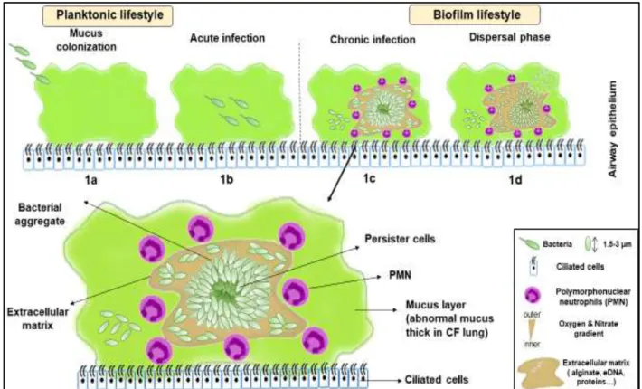
Figure 1. Schematic representation of different steps of formation of mucus dispersed biofilms. The formation begins with the mucus colonization by planktonic PA (a) leading to acute infection (b). During later stage, biofilm is mature causing chronic infection (c), forming characteristics specific to biofilm (bacterial aggregate in self-produced matrix rich in anionic polysaccharides). Finally, some PA begin to detach and the biofilm disperses (d). (Personal creation)
The establishment of mucus dispersed biofilms can be described by several stages presented in Figure 1.
First, planktonic PA colonized the mucus using flagella and Pili IV (Figure 1a) (Sousa and Pereira 2014). Then, in the early infection stage, planktonic bacteria begin to adapt to the mucus environment, in particular to the depletion of oxygen and the response of the host's immune system. In these stages, bacteria still produce virulence factors such as rhamnolipid, proteases and pyoverdine, and are still susceptible to ATBs treatment (Figure 1b) (Sousa and Pereira 2014). Real biofilm development begins at the stage of chronic infection, where bacteria are perfectly adapted to the mucus environment thanks to various characteristics including overproduction of alginate, loss of production of virulence factors and loss of mobility (loss of both pili and flagella). To date, it is unclear when the bacteria switch from planktonic to sessile lifestyle after airway colonization (Sousa and Pereira 2014)
This mature biofilm (Figure 1c) is distinguished by gradients of nutrients and oxygen that decrease from the outside to the inside of the biofilm, which creates a gradient of metabolic activity of bacteria in the same direction.
The last stage of biofilm life cycle corresponds to the dispersal phase (Figure 1d), where alterations in the mucus environment (oxygen, nutrients availability, pH) trigger the biofilm dispersion as planktonic bacteria to colonize a new region in the mucus and establish another biofilm (Kirov et al. 2007; Sousa and Pereira 2014; Woo et al. 2012). This stage is associated to a coordinate release of virulent factors from the bacteria and corresponds to acute exacerbation of the chronic infection, for which planktonic bacteria can also be found in the patient’s mucus.
2) Biofilms adhering to abiotic surfaces
Biofilms adhering to the surface of ETT found especially in patients with VAP are the main biofilms adhering to abiotic surfaces found in the lung (McDaniel et al. 2015) (Figure 2). These biofilms are likely the major microbial reservoir of VAP (Fernández-Barat and Torres 2016). Indeed, 70% of patients with VAP have similar pathogens in their lungs as those that form the biofilm on the ETT (McDaniel et al. 2015). These biofilms can be generated within hours after intubation. Patchy biofilms are generally observed 2-7 days after ventilation initiation on the ETT surface, and 87.5% of ETT surface can be covered by biofilms after 7-10 days. After 10 days, all the ETT surface is generally covered (Boisvert et al. 2016; Diaconu et al. 2018; Fernández-Barat and Torres 2016).
Compared to biofilms suspended in abnormally viscous lung mucus, these biofilms are much larger and can be up to 1000 µm thick. Consequently, steeper O2 and nutriment gradients and higher tolerance to ATBs are observed (Roberts and Stewart 2004; Stewart and Franklin 2008; Bjarnsholt et al. 2013; Høiby et al. 2015). In addition, these biofilms have a more complex architecture than those included in mucus and are often formed by several
microbial species, including different bacteria, fungi and viruses (Orazi et al. 2019; Pericolini et al. 2018). Some of the pathogens which are frequently found in ETT biofilm associated to VAP are from the multidrug-resistant “ESKAPE” group such as PA, A. baumannii, Staphylococcus aureus and Klebsiella pneumoniae (Bauer et al. 2002; Boisvert et al. 2016; Diaconu et al. 2018).
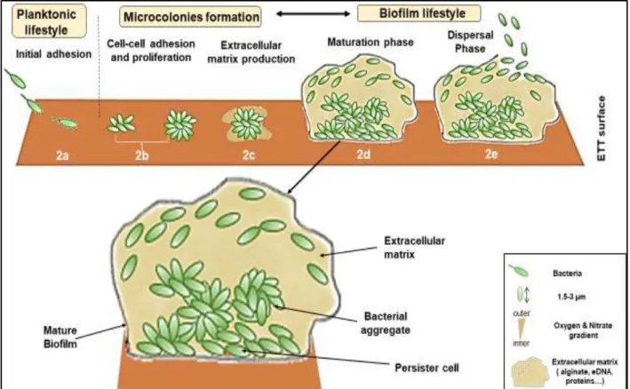
Figure 2. Schematic representation of different steps of formation of the biofilm adherent to abiotic surface. The formation begins with the adhesion of the planktonic cells on abiotic surface (such as endotracheal tube) (a). Then, the bacteria proliferate (b) and start to produce an extracellular matrix (c), leading to the formation of bacterial microcolonies. During later stage, the biofilm is mature forming characteristic structure of three-dimentionnal bacterial communities, bacterial aggregate in self-produced matrix made of polysaccharides (d). Finally, some bacterial cells start to detach allowing the dispersion of biofilm (e) (personal creation).
As for biofilm dispersed in mucus, ETT surface adhering biofilms formation is generally described by several steps presented in Figure 2. The first step is the binding to the ETT surface, mediated in PA by the flagella involved in the swimming of bacteria and by type IV pili needed for their motility by twitching (Figure 2a). The second stage corresponds to the formation of microcolonies, in which the sessile bacteria proliferate and adhere even more to the surface of the ETT, but also to each other, while producing their own EPS (Fernández-Barat and Torres 2016; Maurice et al. 2018; McDaniel et al. 2015). In this step, stability and surface adherence is improved and bacteria division is exponential (Figure 2b and 2c) (Boisvert et al. 2016; Wilson et al. 2012). Both planktonic and sessile bacteria are still present in this
phase (Rasamiravaka et al. 2015; Wilson et al. 2012). The third step concerns the maturation of the biofilm which becomes thicker and more organized (Figure 2d). Microenvironments with bacteria having different metabolic states (aerobic, micro-aerobic and anaerobic) and activity levels (high or low metabolic activity) are created (Fernández-Barat and Torres 2016; McDaniel et al. 2015; Wilson et al. 2012). The last step is the dispersal phase (Figure 2e), where certain bacteria are released from the biofilm to colonize new surfaces that allow its propagation (Bjarnsholt 2013; Diaconu et al. 2018; Fernández-Barat and Torres 2016; Macia et al. 2014). As with biofilms suspended in mucus, this step involved bacterial synchronization using quorum sensing (QS) system and the release of a significant amount of virulence factors (McDaniel et al. 2015).
The risk of development of VAP depends on many factors including the length of the intubation period that increases the risk of biofilm establishment, as well as the type of pathogens that defines the formation of early or late biofilm. For instance, VAP acquired within the first five days after intubation are more likely to be caused by ATBs sensitive bacteria such as methicillin-sensitive Staphylococcus aureus and VAP acquired more than five days after intubation caused by ATBs-resistant bacteria such as PA, A. baumanii and methicillin-resistant Staphylococcus aureus (Diaconu et al. 2018). The ETT commonly used for patient’s intubation is made of polyvinylchloride that constitutes a surface favorable to bacterial adherence and biofilm formation (Fernández-Barat and Torres 2016). Several strategies can be adapted to avoid or reduce the development of biofilms on the surface of ETT. These include replacement of ETT if contamination is suspected, endotracheal aspiration of respiratory secretions, mechanical removal of ETT secretions and biofilm using a mucus razor, and use of antiseptics such as chlorhexidine (Diaconu et al. 2018; Fernández-Barat and Torres 2016; Høiby et al. 2015). More advanced strategies involve modification of the surface of the ETT, such as ETT impregnated with antiseptics or ATBs such as sulfadiazine or silver-coated ETT, which is the only one that showed benefits in clinical trials (Diaconu et al. 2018; Høiby et al. 2015; Raad et al. 2011).
b. Bacterial biofilm regulations
Several interconnected bacterial communication mechanisms have been described as being involved in regulating the evolution of biofilms (Bjarnsholt et al. 2010; Jenal et al. 2017; Liu et al. 2017; Rasamiravaka et al. 2015; Rutherford and Bassler 2012; Valentini and Filloux 2016). Two of them, the bacterial QS system and the second intracellular messenger Bis-(3′-5′) cyclic diguanosine monophosphate (c-di-GMP) have been among the most studied.
1) Bis-(3′-5′) cyclic diguanosine monophosphare
The c-di-GMP is one of the most important bacterial intracellular secondary messenger which regulates the biofilm life cycle (Lin Chua et al. 2017; Valentini and Filloux 2016). High intracellular c-di-GMP concentration enhances biofilm formation via the reduction of motility and production of biofilm matrix, while low c-di-GMP concentration in biofilm cells leads to increased motility and biofilm dispersal. For example, high c-di-GMP level activate the production of Pel, Psl, and alginate in PA (Pestrak et al. 2018; Rasamiravaka et al. 2015; Rutherford and Bassler 2012). The concentration of c-di-GMP in bacteria is controlled by the rate of its synthesis and degradation. Its synthesis from GTP molecules is catalysed by enzymes called diguanylate cyclases and its degradation is catalysed by various phosphodiesterases (PDEs). At least five diguanylate cyclases have been described to specifically control the transition from planktonic to biofilm growth: WspR, SadC, RoeA, SiaD, and YfiN/TpbB, and several PDEs have been linked to biofilm dispersal. Mucoid or SCV phenotype of PA isolate from CF patients have high c-di-GMP concentration due to the overexpression of diguanylate cyclases (Valentini and Filloux 2016).
2) Quorum sensing
QS is a type of bacterial communication mechanism using diffusible molecules that synchronizes gene expression in response to population cell density to regulate population behaviors such as the production of virulence factors and biofilm maturation and dispersion (Lee and Zhang 2015; Solano et al. 2014). Biofilm formation is a cooperative group behavior that involves bacterial populations living embedded in a self-produced extracellular matrix. Only when the population density is high enough (formation of microcolony or microaggregate), the accumulation of the signal in the extracellular environment is sufficient to activate the QS response (Fig. 3). Structurally, QS signal molecules (or autoinducers) are low molecular weight molecules that belong to a wide range of chemical classes including acyl homoserine lactones (AHLs), furanosyl borate diesters (AI2), cis-unsaturated fatty acids (diffusible signal factor), and peptides (Solano et al. 2014). Gram-negative bacteria communicate primarily using two AHLs, (3-Oxododecanoyl)-L-homoserine lactone (3-oxo-C12-HSL or OdDHL) and N-butyryl-L-homoserine lactone (C4-HSL or BHL), which can diffuse through bacterial membranes (Figure 3). PA also produces another membrane permeable signal molecule, the 2-heptyl-3-hydroxy-4-quinolone, which has been designated as the PA quinolone signal or PQS. Four main interconnected QS systems have been identified in PA, namely: Las, Rhl, and PQS and IQS (Lee and Zhang 2015) (Figure 3). Las and Rhl use the AHLs as signaling molecules. The AHL synthases LasI and RhlI are enzymes that produce OdDHL and BHL,
respectively (Girard and Bloemberg 2008; Li and Nair 2012). When AHLs reach critical concentration, they bind to the cytoplasmic regulation proteins (LasR and RhlR), which then act as transcriptional regulators of genes of various virulence factors, genes involved in biofilm evolution, and also activate transcription of AHL synthases, providing a signal amplification mechanism via an auto-induction loop (Lee and Zhang 2015; Li and Nair 2012; Solano et al. 2014).
Figure 3. Scheme presented the characteristics of PA transition from planktonic to biofilm lifestyle, corresponding to PA microevolution during the CF airways infection. In planktonic lifestyle corresponding to the early stage of infection, PA is presented in non-mucoid phenotype and full equipped with cell-associated virulence factors including flagella and pili type IV which are serving for bacterial swimming and twitching motility, type 3 secretion systems (T3SS), and secreted virulence factors including proteases, pyoverdine, and rhamnolipid and exhibit ATBs sensitivity. In biofilm lifestyle corresponding to the chronic stage of infection, PA adapted the CF environment and exhibits a variation in characteristics including the loss of motility and overproduction of alginate, reduced of virulence factors especially the implicated in establishment for initial infection, and PA are tolerant/resistant to ATBs (expression of efflux pumps, restricted penetration…), and trigger the QS system.
Several studies have shown that PA QS systems are involved in the maturation of biofilms. Wild-type PA forms 3D heterogeneous structured biofilms on abiotic surfaces, but LasI mutant strains which cannot produce OdDHL form homogeneous dense flat biofilms (Davies et al. 1998). In PA, LasR activates the production of enzymes involved in the synthesis of the structural Pel polysaccharide of the biofilm matrix (Gilbert et al. 2009). Likewise, PA mutants with an inactive PQS system form thin biofilms containing reduced levels of eDNA compared to the wild type strain (Allesen‑Holm et al. 2006). The Rhl system has been shown to regulate the swarming and twitching motility of PA , which are essential for the development and assembly of PA as adherent abiotic biofilms (Daniels et al. 2004; Rasamiravaka et al. 2015).
In addition, the Rhl system control the production of rhamnolipids in PA (Wood et al. 2018). These glycolipids composed of rhamnose with properties of surfactant and virulence factors are involved in several crucial processes of biofilm evolution. For example, they allow the formation of microcolonies, the maintenance of open channels in the biofilm allowing the transport of nutrients and wastes, and facilitate the dispersion of the biofilm in order to colonize other areas when the environment becomes less favorable. Indeed, PA mutants that produce more rhamnolipids than wild-type exhibit hyper-detaching properties (Schooling et al. 2004; Wood et al. 2018). Lastly, Rhl system also controls the expression of the galactophilic lectins LecA and LecB, virulence factors that contribute to biofilm development in PA (Chemani et al. 2009; Diggle et al. 2006).
Dispersion of biofilm is often associated with the synchronized release by QS systems of virulence factors. In PA cells with low intracellular c-di-GMP concentrations, PQS and Rhl stimulate the production of pyocyanin and rhamnolipids that kill immune cells (macrophage and PMNs) and protect newly formed planktonic cells (Lin Chua et al. 2017; Lu et al. 2018). Mutation of the PQS system decreased the production of virulence factors such as pyocyanin, elastase, PA-IL lectin and rhamnolipids (Diggle et al. 2003; Lee and Zhang 2015; Rybtke et al. 2015).
c. Biofilm tolerance to ATBs
The reduced susceptibility of bacteria in biofilms to antimicrobial agents often leads to treatment failure and is due to both resistance and tolerance of bacteria to these agents (Ciofu et al. 2017; Høiby et al. 2010b). While resistance is associated with bacterial genetic change and increase in minimal inhibitory concentration (MIC), tolerance results in the ability of bacteria to survive antimicrobial treatment using their current genes and biofilms have specific phenotypic features related to ATBs tolerance (Verderosa et al. 2019).
1) Limitation of the ATBs transport through the biofilm
Various components of biofilms are involved in sequestration or reducing the diffusion of ATBs. Mainly, the matrix of bacterial biofilms contains several negatively charged polymers such as exopolysaccharides, proteins and eDNA, which form a mesh that can limit the penetration of certain antimicrobials into biofilms (Tseng et al. 2013). Membrane vesicles and free soluble lipopolysaccharides (LPS) are also negatively charged components that can be abundant within biofilms and can limit diffusion of some ATBs through biofilms (Mann and Wozniak 2012; Yokota et al. 2018). The reduced penetration of ATBs into biofilms is a tolerance mechanism which is considered problematic primarily for polycationic ATBs such as
aminoglycosides and polymyxins due to electrostatic interactions with negatively charged biofilm matrix components (Cao et al. 2016; Chiang et al. 2013; Chirgwin et al. 2019; Davenport et al. 2014; Du et al. 2015; Hatch and Schiller 1998; Nichols et al. 1988; Okshevsky and Meyer 2015; Torres et al. 2019; Tseng et al. 2013; Walters et al. 2003; Yokota et al. 2018). Yet, cationic ATBs such as TOB and COL are among the most widely used for treating lung infections by inhalation in CF patients. Additionally, in these patients, the thick mucus in which the PA biofilms are formed is also composed of negatively charged polymers such as mucins which can further reduce the diffusion of positively charged ATBs (Bahamondez-Canas et al. 2018; Müller et al. 2018; Torres et al. 2019).
Figure 4. Scheme representative of biofilm colony formed on infected tissues within viscous mucus layer, characteristics of a chronic infection in the CF airway, illustrating the restriction of the penetration of cationic ATBs (TOB and COL) due to their interactions with components negatively charged in mucus (eDNA and mucins) and biofilms (anionic polymers including alginate, DNA). Biofilms and mucus constitute barriers for cationic ATBs diffusion and prevent their reaching the target bacterial colonies.(Armijo et al. 2020)
i. Extracellular DNA
Depending on the type of biofilm, the eDNA present in the lung biofilms can come from two sources. First, bacteria, which actively release or secrete eDNA and also released it during bacterial lysis and the formation of outer membrane vesicles (Lewenza 2013). For biofilms
formed in mucus, eDNA also come from PMNs that surround the biofilm. For instance, in CF patients, virulence factors produced by PA such as rhamnolipids and pyocyanin lyse PMNs and release eDNA that can subsequently be incorporated into the biofilms (Alhede et al. 2009; Chiang et al. 2013; Christensen et al. 2012). In addition, during infection, PMNs release granules to form an antimicrobial matrix of extracellular fibril known asneutrophil extracellular traps, also made up of DNA. This neutrophil extracellular traps is likely a major contribution of eDNA at the site of infection (Lewenza 2013).
Extracellular DNA can bind to cationic ATBs, such as aminoglycosides and antimicrobial peptides, and contributes to antimicrobial tolerance in PA biofilm by acting as an antimicrobial shield (Chiang et al. 2013; Okshevsky and Meyer 2015). In addition, eDNA also induces an ATBs resistance due to its ability to bind and sequester divalent metal cations such as Zn2+, Mg2+, Mn2+ and Ca2+. Indeed, the sequestration of Mg2+ by eDNA activates PhoPQ and PmrAB two-component systems in PA. These systems promote the remodeling of lipid A and core region of the LPS of PA by adding 4-aminoarabinose at the end of the phosphate groups. These modifications remove negative charges of the LPS and reduce the binding of ATBs such as antimicrobial peptides, COL, and aminoglycosides to the bacterial outer membrane, increasing the tolerance to these ATBs (Hunt et al. 1995; Lewenza 2013; Mulcahy et al. 2008; Okshevsky and Meyer 2015). To improve the efficacy of cationic ATBs against in vitro and in vivo biofilms, multiple studies have evaluated the used deoxyribonucleases (DNase) to hydrolyze the eDNA and disperse the biofilms to improve ATBs efficacy (Alipour et al. 2009; Baelo et al. 2015; Deacon et al. 2015; Nair et al. 2020; Okshevsky and Meyer 2015; Tetz et al. 2009).
ii. Polysaccharides (Alginate,Psl,Pel)
In PA biofilms, the major polysaccharides produced, i.e. alginate, Pel, and Psl are critical in providing tolerance to various ATBs (Billings et al. 2013; Goltermann and Tolker-Nielsen 2017; Murakami et al. 2017). Indeed, Goltermann et al. demonstrates that overproduction of these polysaccharides in PA aggregates increases the tolerance of bacteria to TOB but also to ciprofloxacin (CIP) (Goltermann and Tolker-Nielsen 2017) . Similarly, Cotton et al. showed that treating abiotic biofilms of PA with alginate lyase increased the efficacy of 1.0 μg/mL gentamicin and 0.25 μg/mL CIP by 30% and 100%, respectively (Cotton et al. 2009). As diffusion of CIP, a relatively small and uncharged molecule, should not be limited by the matrix polysaccharides, these may indicate that the presence of these components altered the susceptibility of the bacteria in aggregates to ATBs by other mechanisms (Ciofu and Tolker-Nielsen 2019). In another study, Billings et al. showed that biofilm made by a PAO1 mutant incapable of producing Psl was 4 times more sensitive to COL than wild-type PAO1 biofilms
(Billings et al. 2013). In contrast, the sensitivity of biofilm of mutants unable to produce alginate or Pel was similar to that of wild type. It is important to mention that Pel and Psl are overexpressed in the SCV of PA often observed in CF patients, which correlate with an increased risk of failure of antipseudomonal treatment with ATBs (Goltermann and Tolker-Nielsen 2017).
Like eDNA, alginate is a polyanion, and one of the arguments to explain its efficiency in providing tolerance to biofilm, in particular to cationic ATBs, would be its ability to bind these ATBs by electrostatic interaction, causing a reduction in their diffusion rate or penetration into the biofilm (Nichols et al. 1988; Tseng et al. 2013; Walters et al. 2003). For example, PA biofilm exopolysaccharides have been shown to limit the diffusion of positively charged aminoglycosides, while neutral CIP can normally enter the biofilm. (Ciofu and Tolker-Nielsen 2019; Goltermann and Tolker-Nielsen 2017). Cao et al, showed on an alginate bead biofilm model that the binding of TOB to a multitude of sites led to a pronounced delay in its penetration into the biofilm (Cao et al. 2016). This filtering of the free TOB concentration inside biofilm beads was expected increasing the survival probability of bacteria residing in the biofilm (Bagge et al. 2004; Ciofu and Tolker-Nielsen 2019).
2) Host mucus can limit the transport the transport of ATBs
In CF patient, PA biofilms are mainly found embedded in the lung mucus (Bjarnsholt et al. 2009a). Lung mucus is a viscoelastic gel that covers the epithelium of the airways and plays a vital role in protecting the lungs against external factors such as bacteria (Müller et al. 2018). Mucins (MUC5AC and MUC5B) are polyanionic glycoproteins that give mucus its mechanical flow properties. In CF, mucus viscosity and elasticity is increased and the mucin MUC5AC is overproduced (Groneberg et al. 2002; Ridley and Thornton 2018; Witten et al. 2019). In addition, pulmonary mucus also contains eDNA and phospholipids. As for the EPS biofilm matrix, all those polyanions can bind polycationic ATBs and provide an additional barrier to their diffusion that might allow the bacteria enough time to adapt to a more tolerant state (Bahamondez-Canas et al. 2018; Ciofu and Tolker-Nielsen 2019; Müller et al. 2018; Witten et al. 2019). For example, Bos et al. showed that TOB binds to mucus with a higher affinity in the presence of eDNA, suggesting that the diffusion of TOB is significantly reduced in mixtures of biofilm/mucus (Bos et al. 2017). While the effect of mucus on reducing the diffusion of aminoglycosides such as TOB has been shown by several consistent studies (Huang et al. 2015; Iglesias and Bambeke 2020; Müller et al. 2018), its effect on the diffusion of polymyxins is more controversial. For instance, Huang et al. found that COL, polymyxin B (PMB), and TOB, were bound at around 80% to pig mucins in dialysis experiments (Huang et al. 2015). When comparing MICs measured with or without added mucin, both polymyxins showed
greater than 100-fold increases in MICs for several Gram-negative bacteria. However, other studies have shown that the efficacy of COL against abiotic biofilms of PA was much less affected by the presence of mucin or human mucus than that of TOB (Müller et al. 2018).
d. Investigated therapies specific to biofilms
Biofilm dispersion is essential to allow bacteria to escape and colonize new niches when nutrients and other resources become limited and waste products accumulate. Therefore, most bacteria can switch between biofilm and planktonic growth. This coordinated dispersal event is mediated by a decrease in concentration in the bacterial intracellular second messenger c-di-GMP (Wille and Coenye 2020). Thus, an antibiofilm strategy has emerged in which dispersing agents are used to initiate the dispersion of the biofilm and allow to eliminate the planktonic bacteria, more vulnerable than the bacteria in the biofilm, by ATBs co-administered with these dispersing agents or by the host immune defenses (Wille and Coenye 2020). As reviewed by Fleming and Rumbaugh (Fleming and Rumbaugh 2017) and Wille and Coeny (Wille and Coenye 2020), one strategy to induce biofilms dispersal is to use molecules or events that decrease the intrabacterial concentration of c-di-GMP, such as a change in concentration in a carbon source, oxygen depletion, administration of nitric oxide (NO) or molecules interacting with bacterial QS. A second strategy to trigger the dispersion of biofilms is the use of enzymes and molecules that act directly on the organization or chemical stability of components of the matrix
.
1) Agents decreasing intracellular c-di-GMP concentration
These agents more or less directly reduce the concentration of c-di-GMP in the bacteria and lead to the production of enzymes that degrade the matrix causing biofilm dispersal. It should be noted that lowering the concentration of c-di-GMP does not always lead to dispersion of the biofilm. For example, doxorubicin reduces the concentration of c-di-GMP in PA biofilms, while it increases biofilm formation by stimulating the release of eDNA (Wille and Coenye 2020).
i. Nitric oxide
NO is the most studied agent for PA biofilm dispersion (Barraud et al. 2014; Fleming and Rumbaugh 2017; Wille and Coenye 2020). At concentrations non-toxic to humans, NO activates several PDEs in PA, resulting in degradation of c-di-GMP and increased expression of enzymes such as the endonuclease EndA, which degrade the matrix (Barraud et al. 2009; Barraud et al. 2006; Wille and Coenye 2020). It also increases the motility of PA cells (Barraud
et al. 2006). Thus, NO-donor molecules have been evaluated in vitro and in vivo for PA biofilm dispersal. For example, NO‑donor sodium nitroprusside applied at 500 nM for 24h reduced the biomass of in vitro abiotic biofilms of PA by 80%. Addition of TOB at 100 µM (0.2 µg/L) caused almost-complete removal of the biofilms (Barraud et al. 2006). Similarly, the NO-releasing prodrug cephalosporin-3′diazeniumdiolate, which is activated by bacterial β-lactamases showed 70% removal of the biofilm of PA strain PA01 in vitro (Barraud et al. 2012). The prodrug was also active against CF isolates of PA, but the cephalosporin released was inactivated by the β -lactamases and additional ATB were needed to eradicate the biofilm (Soren et al. 2020). Nitroxide conjugate were also used as NO-donor. Verderosa et al. synthesized CIP -nitroxide conjugates that has shown efficacy against in vitro PA biofilms (Verderosa et al. 2017; Verderosa et al. 2016). A clinical trial using inhaled NO gas at 10 ppm in air associated to intravenous TOB combined or not to ceftazidime has been performed in CF patients. The treatment of 7 days resulted in PA biofilm clusters reduction and no adverse effects were observed (Howlin et al. 2017). TOB is known to diffuse slowly into the lungs after intravenous administration. We can assume that the combination of NO gas with inhaled TOB instead of intravenous injection could have given more promising results.
ii. Change in nutrient concentrations
As previously described, the biofilm environment is associated with nutrient limitations which reduce the metabolism of bacteria at the center of the biofilm and increase tolerance to ATBs targeting bacterial metabolism such as TOB or CIP (Bjarnsholt et al. 2005; Ciofu and Tolker-Nielsen 2019; Høiby et al. 2010a). Interestingly, an important change in availability of nutrients, such as carbon and nitrogen sources, stimulates the passage of bacteria from biofilm to planktonic lifestyle (Gjermansen et al. 2005; Sauer et al. 2004). For example, the biomass of 4-day-old PA biofilms grown on silicon tubes was reduced by 80 % by a 10-fold increase in the carbon concentration provided by succinate or glutamate in the growth medium (Sauer et al. 2004). The increase in the concentration of glutamate is sensed by the BdlA chemotaxis sensor in PA, which activates multiple PDEs and decrease the c-di-GMP concentration (Li et al. 2014). Similarly, a strong reduction of nutriments can also induce a biofilm dispersion through c‑di‑GMP signaling (Gjermansen et al. 2005). Based on these results, the effect of various excipients generally used to formulate drugs on the dispersal of biofilms and improving the efficacy of ATBs was tested (Bahamondez-Canas and Smyth 2018). Bahamondez-Canas and Smyth screened 190 potential excipients alone, and in combination with TOB against PA strains PA01 and PA14 grown as abiotic biofilms. Amino acids such as and D-alanine, L-proline, or N-acetyl-D-glucosaminitol improved significantly the antibiofilm activity of TOB.
iii. Antibiofilm peptides
Several antibiofilm peptides, natural or synthetic, have shown the ability to disperse existing biofilms (Pletzer et al. 2016). Some of these peptides are thought to indirectly act on diguanylate cyclases and PDEs to reduce intracellular c-di-GMP levels and induce biofilm dispersion. Yet, the exact mechanism has to be identified (Pletzer et al. 2016). Compared to other dispersing agents, most antibiofilm peptides also have intrinsic bacterial activity at concentrations greater than those required for dispersion, potentially allowing them to be used to eradicate biofilms without additional ATB treatment but simply by increasing their administered dose (Dostert et al. 2019; Feng et al. 2013; Overhage et al. 2008; Pletzer et al. 2016). Most of these peptides are positively charged and have a wide spectrum against Gram- negative and Gram-positive bacteria (Dostert et al. 2019; Pletzer et al. 2016). Human cathelicidin (LL-37) produced by PMNs was the first anti-biofilm peptide identified to have dispersing activity against PA and A. baumannii biofilms (Feng et al. 2013; Overhage et al. 2008). Overhage et al. has reported that LL-37 at 0.5 mg/L (1/128 of its MIC) reduced up to 40 % the thickness of PA biofilms formed on 96-well microtiter plates (Overhage et al. 2008). Chennupati et al. found that a high concentration of LL-37 (2.5 g/L) eradicated PA biofilms from the nasosinus mucosa of rabbits (Chennupati et al. 2009). However, these high concentrations of LL-37 have shown proinflammatory and ciliotoxic effects. All these findings encouraged the design and discovery of multiple antibiofilm peptides (Dostert et al. 2019; Feng et al. 2013). For example, synthetic peptides derived from bactenecin have shown disruption of abiotic biofilms of PA and A. baumannii at a sub-inhibitory concentration by inducing the degradation of alarmone guanosine tetraphosphate (ppGpp), a second messenger involved in the development of the biofilm (Dostert et al. 2019; de la Núñez et al. 2015; de la Fuente-Núñez et al. 2014). This second messenger is able to relay signal to the c-di-GMP biofilm dispersal machinery (Díaz-Salazar et al. 2017; Fontaine et al. 2018).
iv. Interaction with quorum sensing
The role of QS systems in biofilm maturation and dispersal offers another strategy that has been intensely studied for the development of a novel antibiofilm therapy using quorum sensing inhibitors (QSI) (Brackman and Coenye 2015; Rasamiravaka et al. 2015; Solano et al. 2014). Primarily, QSIs that target AHL-based QS systems in Gram-negative bacteria have been evaluated for their ability to disperse biofilms using in vitro and in vivo models (Geske et al. 2005; Ishida et al. 2007). For example, analogues of AHL, having additional aromatic fractions such as bromophenyl, have shown inhibitory activity of the Las QS system and reduced the formation of abiotic biofilm of PA PA01 (Geske et al. 2005). Analogues in which