HAL Id: tel-02491370
https://tel.archives-ouvertes.fr/tel-02491370
Submitted on 26 Feb 2020
HAL is a multi-disciplinary open access
archive for the deposit and dissemination of sci-entific research documents, whether they are pub-lished or not. The documents may come from teaching and research institutions in France or abroad, or from public or private research centers.
L’archive ouverte pluridisciplinaire HAL, est destinée au dépôt et à la diffusion de documents scientifiques de niveau recherche, publiés ou non, émanant des établissements d’enseignement et de recherche français ou étrangers, des laboratoires publics ou privés.
the control of nociceptive transmission in the dorsal
horn of the spinal cord : an optogenetic study in
different pathophysiological contexts
Franck Aby
To cite this version:
Franck Aby. Serotonergic neurons of the nucleus raphe Magnus in the control of nociceptive transmis-sion in the dorsal horn of the spinal cord : an optogenetic study in different pathophysiological contexts. Neurons and Cognition [q-bio.NC]. Université de Bordeaux, 2019. English. �NNT : 2019BORD0354�. �tel-02491370�
POUR OBTENIR LE GRADE DE
DOCTEUR DE
L’UNIVERSITÉ DE BORDEAUX
Ecole Doctorale des sciences de la vie et de la santé
Spécialité Neurosciences
Par Franck Aby
Les neurones sérotoninergiques du noyau raphé
Magnus dans le contrôle de la transmission
nociceptive dans la corne dorsale de la moelle
épinière : une étude optogénétique dans différents
contextes pathophysiologiques
Sous la direction de : Pr Pascal Fossat
Soutenue le 13 Décembre 2019
Mme. Nadjar, Agnès Professeur NutriNeuro Bordeaux Présidente
Mme. Pezet, Sophie Maître de conférences ESPCI Paris Rapportrice
Mr. Poisbeau, Pierrick Professeur INCI Strasbourg Rapporteur
Mme, Antri, Myriam Maître de conférences Neuro-Dol Clermont-Ferrand
Examinatrice
Mr. Herry, Cyril Directeur de
recherche
Neurocentre
Magendie Bordeaux
Membre invité
POUR OBTENIR LE GRADE DE
DOCTEUR DE
L’UNIVERSITÉ DE BORDEAUX
Ecole Doctorale des sciences de la vie et de la santé
Spécialité Neurosciences
Par Franck Aby
Serotonergic neurons of the nucleus raphe Magnus in
the control of nociceptive transmission in the dorsal
horn of the spinal cord: an optogenetic study in
different pathophysiological contexts
Under the supervision of: Pr Pascal Fossat
Defended on december, 13th, 2019
Mme. Nadjar, Agnès Professeur NutriNeuro Bordeaux Chair
Mme. Pezet, Sophie Maître de conférences ESPCI Paris Rapporteur
Mr. Poisbeau, Pierrick Professeur INCI Strasbourg Rapporteur
Mme, Antri, Myriam Maître de conférences Neuro-Dol Clermont-Ferrand
Reviewer
Mr. Herry, Cyril Directeur de
recherche
Neurocentre
Magendie Bordeaux
Invited member
dans le contrôle de la transmission nociceptive dans la corne
dorsale de la moelle épinière : une étude optogénétique dans
différents contextes pathophysiologiques.
Résumé :
La douleur est une sensation et une expérience émotionnelle désagréable résultant de stimulations potentiellement nuisibles pour protéger l'intégrité du corps. Un mécanisme endogène impliquant le système PAG-RVM, module la sensation de douleur en filtrant les entrées nociceptives. Un équilibre entre des influences excitatrices et inhibitrices contrôle la transmission nociceptive et une perturbation de cet équilibre conduit à l’installation de douleurs pathologiques. Dans ce travail, nous avons utilisé une approche optogénétique pour cibler spécifiquement les neurones sérotoninergiques (5-HT) du noyau du raphé Magnus (RMg) projetant sur la corne dorsale de la moelle épinière. Nous avons montré que ces neurones exerçaient une action analgésique tonique par une diminution de l'excitabilité des neurones de projection de la corne dorsale de la moelle épinière. Cet effet étant indépendant du sexe. Nous avons également observé que les neurones sérotoninergiques (5-HT) sont indirectement liés aux neurones de projection par l'intermédiaire d'interneurones inhibiteurs locaux. Puis, nous avons montré que les neurones sérotoninergiques (5-HT) du RMg recevaient des projections des neurones à somatostatine du ventro-latérale de la substance grise périaqueducale (vlPAG) exerçant une facilitation descendante de la transmission nociceptive. Fait intéressant, nous montrons que dans un modèle de neuropathie périphérique, l'action inhibitrice des neurones à sérotonine (5-HT) du RMg est transformée en influence excitatrice, aussi bien chez les mâles que les femelles, en raison d'un déplacement de l'équilibre du chlore au sein de la moelle épinière. Ces résultats suggèrent que la même voie descendante peut être à la fois excitatrice et inhibitrice dans des conditions pathologiques, révélant des informations cruciales sur les changements à long terme associés à la douleur chronique.
Mots clés :
Douleur, moelle épinière, réseaux neuronaux, 5-HT, SST, KCC2, WDR,excitabilité, douleur neuropathique, genre, électrophysiologie in vivo, optogénétique, pharmacologie.
Unité de recherche
Institut Interdisciplinaire de Neurosciences CNRS UMR5297
control of nociceptive transmission in the dorsal horn of the spinal
cord: an optogenetic study in different pathophysiological
contexts
Abstract:
Pain is an unpleasant sensation and emotional experience elicited by potentially harmful stimulations to protect the integrity of the body. An endogenous mechanism involving the PAG-RVM modulatory system control pain sensation by filtering nociceptive inputs. A balance between both excitatory and inhibitory influences control nociceptive transmission and impairment in this balance leads to the development of pathological pain. In the present study, we used an optogenetic approach to specifically target serotoninergic neurons (5-HT) that projected to the dorsal horn of the spinal cord. We showed that these neurons exerted a tonic analgesic action through a decreased excitability of projection neurons of the dorsal horn of the spinal cord. This effect is gender independent. We also observed that 5-HT neurons are indirectly connected to projection neurons through local inhibitory interneurons. Then, we showed that 5-HT neurons of the RMg received descending inputs from the SST neurons of the ventro-lateral part of the periaqueductal gray (vlPAG) that exerted downward facilitation on pain transmission. Interestingly, we show that 5-HT inhibitory action is switched to an excitatory influence in a model of peripheral neuropathy due to a spinal chloride equilibrium shift. These results suggest that the same descending pathway can be both excitatory and inhibitory upon pathological conditions, providing crucial insights about long-term changes associated with chronic pain.
Keywords:
Pain, spinal cord, neural networks, 5-HT, SST, KCC2, WDR, excitability,neuropathic pain, gender, in vivo electrophysiology, optogenetics, pharmacology.
Unité de recherche
Institut Interdisciplinaire de Neurosciences CNRS UMR5297
La douleur est un système d'alerte essentiel qui protège l’organisme contre les lésions tissulaires réelles ou potentielles. La sensation de douleur est généralement désagréable mais s’atténue une fois le corps averti du danger et la zone endommagée sûre. Ce contrôle de la douleur est dû à l'existence d'un système endogène capable de moduler la transmission douloureuse. Ce système endogène est mobilisé aussi bien dans un contexte de stress aigu, dans lequel la sensation de douleur peut être efficacement diminuée ou supprimée, que par un contrôle inhibiteur nociceptif diffus (DNIC). Le système de contrôle endogène de la douleur est principalement constitué par une voie descendante contrôlant la transmission nociceptive directement au niveau de la corne dorsale de la moelle épinière, premier relais par lequel passent les informations nociceptives. Il est considéré dans la littérature que les voies descendantes excitatrices et inhibitrices contrôlent toutes deux la transmission nociceptive. Cependant, l'équilibre entre les deux influences est altéré dans le cas de douleur pathologique, ce qui favorise les facilitations plutôt que les inhibitions. La voie descendante de la douleur implique différents noyaux du tronc cérébral qui projettent directement ou indirectement sur la moelle épinière. Les projections indirectes, notamment ceux provenant de la partie ventro-latéral de la substance grise périaqueducale (vlPAG), ont généralement un relais sur le bulbe rostral ventromedial (RVM) d’où projettent des influences inhibitrices et excitatrices sur la corne dorsale de la moelle épinière. Au sein de la RVM, les cellules sérotoninergiques (5-HT) constituent une importante population neuronale se limitant à des noyaux précis (noyau obscurus, paragigantocellularis et raphé Magnus). Les neurones 5-HT du noyau raphé Magnus (RMg) projettent vers la partie dorsale de la moelle épinière et sont impliqués dans le contrôle de la douleur. Ce contrôle de la transmission nociceptive est fortement dépendant du contexte physiopathologique. De plus, il existe une grande diversité de récepteurs 5-HT affectant différemment les neurones de la corne dorsale. Cependant, à ce jour, le rôle exact des
neurones 5-HT du RMg dans la transmission nociceptive n'est toujours pas éclairci. Afin de déterminer le rôle exact des neurones 5-HT du RMg dans la transmission nociceptive, nous avons utilisé une approche à la fois virale, pharmacologique et
électrophysiologique chez les souris ePet-cre qui exprime la cre-recombinase spécifiquement dans les neurones à 5-HT.
Tout d’abord, nous avons confirmé la bonne expression de la cre-recombinase limitée aux neurones 5-HT dans le raphé Magnus (RMg) ainsi que dans le raphé dorsal (DR) et le raphé médian (MR) en croisant des souris ePet-cre à des souris rapportrices Ai9. Par un marquage
localisation avec des neurones cre positif s’élevant à 83,8%. Nous avons également observé une population dense de fibres 5-HT dans la corne dorsale de la moelle épinière confirmant ainsi les projections 5-HT sur la moelle épinière. Puis, en utilisant une souris Gad67-GFP, nous avons confirmé que les neurones 5-HT du RMg ne sont pas des neurones GABAergiques mais présentaient un degré élevé d'interaction avec eux.
Ainsi nous nous sommes rendus compte que les souris ePet-cre nous donnaient un outil crucial pour étudier le rôle précis de ces neurones dans la transmission nociceptive.
Dans un second temps, afin de valider notre approche virale, nous avons réalisé une injection stéréotaxique d’un virus inductible permettant ainsi l’expression d’opsine ChR2 dans les neurones 5-HT du RMg. Puis trois semaines après, nous avons réalisé des enregistrements de neurones 5-HT exprimant les opsines en patch-clamp en courant imposé sur tranches de RMg. Nous avons observé que la stimulation optogénétique à 475nm induisait d’une part une dépolarisation neuronale rapide et reproductible et qu’un mode de stimulation à 5Hz/5ms induisait d’autre part un train de potentiels d'action suivant fidèlement la stimulation optogénétique. En revanche, la stimulation optogénétique à 525nm induisait une forte hyperpolarisation qui s’estompait avec l’arrêt de la stimulation confirmant ainsi que notre
approche optogénétique nous permettait de moduler l'activité des neurones à 5-HT du RMg. De plus, en accord avec la littérature, nous avons observé des projections 5-HT du RMg
visibles dans les couches profondes de la corne dorsale de la moelle épinière.
Afin d’étudier les conséquences d’une manipulation des neurones 5-HT du RMg, nous avons effectué une stimulation optogénétique de ces neurones à l’aide de fibres optiques placées aussi bien au-dessus du RMg que de la partie lombaire de la moelle épinière. En comportement, chez des souris 5-HT cre en absence de lésion nerveuse, nous avons observé que l'inhibition optogénétique sélective des neurones 5-HT que ce soit au-dessus de la RMg ou de la corne dorsale spinale induisait une importante hypersensibilité mécanique et thermique confirmant ainsi que les neurones RMg 5-HT étaient toniquement actifs et jouaient un rôle crucial dans le contrôle de la transmission nociceptive. En revanche, l'activation optogénétique des neurones 5-HT ou de leurs projections, au-dessus de la RMg ou de la corne dorsale de la moelle épinière, engendrait une importante analgésie mécanique et thermique. Par conséquent, nous avons pu
en conclure que les neurones RMg 5-HT exerçaient une inhibition tonique descendante de la transmission nociceptive constituant ainsi un des contrôle inhibiteur descendant endogène de la douleur.
avons comparé la manipulation des neurones 5-HT chez les sujets mâles et femelles et n'avons observé aucune différence entre les deux.
Pour confirmer les cibles spinales de la manipulation des neurones 5-HT, nous avons effectué des enregistrements électrophysiologiques in vivo des neurones de la corne dorsale en ciblant spécifiquement les neurones à convergence (WDR) qui sont des neurones de projection recevant entre autres des entrées nociceptives des fibres C. Nous avons d'abord évalué la conséquence d'une inhibition des neurones RMg 5-HT sur l'activité des WDR, et nous avons observé que l'inhibition optogénétique des projections 5-HT entraînait une augmentation de l'activité spontanée des WDR et de leur réponse aux entrées nociceptives de fibres C ainsi qu'une augmentation de leur capacité à être sensibilisé. D'autre part, l'activation optogénétique des fibres descendantes RMg 5-HT directement au-dessus de la moelle épinière induisait une diminution significative des réponses évoquées des neurones WDR aux entrées nociceptives et une diminution significative de leur capacité à être sensibilisés. Par conséquent, nous
montrons que les neurones 5-HT du RMg projetant sur la corne dorsale de la moelle épinière, inhibent constitutivement la transmission nociceptive en diminuant l'excitabilité des WDR.
Dans le but de déterminer les cibles des neurones 5-HT du RMg sur les microcircuits de la corne dorsale, à l’aide de marquages immunohistochimiques de la tryptophane hydroxylase 2 (TPH2), chez des souris Gad67-GFP, nous avons observé des potentiels boutons synaptiques de terminaison de neurones 5-HT sur des neurones GABA dans les couches profondes de la corne dorsale de la moelle épinière. Pour confirmer ces connexions au réseau inhibiteur local, nous avons dans un premier temps comparé, chez des souris 5-HT cre injectées avec un AAV inductible marqué à la GFP, les appositions entre les neurones GFP positifs et les neurones excitateurs (TLX3) ou inhibiteurs (PAX2) et nous avons constaté que les projections 5-HT sont nettement plus en contact avec les interneurones inhibiteurs. Puis, nous avons confirmé la présence de contacts réels entre les neurones RMg 5-HT et les neurones inhibiteurs, en utilisant des souris Gad 67-GFP*5-HT cre dans lesquelles SynMYC-revWPRE (marqueur de terminaison synaptique) a été exprimé dans les neurones RMg 5-HT. À l’aide d’un immunomarquage sur une fine coupe transversale de la partie lombaire de la moelle épinière, nous avons trouvé des boutons synaptiques des projections 5-HT sur les neurones à GABA et à parlvalbumine. Dans un second temps, afin de déterminer le rôle fonctionnel de ces contacts, nous avons éliminé les inhibitions induites par les neurones GABAergique/glycinergiques dans
optogénétique des projections RMg 5-HT n’induisait plus une inhibition de la transmission nociceptive. En effet, en réalisant un enregistrement électrophysiologique associé à une stimulation optogénétique des projections 5-HT au-dessus de la moelle épinière, nous avons observé qu’un blocage de la neurotransmission GABAA/GlyR suite à une injection intrathécale de picrotoxine, n'induisait plus de diminution de la réponse évoquée des WDR aux stimuli nocifs. Par conséquent, nous avons montré que l'inhibition tonique descendante de 5-HT
du RMg sur la transmission nociceptive passe par une excitation directe des interneurones inhibiteurs GABAergic/glycinergic dans les couches profondes de la moelle épinière qui à leur tour inhibent les neurones WDR de la corne dorsale de la moelle épinière.
Dans un deuxième temps, en collaboration avec l'équipe de Cyril Herry, nous avons essayé de déterminer comment l'activité des neurones RMg 5-HT était modulée. Pour ce faire, à l’aide d’immunomarquages nous avons observé des potentiels boutons synaptiques de terminaison de neurones GABAergiques, glutamatergiques et exprimant de la somatostatine (SST) sur des neurones 5-HT. Nous avons d'abord établi le rôle fonctionnel des neurones
SST du vlPAG sur la transmission nociceptive spinale en utilisant des souris SOM-IRES-cre. Dans une approche comportementale, nous avons effectué des manipulations
optogénétiques de ces neurones en utilisant des fibres optiques implantées bilatéralement au-dessus du vlPAG. L'inhibition optogénétique des neurones SST a provoqué une importante analgésie mécanique et thermique, ce qui démontre que les neurones SST du vlPAG exercent une facilitation tonique descendante de la transmission nociceptive. Cet effet facilitateur est confirmé par l'activation optogénétique des neurones SST qui provoque une hypersensibilité mécanique et thermique significative. Par conséquent, nous déterminons que les neurones
SST du vlPAG exercent une facilitation tonique descendante sur la transmission nociceptive qui est opposé à l’action des neurones 5-HT sur la transmission nociceptive.
Ensuite, nous avons réalisé des enregistrements électrophysiologiques des neurones de la corne dorsale, centré également sur les neurones à convergence. Nous avons observé que l'inhibition optogénétique bilatérale des neurones SST du vlPAG induisaient à la fois une diminution significative de la réponse des neurones WDR en réponse aux entrées nociceptives ainsi qu’une diminution significative de leur capacité de sensibilisation. En revanche, l'activation optogénétique bilatérale des neurones SST du vlPAG a induit une augmentation significative de la réponse WDR aux entrées de fibres C nociceptives ainsi qu'une augmentation de leur capacité à être sensibilisés. Par conséquent, nous avons montré que les neurones SST
de la corne dorsale.
Puis, afin de déterminer les circuits neuronaux impliqués dans cet effet, nous avons évalué, dans un premier temps, les cibles des neurones SST vlPAG dans la RVM et en particulier dans le RMg. En développant une stratégie virale et un marquage immunohistochimique, nous avons observé une apposition entre les projections SST du vlPAG et les neurones 5-HT de RMg projettant sur la moelle épinière. Nous avons également observé l’existence de projection de neurones SST du vlPAG en faible quantité dans la corne dorsale de la moelle épinière, en particulier dans la couche superficielle. On peut donc conclure que parmi les projections du vlPAG sur le RMg, il existe un circuit de neurones SST du vlPAG qui
projettent vers des neurones 5-HT du Raphé Magnus projetant sur la moelle épinière (voie indirecte). De plus, certains neurones SST établissent aussi des connections directes de longue portée sur la moelle épinière (voie directe).
Afin de déterminer le rôle fonctionnel des différentes projections (directe et indirecte) des neurones SST du vlPAG sur la transmission nociceptive, nous avons tout d’abord évaluer le rôle fonctionnel du circuit SST du vlPAG - 5-HT du RMg sur la transmission nociceptive (voie indirecte), chez des souris SOM-IRES-CRE en comportement, nous avons réalisé une activation optogénétique des projections SST du vlPAG au-dessus du RMg et observé une hypersensibilité mécanique et thermique significative. Puis, nous avons observé que l'activation optogénétique des projections SST du vlPAG au-dessus de la RMg entraînait également une hyperexcitabilité des neurones WDR. Cependant, en évaluant le rôle fonctionnel des projections directes des neurones SST du vlPAG sur la moelle épinière par des enregistrements électrophysiologiques unitaires ciblant les WDR associés à une stimulation optogénétique au-dessus de la moelle épinière, nous n'avons observé aucun changement dans l'excitabilité des WDR. Par conséquent, nous pouvons en conclure que les neurones SST du vlPAG exercent
une facilitation tonique descendante sur la transmission nociceptive via une action probable sur des neurones 5-HT du RMg.
Dans un troisième temps, nous avons étudié les conséquences de la modulation des neurones 5-HT du RMg sur la transmission nociceptive en condition neuropathique.
La 5-HT est connue pour faciliter la transmission de la douleur dans un contexte pathologique. Nous avons utilisé le modèle de neuropathie périphérique de lésions nerveuses partielles (SNI) pour étudier les conséquences de la manipulation optogénétique des neurones 5-HT du RMg sur la transmission nociceptive. Nous avons d'abord confirmé que la procédure SNI provoque une hypersensibilité mécanique et thermique à la douleur. Il est intéressant de noter que suivant
optogénétique des neurones 5-HT ou de leurs projections au-dessus de la RMg ou de la corne dorsale de la moelle épinière respectivement ne produisent plus aucun changement dans la sensibilité mécanique et thermique, ce qui suggère que dans un contexte pathologique, les neurones 5-HT du RMg perdent leur effet analgésique tonique. En revanche, l'activation optogénétique au-dessus de ces mêmes neurones ou de leurs projections, curieusement, a induit une diminution significative du seuil de douleur mécanique et de la latence thermique de la douleur. Cet effet était également indépendant du sexe puisque aucune différence n'a été
observée chez les mâles et les femelles testés. Ainsi, dans la douleur neuropathique, les neurones 5-HT du RMg perdent leur effet analgésique et exercent une facilitation descendante sur la transmission de la douleur. En effectuant des enregistrements
electrophysiologiques, nous avons montré que l'inhibition optogénétique des projections 5-HT du RMg n’engendrait aucun changement dans l'excitabilité des WDR, tandis que l'activation optogénétique des projections 5-HT du RMg induisait une hyperexcitabilité des WDR. Par
conséquent, en cas de douleur neuropathique, nous avons montré que les neurones 5-HT se projetant sur la corne dorsale facilitent la transmission nociceptive en augmentant la réponse des WDR aux entrées de fibres C, ce qui entraîne une hyperexcitabilité des WDR.
Ensuite, nous avons étudié si la mise en place de la neuropathie pouvait induire des modifications des cibles des neurones 5-HT sur la corne dorsale de la moelle épinière pouvant ainsi expliqué l’effet opposé de ces neurones à 5-HT sur la transmission nociceptive. Nous avons constaté que les fibres 5-HT se projettent toujours principalement sur les interneurones inhibiteurs. Ayant précédemment montré que l’effet analgésique des neurones à 5-HT en condition de douleur aiguë dépend des interneurones inhibiteurs et des courants chlorure médiés par les récepteurs canaux GABA/glycine, l’homéostasie des ions chlorures des interneurones
inhibiteurs de la corne dorsale pourrait être impliquée dans la facilitation médiée par les neurones 5-HT du RMg en condition de douleur neuropathique.
Dans un modèle de douleur neuropathique incluant le SNI, il a été démontré que les mécanismes de désinhibition médiés par une déficience dans l’activité des co-transporteurs (Na+-K+-Cl-) KCC2 (Doyon, 2011 ; Kaila et al., 2014) sont responsables d'une partie de l'hyperexcitabilité neuronale et de l'hypersensibilité de la douleur (Coull et al., 2005 ; Beggs et al., 2012). Nous avons donc évalué la conséquence du changement de l'équilibre en
chlorure des fibres afférentes ou des neurones WDR sur l'entraînement sérotoninergique opposé observé chez les souris témoins et les souris SNI, en utilisant une approche pharmacologique pour stimuler le co-transporteur KCC2 chez la souris SNI. En effet,
une hypersensibilité mécanique et thermique, la même stimulation optogénétique des projections 5-HT du RMg chez les mêmes animaux provoque une analgésie mécanique et thermique significative, absente lors de l’utilisation du véhicule seul. Cet effet inhibiteur n'est pas lié au sexe puisqu’il est équivalent chez les mâles et les femelles. Ainsi, dans les conditions neuropathiques, nous avons montré que la facilitation induite par la RMg 5-HT est due au déséquilibre du gradient de chlore résultant de la diminution de l'activité du co-transporteur KCC2 et qu'une augmentation de l’activité du co-transporteur KCC2 peut inverser les conséquences du SNI. Puis, nous avons observé que la superfusion de CLP290 au-dessus de la moelle épinière supprime l'augmentation de l'excitabilité des WDR induite par l'activation optogénétique des projections 5-HT au-dessus de la moelle. Par conséquent, dans des
conditions de douleur neuropathiques, nous avons montré que l'hyperexcitabilité WDR induite par la 5-HT du RMg est une conséquence de la diminution de l'activité du cotransporteur KCC2.
Enfin, nous avons évalué les conséquences d'un blocage de KCC2 chez des souris naïves par injection ip de furosémide. Nous avons observé que l'activation optogénétique des projections 5-HT du RMg provoquait une hypersensibilité mécanique et thermique. Cet effet était équivalent chez les mâles et les femelles.
En conclusion, la manipulation de KCC2 a orienté le signe de l'influence
descendante de 5-HT sur la transmission nociceptive, qui passe de l'inhibition de la douleur à la facilitation dans un contexte pathologique. L'ensemble de ces résultats suggère fortement que les changements dans l'équilibre en chlorure de la corne dorsale de la moelle épinière des souris neuropathiques influencent le contrôle de la douleur endogène.
Je tiens tout d’abord à remercier sincèrement les membres du jury de thèse : le Dr. Pierrick Poisbeau et le Dr. Sophie Pezet pour avoir accepté de juger ce travail et d’en être les rapporteurs, Dr. Antri Myriam pour avoir bien voulu examiner ce travail, Dr. Herry Cyril pour cette collaboration ainsi que toute l’assistance et conseils apportés au cours de ces années et le Dr. Nadjar Agnès pour avoir accepté de présider cette soutenance. Au détour d’un congrès, d’un symposium ou d’une conversation privée, comme ce fut déjà le cas par le passé, j’aurai toujours plaisir à vous saluer et discuter de nos travaux.
Je tiens à exprimer ma profonde gratitude et mon estime au Dr Marc LANDRY, directeur du laboratoire, qui m’a chaleureusement accueilli au sein de son équipe et qui s’est toujours rendu disponible pour échanger et partager que ce soit sur des sujets de recherche que sur des sujets relatifs à l’expérience de la vie.
Je remercie bien évidemment tous les membres de la Team LANDRY, en commençant par notre aîné le Pr. André CALAS, pour toute l’expérience partagée, les conseils prodigués ainsi que pour toutes ses anecdotes. Merci au Dr. Yves LE FEUVRE, pour ses conseils et son aide, au Dr. Alexandre FAVERAUX, pour tous les moments de partage autour d’une table ou d’un ballon de basket, au Dr Christel BAUDET pour tous ses conseils, les moments de rigolade et de partage.
Je tiens à adresser des remerciements bien particuliers au Dr Rabia BENAZZOUZ ; je n’aurai que trop peu de mots pour définir tout ce qu’elle a su m’apporter, je me contenterai juste de lui dire merci, merci d’être qui elle est et merci pour tous ces moments passés et j’espère à venir. Merci Rabia, merci pour tout.
Je porte une attention toute particulière à mon cher et tendre Directeur de thèse Pascal FOSSAT pour « TOUTE » la « PATIENCE » dont il a fait preuve durant ces 5 années. Merci de t’être autant investi durant toutes ces années. Outre le fait de m’avoir enseigné l’art délicat de l’électrophysiologie unitaire in vivo, je te remercie pour toute la rigueur scientifique inculquée, pour ton soutien en toutes circonstances et ce malgré les très nombreux « IL VA
FINIR PAR ME TUER UN JOUR ». Merci d’avoir répondu présent à toutes ces idées les
plus farfelues les unes que les autres que j’ai pu te présenter.
Sur un plan plus personnel, merci pour ton amitié, tes conseils, ton aide et encore une fois tout ton soutien. Merci d’être mon Pascal (pour le reste on se verra en aparté).
Yazid, Amanie, Mariam, Nino, Constantin, Laura, Anthony, et j’en oublie certainement. Et merci à ceux qui le partagent encore et qui prendront la relève : Zoé, Yadali, Cynthia et Thibault, merci pour ces moments de joie et de folie.
Un merci tout particulier à Sara et à Walid qui ont fait leur stage de master1 et 2 sur le projet 5-HT, merci pour votre aide et merci d’avoir été des amours.
Je n’oublie pas ceux qui nous ont précédés ; Houda et ses petites questions, Otmane mon frère pour tous ces moments de partage, de souffrance, de rigolade et j’en passe, Joao, qui a commencé sa thèse en même temps que moi et à qui je souhaite le meilleur pour la suite, Olivier pour son amitié et son aide, Maria, Charline et Anaïs pour tous nos moments d’échange et de partage, Sébastien pour nos discussions, nos matchs de basket et pour son aide.
Petite pensée pour tous les thésards de 3ème ou 4ème année qui ont soutenu ainsi que ceux qui vont le faire dans les années à venir (Thiago, Thomas et j’en oublie) avec une attention toute particulière pour mon Vladimir, merci à toi d’être qui tu es.
Un grand merci aux autres équipes IINS pour leur aide, avec une mention spéciale pour la TEAM MULLE (remerciements particuliers à Séverine, Gael, Dario, Noel, Sandrine), la TEAM GAMBINO (Frédéric, Elisabetha, Nicolas), la TEAM GROC (Julien, François, Joana), la TEAM ROUX (Camille, Edith, Pascal, Tiphaine et Lisa) et la TEAM HUMEAU (Urielle, Yann et anciennement Hajer).
Un grand merci à la plateforme in vivo et plus particulièrement à Audrey, Mélissa Amandine, Guillaume, Laetitia et Elisabeth qui ont pris soin de nos lignées de petites souris et pour le super travail qu’ils font au quotidien.
Je remercie également Christel (merci pour tout), Fabrice, Sébastien, Magali, Monica du BIC pour m’avoir formé et apporté leur aide sur l’utilisation des différents plateformes et logiciels, Mélina et Sabrina pour toute leur expérience partagée en microscopie électronique. Merci pour ces moments de fous rires.
Je tiens à remercier chaleureusement Rémi, pour nos moments d’échange et d’entraide. Merci également à Jessica et sa relève pour l’énorme travail qu’ils effectuent et ont effectué au sein de l’institut et pour leur aide précieuse. Merci de nous mettre dans d’aussi bonnes conditions de travail !
Merci à toute l’équipe administrative; Cédric, Mélanie, Marie-France, Marie-Noelle, Arlette, Martine, Pauline, Philippe, Laurent et jean François qui font un travail remarquable et sont toujours disponibles pour nos nombreuses questions et autres angoisses au quotidien.
Un grand merci aux autres équipes du Campus notamment celles de Magendi avec une attention toute particulière pour l’équipe FRICK (Mélanie et Andréas), l’équipe HERRY (attention spéciale à Cyril, Nanci, Ha Rang, Suzana, Marianne, Valentine), l’équipe OLIET (attention spéciale à Aurélie, Philippe, Aude, Nadège, et Stéphane, l’équipe de l’IMN avec une mention spéciale pour la Team BENAZZOUZ (Abdelhamid BENAZZOUZ, Lamiaa, Frédéric et Kéri-Ann), l’équipe de l’école des neurosciences (Sandrine, Antonella, Mariella…). Merci à vous pour tous ces moments partagés au cours de cette thèse.
Un grand merci à vous mes intimes ; ceux qui m’accompagnent depuis ma toute petite enfance mais aussi ceux qui ont croisé mon chemin un peu plus tard et qui ne l’ont plus quitté depuis. Merci pour votre présence, vos encouragements, votre soutien, vos appels et votre amour. On est ensemble !
Elsa, merci pour ton soutien, ton amour, ta folie et ta joie de vivre qui rendent mon quotidien tellement plus agréable. Je te remercie de bien vouloir me suivre dans ma folie, merci de savoir me canaliser et lire en moi. Merci d’avoir voulu m’accompagner dans cette expérience aussi bien professionnelle que personnelle.
Spéciale dédicace à ma famille, merci à vous tous pour tout ce que vous avez fait et continuez de faire pour moi. Merci pour vos conseils, votre soutien, votre amour, votre force et votre présence au quotidien à mes côtés. Que dire de plus que vous ne savez déjà, sachez juste que je vous aime toujours un peu plus que la vieille et un peu moins que le lendemain et que cette thèse est aussi la vôtre.
Pour terminer, je tiens à partager une pensée toute particulière pour ceux qui m’ont quitté (Mes amours M. et Mme ALEXANDRE, Tonton Johnson, Tonton et Tantie Cole, Tantie Marina et vous mes ami(e)s parti(e)s si tôt…). Merci à vous pour tout ce que vous avez fait
pour moi ; merci pour votre amour, votre soutien, votre présence… Sachez que je vous aime et que les promesses faites seront tenues.
Avant- propos
L’ensemble des travaux de thèse présenté dans ce manuscrit a été réalisé au sein de l’Institut Interdisciplinaire de Neurosciences CNRS UMR5792 dirigé par Dr. Daniel Choquet dans l’équipe « Mécanisme central de la sensibilisation à la douleur » dirigée par le Pr. Marc Landry sous la direction du Pr. Pascal Fossat et les résultats obtenus au cours de ce projet sont en cours de publication. Cependant au cours de ma thèse, j’ai contribué à la réalisation d’autres projets qui ont donné lieu aux publications suivantes :
Publications
Causal link between developmental n-3 PUFA deficiency and motivation deficits.
Ducrocq, F and Walle, R and Contini, A and Oummadi, A and Caraballo, B and Van der Veldt, S and Boyer, M and Aby, F and Tolentino-Cortez, T and Helbling, JC and Martine, L and Grégoire, S and Cabaret, S and Vancassel, S and Layé, S and Kang, JX and Fioramonti, X and Berdeaux, O and Barreda-Gómez, G and Masson, E and Ferreira, G and Ma, D W. L. and Bosch-Bouju, C and De Smedt-Peyrusse, V and Trifilieff, P.
En revision pour Cell Metabolism, DOI: 10.2139/ssrn.3382436
Windup of Nociceptive Flexion Reflex Depends on Synaptic and Intrinsic Properties of Dorsal Horn Neurons in Adult Rat.
Aby, F; Bouali-Benazzouz, R; Landry, M; Fossat, P.
Preprints 2019, 2019100006 (doi: 10.20944/preprints201910.0006.v1).
Inflammatory-induced spinal dorsal horn neurons hyperexcitability is mediated by P2X4 receptors.
Aby F, Whitestone S, Landry M, Ulmann L, Fossat P.
Pain Rep. 2018 May 23;3(3):e660. doi: 10.1097/PR9.0000000000000660.
Calcium signalling through L-type calcium channels: role in pathophysiology of spinal nociceptive transmission.
Roca-Lapirot O, Radwani H, Aby F, Nagy F, Landry M, Fossat P.
Br J Pharmacol. 2018 Jun;175(12):2362-2374. doi: 10.1111/bph.13747. Epub 2017 Mar 24. Review.
Group I metabotropic glutamate receptor plasticity after peripheral inflammation alters nociceptive transmission in the dorsal of the spinal cord in adult rats.
Radwani H, Roca-Lapirot O, Aby F, Lopez-Gonzalez MJ, Benazzouz R, Errami M, Favereaux A, Landry M, Fossat P.
LIST OF ABBREVIATIONS ... 19
I. INTRODUCTION... 21
A. PAIN ... 21
1. Concept and Definition ... 21
2. Pain physiology ... 26
Physiological pain ... 26
Sensitization ... 27
Pathological Pain ... 28
Pain State Classification ... 29
Multidimensional Classification of Pain ... 33
3. Anatomy and physiology of the pain ... 34
From nociception to pain sensation ... 34
Endogenous pain modulation mechanism ... 51
B. RAPHE MAGNUS DESCENDING CONTROL ... 55
1. The Periaqueductal Gray ... 55
2. Nucleus Raphe Magnus (RMg) of the RVM ... 56
The Serotonin 5-HT ... 58
Other neurotransmitters ... 81
3. Pain modulation as part of adaptative responses to physiological challenges ... 82
C. AIM OF THE THESIS ... 83
II. MATERIALS AND METHODS ... 84
A. EXPERIMENTAL APPROACHES ... 84
1. Ethical Statement ... 84
2. Animals ... 84
Epet-Cre (+/-) mice (5-HT-cre mice) ... 84
5-HT cre* Ai9 tdTomato mice ... 85
GAD 67-GFP* 5-HT cre mice ... 85
SST-Cre (+/+) mice and SST-Cre (+/-) mice ... 86
3. Surgical procedure ... 86
Viral and tracer strategy ... 86
Fibre Optic Cannula Implantation ... 90
Pathological pain model: Spared Nerve Injury ... 94
4. Pain Behaviour ... 95
Mechanical Sensitivity ... 95
Heat assessment ... 97
5. Pharmacological approach ... 97
Intraperitoneal injection of furosemide or vehicle ... 97
Intrathecal injection of Picrotoxin, Granisetron or vehicle ... 98
Per Os administration of CLP 290 or vehicle ... 98
6. Electrophysiology recording ... 98
In vivo extracellular recordings ... 98
In vitro Patch-clamp recordings ... 99
7. Anatomical analysis ... 101
Virus expression ... 101
Immunohistochemical study ... 102
8. Statistical analysis ... 105
9. Table of Resources ... 106
B. RESULTS OF EXPERIMENTAL APPROACHES ... 109
1. Validation of animal model ... 109
Characterisation of Cre-recombinase in the RMg ... 109
2. Validation of our viral strategy ... 113
3. Assessment of the optical fibre implantation on mechanical modality ... 117
1. Consequences of optogenetic manipulation of 5-HT neurons in acute pain ... 121
Inhibition of RMg 5-HT neurons induces mechanical hyperalgesia, mechanical allodynia and thermal hyperalgesia ... 121
Activation of RMg 5-HT neurons induces mechanical and thermal analgesia ... 123
2. Consequences of optogenetic manipulation of 5-HT neurons on dorsal horn neurons. ... 125
RMg 5-HT neurons modulation modify WDRs integration property to noxious stimuli ... 126
3. RMg 5-HT inhibitory influence is not gender dependent ... 129
4. Target of RMg 5-HT neuron on the dorsal horn of the spinal cord ... 130
RMg 5-HT descending inhibition on pain transmission is mediated by dorsal horn inhibitory interneurons. 136 RMg 5-HT descending action on GABAergic/glycinergic inhibitory interneuron does not pass through 5-HT3 receptors activation. ... 138
B. DESCENDING INFLUENCE OF SST NEURONS OF VLPAG ON PAIN TRANSMISSION. ... 143
1. Consequences of optogenetic manipulation of SST neurons in acute pain ... 143
Inhibition of vlPAG SST neurons induces mechanical and thermal analgesia ... 143
Activation of vlPAG SST neurons induces mechanical hyperalgesia and allodynia and thermal hyperalgesia145 2. Consequence of optogenetic manipulation of vlPAG SST neuron on dorsal horn neurons ... 147
vlPAG SST neuron modulation modifies WDRs integration property to noxious stimuli ... 148
vlPAG SST facilitation partly passes through RMg 5-HT neuron ... 150
vlPAG SST-induce facilitation does not pass through dorsal horn spinal cord. ... 158
C. DESCENDING RMG 5-HT MODULATION ON SPINAL NOCICEPTIVE TRANSMISSION IN NEUROPATHIC PAIN. ………. 159
1. SNI leads as expect to mechanical and thermal hyperalgesia ... 160
2. Consequences of optogenetic manipulation of 5-HT neurons in neuropathic pain ... 162
Inhibition of RMg 5-HT neurons does not influence the nociceptive transmission ... 162
Activation of RMg 5-HT neurons induces mechanical hyperalgesia and allodynia and thermal hyperalgesia. ………..164
RMg 5-HT descending facilitation is not influenced by gender ... 167
3. Consequences of optogenetic manipulation of 5-HT neurons on dorsal horn neurons. ... 168
In neuropathic pain condition, the modulation of 5-HT neurons of the RMg modify differently DHN excitability ... 168
4. RMg 5-HT neurons target also inhibitory interneuron in the dorsal horn of the spinal cord in SNI condition ... 171
D. INFLUENCE OF DHN CHLORIDE BALANCE ON RMG 5-HT MODULATION. ... 173
1. RMg 5-HT-induced pain facilitation is mediated by dorsal horn KCC2 cotransporter in SNI ... 173
CLP 290 reverse the RMg 5-HT facilitation on pain transmission ... 173
CLP 290 induced RMg 5-HT inhibition is not influenced by gender... 175
CLP 290 suppresses WDR hyperexcitability... 176
2. RMg 5-HT-induced inhibition on pain transmission is mediated by dorsal horn KCC2 cotransporter in acute pain ... 178
Blockade of the spinal KCC2 reverse RMg 5-HT induced inhibition in acute pain ... 178
KCC2 impairment induced facilitation is not influenced by gender ... 180
IV. GENERAL DISCUSSION ... 182
A. CHARACTERIZATION OF THE ROLES OF RMG 5-HT NEURONS ON SPINAL NOCICEPTIVE TRANSMISSION IN PHYSIOLOGICAL CONDITION ... 182
B. 5-HT NEURONS OF THE RMG SEEMS TO BE CONNECTED TO THE STRESS INDUCED ANALGESIA NETWORK. ………. 186
C. CHARACTERIZATION OF THE ROLES OF RMG 5-HT NEURONS ON SPINAL NOCICEPTIVE TRANSMISSION IN NEUROPATHIC PAIN CONDITION ... 190
D. IMPAIRMENT OF CHLORIDE BALANCE RESULTS IN THE SHIFT OF RMG 5-HT MODULATION FROM INHIBITION TO EXCITATION ON PAIN TRANSMISSION. ... 192
E. RMG 5-HT INFLUENCE ON PAIN TRANSMISSION IS NOT GENDER DEPENDENT ... 193
5-HT: 5-hydroxytryptamine
(serotonin)
DRG: Dorsal Root Ganglia P2X: Purinergic Receptor
Ionotropic
AAV: Adeno-Associated
Virus
FG: Fluorogold PAG: Periaqueductal grey
ACC: Anterior Cingulate
Cortex
GABA:
Gamma-Aminobutyric Acid
PBS: Phosphate-buffered
saline
ACd: dorsal Anterior
cingulate Cortex
GAD 65-67:
Gamma-Aminobutyric Acid gene promoter 65 and 67
PFA: Paraformaldehyde
ACH: Acetylcholine GAD67:
Gamma-Aminobutyric Acid gene promoter 67
PWT: paw withdrawal
threshold
ACSF: Artificial Cerebro
Spinal Fluid
GFP: Green Fluorescent
Protein
rACC: rostral Anterior
Cingulate Cortex
ArchT: Archaerhodopsin Glu: Glutamate SERT: Serotonin transporter
BSA: Bovine Serum
Albumin
Gly: Glycine SNI: Spared nerve injury
CAG: Chicken β Actin
promoter
IASP: International
Association for the Study of Pain
SOM or SST: somatostatin
CEA: Central Amygdala IP: Intraperitoneal SUDO: simplified up and
down
ChR2: Channelrhodopsin 2 IT: Intrathecal Syn: Synaptaphysin
DA: Dopamine KCC2 Potassium‐chloride
transporter member 5
TPH2: Tryptophan
DIO: Double-floxed Inverse
Open reading frame
MR: Median Raphe VGLUT1-3: Vesicular
transporter of glutamate 1 and 3
DMSO: Dimethyl sulfoxide RMg: Raphe Magnus vlPAG SST: somatostatin
neuron of the Periaqueductal grey ventrolateral
DR: Dorsal Raphe RMg 5-HT: Serotonin
neurons of the raphe Magnus
WDR: Wide dynamic range
eGFP: Enhanced Green
Fluorescent Protein
RVM: medulla rostral
ventral
YFP: yellow-fluorescent
I. INTRODUCTION
A. PAIN
1. Concept and Definition
From ancient times, pain has always been a major issue of mankind, and therefore became the centre of attention and the object of ongoing struggle to understand and control it. Time after time, pain has been conceptualized in different ways, reflecting the contemporary spirit of the age and, therefore, has changed over recorded history with changing world views. It appeared that for primitive humans, there is a causal relationship between painful disease or pain caused by injury and magic fluids intrusion, evil spirits, or demons into the body (Tainter, 1948). By contrast, for the ancient Egyptians (Figure 2), painful adversity was most related to Lord’s will or dead spirits (Leipzig, 1875), especially according to our good deeds we felt more or less evil. Idea behind the concept that the heart symbolizing our good deeds, was the centre of sensation.
Figure 2 : Illustration of the doctors reciting an incantation and praying the gods to assist the patient’s healing before initiating any treatment (from School History, 2018).
For ancient Chinese (Figure 3), pain has been considered to be due to an imbalance of two opposing unifying forces the Yin (passive force) and the Yang (active force), which result to a major disruption of flow of the chi, the vital energy associated with these two forces which
diffuse along the body through a 14 meridians network each linked to an internal organ or function and then affecting the organ or function associated (Saunders, 1967).
Figure 3 : Illustration of meridians in Traditional Chinese Medicine (from TCM, Woodbridge, 2019). Meridians in traditional Chinese medicine (TCM) are interconnected channels of the human body through which Chi, the body’s vital energy flows. There are several types of them related to the theories the anma, the Ying and the Yang and the five elements. TCM aims first and foremost to maintain the harmony of energy within the body as well as between the body and external elements base on balance within the meridians. Health is related to the body's ability to maintain the dynamics necessary to cope with aggression. On the other hand, the disease occurs when the body has lost its ability to adapt.
Concept close to the theory of the “Corpus Hippocratium” with the idea of a balance of the four moods, blood, phlegm, yellow bile and black bile in which pain occurred when there is an imbalance in one of these humours (Keirsey, 1998), theory-based on ancient Greece belief where the nature of sensory inputs and sensory organs of the body was put at the centre of their concerns (Keele, 1957). Starting in the Middle Age, in Europe, the concept that the centre of the sensory perception is more the brain than the heart appeared notably with the work of Albertus Magnus (Fulop-Miller, 1938) on the localisation of “the sensorium commune” in the anterior cerebral ventricle and suggest that more than being the centre of the sensation, the brain had the ability to temper the heart. This concept evolved notably through the contribution of great scientists such as Leonardo da Vinci, Vesalius (Vesalius et al., 1543) and Varolius (Keele, 1957) with the idea that pain sensation was related to touch sensibility and nerves were considered as tubular structures. It is only centuries later (nineteenth century) with the
development of the pain research that emerged “The doctrine of Specific Nerve Energies” a theory in which external sensation or information from different part of the body are only provided by sensory nerves from each of the five senses (sight, hearing, smell, taste and touch) to the brain (Müller, 1838). From the seventeenth and during the remainder of the nineteenth century, the concept of pain evolved approaching the current concepts with different theories as the intensive theory (summation) where pain is considered, not as a unique sensory experience but rather as an outcome of any type of strong sensory stimulation, which suggested that intense activation of any sensory modality (stronger than usual) is unpleasant (Erb, 1874) or the specificity theory where the information is sent out from the periphery to the higher brain centre and come back in motor command without any alteration (Figure 4), but also with the Von Frey’s theory which suggests that pain is conveyed by specific fibres from the receptors at the periphery to spinal cord and following specific pain pathways in the neuraxis and even insinuates the existence of a pain centre.
Figure 4 : Illustration of Descartes ‘pain pathway: “Particles of heat” (A) activate pain nerves in a spot of skin (B) and these signals are transmitted (C) to the brain (de) where they are reflected into motor nerves that draw the foot away from the heat stimulus. (Descartes, 1648)
Thereafter, the pattern theory integrated the idea of a modulation of the afferents transmission in the substantia gelatinosa of the spinal cord dorsal horn before prior to full integration by the higher brain centre and a comeback in motor command which is more commonly known as the spinothalamic tract (Goldscheider, 1884). It is only recently that pain has been considered as
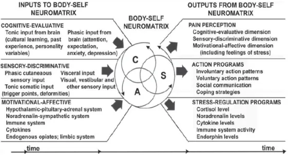
the outcome of complex interactions between central and peripheral nervous system notably thanks to the work of the physiologists Ronald Melzack and Patrick Wall in 1965 and their "Gate control theory" (Figure 5, Melzack and Wall, 1965) where a painful, nociceptive stimulus carried by nociceptive fibre is modulated in the substantia gelatinosa of the spinal cord through presynaptic inhibition from incoming beta-fibres. This mechanism suggests the existence of a balance between painful inputs form small diameter afferent fibres (nociceptor) and non-painful inputs coming from large diameter afferent fibres (non-nociceptor), and all this is under the control of supraspinal sites that could further modulate pain. Then, a couple of year later, in 1992, the neuromatrix theory, evolving from the gate control theory of pain, described pain as “a multidimensional experience produced by characteristic neurosignature patterns of nerve impulses generated by a widely distributed neural network—the body-self neuromatrix—in the brain” (Figure 6, Melzack, 2001). Theory in which the pain felt is not only due to the integration of the painful information coming in over the nociceptive fibre to the pain center in the brain but through the balance between the painful information and the generation of subjective experiences involving a complex network of systems that interact to modify and influence the perception and response to noxious stimuli, and explain how pain could persist in the absence of noxious stimuli.
Figure 5 : Schematic diagram of the Melzack-Wall gate control theory of pain mechanisms (1965). Aβ (Large-diameter) and C-fibre (small-diameter) afferent fibres project to the substantia gelatinosa (SG) and first central transmission (T) cells. The inhibitory effect (-) of SG on the afferent terminals is increased (+) by activity in Aβ fibres and decreased by activity in C-fibres. A specialized system of Aβ fibres (the central control trigger) activates certain cognitive processes that influence the modulating properties of the spinal gating mechanism via descending fibres (Melzack and Wall, 1965).
Figure 6 : Factors that contribute to the patterns of activity generated by the body-self neuromatrix, which comprises sensory (S), affective (A), and cognitive (C) neuromodules (Melzack, 2001).
Thus, as history as proven, behind pain sensation, different concepts have evolved, substantially shaped by the advances in research resulting in a definition accepted by the majority and proposed by the International Association for the Study of Pain (Merskey and Bogduk, 1994), Pain is now defined as “an unpleasant sensory and emotional experience associated with actual or potential tissue damage, or described in terms of such damage” (Figure 7). Therefore, pain is considered as a personal experience which involves neural mechanisms that arbitrate both sensory and hedonic functions. That’s the reason why pain is subjective and its study is delicate and complex.
Figure 7 : Description of non-exhaustive factors which participate in pain perception. Modified from Dr Caleb Burgess (Burgess, 2019)
2. Pain physiology Physiological pain
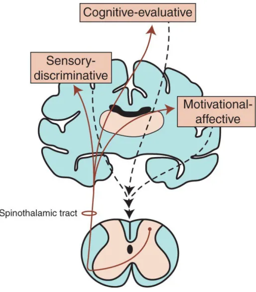
Pain, notably its physiological aspect, can be described as a warning mechanism with a protective role, with the aim to keep tissue damage to a minimum following a noxious and potentially damaging stimulus. It corresponds to a cascade of modifications increasing with the intensity of the noxious stimulus, occurring in the peripheral and central nervous system, and responsible for the perception of pain. However, neuronal excitation in nociception is not only a static process of proportionality between intensity of the stimulus and the behavioural responses, but some dynamic changes can also occur as the result of a leftward shift of the stimulus-response curve, which takes place both at the peripheral or at the central nervous system (as known as the sensitization). To get a real grasp of this complex phenomenon, Melzack and Casey put forward the idea of seeing pain as a three dimensions: The sensory discriminative component which refers to the perception of pain including the location, intensity, characteristic and duration; the motivational affective component referring to the unpleasantness of pain; and the cognitive component which refers to all the factors in terms of past experiences and probability of outcome based on individual’s beliefs (e.g., culture, past experiences etc…) related to pain which can influence both the sensory discriminative and the motivational affective dimensions negatively or positively (Figure 8, Melzack and Casey, 1968). In contrast, it is important to remember that a greater distinction should be made between pain and nociception. Nociception, unlike the pain, is the neural process of encoding and processing noxious stimuli (IASP, 2019a) resulting from a thermal, mechanical, or chemical energy acting on specialized nerve ending (nociceptors) to the central nervous system and it is only when sensory information reaches the cerebral cortex that the perception of pain takes place.
Unfortunately, a dysregulation of pain mechanism may occur and become pathological, notably when it loses its safety aspect and becomes no longer helpful for the protection of the integrity of the organism as an acute warning mechanism but on contrary, becomes chronic and debilitating.
Figure 8 : The dimensions of pain as outlined by (Melzack and Casey, 1968) described in the text above.
Sensitization
Neuronal excitation in nociception is not only a static process of proportionality between intensity of the stimulus and the behavioural responses but it may be subjected to dynamic changes, known as the sensitization, which is described as “a phenomenon which increased responsiveness of nociceptive neurons to their normal input, and/or recruitment of a response to normally subthreshold inputs.”(IASP, 2019a), including a modification (decrease) of the threshold and an increase in suprathreshold response. Phenomenon, which can occur both at peripheral and at the central nervous system (defined respectively as the peripheral and central sensitization), considered to be a key element of the physiological pain, but at some point can participate in emphasising the dysregulation of physiological pain, in chronic pain.
Pathological Pain
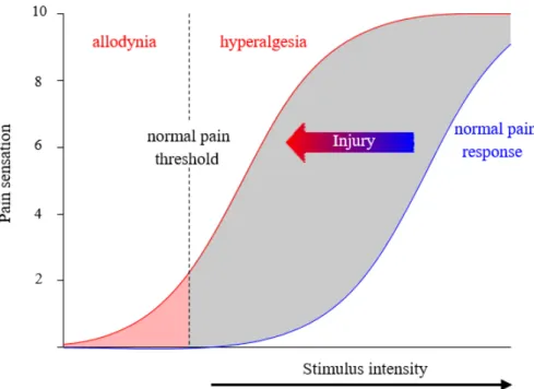
Nevertheless, other manifestations of pain related to tissue injury can occur, notably when its perception is no more proportional to the intensity of the noxious stimulus and becomes chronic and incapacitating (Cervero and Laird, 1996). Alterations of the pain pathway, which lead to hypersensitivity characterised by an exaggerated response to noxious stimuli associated with a lowered threshold to noxious stimuli which is defined as a hyperalgesia, both at the site of the tissue damage (primary hyperalgesia), and surrounding the area of primary tissue or nerve injury bordering uninjured tissue (secondary hyperalgesia) (Treede et al., 1992) and the appearance of pain response to innocuous stimuli (allodynia); (Figure 9). Hyperalgesia and allodynia highlight changes in either the peripheral or central nervous systems, referred as peripheral or central sensitization, respectively. This phenomenon implies several factors such as genetic and environmental factors, which contribute to sensitization resulting in persistent (chronic) pain in some individuals (even after healing has normally taken place) characterized also by the abnormal state and function of the spinal cord neurons, which become hyperactive. Under normal condition, the nociceptive sensory system returns to a baseline functional state as soon as healing takes place. Unfortunately, many components of sensitization can persist and manifest themselves as chronic pain and hyperalgesia, especially when the nervous system itself is injured leading to chronic neuropathic pain.
Figure 9 : Schematic representation of sensitization of nociceptors response to a peripheral stimulus induces by a tissue injury. In blue, weak or innocuous stimuli do not evoke pain sensation in normal condition. In red, sensitization in response to a stimulus after tissue damage, shifting the response function to the left. A moderate painful stimulus is now perceived as intense (i.e., pain hyperalgesia) and a previous innocuous stimulus can be perceived as painful (i.e., allodynia). Adapted from (Cervero and Laird, 1996).
Pain State Classification
Pain is the most commonly symptom observes in medicine, which may be expressed in different ways, can occur in any part of the body and affect any system, be acute or chronic, sporadic or constant. That is why, in the aim of a better understanding of pain disorders and to guide assessment or treatment, it is important to classify the different pain state.
i.According to the Mechanistic/Etiology
Six different types of pain can be distinguished based on the physiopathological mechanism. - Nociceptive pain
Nociceptive pain, defined as “pain that arises from actual or threatened damage to non-neural tissue and is due to the activation of nociceptors”(IASP, 2019a), represents the normal response to noxious stimuli or injury of tissues (skin, muscles, visceral organs, joints, tendons, or bones). It is caused by ongoing activation of nociceptive afferent fibres (Aẟ and C nociceptor) through mechanical, thermal, or chemical stimulation (Costigan et al., 2009). Depending on the location of the nociceptors involved,
nociceptive pain may also be subdivided into visceral pain, deep somatic pain, and superficial somatic pain.
- Neuropathic Pain
Neuropathic pain, defined by the IASP as “pain caused by a lesion or disease of the somatosensory nervous system” and in such of terms as “burning, stabling, electrical, shooting pain…” by the patient, is caused by a primary lesion or dysfunction affecting the somatosensory nervous system (Merskey and Bogduk, 1994) both from peripheral nerves (peripheral neuropathic pain) to the central nervous system which is a regional pain associated with aberrant sensibility to temperature and to noxious stimulation (Bouhassira et al., 2005).
- Nociplastic Pain
Recently adopted, nociplastic pain is defined by the IAPS as “a pain that arises from altered nociception despite no clear evidence of actual or threatened tissue damage causing the activation of peripheral nociceptors or evidence for disease or lesion of the somatosensory system causing the pain”(IASP, 2019a).
- Psychogenic Pain
Pain disorder associated with psychological, emotional or psychosocial factors such as depression, anxiety and bipolar disorder, obsessive-compulsive behaviour, which describes both short- and long-term episodes of pain that occur usually in the absence of any objective inflammatory or physical pathology that could explain the pain sensation (Toda, 2007), People experiencing psychogenic pain even if brief episodes, as well as persistent symptoms, described it as very real and painful.
- Mixed Pain
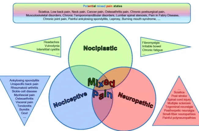
Pain defines as a “complex overlap of the different known pain types (nociceptive, neuropathic, nociplastic) in any combination, acting simultaneously and/or concurrently to cause pain in the same body area (Figure 10). Either mechanism may be more clinically predominant at any point in time. Mixed pain can be acute or chronic (Freynhagen et al., 2019).
Figure 10 : Representation of “mixed pain” defined as the overlapping of the nociplastic, nociceptive and neuropathic pain (Freynhagen et al., 2019)
- Idiopathic Pain
Idiopathic pain is defined as a pain that occurs when there is no known physical or physiological cause or that persists after the trauma or pathology has healed (Bouhassira and Attal, 2012; Cheng, 2018). It arises generally from neurological dysfunction, not damage (such as fibromyalgia, temporomandibular disorders…).
ii.According to the Anatomic perception
It is a conventional classification of pain based on the anatomical location from which pain is perceived, allowing in some cases to determine the possible cause (e. g. low back pain, headaches, joint pain, cardiac pain... each referring to the specific location of the symptoms). Nevertheless, some criticism can be raised on the importance of such a classification on setting up a therapy notably due to the lack of anatomically defined specificity in the neurophysiology of pain. Indeed, pain is not always associated with a precise anatomical location but rather as a relaying pain. For example, pain from internal organs, which do not have nociceptors, is not well localized and may masks the origin of this pain, particularly in the case of cardiac pain,
which can be felt in the left shoulder of the body without necessarily being associated with chest pain.
iii. According to the Duration
Traditionally, one common way to classify pain is to differentiate it according to a temporal criterion. It is a measurable characteristic allowing differentiation between acute (including pain associated with tissue damage, inflammation, or a disease process that is of relatively brief duration regardless of how intense) and chronic pain (for pain that persists for extended periods of time). Conventionally, considering the duration of the symptoms, it has been well admitted that pain can be divided into groups (Backonja et al., 2010; Swieboda et al., 2013):
- Acute pain, which is defined as a pain that lasts less than 3 to 6 months.
- Chronic pain, which is defined as a pain lasting for more than 3 to 6 months, or persisting after a complete tissue healing or beyond the course of an acute disease. - Survived pain, which describes a persisting pain despite the healing of the tissue
damage, which resulted in acute pain
However, this criterion may lead to a lack of understanding of pathology because a single dimension of duration is not necessarily link to pathological factors.
iv.According to the Intensity
It is a conventional pain classification based on the intensity of perceived patient pain. Although the fact that the sensation of pain is subjective, making it the most difficult feature to assess, a common point of the intensity of pain is its tolerance. That is why, in the aim to evaluate intensity visual, categorical or analogue scales are used to compare pain with the worst pain than the patient ever suffered (Figure 11).
Figure 11 : Comparative pain scale chart used to assess pain by an increasing factor. Scales consisting of a horizontal line, more or less 10 cm in length, one hand of which is “0”, meaning literally no pain and at the opposite end “10”, meaning the strongest pain endured in life. (Moyle, 2015)
Multidimensional Classification of Pain
Pain experience, as described above, covers different components such as physical, psychological or social component often resulting in suffering. Suffering which can be described as a threat to the intactness of individual’s self-concept and integrity such as our desire of happiness and health (Schmidt and Willis, 2007) resulting from aggression to the physical or emotional components of pain including loss of physical function, social isolation, family distress, and a sense of inadequacy or spiritual loss (Benzon et al., 2018).
Therefore, pain approach should be multidimensional to encompass all these different modalities. As an alternative to the unidimensional approaches, a comprehensive taxonomy of pain should consider multifactorial assessment (Table 1) rather than a single dimension such as
(Serlin et al., 1995; Cleeland et al., 1996; Sela et al., 2002) : - Physical effects and symptoms
- Functional effects (such as social consequence) - Psychological factors (anxiety, fear …)
Table 1 : Proposal of a comprehensive taxonomy of pain based upon multifactorial assessment (Turk and Okifuji)
3. Anatomy and physiology of the pain From nociception to pain sensation
Pain, as described above, is considered a physiological protective system essential to the survival, well-being, learning and adaptation of human being. However, it can become pathological, notably when, in absence of tissue damage or following appropriate healing of injured tissue, it occurs or persists and then become no longer useful as a protective system and become chronic and debilitating. Thus, an understanding of the physiological mechanism underlying pain perception will provide key elements to apprehend the mechanisms of acute and chronic pain.
One approach to understanding the pain sensation is to follow the nociceptive signal pathways also called nociception (a physiological process of activation of neural pathways by stimuli that are potentially or currently damaging to tissue) from the periphery (detected by specialized peripheral sensory neurons as known as the nociceptor) to the brain through the spinal cord.
Four distinct processes are necessary for a nociceptor to convey noxious information to the CNS, with the starting point being signal transduction, which occurs in the peripheral terminals of primary afferent neurons.
Pain Parameters: Anatomy/System
Duration/Intensity/Quality
Associated Abnormality (physical/psychological) Underlying Diseases:
Signs/Symptoms Pain Mechanisms:
NEUROPHYSIOLOGICAL
Primary afferent involvement CNS involvement
Psychological
Cognitive–Affective–Behavioral Involvement Cognitive appraisal of pain
Coping Affect/mood Environment
i.Sensory afferent fibres from the periphery to spinal cord
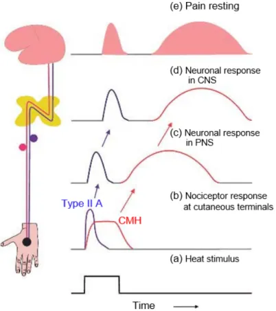
Sensory afferent fibres originating in the peripheral terminal group together to form spinal nerves, each supplying a distinct area of skin (dermatome), and overlapping more or less the dermatomes of neighbouring spinal nerves and later splitting to form a ventral and dorsal roots. Dorsal roots exclusively relate to sensory information. These sensory afferent fibres can be classified physiologically according to their physical characteristics such as axon diameters, conduction velocity and function into different groups: A fibre group with subtype Aα (or group I; thickly myelinated fibres, muscle proprioceptors), Aβ (or group II; less myelinated fibres, low-threshold mechanoreceptors), Aδ (or group III; thinly myelinated fibre, mechanoreceptor-thermoreceptor), and C fibre group (or group IV; unmyelinated fibres, polymodal receptor). It is generally admitted that transduction of noxious signals from most spinal cord innervated tissues is mediated by the thinly myelinated Aδ fibres and unmyelinated C fibres with free nerve ending (nonspecialized); nevertheless, to a lesser extent, thickly myelinated fibres may also play a role in pain sensation (especially from the skin). To get more in detail we will mainly focus our attention on one type of nociceptor (mechano-heat-sensitive nociceptor), the cutaneous fibres which are able to respond to stimuli that are potentially or actually tissue-damaging (noxious stimuli), such as intense mechanical stimuli (pinch, pressure, indentation), algesic chemical, or elevated thermal (> 45 ° C) stimuli.
The Aα/β fibres, as described above, are large-diameter myelinated fibres with the fastest peripheral conduction velocity. They are mainly involved in the transmission of non-nociceptive input, such as hair movement, pressure or small touch and also seem to play a tonic inhibitory role on the nociceptive input through the recruitment of inhibitory interneurons in the substantia gelatinosa of the spinal cord thus modulating nociceptive input from the same spinal segment (main mechanism of the gate control theory (Melzack and Wall, 1965).
The Aδ fibres are, to a lesser extent compare to Aα/β fibres, thinly myelinated fibres with intermediate velocity. Depending on the specificity of their response to stimulation, Aδ fibres can be divided into two types (Treede et al., 1998): On one hand, the mechano-heat nociceptors which mainly respond to intense and potentially harmful mechanical and heat stimulation, and on the other hand the polymodal Aδ fibres which respond both to mechanical, thermal, and chemical stimulation. However, it has been shown that the mechanonociceptor Aδ fibres neuronal activity may lead to hyperalgesia through the increase of their discharge resulted from thermal stimulation. In addition, the Aδ fibres are considered to be responsible for the first pain sensation, pricking pain, sharpness, and transient sensation (Basbaum et al., 2009).