ﺔﯿﺒﻌﺸﻟا ﺔﯿطاﺮﻘﻤﯾﺪﻟا ﺔﯾﺮﺋاﺰﺠﻟا ﺔﯾرﻮﮭﻤﺠﻟا و ﻲﻟﺎﻌﻟا ﻢﯿﻠﻌﺘﻟا ةرازو ﻤﻠﻌﻟا ﺚﺤﺒﻟا
ﻲ
Popular Democratic Republic of Algeria Ministry of Higher Education and Scientific Research
University of Mohammed Seddik Benyahia- Jijel- Faculty of Natural Sciences and Life Cellular and Molecular Biology Department
Graduation Thesis
Submitted By Mehdi DAD
For A Master's Degree In Pharmacology Sciences
Topic
Assessment by the Micronucleus Assay of the Genotoxicity
among Hospitalized Breast Cancer Patients Subjected to
Antineoplastic Polychemotherapy.
Jury composed from Members
President : Dr Nada ZABAIOU Supervisor : Pr Mesbah LAHOUEL Examiner : Dr Lamia BENGUEDOUAR
Acknowledgements
First, I want to thank the God, my creator for giving me the power that I needed to accomplish this work, my big gratitude for Allah.
I also want to thank Allah for making the professor Mesbah Lahouel my supervisor, it was really bless from god and I thank professor Mesbah Lahouel for accepting to be my supervisor although he had many responsibilities but he was always there for me with his advices, philosophy and goodness. His name is Mesbah and he was really “Mesbah” a light against the darkness of my ignorance.
My gratitude for Mister Kamel Boumeslat my co-supervisor for helping me in the experimental study he gave me everything I needed and he was always there for help so thank you so much.
I also thank Mister Ziad Beghoul he was always present in the research laboratory to gives us everything we need for our study.
My gratitude for the jury members for accepting to judge this work. I Thank Dr. Nada Zabaiou for accepting to be the president of the jury.
I would also like to thank Dr. Lamia Benguedouar for accepting to be the examiner.
Dedicates
With all my gratitude and deep feeling of respect, I dedicate this work:
To My parents who were always there for me with their support and encouragement, they believed in me when nobody does and they gave me everything I needed to follow my dreams.
To My sisters and my brother I hope god bless them all.
To all my friends especially Ahmed Mat, Fares, Islam, Med Amin, Abdu, Sofiane, Yasser and Farok.
To my classmate especially Aida, Sara, Sabrina, Amira, Nabila, Manel and Omaima.
Table of contents
Acknowledgement and dedicatesIntroduction………...………1
Part I: Literature synthesis Chapter 1: General information about cancer 1. Pathophysiology of cancer……….……….3 2. Cancer therapy………3 2.1 Surgery………..3 2.2 Radiation………...…4 2.3 Chemotherapy………...……4 2.4 Hormonotherapy………..…….5 2.5 Immunotherapy……….………6
3. Chemotherapy side effects………..………6
3.1 Nausea, vomiting and hair loss……….………6
3.2. Extravasation with tissue necrosis………...……7
3.3. Bone marrow suppression and infections………7
Chapter 2: General information about genotoxicity I. The Genotoxicity………..………..8
1. Genotoxins ………...8
1.1 Chemical genotoxins ………...8
1.2 Physical genotoxins ……….9
1.3 Biological genotoxins.……….9
2. Mechanism of genotoxicity and mutagenicity ………...9
2.1 Primary DNA lesions……….………..9
2.2 Gene mutations………..………10
II. Genotoxicity assays ………...………..11
1. Micronucleus assay………..11
2. Comet assay ………...………….13
3. Ames test………..14
4. Chromosomes aberration test………..……….15
III. Reminder about erythropoiesis………...………15
Part II: Experimental study 1. Patients………..16
2. Material………...………..16
3. Methods……….17
3.1 Obtaining blood smears………..17
3.2 Blood smears coloration……….……….18
3.3 Microscopic observation……….………18
Part III: Results and Discussion 1. Case study……….20
2. Statistical analysis……….20
2.1 Characteristic of control population………21
2.2 characteristic of patient’s population………..21
3. Micronucleus frequency among patients under chemotherapy. ...………21
Introduction
INTRODUCTION
Cancer caused 9.6 million deaths worldwide in 2018 with 18.1 million new cases each year
(OMS, 2018). Lung cancer is the most common cause of cancer deaths worldwide, accounting
1.37 million deaths in 2008 (Dent et al., 2013). In addition to lung cancer, breast cancer is the
most common malignant disease in western women. In these patients, it is not the primary
tumour but metastases at distant sites that are the main cause of death (Weigelt et al., 2005).
The use of chemotherapy to treat cancer began at the start of the 20th century with attempts to
reduce chemicals that might affect the disease by developing methods to screen chemicals
using transplantable tumors in rodents (Devita and Chu., 2008).
Chemotherapy remains a great hope in oncology. However, if its side effects on many tissues
are well documented, research on its genotoxic risks is limited. Genotoxicity of anticancer
drugs to normal cells is one of the most serious problems of chemotherapy due to the
possibility of inducing secondary malignancies (Blasiak et al., 2002). Chemicals that can
interact with the genetic material and generate critical DNA lesions are called tumour
initiating agents or genotoxic carcinogens (Lutz and Maier., 1988).
Genotoxicity is a word in genetics defined as a destructive effect on a cell's genetic material.
Regulatory authorities all over the world require data on the genotoxic potential of new drugs,
as part of the safety evaluation process (Shah., 2012). Genotoxicity tests can be defined as in
vivo or in vitro tests designed to detect the compounds which induce genetic damage directly
or indirectly by various mechanisms (Rao et al., 2008).
The objective of this work is to evaluate by the micronucleus assay the genotoxic potential of antineoplastic drugs (Docétaxel, cychlophosphamide, Adriamycin and capiritabine) used in the treatment of breast cancer patients and compare those results with non-exposed population.
The experimental study was conducted at the molecular toxicology laboratory of Jijel-Mohammed Seddik Benyahia University. The period of our experimental study is two months was from 21 April 2019 to 20 June 2019.
This work is divided to two parts. The first is the literature synthesis, which we are going to
talk about cancer, cancer therapy and genotoxicity. In the second part we are going to
represent and discuss the results of the micronucleus assay to conclude about the genotoxicity
Chapter 1:
1. Pathophysiology of cancer
Pathogenesis of cancer depends on both environmental (e.g. exposure to carcinogens) and genetic factors which derange the molecular mechanisms that control cell proliferation. The hallmarks of a malignant cell are autonomous growth signaling coupled with insensitivity to anti-growth signals, immortalization, invasion and metastasis, evasion of apoptosis, sustained angiogenesis and DNA instability. Some cancer cells overexpress oncogene. Oncogene encodes growth factors and mitogenic factors that regulate cell cycle progression and cell growth (Ritter et al., 2008).
Breast cancer is genetic, in the sense that breast tumor development is initiated by alterations, Mutations, somatic recombinations, duplications, and so on of DNA sequences in breast epithelial cells. These alterations may be inherited in the germ line, or may be due to somatic events induced by environmental mutagens (King, 1985), between 10% and 20% of breast cancer cases are ascribed to hereditary factors. Inherited mutations inactivating the BRCA1 and BRCA2 genes (encode proteins involved in DNA repair) lead to an up to 80% life-time risk of breast and /or ovarian cancers (Schulz, 2015).
Breast cancer starts as a local disease, but it can metastasize to the lymph nodes and distant organs, Invasive breast cancer probably evolves from normal epithelium of the terminal duct or lobular unit through a series of increasingly abnormal proliferative lesions including typical and atypical hyperplasia and noninvasive carcinoma (Berardo et al., 1998).
2. Cancer therapy
With present methods of treatment, about one third of patients are cured with local treatment Strategies, such as surgery or radiotherapy, when the tumor remains localized at the time of diagnosis. Earlier diagnosis might lead to increased cure rates with such local treatment. In the remaining cases, however, early micrometastasis is a characteristic feature, indicating that a systemic approach with chemotherapy is required for effective cancer management.
2.1 Surgery
Local control of the tumour, which means the total eradication of the primary tumour and disease involving regional lymphatics, is indispensable for obtaining a cure. Surgery is often the most appropriate procedure for obtaining this goal and, from this point of view, remains the cornerstone in treatment (Bremers et al., 1999).
2.2 Radiation
About half of all cancer patients receive radiation therapy, either alone or with surgery or chemotherapy. Radiation therapy bombards cancer with X-rays, gamma rays, and subatomic particules (electrons, protons, and neutrons). Radiation therapy is effective on cancer cells because they divide continually. Radiation damages DNA, and the DNA in continually dividing cells does not repair itself as well as the DNA in a normal cell, and damage accumulates. Eventually, the severely damaged DNA cannot reproduce itself, and the cancer Cell dies while dividing. Radiation also damages cells in normal tissues, but fewer normal Cells are damaged because they are dividing less frequently than cancer cells (lyons, 2007).
2.3 Chemotherapy
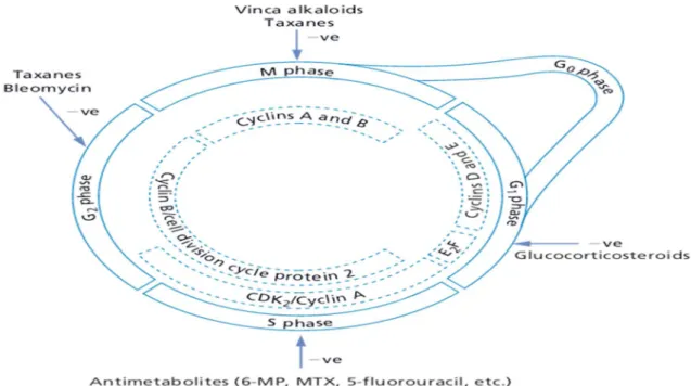
In the early 1900s, the famous German chemist Paul Ehrlich set about developing drugs to treat infectious diseases. He was the one who coined the term ‘‘chemotherapy’’ and Defined it a the use of chemicals to treat disease (Devita et al., 2008), Chemotherapy Included a number a families defined by both their chemical structure and mechanism of action : alkylating agents, antibiotics, antimetabolites, topoisomerase I and II inhibitors, mitosis inhibitors, platinum compounds and others (Espiinosa et al., 2003).
Figure 1: The cell cycle regulatory systems and sites of anticancer drug actions. 6-MP, 6- mercaptopurine; MTX, methotrexate (Ritter et al., 2008).
To focus more about our patients, we are going to talk briefly about the antineoplastic drugs they took as chemotherapy protocol, they were subjected to combination of Adriamycin and Cyclophosphamide.
Adriamycin: (Doxorubicin), an anthracycline antibiotic, is one of the most popular anticancer drugs. Adriamycin is used for the treatment of human cancers including a variety of solid cancers Although the main anticancer action of adriamycin is believed to involve DNA damage through topoisomerase II inhibition and free radical generation (Mizutani et al., 2005).
Doxorubicin is not absorbed orally, and because of its ability to cause tissue necrosis must not be injected intramuscularly or subcutaneously. Distribution studies indicate rapid uptake in all tissues except the CNS. Extensive tissue binding, primarily intranuclear, accounts for the prolonged elimination half-life.The drug is extensively metabolized in the liver to hydroxylated and conjugated metabolites and to aglycones that are primarily excreted in the bile (Sikic, 2002).
Cyclophosphamide: is an Alkylating agents compose a diverse group of electrophiles which reacts with DNA to form various adducts, of which interstrand DNA cross-links are particularly significant. Under favourable circumstances, the presence of these adducts either initiates apoptosis or cell cycle arrest (Estlin et al., 2000).
Cyclophosphamide can be given orally, intramuscularly,or intravenously. The plasma half-life of intact cyclophosphamideis 6.5 hours.Only 10 to 15% of the circulating parent drug is protein bound, whereas 50% of the alkylating metabolites are bound to plasma proteins. Since cyclophosphamide and its metabolites are eliminated primarily by the kidneys, renal failure will greatly prolong their retention (Sikic, 2002).
2.4. Hormonotherapy
The growth of number of cancers is hormone dependent or regulated by hormones. Research in The fields of fertility, birth control, and menopause has yielded valuable hormone analogs and Antagonists for the treatment of both breast and prostate cancer. these molecules interrupt
the Stimulatory axis created by systemic pools of androgens and estrogens, inhibit hormone Production or binding to receptors, and ultimately block the complex expression of genes that promotes tumor growth and survival these drugs have proven effective in extending survival
and delaying or preventing tumor recurrence in breast cancer and prostate cancer (Moy et al., 2011) .
Many breast cancers show estrogen-dependent proliferation. Administration of anti-estrogens can therefore be expected to cause tumor mass reduction. The most well established drug for hormone therapy is tamoxifen (Nolvadex®). This is a first-line drug for the treatment of breast cancer in postmenopausal patients (Ito, 2002).
2.5 Immunotherapy
These agents influence the biological response to the tumour. They may act indirectly to mediate anti-tumour effects, for example stimulate the immune response against the transformed neoplastic cells or directly on the tumour like by modulating tumour differentiation. Drugs with proven anti-cancer clinical efficacy in this class are interleukin-2 and interferon-alfa 2b (Ritter et al., 2008).
3. Chemotherapy side effects
Side effects of anticancer chemotherapy can be so difficult to live with that some patients regard them as worse than the disease itself (Lerman et al., 1990).
3.1 Nausea, vomiting and hair loss
Nausea and vomiting have been reported by patients, nurses and Physicians as the most distressing side-effects of chemotherapy (de Boer-Dennert., 1997). Chemotherapy induced hair loss (alopecia) is a common side effect of adjuvant and Metastatic chemotherapy regimens. The likelihood of alopecia is related to the type of drug used and its schedule of administration (Lemieux et al., 2008).
Anticancer drugs ruin mitotic and metabolic processes in actively growing hair follicles leading to the thinning of hair shaft, which becomes fragile and susceptible to fracture with minimal trauma (Botchkarev, 2003).
3.2. Extravasation with tissue necrosis
Most chemotherapeutic agents are given by intravenous administration and some drugs are available orally. When given intravenously, these drugs cause few side-effects at the site of injection. However, when they are injected or leak into the surrounding tissue, a tissue reaction varying from irritation to necrosis may arise (Schrijvers, 2003).
3.3. Bone marrow suppression and infections
Chemotherapeutic regimens were found to be a strong predictor of developing short and long term bone marrow toxicities including aplastic anemia (Nurgalieva et al., 2010).
Cancer chemotherapy causes immunosuppression and serious infections can result Neutropenia and hypogammaglobulinaemia are known Risk factors for infections in cancer patients (Peterslund et al., 2001).
The side effects of antineoplastic drugs taken by our patients are:
Adriamycin: acute Nausea, red urine and delayed cardiotoxicity, alopecia, myelosuppression, stomatitis (Chu & Sartorelli., 2012).
Cyclophosphamide: Acute nausea, vomiting and delayed myelosuppression, alopecia, hemorrhagic cystitis, cardiotoxicity (high dose) (Cornett and Dea., 2017).
Chapter 2
I. The Genotoxicity
Genotoxicity is a word used in genetics that describes the possession of substance that has destructive effect on the genetic material of the cell (DNA, RNA) thus affecting the integrity of the cell. Genotoxins are mutagens that can cause genotoxicity leading to the damage of DNA or chromosomal material thus causing mutation (Saks et al., 2017).
Genotoxicity Tests can be defined as in vivo in vitro tests designed to detect the compounds which induce genetic damage directly or indirectly by various mechanisms (Rao et al., 2008).
1. Genotoxins
Genotoxins are defined as agents that interact with DNA, either inducing mutations or damaging DNA (Kohn et al, 1998). Agents that damage DNA can be classified in several different ways: they can be endogenous (reactive by products from processes such as metabolism or inflammation), exogenous (agents present in food, water, or the air) and they can be physical such as ultraviolet (UV) light and ionizing radiation or chemicals (Swift et al., 2014).
1.1 Chemical genotoxins
Chemical genotoxins work as reactive electrophiles. These compounds form covalent adducts with the nucleophilic sites of DNA, RNA, or proteins. Chemical genotoxic carcinogens are divided into two main groups: direct-acting carcinogens and indirect-acting carcinogens. Direct-acting carcinogens cause DNA damage without metabolic activation, as they damage DNA from within. The most common are epoxides, imines, and alkyl and sulphate esters. Indirect-acting carcinogens become carcinogenic after metabolic activation. Typical indirect carcinogens are polycyclic aromatic hydrocarbons, nitrosamines, nitrosoureas and aromatic amines (Plošnik et al., 2016).
Chemicals genotoxins can be reactive oxygen species (ROS), intercalating agents, alkylating agents and base analogues (Swift et al., 2014).
1.2 Physical genotoxins
The two main physical genotoxic agents are the ultraviolet radiation and the ionizing radiation. Exposure to UV light induces a number of cellular changes, including the generation of DNA lesions, the induction of stress proteins (such as p53 and p21), and the initiation of cell cycle checkpoint arrest in cycling cells. in other side Ionizing radiation has been demonstrated to induce mutations and cause cancer in a dose-dependent manner IR damages all components of the cell and is known to produce more than 100 distinct DNA adducts (Shackelford et al., 1999).
1.3 Biological genotoxins
Biological genotoxins can be bacterial like Helicobacter pylori, or viral like Epstein Barr virus, human T lymphotropic viruses I and II, human papilloma virus and the hepatitis B virus, or parasites such as Schistosoma haemotobium, Clonorchis sinensis and Opisthorchis vivarium; or growth factors (Oliveira et al., 2007).
2. Mechanism of genotoxicity and mutagenicity
The damage to the genetic material is caused by the interactions of the genotoxic substance with the DNA structure and sequence. These genotoxic substance interact at a specific Location or base sequence of the DNA structure causing lesions, breakage, fusion, deletion, mis-segregation or non-disjunction leading to primary DNA damage and mutation (Saks et al., 2017)
2.1 Primary DNA lesions
DNA in human cells is continuously subject to damage, it is in most cases appropriately repaired, leaving relatively few permanent changes. The various kinds of primary DNA damage comprise chemical modification or loss of DNA bases, single strand or double strand
breaks as well as intra- and interstrand crosslinks. Each type of damage can lead to mutations
Figure 2: Differents types of DNA primary lesions (Bickman and Smolen., 1994).
2.2 Gene mutations
Single point mutations or gene mutations are small changes in the DNA at the level of the bases and genes, it includes Base pair substitutions and Addition or deletion of bases (nagarathna et al., 2013).
Figure 3: Different types of gene mutations (Hanna, 2015).
Base modification
Double strand breaks Intercalation
Single strand breaks
Adducts
Intrastrand cross-link
Substitutions
Deletion
2.3 Chromosome mutations
Two types of chromosomal aberrations can occur in cells: changes in chromosome structure and changes in chromosome number. Structural changes include:
Deletions are chromosomal changes in which one or more genes or chromosomal segments are lost (Benhacine and Sahil., 2016).
Duplications occur when one or more copies of a chromosomal segment are present on the same or different chromosomes (nagarathna et al., 2013).
Inversions occur when a breakage in one of the chromosomes occurs and the segment rotates 180ᵒ before it rejoins (Dutta et al., 2016).
Translocations take place when nonhomologous chromosomes break and exchange segments in diploid (2n) organisms.
There are two major types of chromosomal aberrations that are the result of changes in chromosome number, These are polyploidy and aneuploidy.
Polyploidy results when cells acquire one or more sets of chromosomes beyond the “normal” Number of sets. Aneuploids are result of changes in the individual number of homologous Chromosomes in a set (stansfield et al., 2003).
II. Genotoxicity assays
It is very important to do genotoxicity studies to avoid the potential damage that can be caused by it. These genotoxicity tests are done to identify if a drug or other substance have the potential to cause mutation and genotoxicity. By doing so they help us identifying the hazards in the early stage of drug development itself. Identification of the genotoxic agents helps us understand the mechanism of the mutation and genotoxicity thereby paving us way to better prevent the frequency of such mutation and genotoxicity (Saks et al., 2017).
The four most widely used genotoxicity assays are: the Ames test, comet assay, the micronucleus assay and chromosomal aberration assay.
1. Micronucleus assay
Micronucleus formation results from fragments of chromosomes that are not present in the daughter nucleus at the time of mitosis (Schmid, 1975). Absence of centromere due to
formation of these chromosome fragments. Three criteria are consider: the size of the micronucleus, the DNA content, and the percentage of micronuclei having positive C bands (Vanparys et al., 1990), C bands correspond to the pericentromeric DNA and their frequency is increased in the presence of aneuploidogens (Van Hummelen et al., 1992). This test has the advantage of highlighting both aneugenous lesions (abnormalities in the number of chromosomes) and clastogenic lesions (Tarantini, 2009). A simple technique directly uses target cells of certain genotoxic (Pillière and Falcy, 1991). However, this test gives less information about the nature of aberrations if not associated with other tests (Parry and Sors, 1993).
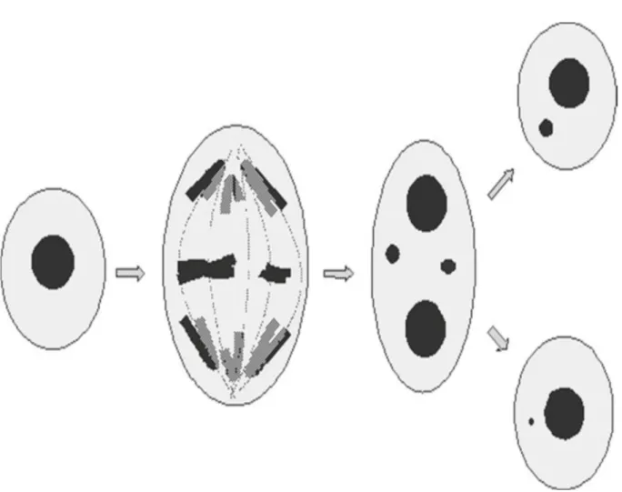
Figure 4: Overview of micronucleus formation with micronuclei originating from either a whole chromosome or a chromosomal fragment, in binucleate and mononucleate cells (Doherty, 2012).
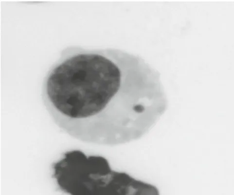
Figure 5: Photograph of a micronucleus in a mononucleate L5178Y cell (the image was captured from an acridine orange preparation in fl uorescent colours and then negative image was used to convert it into grey scale) (Doherty, 2012).
2. Comet assay
The comet assay is a simple, fast, and sensitive technique for quantifying single and double strand breaks in DNA and alkali-labile sites at individualized cells. It makes it possible to measure the breaks induced directly by a genotoxic agent or during enzymatic damage repair processes or during secondary processes of DNA fragmentation occurring for example during programmed cell death. This test consists in separating the nuclei of the isolated cells in an electrophoretic field, in a strongly alkaline medium. Nuclei whose DNA has undergone breakage then take on a comet form while nuclei whose DNA is undamaged appear as a regular disc. A semi-quantitative (four-category classification) or quantitative (moment-size assessment) assessment of injury rates can then be performed (Ostling and Johanson, 1984).
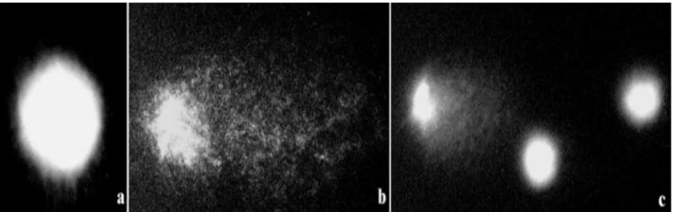
Figure 6: Comet assay in Crepis capillaris cells: a - control nuclei, not damaged; b, c- cells treated by mutagen (MH), nuclei with different level of DNA
damage are shown (Maluszynska and Juchimiuk, 2004).
3. Ames test
Bacterial mutagenesis assay: (Ames test): This is the preliminary test performed to detect the carcinogenic potential of an entity using bacteria. This test detects the point mutation or frame shift mutation. In the Ames test, a strain of Salmonella typhimurium auxotrophic (deficient) for histidine (his-), and which requires exogenous histidine, is used. Hence the bacteria are unable to survive in a medium devoid of Histidine (Rao et al., 2008).
4. Chromosome aberration test
Typically, in the in vitro chromosome aberration (ABS) test, CHO or V79 cells are treated with the test chemical for 1–1.5 cell cycles. The cells are transferred, after exposure to hypotonic treatment and fixation, to microscope slides and stained, and their chromosomes are examined microscopically in their first metaphase after treatment for the presence of breaks or rearrangements. The data are evaluated as the percentage of cells containing aberrations and sometimes as aberrations per cell. Compared with cells containing point mutations where the damage is measured in subsequent generations of cells, most cells containing chromosome damage will not survive to complete mitosis or will produce nonviable daughter cells (Zeiger, 2001).
III Reminder about erythropoiesis
The Erythropoiesis is a process that produce mature erythrocytes in the bone marrow starting with stem cells after successive series of differentiations and under the influence of hormone called erythropoietin (Suzuki et al., 2015). The different stages of erythropoiesis are:
Erythropoiesis start with the CSH where it gives the proerythroblasts (it is a big nucleated cell with diameter of 20 Micrometer), next this proerythroblast differentiate to erythroblast first basophil, on polychromatophil and then acidophil and at the end to reticulocyte known by the condensation of the nucleus and its expulsion to give at the end two mature erythrocyte (Breda et Rivella., 2014).
1. Patients
Nine breast cancer patients under chemotherapy treatment were chosen for the experimental study and three non-exposed person as controls.
patient Age Type of cancer Treatment
1 43 Breast cancer Adriamycin+cyclophosphamide 2 65 Breast cancer Adriamycin+cyclophosphamide 3 55 Breast cancer Adriamycin+cyclophosphamide 4 51 Breast cancer Adriamycin+cyclophosphamide 5 50 Breast cancer Adriamycin+cyclophosphamide 6 47 Breast cancer Adriamycin+cyclophosphamide
7 38 Breast cancer Cyclophosphamide + Docetaxel+ Capiritabine 8 65 Breast cancer Cyclophosphamide + Docetaxel+ Capiritabine 9 55 Breast cancer Cyclophosphamide + Docetaxel+ Capiritabine
Table 1: Breast cancer patients.
Non-exposed population sex Age
Control 1 Female 37
Control 2 Female 23
Control 3 Female 22
Table 2: Non-exposed population.
2. Material
For obtaining blood smears For blood coloration For blood slides observation
Total blood, gloves, slides, micropipette, compress (Gaz).
May Grunwald Giemsa reagent, distilled water
Microscope, immersion oil.
3. Methods
3.1 Obtaining blood smears
A small drop of whole blood was placed on a very clean slide (sterilized by alcohol) using 10µl micropipette. Then we took second slide at the angle of 30 – 45 °, while maintaining Contact wih the buttom slide we pulled the top slide back to contact the drop, which were spreading by the Capillary action, maintaining contact with the buttom slide and pushing the top slide in one motion to produce smear, we left out the slide dry in the free air and after that the slide was ready for coloration.
Figure 8: Different steps for obtaining smears.
3.2 Blood smears coloration
First step is fixation by methanol during 2-5 min, next coloration with may-grunwald reactive must be performed during 3 min, the final step is to move the slide on geimsa 5-10% during 20 min and dont forget to rinse with water the slide after every step.
When the slides are dried you are ready for microscopic observation.
Picture 2: may-grunwald geimsa reagent.
3.3 Microscopic observation
After looking for a good field in the slide, we started counting the whole number of the erythrocytes and the number of the micronucleated erythrocytes and then, we changed the field in the same slide and after counting 10 fields we calculated the frequency of the micronucleus formation.
Part III: Results and Discussion
1. Case study
Chemotherapy is the treatment of choice for hundreds of thousands of cancer patients diagnosed each year. Its frequent use with cancer patients is the result of recent advances in antineoplastic medication, new and more effective medications have increased the life expectancy for many patients and, in some cases, have resulted in remission and cure. Unfortunately, such long-term gain can come at considerable short-term cost to the cancer patient in the form of aversive and debilitating side effects (Carey and Burish, 1988).
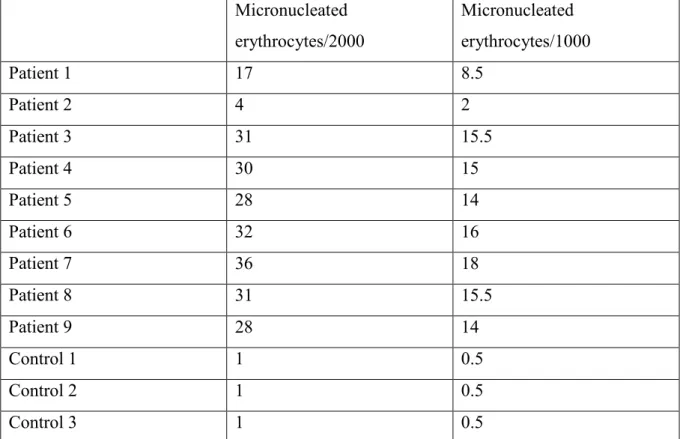
The following table represent the results of the Micronucleus assay of our experimental study:
Micronucleated erythrocytes/2000 Micronucleated erythrocytes/1000 Patient 1 17 8.5 Patient 2 4 2 Patient 3 31 15.5 Patient 4 30 15 Patient 5 28 14 Patient 6 32 16 Patient 7 36 18 Patient 8 31 15.5 Patient 9 28 14 Control 1 1 0.5 Control 2 1 0.5 Control 3 1 0.5
Table 4: results of the micronucleus assay for the patients and non-exposed population (control).
2. Statistical analysis
We used student test (T) to compare between the patients population and the non-exposed population. The difference between three populations is considered according to the student test (T):
P<0.5: significant (*);
P<0.01: very significant (**);
P<0.001: very high significant (***) ; 2.1 Characteristic of control population
The medium age of the non-exposed population is 27 years old, this population represented just by women who have no contact with cytotoxic products.
2.2 characteristic of patient’s population
The medium age of patients population is 52 years, all of them are breast cancer patients, this population achieved many cures cases thanks to Adriamycin and cyclophosphamide.
3. Micronucleus frequency among patients under chemotherapy
For the patients treated with chemotherapy we notice frequency of (11.83%ᵒ), this result is very high significant compared with controls population (p<0.001) which denote the genotoxic character of antineoplastic drugs.
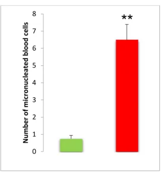
For the exposed population, the rate of micronulated erythrocytes with an estimated average of [5.81 ± 0.81] was significantly elevated compared to the control population. This result corroborates with that obtained by (Brahem et al, 2010) who evaluated the frequency of micronuclei on the blood cells of 20 nurses administering and reconstituting the antineoplastics. Micronucleated red blood cells levels were significantly higher in the population exposed only in the control population (Very highly significant).
Figure 9: The percentage (/1000) of micronucleated erythrocytes in the exposed population (red color) compared to the negative controls (green color).
0 1 2 3 4 5 6 7 N u m b e r o f m ic ro n u cl e at e d b lo o d ce lls
***
In a parallel study conducted in our laboratory by students from the paramedical school showed that the rate of genotoxic risk of the antineoplastic drugs is even greater in the subgroup of manipulators who prepare and administer anticancer drugs with an estimated average of [6.50 ± 0.89] and this despite the strict respect of protective measures with particular systematic wear (fig 10)..
Those results accord with the results obtained by (Rekhadevi et al., 2007) about the high genotoxic risk among the manipulators and nurses of oncology service.
Figure 10: The percentage (/1000) of micronucleated erythrocytes in the exposed population (red color) compared to the negative controls (green color).
The comparaison of the two treatments (Cyclophosphamide+adriamycine versus Cyclophosphamide + Docetaxel+ Capiritabine) shows a significant increase of micronuceated cells in the group of patients receiving Docetaxel and Capiritabine (fig 11). It seems that docetaxel is most toxic than the other drugs.
This result accord with the result of (Hesketh, 2008) which shows that docetaxel and capiritabine have more genotoxic potential than adriamycine.
0 1 2 3 4 5 6 7 8 N u m b e r o f m ic ro n u cl e at e d b lo o d c e lls
**
Figure 11: The percentage (/1000) of micronucleated erythrocytes in the population receiving cyclophosphamide+adriamycine (green color) and those receiving cyclophosphamide + Docetaxel+ Capiritabine (red color).
0 1 2 3 4 5 6 7 8 1 2 N u m b e r o f m ic ro n u le at e d b lo o d c e lls
CONCLUSION
Considering all the previously mentioned results, we speculated that the antineoplastic drugs could de due to their ability to alter many signaling pathways including stimulation of necrosis and induction of apoptosis through DNA damage. As a consequence, Adriamycin and cyclophosphamide were able to induce genotoxicity and damage to the genetic material of the cell leading to mutations, apoptosis and cell cycle disorder.
Our results are of major interest and open up great prospects:
There are several genotoxicity assays that can help us to assess the genotoxic potential of drugs and other substances like :
Micronucleus assay Comte assay
and thus help us to determine the less toxic dose of administration.
As an extension of this work and to further support our results, it would be interesting to study and analyze the possible genotoxicity of others anti-neoplastic agents like conventional chemotherapy or the new therapies and immunotherapy.
Benhacine Louiza et Sahil Nassima, 2016, Etude de la génotoxicité des extaits de Pistacia lentiscus par le test d’Ames. Génetique Appliquée, Université A. MIRA – Bejaia.46.
Breda, L., & Rivella, S, 2014, Modulators of erythropoiesis: emerging therapies for hemoglobinopathies and disorders of red cell production. Hematology/Oncology Clinics,
28(2), 375-386.
Bickman, J.W; Smolen, M.J, 1994. Somatic and heritable effects of envi-ronmental genotoxins and the emergence of evolutionary toxicology. Envi-ronmental Health Perspectives, 102: 25-28.
Blasiak Janusz, Ewa Gloc and Mariusz Warszawski, 2002, a comparison of the in vitro genotoxicity of anticancer drugs idarubicin and mitoxantrone. ABP Vol. 49 No 1: 145-155.
Burcham C. Philip, 2014, an introduction to toxicology. Springer: 221-256.
Botchkarev Vladimir. A, 2003, Molecular Mechanisms of Chemotherapy-Induced Hair Loss. The Society for Investigative Dermatology, vol 8: 72-75.
Bremers AJ, Rutgers EJ, van de Velde CJ (1999) Cancer surgery: the last 25 years. Cancer Treat Rev, 25: 333-353.
Chu Edward and Alan C. Sartorelli, 2012, Cancer Chemotherapy. McGraw Hill 12th edition: 949-975.
Cornett A. Patricia and Tiffany O. Dea, 2017, cancer. Mc Gray Hill education: 1607-1675.
De Boer-Dennert M , R de Wit , Pim Schmitz, J Djontono, V Beurden, G Stoter and J Verweij, 1997, Patient perceptions of the side effects of chemotherapy: the influence of (5HT3) antagonists. British Joumal of Cancer 76(8): 1055-1061.
Dent G. Annette, Tom G. Sutedja, Paul V. Zimmerman, 2013, Exhaled breath analysis for lung cancer. AME Publishing Company: 125-137.
DeVita. Vincent T, Jr. and Edward Chu, 2008, a History of Cancer Chemotherapy. Cancer Res, 68: 8643-8653.
Doherty T. Ann, 2012, the in vitro micronucleus assay. humana press: 121 -141.
Dutta, D., Gunasekera, D., Ragni, M. V., & Pratt, K. P, 2016, Accurate, simple, and inexpensive assays to diagnose F8 gene inversion mutations in hemophilia A patients and carriers. Blood advances, 1(3), 231-239.
Espinosa Enrique, Pilar Zamora, Jaime Feliu and Manuel Gonzalez Baron, 2003, classification of anticancer drugs a new system based on therapeutic targets. cancer treatment reviews, 29: 515–523.
Estlin. e. j, m. ronghe, g. a. a. burke, s. m. yule, 2000, the clinical and cellular pharmacology of vincristine, corticosteroids,l-asparaginase, anthracyclines and cyclophosphamide in relation to childhood acute lymphoblastic leukaemia. british journal of haematology. 110: 780-790.
Hanna,K. (2005), Chromosome breakage at high dose rates. Mutation Research, 182: 270-271.
Ito yoshinori, 2002, Chemotherapy and Hormone Therapy for Breast Cancer: Current Status and Perspective. Journal of the Japan Medical Association, 45(10): 424–433.
King mary claire, 1985, genetic analysis of breast cancer in families of mice and women. Martinus Nijhoff Publishing: 227-236.
Kohn.A, M. donchin, J.Y. jacobs, Y. horn, Y. gibor, F. fish,G. riesenfeld, F. kirenberg, I. lampert, A. halachm and M. Herzberg, 1988, Detection of Genotoxic Substances in Cancer Patients Receiving Antineoplastic Drugs. Annals new york academy of sciences: 776-791.
Lemieux Julie, Elizabeth Maunsell and Louise Provencher, 2008, chemotherapy-induced alopecia and effects on quality of life among women with breast cancer: a literature review. Psycho-oncology, 17: 317–328.
Lerman Caryn, Barbara Rimer, Barbara Blumberg, Suzanne Cristinzio, Paul F. Engstrom, Norma Macelwee, Karen O’connor and Janet Seay, 1990, effects of coping style and relaxation on cancer chemotherapy side effects and emotional responses. Cancer nursing, 13(5): 308-315.
Lutz W. K. and P. Maier, 1988, Genotoxic and epigenetic chemical carcinogenesis: one process, different mechanisms. elsevier publications: 322-326.
Lyons Lyman, 2007, Diagnosis and Treatment of Cancer. chelsea house publishers: 65-77.
Maluszynska Jolanta and Jolanta Juchimiuk, 2004, plant genotoxicity: a molecular cytogenetic approach in plant bioassays. Arh Hig Rada Toksikol 2005, 56:177-184.
Mizutani Hideki, Saeko Tada-Oikawa, Yusuke Hiraku, Michio Kojima, Shosuke Kawanishi, 2005, Mechanism of apoptosis induced by doxorubicin through the generation of hydrogen peroxide. Life Sciences 76: 1439–1453.
Moy Beverly, Richard Lee and Matthew Smith, 2011, natural products in cancer chemotherapy: hormones and related agents. McGraw hill 12th edition: 1755-1770.
Nagarathna, P. K. M., Wesley, M. J., Reddy, P. S., & Reena, K, 2013, Review on genotoxicity, its molecular mechanisms and prevention. Int J Pharm Sci Rev Res, 22(1), 236-43.
Nurgalieva Zhannat, Chih-Chin Liu, Xianglin L. Du, 2010, Chemotherapy use and risk of bone marrow suppression in a large population-based cohort of older women with breast and ovarian cancer: 43-54.
Oliveira A. Paula, Aura Colaço, Raquel Chaves, Henrique Guedes-pinto, Luis F. De-la-cruz P and Carlos Lopes, 2007, chemical carcinogenesis, Anais da Academia Brasileira de Ciências. 79(4): 593-616.
Ostling O, Johanson KJ, 1984. Microelectrophoretic study of radiation-induced DNA damages in individual mammalian cells. Biochemical and Bio-physical Research Communications, 123: 291-298.
OMS, 2018, Dernières données mondiales sur le cancer. Communique de press, 263 : 1.
Parry J.M. et Sors A. (1993). The detection and assessment of the aneugenic potential of environmental chemicals: the European Community Aneuploidy Progress. Mutaion Research287: 3-15.
Peterslund Niels Anker, Claus Koch, Jens C Jensenius, Steffen Thiel, 2001, Association between deficiency of mannose-binding lectin and severe infections after chemotherapy. The lancet, 358: 637–638.
Pillière F. et Falcy M. (1991). Exposition aux produits chimiques génotoxiques: marqueurs biologiques pour la surveillance des salariés. Documents pour le médecin du travail. 336p.
Plošnik Alja, Marjan Vračko and Marija Sollner Dolenc, 2016, Mutagenic and carcinogenic structural alerts and their mechanisms of action. Arh Hig Rada Toksikol, 67: 169-182.
RAO, S. H., Vyawahare, N. S., Ghaisas,A. B., Hanamsagar, R.M.and Chothe, A. S., 2008, genotoxicity testing: a review. Electronic journal of environmental sciences vol 1, 37-48.
Ritter James M, Lionel D Lewis, Timothy GK Mant and Albert Ferro, 2008, a Textbook of Clinical Pharmacology and Therapeutics. Companion web fifth edition: 368-385.
Schmid W. (1975). The micronucleus test, Mutation Research, 31: 9-15.
Schrijvers D. L. 2003, Extravasation: a dreaded complication of chemotherapy. Annals of Oncology 14: 26-30.
Schulz Wolfgang Arthur, 2005, molecular biology of human cancers. Springer: 357-382, 47-70.
Shackelford Rodney E., William K. Kaufmann and Richard S. Paules, 1999, Cell Cycle Control, Checkpoint Mechanisms, and Genotoxic Stress. Environmental Health Perspectives, Vol 107: 5-24.
Shah Shaily Umang, 2012, importance of genotoxicity & S2A guidlines for genotoxicity testing for pharmaceuticals. Journal of Pharmacy and Biological Sciences , ISSN : 2278-3008.
Sikic branimir, 2002, antineoplastic agents. Lippincott fifth edition: 639-656.
Stansfield.d william, Jaime.s colomé, raul j. cano, 2003, molecular and cell biology. schaum’s easy outlines: 60-67.
Swift Lucy H. and Roy M. Golsteyn, 2014, Genotoxic Anti-Cancer Agents and Their Relationship to DNA Damage, Mitosis, and Checkpoint Adaptation in Proliferating Cancer Cells. International Journal of Molecular Sciences, 15: 3403-3431.
Suzuki, N., Mukai, H. Y., & Yamamoto, M, 2015, In vivo regulation of erythropoiesis by chemically inducible dimerization of the erythropoietin receptor intracellular domain. PloS
one, 10(3), e0119442.
Tarantini A. (2009). Modulation de la génotoxicite des hydrocarbures aromatiques polycycliques (HAP) en mélanges. Thèse de doctorat, Ecole doctorale Ingénierie pour la Santé, la Cognition et l’Environnement de Grenoble. 174p.
Van Hummelen P., Deleener A., Vanparys Ph. et Kirsch-Volders M. (1992). Discrimination of anenploidogens from clastogens by c-banding, DNA and area measurements of micronuclei from mouse bone narrow. Mutation Research. 271: 13-28.
Vanparys P., Vermeiren F., Sysmans M. et Temmerman R. (1990). The micronucleus assay as a test for the detection of aneugemic activity. Mutation Research. 244: 95-103.
Weigelt Britta , Johannes L. Peterse and Laura J. van ’t Veer, 2005, breast cancer metastasis: markers and models. nature reviews cancer, vol 5: 591 -602.
Zeiger Errol, 2001, Genetic Toxicity Tests for Predicting Carcinogenicity. Marcel Dekker: 29-47.
Abstract:
After cardiovascular diseases, cancer is the second cause of death amongst the global Population. Cancer therapy including surgery, radiotherapy, chemotherapy, hormonotherapy and immunotherapy, can treat cancer or reduce tumor growth. Because of the cytotoxicity and the genotoxicity of chemotherapeutic drugs, cancer patients suffer from several side effects like nausea, vomiting and alopecia. The genotoxicity is a word used in genetic to describe the destructive effect on the genetic material of the cell caused by substances called genotoxins. There are many assays to evaluate the genotoxicity. We used in this study the micronucleus assay to evaluate the genotoxic potential of antineoplastic drugs used by breast cancer patients in the oncology department of Jijel hospital. The result was as we expected the antineoplastic drugs have high genotoxic potential. We observed a high number of micronucleated erythrocytes in the blood of cancer patients subjected to chemotherapy combination cyclophosphamide + Adriamycin and cyclophosphamide + docetaxel+ cytarabine..
Key words: cancer, chemotherapy, genotoxicity, micronucleus assay.
ﺺﺨﻠﻤﻟا و ﺐﻠﻘﻟا ضاﺮﻣا ﺪﻌﺑ ﻦﯿﯾاﺮﺸﻟا , نﺎطﺮﺴﻟا جﻼﻋ .نﺎطﺮﺴﻟا ﻮھ ﻢﻟﺎﻌﻟا ﻲﻓ تﺎﯿﻓﻮﻠﻟ ﺐﺒﺳ ﻲﻧﺎﺛ ﺼﺌﺘﺳا ﮫﯿﻓ ﺎﻤﺑ ﺔﯿﺣاﺮﺟ ﺔﯿﻠﻤﻌﺑ ﮫﻟﺎ ﺔﻌﺷﻷﺎﺑ جﻼﻌﻟا . ﻲﺋﺎﯿﻤﯿﻜﻟا جﻼﻌﻟا و ﻲﻧﻮﻣﺮﮭﻟا جﻼﻌﻟا ، ﺑ .مرﻮﻟا ﻢﺠﺣ ﻦﻣ ﻞﻠﻘﯾ وا ﮫﺠﻟﺎﻌﯾ نا ﻦﻜﻤﯾ ﻲﻋﺎﻨﻤﻟا جﻼﻌﻟا ﺔﯿﻤﺴﻟا ﺐﺒﺴ اﻟ ﻷ ﺔﯿﻨﯿﺠﻟاو ﺔﯾﻮﻠﺨ ﺔﯾودﻻا هﺬﮭﻟ ﺔﯿﺒﻧﺎﺟ ضاﺮﻋأ ةﺪﻋ ﻦﻣ نﺎطﺮﺴﻟا ﻰﺿﺮﻣ ﻲﻧﺎﻌﯾ نﺎطﺮﺴﻟا جﻼﻋ ﺔﯾود و ﺊﯿﻘﺘﻟﺎﻛ ا و نﺎﯿﺜﻐﻟ ﻒﺻﻮﻟ ﺔﺛارﻮﻟا ﻢﻠﻋ ﻲﻓ ﻞﻤﻌﺘﺴﺗ ﺔﻤﻠﻛ ﻲھ ﺔﯿﻨﯿﺠﻟا ﺔﯿﻤﺴﻟا .ﺮﻌﺸﻟا ناﺪﻘﻓ ا ﺔﯿﻠﺨﻠﻟ ﺔﯿﺛارﻮﻟا ةدﺎﻤﻟا ﻲﻓ ﻒﻠﺘﻟا داﻮﻣ ﮫﺒﺒﺴﺗ يﺬﻟ ةﺪﻋ كﺎﻨھ .ﺔﯿﻨﯿﺟ مﻮﻤﺳ ﻰﻤﺴﺗ ﺮﯾﺪﻘﺘﻟ برﺎﺠﺗ ﺔﯿﻨﯿﺠﻟا ﺔﯿﻤﺴﻟا ﺧا ﺔﺳارﺪﻟا هﺬھ ﻲﻓ ﺎﻨﻠﻤﻌﺘﺳا . ﻐﺼﻟا ىﻮﻨﻟا رﺎﺒﺘ ةرﺪﻗ ﺮﯾﺪﻘﺘﻟ ةﺮﯿ ﺖﻠﻤﻌﺘﺳا ﻲﺘﻟا نﺎطﺮﺴﻟا تادﺎﻀﻣ ا ﻰﻠﻋ ﻞﺠﯿﺟ ﻰﻔﺸﺘﺴﻣ ﻲﻓ ﻰﺿﺮﻣ ﻞﺒﻗ ﻦﻣ ﺖﻧﺎﻛ ﺔﺠﯿﺘﻨﻟا .ﺔﯿﻨﯿﺟ ﺔﯿﻤﺳ ثاﺪﺣ ﻊﻗﻮﺘﻣ نﺎﻛ ﺎﻤﻛ ﻟا تﺎﯾﺮﻜﻠﻟ ﺮﯿﺒﻜﻟا دﺪﻌﻟا ﺔﯾؤر ﺪﻌﺑ اﺬھ لﻮﻗأ ﺔﯿﻨﯿﺟ ﺔﯿﻤﺳ ثاﺪﺣا ﻰﻠﻋ ةﺮﯿﺒﻛ ةرﺪﻗ ﺎھﺪﻨﻋ نﺎطﺮﺴﻟ ةدﺎﻀﻤﻟا ﺔﯾودﻻا ﺔﯾﻮﻣﺪ .ﻲﺋﺎﯿﻤﯿﻜﻟا جﻼﻌﻠﻟ اﻮﻌﻀﺧ ﻦﯾﺬﻟا نﺎطﺮﺴﻟا ﻰﺿﺮﻣ مد ﻦﻣ ةذﻮﺧﺄﻤﻟا ةﺮﯿﻐﺻ تﺎﯾﻮﻧ يﻮﺘﺤﺗ ﻲﺘﻟا ءاﺮﻤﺤﻟا تﺎﻤﻠﻛ ﺔﯿﺣﺎﺘﻔﻣ : ،نﺎطﺮﺴﻟا ,ﻲﺋﺎﯿﻤﯿﻜﻟا جﻼﻌﻟا .ةﺮﯿﻐﺼﻟا ىﻮﻨﻟا رﺎﺒﺘﺧا ،ﺔﯿﻨﯿﺠﻟا ﺔﯿﻤﺴﻟا