RESEARCH OUTPUTS / RÉSULTATS DE RECHERCHE
Author(s) - Auteur(s) :
Publication date - Date de publication :
Permanent link - Permalien :
Rights / License - Licence de droit d’auteur :
Bibliothèque Universitaire Moretus Plantin
Institutional Repository - Research Portal
Dépôt Institutionnel - Portail de la Recherche
researchportal.unamur.be
University of Namur
Optical structure and function of the white filamentary hair covering the edelweiss
bracts
Vigneron, Jean-Pol; Rassart, Marie; VERTESY, Zofia; KERTESZ, Krisztian; Sarrazin,
Michael; BIRO, Laszlo; ERTZ, Damien; Lousse, Virginie
Published in: Physical Review E
Publication date: 2005
Document Version
Early version, also known as pre-print
Link to publication
Citation for pulished version (HARVARD):
Vigneron, J-P, Rassart, M, VERTESY, Z, KERTESZ, K, Sarrazin, M, BIRO, L, ERTZ, D & Lousse, V 2005, 'Optical structure and function of the white filamentary hair covering the edelweiss bracts', Physical Review E, vol. 71, no. 011906, pp. 1-8.
General rights
Copyright and moral rights for the publications made accessible in the public portal are retained by the authors and/or other copyright owners and it is a condition of accessing publications that users recognise and abide by the legal requirements associated with these rights. • Users may download and print one copy of any publication from the public portal for the purpose of private study or research. • You may not further distribute the material or use it for any profit-making activity or commercial gain
• You may freely distribute the URL identifying the publication in the public portal ?
Take down policy
If you believe that this document breaches copyright please contact us providing details, and we will remove access to the work immediately and investigate your claim.
arXiv:0710.2695v1 [physics.optics] 14 Oct 2007
it reaches the cellular tissue. Calculations based on a photonic-crystal model provides insight on the way radiation can be absorbed by the filamentary threads.
PACS numbers: 42.70.Qs, 42.66.-p, 42.81.-i, 42.81.Qb, 87.19.-j
I. INTRODUCTION
Leontopodium nivale subsp. alpinum (Cass.) Greuter (≡ Leontopodium alpinum Cass.)[1] is a perennial herba-ceous plant which lives in european mountains, where it can be found (in the Alps) up to an altitude of 3400 m. The whole plant, including stems, leaves and bracts sur-rounding flowers are abundantly felted with white hairs. The inflorescence is composed of two to ten small capitula crowded together at the apex of the stem and subtended by a star of five to nine densely white-downy foliaceous bracts (see Fig. 1). The white hair covering the plant is thought to limit water evaporation because the plant distributes over very dry and windy regions. The plant is indeed particularly resistent to drought. The cells which make the living tissue of the plant are known to be an absorber of ultraviolet (UV) radiation. It was even sug-gested in a recent patent[2] that this could be used in a preparation produced from dedifferenciated cells grown in vitro for the protection of the human skin against agressive ultraviolet radiation. The real benefit of the compounds found in the genus Leontopodium cells for cosmetic or medical applications[3] is still a matter of discussion, but the sensitivity of these cells to penetrat-ing UV radiation raises quite interestpenetrat-ing questions. One of these is an issue of protection[4] : how does the plant resist the high flux of the energetic and harmful irradia-tion to which it is exposed in the high-altitude rarefied atmosphere?
The way ultraviolet light is handled by the plant may be better understood if we consider in more detail the structure of the wooly layer covering the foliaceous bracts
∗Electronic address: [email protected]
surrounding the inflorescence. However, in this case, the optical properties of this filamentary pads seem to be de-termined in part by its submicronic structure, so that examination of these objects using scanning electron mi-croscopy is required. This observation provides an oppor-tunity to demonstrate that this plant carries an optical structure which, from many points of view, is reminis-cent of rereminis-cently described artificial photonic-crystal opti-cal fibers[5, 6, 7]. The short-range order and long-range
FIG. 1: (Color online) Leontopodium nivale (edelweiss) are herbaceous plants which develop a characteristic star of silver-white foliaceous bracts particularly dense on the side facing the sky. The reflectance of this wooly layer is high at all visible wavelengths but, as demonstrated in the present work, shows a strong deficiency in the near ultraviolet range. Picked up too frequently in the last centuries, the edelweiss has become a rather rare plant, with the consequence of a protection in most countries where it can be found. All material used in the present work has been taken from cultivated specimens.
2
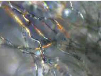
FIG. 2: (Color online) Optical microscope image of the criss-cross of transparent filaments which form the white cottony pads covering the edelweiss bracts. The image was recorded in transmitted polarized light. The diameter of the filaments can be estimated near 10 µm in diameter. A slight iridescence can be noted.
disorder found in this wooly material somewhat compli-cate the identification of the mechanisms which lead to the energy absorption but, as an attempt of clarification, we will report on optical reflection and transmission ex-periments, focusing on the properties of the thin wooly coating of the plant, rather than on the cells forming the living tissue. Simulations will be carried out in support of the understanding of the outcome of these measure-ments. The cultivated specimen of Leontopodium nivale used in this study has been obtained from the “Jardin Alpin du Lautaret”, France. It is kept in the herbar-ium of the National Botanic Garden of Belgherbar-ium (BR ; Vigneron & Ertz no
7065).
II. MICROSCOPIC STRUCTURE OF A
LEONTOPODIUM PUBESCENT BRACT
SURFACE
The surface of the bracts is covered by a macroscopic layer of white cottony pads. The thickness and density of this hair layer is highly variable. Our experiments were conducted on cultivated samples with a rather low hair density. The optical microscope easily reveals the dis-ordered criss-cross structure of the transparent filaments which forms this white pads layer. The image on Fig. 2 is taken on the border of a bract, where the filaments are rarefied, under transmission illumination. A linear polarization of the illuminating light resulted in an in-crease of the image contrast. This polarized image also shows some iridescence, which may indicate an optical structure at a finer scale. Fig. 3 shows a slightly dif-ferent view of the criss-cross structure. Here, the white pads has been detached from the bract, and observed as a suspension in water. The filaments are clearly seen to
FIG. 3: (Color online) Optical microscope image of the white pads, detached from the edelweiss bract, suspended in water and illuminated in transmission. The filaments clearly appear transparent. From this image, it can be inferred that the transparent materials making the filaments has a refractive index very slightly higher than that of water.
be transparent, with a refractive index not very different from that of water. This picture indicates that the mate-rial which makes the filament exhibits a refracting index close to 1.4, a value which later in this work, will be used to support simulations.
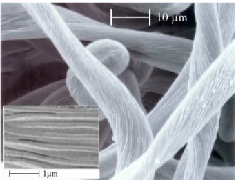
Optical properties are however not determined at this length scale. In order to better know how light waves behave in the white pads, it is necessary to obtain in-formation at magnifications showing submicron details. A scanning electron microscope (SEM) can be used for this purpose. Fig. 4 shows the filaments under the SEM, revealing their longitudinal structure.
The filament transverse section does not show a con-stant shape: the changes of apparent diameter along the length of the filament actually correspond to rather strong deformations. The filaments appear to change their section shape along their length. This is only pos-sible because the filaments are found to be hollow. This has been confirmed by carefully shaving the bract surface and examining the remaining stump (see Fig. 5). This observation indicates a wall thickness below the microm-eter significantly smaller than the filament diammicrom-eter.
The second observation is the transverse structuring of the filament, whose surface clearly shows a peripheral array of parallel fibres. The diameter of these fibres is of the order of 0.18 µm, the order of magnitude of the wavelengths of near ultraviolet radiation. The filament is then possibly a photonic structure whose optical proper-ties may influence the UV reflectance and transmittance of the hair layer covering the bract.
In the longitudinal direction, the spatial structure varies slowly, when compared to the variations found in the transverse direction. A reasonable view of this structure is then to consider it as a two-dimensional pho-tonic structure, though recognizing that, in reality, some
FIG. 4: Scanning electron microscope picture of the fila-ments covering the leaflets surrounding the Leontopodium ni-vale bracts. Note the submicron structure of the filaments (inset) : with a high-magnification the filament surface ap-pears to be outfitted with an array of thin parallel fibres, about 0.18 µm in diameter.
FIG. 5: Scanning electron microscope picture of the remains of the filaments after the hairs have been cut out. The base of each filament appears as a small crater, indicating that the filament is a hollow structure (upper inset). The lower inset shows the bottom of the scar left by the removal of a hair.
roughness exists along the “invariant” direction. A sim-plified view of the filaments which constitutes the white hair layer considers two distinct length scales : the large scale structure, with typical inhomogeneities spanning 10 µm or more, and the small scale structure, with typ-ical lengths well under a micrometer, which appears as a much more ordered structure reminiscent of photonic-crystal optic fibers.
FIG. 6: (Color online) Reflectance of light, for normal in-cidence, on an edelweiss bract. The dashed line reports a specular reflection, while the solid line reports a reflection in-tegrated over all outgoing directions. The reflectance is nearly constant over the whole visible range of wavelengths (explain-ing the white appearance of the bracts). It is found very weak in the ultraviolet range, just below 400 nm. In this range, the bracts do not transmit any wavelength, so that the reflectance deficiency indicates a global radiation absorption.
III. REFLECTANCE AND ABSORPTION IN A
COMPLETE EDELWEISS BRACT
The reflectance spectra of a complete leaflet taken from the star-like organ surrounding the edelweiss flowers have first been investigated using an Avaspec 2048/2 fiber op-tic spectrometer. Measurements were performed, under quasi-normal incidence, in the specular direction and, us-ing an integration sphere, accumulatus-ing all scatterus-ing di-rections. The results are shown on Fig. 6. Specular and integrated reflectances are very similar and show a constant value over the whole visible range, except for a slight jump observed near 700 nm. This spectral profile explains in a very direct way bright-white appearance of the bracts. But the most spectacular reflection variation occurs when entering the near-ultraviolet region, where it is found that no light is actually back-scattered.It is important to note that bracts are rather thick leafs and, in transmission, they turn out to be opaque to all opti-cal radiations. The missing ultraviolet radiation is then actually absorbed and this may be harmful to the plant, if the ultraviolet light penetrates into the living cells of the leaf and produces damage there.
An important issue is then to know whether, in nature, the absorption takes place preferentially in the cells or in the wooly hair on the bracts. We have tried answering this question by separating the green living part of the bract from the white hair layer and perform separate op-tical measurements on these isolated materials. As shown on Fig. 7, the green leaf substrate essentially reflects the green band left out with the living-vegetal chlorophyll activity. Note also the reflection band for wavelengths larger than about 700 nm, which can explain the jump of
4
FIG. 7: Light reflectance, for normal incidence, on an edel-weiss bract removed from its hair layer. The spectrum, col-lected in an integration sphere, shows the characteristic green reflection of vegetal cells. Absorption can take place at other wavelengths, including the near ultraviolet range, below 400 nm, exposing the cells to radiation damages.
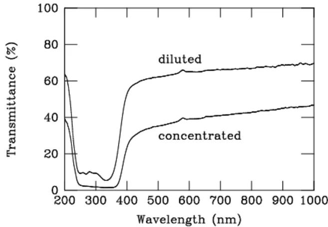
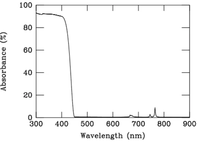
reflectance found on Fig. 6. The surface of the bare leaf is ready to absorb other wavelengths, including the near ultraviolet radiation. This absorption capability could be confirmed by preparing a bracts extract, using a proce-dure described in Martin et al.[2]. Fresh bracts wetted with ethanol, ground in a mortar, were first filtered us-ing Filtrak 1388 filter paper. The transmission spectrum extract(Fig. 8) was recorded after a few days of chloro-phyll photodegradation. The extract could be dried and re-dissolved in ethanol without altering the transmission spectrum. This measurement confirms the sensitivity of the living cells to the UVA radiations.
The observation of absorption in the leaf cells justifies the assumption of possible chemical damages occurring at these UVA wavelengths, so that the plant does need a protection in the high-altitude environment. In the next section, we investigate more specifically the possi-ble shielding offered by the wooly hair layer covering the bract.
IV. REFLECTANCE AND TRANSMITTANCE
OF THE LAYER OF WHITE FILAMENTARY PADS
The layer of cottony hair covering the bract was care-fully removed and rearranged on a glass plate so that the thickness and the density of the layer do not exceed those found on the living plant. Reflectance and transmittance measurements were then performed, using an integration sphere. By recording the transmission of the bare glass slides under identical conditions we checked that the sup-porting glass did not induce detrimental absorption till 300 nm UV radiation. The reference beam was defined in presence of the glass support, so that any variations of optical properties due to the wavelength dependence of
the glass substrate was corrected for. The result is shown on Fig. 9 and Fig. 10 for reflectance and transmittance, respectively. The distribution of the reflectance over the near-ultraviolet and visible spectra does not differ much from that obtained in vivo, when the filaments layer rests on the naked plant leaf. The reflectance is still constant over the whole visible spectrum, above 400 nm. The same disappearance of reflectance is also noticed in the ultraviolet band between 300 and 400 nm. The prepara-tion could be kept for several weeks without noticeable changes of its optical properties.
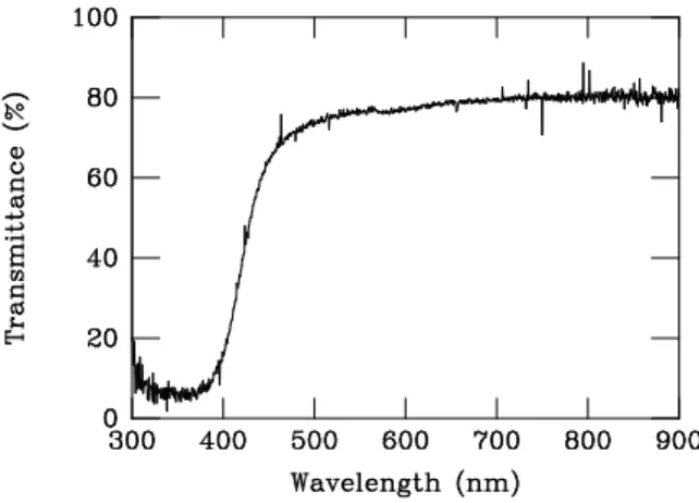
Even more instructive, we find the same kind of filter-ing function in transmission. The transmission through the wooly layer is rather uniform over the whole visible range but presents a pronounced dip in the ultraviolet region between 300 nm and 400 nm. The deficiency of reflectance and transmittance in this range of ultraviolet radiation means that a strong absorption occurs there. Note that by “absorbed” we mean radiation which looses energy while transiting through the filament wall, but also, eventually, radiation which first turns into guided waves in a Fano resonance process (see below) and then disappears with the lifetime of the guided mode. In the visible part of the spectrum, the sum of reflectance and transmittance is close to 100%, so that very little absorp-tion is actually expected. The wooly layer tends to pro-tect the plant from near ultraviolet (UVA, from 320 nm to 400 nm) but does not hinder the exposition to visible radiation needed to sustain biological processes. The at-tenuation of the visible spectrum is essentially controlled by the reflection and not by the absorption.
The sources and setup used in the present experiments did not provide access to other ultraviolet ranges (UVB, from 290 nm to 320 nm and UVC, from roughly 100 nm to 290 nm) so that we do not bring any conclusion for
FIG. 8: Transmittance of an extract from the cells found in the bracts living tissue. The “diluted” preparation is a mix of the “concentrated ” extract with an equivalent volume of ethanol. The measurement is done through a quartz cell of-fering an extended transparency in the UV range. The nor-malizing intensity refers to pure ethanol tested in the same conditions.
FIG. 9: Reflection from an isolated layer of white pads de-posited on a glass substrate. The reflectance was measured using an integration sphere at near-normal incidence. Note the reflectance deficiency in the near ultraviolet region.
FIG. 10: Transmission through an isolated layer of white pads deposited on a glass substrate. The transmittance was measured using an integration sphere at near-normal inci-dence. Note the transmittance deficiency in the near ultravi-olet region. Deficiencies in transmittance and reflectance at the same wavelengths implies, ultimately, absorption at these wavelengths.
these ranges of deeper UV radiations. In order to better understand the mechanisms involved in the optical prop-erties just described, we performed a series of numerical simulations based on a simple model of a filamentary sur-face. These simulations are reported in the next section.
V. THEORETICAL MODEL FOR THE
ABSORBANCE OF FILAMENTARY SURFACES
The structure described by the scanning electron mi-croscope is highly complex because it exhibits inhomo-geneities at very different length scales. The submicronic fibres in the filament is only a fraction of micrometer in diameter; the filament itself has a diameter of about
FIG. 11: Filament cross section model, as deduced from field-emission scanning electron microscopy. The structure is a hollow tube, about 10 µm in diameter. An array of well-separated parallel fibers with a lateral diameter of 176 nm is attached to the external surface of the tube. In the calcu-lations, the curvature of the wall is neglected. The wall is viewed as a grating which is shown on the right. The grat-ing period is taken to be a = 420 nm. The other parameters h= 410 nm and d = 400 nm have less incidence on the grat-ing optical properties. The refractive index of his structure is chosen to be 1.4. The incidence plane orientation with re-spect to the direction of the grating corrugation is measured by the angle φ, as shown.
ten micrometers, and the filaments form a disordered medium which extends over distances a thousand times larger. We will not attempt to produce a model which accounts for all aspects of light scattering at these various length scales, but rather focus on the role of the submi-cronic surface corrugation on the radiation absorption.
We then only consider the surface structure of the fil-ament, which we assume perfectly ordered. The filament is actually a tube, with a wall a fraction of a micron thick (see Fig. 11). The tube has a diameter of about 10 µm, so that the wall has a rather large curvature radius. The curvature can then be neglected, and we end up repre-senting the wall as a planar slab bearing a grating-like surface corrugation. As the inset of Fig. 4 shows, the submicronic structure of the filament surface can be de-scribed as a collection of parallel fibers with a very small and constant diameter, attached on a more or less flat surface. The examination of these fibers allows to esti-mate their average diameter, 0.176 µm. The refractive index of the cylinders is taken to be 1.4, slightly higher than that of water in the visible. The geometry of the model is summarized on Fig. 11. From the filament cross-section sketched on the left, the ideal grating pro-file is constructed and shown in perspective, on the right inset. The curved parts of this profile are semi-circles and the protrusions width is given the value 0.176 µm, as mentioned before. Keeping the same typical dimen-sions, a slightly different profile (using a gaussian shape
6 of the protrusions) has also been studied, with the
con-clusion that minor modifications of the profile could not bring any significant change in the grating response.
We focus the investigations on the reflection of visi-ble and ultraviolet electromagnetic waves. It should be repeated that this model does not contain all features shown on Fig. 4 and that characteristics such as the fi-bre longitudinal irregularities or the randomness of the filament orientation are not accounted for.
The reflectance of this photonic structure is computed using a transfer matrix method in a plane wave represen-tation. A brief account of the method has been presented elsewhere[8, 9, 10] so that we will not recall any techni-cality about these computations.
We consider a monochromatic plane wave impinging on the wall surface. The incidence plane crosses the fi-bres at right angle and the angle of incidence (from the normal) is a typical 45o
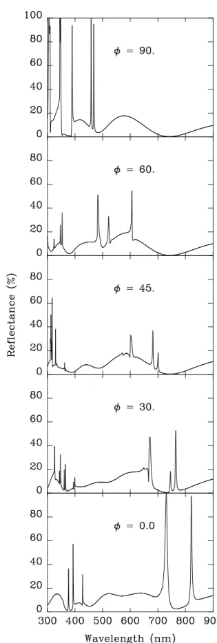
. In all the results described here, the transverse-electric (TE) incident-wave polarization is presented. Transverse-magnetic (TM) polarization was also examined but will not be reported in detail : the spectra are very similar and this analysis brings no fur-ther insight. The reflectance is shown on Fig. 12, as a function of the incident wavelength for several orienta-tions of the incidence plane (φ is the angle made by the incidence plane with the direction of the corrugation). These spectra show rather sharp structures superimposed to a broader background which reaches a high value of about 20%.
For our purpose, the most noticeable part of the spec-trum is the near-ultraviolet region, between 300 and 400 nm. The sharp, asymmetric lines which appear there are related to the Bragg scattering of modes which conduct light through the network of peripheral fibres. Long-lived guided modes are normally not seen in the re-flectance spectrum of a flat film, as they are associated with evanescent waves in the incidence and emergence re-gions surrounding the film. As is well known the separa-tion of guided modes and radiative modes in a planar slab or a perfect wave guide is a consequence of the full trans-lational invariance of the film when displaced parallel to itself. A lateral roughness makes this separation inappro-priate, and allows some degree of hybridization between these two distinct classes of modes. As a consequence, the sharp guided modes become leaky and, conversely, can be loaded with energy by an external radiation. It was shown[11] that the superposition of the guided pho-tonic modes with the non-resonant transiting radiation give rise, in the transmission and in the reflection spec-tra, to asymmetric lines whose shape has been referred to as Fano profiles[12]. The presence of these easily rec-ognizable structure denote the possible energy transfer from incident waves into guided modes.
As seen on Fig. 12 these modes accumulate in the short-wavelength, near-UV, part of the spectrum, dis-turbing the regular pattern of Fabry-Perot resonances. The presence of these peaks in the UV part of the spec-trum simply means that the “176 nm” fibers attached
FIG. 12: Reflectance of a slab structured as shown on Fig. 11, for a light wave impinging at 45o
from normal in an inci-dence plane perpendicular which makes various angles φ with direction of the grating corrugation (TE polarization).
bly leads to absorption in both ranges (UV and visible) of the spectrum. The absorption actually depends on the dissipation capabilities of the material traversed by the radiation, once entered in the filament. This issue is further discussed in the next section.
VI. RADIATION ABSORPTION
It is unclear whether the ultraviolet absorption can take place in the filament wall itself (which should then be characterized by a complex value of its refractive in-dex) or whether the filament hollow core actually con-tains an absorbing agent. However, the absorption of these high-energy radiation (3-4 eV) is likely to cause chemical damage and this mechanism should be confined to wavelengths beyond the high-energy end of the visible spectrum.
A simple plausible mechanism of absorption involves the possible presence of water inside the hollow filaments. Pure water is known to absorb all electromagnetic radi-ations in the wavelengths range from kilometers to fem-tometers, except for those falling in the narrow window of the visible spectrum[13]. The absence of absorption in this biologically important range is just due to the lack of excitation mechanisms, between vibrational and electronic transitions of the water molecule. The absorp-tion coefficient increases rapidly below 400 nm and will reach very high values (up to 106 cm−1) near 150 nm. In the near-UV range, molecular electronic excitation al-ready contributes to destroy transparency so that water could well be the “agent” suggested above. However, it is somewhat doubtful that the absorption coefficient of pure water can already reach the values of α ≃ 104 or 105 cm−1 needed to turn 10 µm thick filaments into efficient absorbers, above 300 nm. For deeper UV ra-diation, UVB and UVC, there is no doubt that such a mechanism would be quite efficient but it remains ques-tionable for UVA. On the other hand, it is also unlikely that the physiological liquid assumed to be filling the fil-ament would actually be pure water. Unfortunately, the detection and analysis of the content of the filaments will not be an easy operation, due to the very small size of
the infrared and visible recordings have failed to show any significant absorption, while the UVA absorption ap-proaches 100%. As a consequence, a constant value of ε′′ is not realistic and a dissipative response which rapidly shifts from high to low values at the UV onset would better represent the observed absorption. Without such a shift, the edelweiss bracts would absorb nearly as much in the visible range as it absorbs the near UV and, most likely, would appear dark. Fig. 13 describes the com-puted absorption of the structure shown on Fig. 11, when the dielectric function imaginary part ε′′ shifts from a value 0.4 in the UV range below 400 nm to a “trans-parent” value of 0.001 maintained above 450 nm in the visible and infrared ranges. The molecular origin of this abrupt change is more than likely. As we can see, the physical origin of the whitening of the plant leaves in-volves the sharp disappearance of a dissipative response between the near-UV and visible ranges. This means that, besides the purely structural effects the “pigmen-tary” (i.e. material-based rather than structural) mech-anisms play a significant role in determining the bracts coloration. The detailed analysis of this aspect of the ab-sorption process would require a detailed in vivo study of the nature of the filament wall which is not yet available to date.
VII. CONCLUSION
Photonic structures have been often found in nature[14, 15]. Classic examples include the iridescence of insect cuticle[16, 17] (mainly butterflies, beetles, wee-vils, ...) or the changing colors of bird feathers[18]. These photonic structures are most often the origin of bright signalling colors but, occasionally, they can be in-volved in the production of criptic colors, as in the but-terfly Cyanophrys remus[19]. In other cases, the pho-tonic structure serves more physiological purposes, like the control of thermal energy exchange[20].
Photonic structures are not so common in plants, al-though some examples have been clearly described[21, 22, 23]. The observations presented here tend to show that Leontopodium nivalecould, from both the structural and
8
FIG. 13: Absorption (in percents) of radiation on the model slab structured shown on Fig. 11, for an incidence transverse-electric wave impinging at 45o
from normal in an incidence plane which makes a angle φ = 300
with the direction of the grating corrugation. The material in the model is given a complex dielectric constant, with an imaginary part which shifts from the value ε′′= 0.4 adequate for ultraviolet regions
below 400 nm to ε′′ = 0.001 adequate for the infrared and
visible ranges, above 450 nm.
optical points of view, be seen as another example. This plant has developed a layer of filamentary hair which cov-ers the bracts surrounding the flowerheads and, with less density, the entire aerial part of the plant. This fleece protects from dehydration and cold, but also turns out to shield the covered living cells from harmful ultravio-let radiations. This protection is not obtained, as some-times mentioned, by reflection but rather by absorption within the protective hair layer. The picture suggested here to explain the absorption involves a selective ultra-violet dissipation assisted by diffraction effects : the fi-brous structure of the external surface of the filaments not only provides a guiding support of ultraviolet radi-ation but also brings the roughness required for energy exchange between the incident waves and these guided modes.
With this structure, nature may have found here a
clever solution to an engineering problem of wide appli-cation. Ultraviolet screening is of prime importance for the design of many functionalized materials. Examples can be given for packing materials, anti-ultraviolet sun screener for cosmetics, anti-ultraviolet powder for car or construction paints where the ultraviolet absorption is of paramount importance. Many of these applications rely on TiO2 nanoparticles (∼ 10 − 50 nm) which may be difficult to handle, especially under the requirement of biocompatibility. The kind of structure developed by Leontopodium nivale may indicate an interesting way to provide a strong ultraviolet absorption with larger (but structured) particles whose location can be much more easily stabilized.
Acknowledgments
This work was carried out with support from EU5 Centre of Excellence ICAI-CT-2000-70029 and from the Inter-University Attraction Pole (IUAP P5/1) on “Quantum-size effects in nanostructured materials” of the Belgian Office for Scientific, Technical, and Cultural Affairs.
We acknowledge the use of Namur Interuniversity Sci-entific Computing Facility (Namur-ISCF), a common project between the Belgian National Fund for Scientific Research (FNRS), and the Facult´es Universitaires Notre-Dame de la Paix (FUNDP).
M.R. has benefitted from a grant for research train-ing at the Laboratoire de Physique du Solide (LPS) of the Facult´es Universitaires Notre-Dame de la Paix (FUNDP).
ZV, LPB and KK wish to thank the Hungarian Academy of Sciences and the Belgian FNRS for finan-cial support.
V.L. was supported as research Fellow by the Belgian National Fund for Scientific Research (FNRS). She is a recipient of a postdoctoral fellowship from the Belgian-American Educational Foundation.
The authors thank Amand Lucas (FUNDP) and Paul Thiry (FUNDP) for critically reading the manuscript and for stimulating discussions.
[1] W. Greuter, Willdenowia 33, 239 (2003).
[2] R. Martin, B. Belcourt, P. Hilaire, and R. Rozot, United State Patent Application Publication US 2003, 0082117/A1 (2003).
[3] M. Dobner, S. Schwaiger, I. Jenewein, and H. Stuppner, Journal of ethnopharmacology 89, 301 (2003).
[4] C. Starr and R. Taggart, Biology: The Unity and Di-versity of Life (Thomson - Brooks/Cole 10650 Toebben Dr. Independence KY41051 (ISBN: 0-534-38800-0) Tenth Edition, 2004).
[5] J. C. Knight, T. A. Birks, P. S. J. Russel, and D. M. Atkin, Opt. Lett. 21, 15471549 (1996).
[6] J. C. Knight, T. A. Birks, and P. S. J. Russel, Opt. Lett.
22, 961963 (1997).
[7] J. D. Joannopoulos, R. D. Meade, and J. N. Winn, Mold-ing the Flow of Light (Princeton University Press, Prince-ton, 1995).
[8] V. Lousse and J. P. Vigneron, Phys. Rev. B 69, 155106 (2004).
[9] J.B. Pendry, and A. MacKinnon, Phys. Rev. Letters 69, 2772 (1992).
[10] V. Lousse, Etude th´eorique des ph´enom`enes de bistabilit´e dans les structures optiques non lin´eaires int´egrant des cristaux photoniques (PhD thesis) (Presses Universitaires de Namur, Namur, Belgium, 2003).