HAL Id: tel-03049056
https://tel.archives-ouvertes.fr/tel-03049056
Submitted on 9 Dec 2020
HAL is a multi-disciplinary open access
archive for the deposit and dissemination of sci-entific research documents, whether they are pub-lished or not. The documents may come from teaching and research institutions in France or abroad, or from public or private research centers.
L’archive ouverte pluridisciplinaire HAL, est destinée au dépôt et à la diffusion de documents scientifiques de niveau recherche, publiés ou non, émanant des établissements d’enseignement et de recherche français ou étrangers, des laboratoires publics ou privés.
Valorization of natural drug products : from extraction
to encapsulation
Duc Hung Nguyen
To cite this version:
Duc Hung Nguyen. Valorization of natural drug products : from extraction to encapsulation. Pharma-cology. Université Bourgogne Franche-Comté, 2020. English. �NNT : 2020UBFCJ003�. �tel-03049056�
Duc Hung NGUYEN
A thesis presented for the degree of
Doctor of Pharmacy
Speciality: Pharmaceutical Technology
Members of jury:
Prof. Dominique Laurain-Mattar
Université de Lorraine President/
Rapporteur
Prof. Anne Sapin-Minet
Université de Lorraine Rapporteur
Prof. Odile Chambin
UBFC
Supervisor
Prof. Anne-Claire Mitaine-Offer
UBFC
Co-supervisor
THESE DE DOCTORAT DE L’ETABLISSEMENT UNIVERSITE BOURGOGNE FRANCHE-COMTE PREPAREE AU LABORATOIRE DE PHARMACOGNOSIE PEPITE EA 4267
Ecole doctorale
Environnements - Santé
Doctorat de Pharmacie, spécialité Pharmacognosie
Par
Nguyen Duc Hung
Thèse présentée et soutenue à l’UFR Sciences de Santé de Dijon, salle R46, le 09 Novembre 2020 Composition du Jury:
Prof. Dominique Laurain-Mattar Université de Lorraine Président/ Rapporteur Prof. Anne Sapin-Minet Université de Lorraine Rapporteur
Prof. Odile Chambin UBFC Directeur de thèse
Prof. Anne-Claire Mitaine-Offer UBFC Co-Directeur de thèse
Titre : Valorisation des produits médicamenteux naturels: de l'extraction à l'encapsulation Mots clés : Saponine, Cytotoxicité, Weigela, Cordyline, Dracaena, Curcumine,
Microencapsulation, Activité antioxydante. Cette thèse s’inscrit dans la cadre de la thématique du Laboratoire de Pharmacognosie et du Laboratoire de Pharmacie Galénique de l’UFR Sciences de Santé, circonsription Pharmacie, au sein de l’Université de Bourgogne Franche-Comté, afin de trouver de nouvelles molécules naturelles bioactives à encaspuler. D’une part, nous nous sommes concentrés sur la recherche de saponines naturelles de plantes issues de la biodiversité vietnamienne et de l’horticulture française du genres Dracaena,
Cordyline (Asparagaceae) et Weigela
(Caprifoliaceae). Les travaux menés ont conduit à isolement de 35 saponines naturelles en utilisant différentes techniques chromatographiques. Les structures ont été déterminées par des méthodes de spectrométrie de masse en source ESI et de spectroscopie RMN. Parmi les 17 composés purs obtenus des 3 espèces appartenant au genre Weigela, 9 sont des glycosides de l’acide oléanolique et de l’hédéragénine de structure nouvelle. A partir des espèces
Dracaena braunii et Cordyline fruticosa
“Fairchild red”, nous avons isolé et caractérisé 18 saponines stéroïdiques dont 7 nouvelles de type spirostane et 6 nouvelles de type furostane. Les activités cytotoxiques de la majorité des saponines isolées ont été évaluées sur trois lignées cellulaires : CT26 (cellules tumorales coliques murines), B16 (cellules de mélanome murin) et HepG2 (cellules d’hépatocarcinome) par le test de prolifération cellulaire MTS. Des relations structure/ activité ont ainsi été proposées.
D’autre part, nous avons sélectionné une molécule naturelle bien connue afin de mettre au point les essais d’encaspulation. La curcumine possède des propriétés thérapeutiques très intéressantes mais elle présente à la fois une faible solubilité et une faible biodisponibilité, limitant l'administration par voie orale. Dans cette partie de la thèse, nous avons cherché à améliorer la stabilité et la biodisponibilité de la curcumine ansi qu’une libération contrôlée dans le tractus gastro-intestinal. Des billes de pectinate de calcium ont préparées en utilisant la gélification ionique en présence de différents tensioactifs. Elles ont été caractérisées grâce à leur propriétés physico-chimiques et leur profils de dissolution in vitro. Leur activité antioxydante a été évaluée par le test DPPH. Le Kolliphor® HS 15 est le tensioactif le plus prometteur pour optimiser les propriétés de la curcumine.
Title : Valorization of natural drug products: from extraction to encapsulation
Keywords : Saponins, Cytotoxicity, Weigela, Cordyline, Dracaena, Curcumin, Microencapsulation, Antioxidant actitivy.
This thesis was carried out at the Laboratory of Pharmacognosy and the Laboratory of Pharmaceutical technology, at the UFR Sciences de Santé, circonscription Pharmacy, in the University of Burgundy Franche-Comté, to find new natural molecules to encapsulate. First of all, we focused on the natural saponins from plants of the Vietnam biodiversity and the French horticulture, belonging to the three genera
Dracaena, Cordyline (Asparagaceae) and Weigela (Caprifoliaceae). The work led to
the successful isolation and elucidation of 35 natural saponins using various chromatographic techniques. The structures were determined by ESI mass spectrometry and NMR spectroscopy. Among the 17 pure compounds obtained from three species of the Weigela genus, 9 oleanolic acid and hederagenin glycosides are previously undescribed ones. From the two species
Dracaena braunii and Cordyline fruticosa
“Fairchild red”, we isolated and characterized 18 steroidal saponins including 7 new spirostane-types and 6 new furostane-types ones. The cytotoxic activities of the majority of isolated saponins were evaluated against mouse colon cancer (CT26 cells), mouse melanoma (B16 cells) and human liver cancer (HepG2 cells) by MTS assays. The structure / activity relationships were also proposed.
On the other hand, we selected a well-known natural molecule to develop encapsulation tests. Among the natural products, curcumin has very interesting therapeutic properties but exhibits both a poor solubility and a low bioavailability, limiting the administration by the oral route. The purpose of this study was to improve the solubility and bioavailability of curcumin as well as simultaneously achieve controlled release in gastrointestinal tract. Pectinate gel beads were prepared based on ionotropic gelation method with the presence of vaious surfactants. After drying, these beads were investigated for physicochemical characteristics (morphological aspects, encapsulation efficiency, stability, physical state), dissolution kinetics (in vitro release) and antioxidant activity was determined with DPPH assay. Kolliphor® HS 15 seems to be the best promising surfactant to increase stability and bioavailability of curcumin.
Université Bourgogne Franche-Comté 32, avenue de l’Observatoire
AKNOWLEDGEMENTS
My sincere appreciation is addressed to my Ph.D supervisor Prof. Odile Chambin. I would like to express my deep and sincere thankfulness for giving me the opportunity to work on my thesis in such creative research environment. Her continuous support, guidance and encouragements help me stand on my feet and climb through the ups and downs of my PhD journey which would has never been this far without her wise advices. I am also deeply grateful to my second supervisor, Prof. Anne-Claire Mitaine-Offer, who coordinated, guided and inspired me through my research work. With her enthusiasm, cheerful character and excellent encouragement, she always showed me how to see problems during my research and taught me how to find solutions.
I owe my most sincere gratitude to Prof. Marie-Aleth Lacaille-Dubois, Director of Laboratory of Pharmacognosy, Université de Bourgogne. She was always willing to help with her experience and supported me with tips and analytical tricks throughout my research.
I also express my thankfulness to Prof. Marie-Pierre Flament and Prof. Yves Wache, two members of thesis supervisory committee. They offered advices about and assessment of my research.
I would like to thank all colleagues in Université de Bourgogne for providing a stimulating and nice environment. I also warmly thank the Vietnamese Government, for providing me the scholarship, and my colleagues in Department of Biology, University of Education, Thainguyen University, Vietnam for their encouragement.
The laboratory of PAM, especially to Physico-chemistry of Food and Wine (PCAV) team, Agrosup Dijon and Dr. Gaëlle Roudaut are acknowledged for inspiring me at the beginning of PhD journey.
Special thanks go to Mr. Bastien Petit, for being a great lab mate throughout my whole PhD journey in Dijon. Bastien, my friend, I will never forget the words “Paradise” and “Cafélisto” for the rest of my life. Thank you for all the crazy moments we have ever done!
I also warmly thank Mr. David Pertuit, a great technician, for supporting me during the fraction and isolation process. I am sincerely grateful for his patience and long hours spent to teach me how to use all equipments.
Futhermore, I am also greatful to the colleagues of laboratory of Pharmacognosy: Dr. Tomofumi Miyamoto, Dr. Chiaki Tanaka, Dr. Thomas Paululat for the measurements of the diverse NMR and MS experiments. Thankfulness is given to Dr. Bertrand Collin and Dr. Pierre-Simon Bellaye for help in cytotoxic activity.
To my lovely wife…
My wonderful wife Huyen could not be here during my PhD journey but she has always encouraged me and offered me the support that I needed to complete this thesis. I would have dropped out this journey and would not have written these words if I had lived without her. Huyen, my darling, you showed me what it means to love and to be loved, I am a lucky person with you. In the deepness of my heart, I give you a great love and appreciation for your faithful patience and all the moments we shared together, I thank you for being a half of my life.
…my sons…
Nam, my second son, I started my PhD journey while you were still in your mother’s womb. I am sorry you, my son, for one reason that I could not be there when you were born. It can be the biggest regret of my life.
To Lam, my first son, I still remember the day I had to say goodbye to you. It has been an unforgettable in my mind. I hope you will understand my situation and excuse my absence at home.
My gratefulness address to my parents who have been supporting me in each and every step and breath I take, to my mother Ngan who gives me encouragement through her never-ended good wished, pray and emotional support, and to my father Hong whose kindness, inspiration always be in my heart. My gratitude is also forwarded to my big brother Ha and his wife Thao for their continuous tenderness and encouragement. To my mother-in-law Chien, my aunts-in-law Khay and Chin, my sister-in-law Ha and her husband Dat, receive my deep gratitude and love for your dedication and trust in me.
I thank to my aunt Hoa and two my uncles Tuan and Dung, you showed me the importance of education and helped me find the right way on every step of my life.
Last, but not least, to Madam Agnès Roy, receive my great gratitude and love for your supports during my whole schooling. Not only did you supply me with energy and motivation every time I depressed but also you taught me languages and the significance of life and love. Agnès, you have always been the one to make me better. For your love and endless support, I am happy to dedicate this thesis to you.
Finally, to someone who have not been here for any reason, I want you know that you have been always the ones to help me to reach this far in my career. Thank you for being a part of my life.
Duc Hung NGUYEN Dijon, 2020
Dedication
I am so proud to dedicate this thesis
To my lovely wife and my children who have always encouraged me throughout my
PhD journey…
To my whole family...
GENERAL INTRODUCTION
Nowadays, chemistry and biology laboratories around the world are studying on natural herbal medicines due to their safe to treat various diseases. There are several evidences to prove the use of traditional medicine in different cultures. Despite this historical importance of plants and considerable contemporary research into the identification of new naturally occurring chemical compounds, many of those are still not revealed both chemical structures and pharmacological activities. Saponin, a naturally occurring surface active glycoside, has been reported in recent years of publications about their properties. These compounds were discovered for their biological activities, from traditional uses to pharmaceutical applications. Saponins possess a various of pharmaceutical potentials, such as antitumor, anti-inflammatory, antiviral, antioxidant and antibacterial (Moghimipour and Handali, 2015). In this context, the Laboratory of Pharmacognosy, EA 4267, at the University of Burgundy Franche-Comté, is focusing on research of biologically active saponins from various plant families. Our research strategy consists in the selection of families and genera known for their richness in saponins, based essentially on chemotaxonomic criteria. Then, after extraction, isolation and structural characterization, the natural molecules are subjected to in vitro biological evaluation mainly in the field of cancerology. Due to these steps, three genera Dracaena, Cordyline and Weigela of two families Asparagaceae and Caprifoliaceae, were chosen for phytochemical investigation. It is interesting to note that many traditional uses of several species belonging to these genera present a potential in the pharmaceutical domain. Saponins were extracted and identified by using various chromatographic methods. These compounds were further evaluated for their cytotoxic activities and the analysis of relationships between structure and activity were carried out.
Aglycon (or sapogenin), an hydrophobic portion of saponin, has gained importance as a novel drug. But, their major limitation of its poor aqueous solubility leads to a relatively short half-life, low bioavailability and less permeability and degradation during human digestion. These problems are able to be overcome by various encapsulation method (Anand et al., 2007). However, extracted saponins is not enough to provide a good quantity,
so encapsulation are not carried out with this compound. Fortunately, curcumin, the principal curcuminoid found in turmeric which exhibits a wide spectrum of biological and pharmacological effects, is also a hydrophobic natural compound with low water solubility quite similar to saponins. We pointed out here an idea of encapsulation of curcumin instead of sapogenin in order to improve the solubility and bioavailability of hydrophobic compounds. This study was done using ionotropic gelation method with the presence of surfactants. Physicochemical characteristics, dissolution kinetics and antioxidant activity were further evaluated for efficacy of encapsulation to enhance the solubility and protect antioxidant capacity of curcumin, in view of future food applications.
This present thesis is structured by two parts:
- The first part will be carried out on saponin including three chapters. The first chapter will concern a literature review about botanical study and previous phytochemical works. The second chapter will give phytochemical investigations on selected plants. The third chapter corresponds to biological study on isolated compounds, in collaboration with Centre Georges-François Leclerc, ICMUB UMR CNRS 6302, using MTS colorimetric assay on different colon cancer cell lines.
- The second part will give a study on encapsulation of curcumin including three chapters. The first chapter will inform about chemical profile of curcumin and general background about some encapsulation methods. The second chapter will present the methodological and material tools used to manufacture beads, together with investigations of physicochemical characteristics, dissolution kinetics and antioxidant activity of beads. The third chapter will show the results and discussion.
A general conclusion also will be carried out at the end of this thesis providing an overview on results and propose perspectives.
CONTENT
Introduction ... 1
Chapter 1. Botanical studies and previous phytochemical works ... 3
1. Botanical study ... 4
1.1. Order Dipsacales ... 4
1.1.1. Family Caprifoliaceae ... 4
1.1.2. Genus Weigela ... 5
1.1.3. Some horticultural species of genus Weigela ... 6
1.2. Order Asparagales ... 7
1.2.1. Family Asparagaceae ... 8
1.2.2. Genus Dracaena ... 9
1.2.3. Genus Cordyline ... 10
2. Previous phytochemical analysis ... 11
2.1. Saponin generalities ... 11
2.2. Saponins isolated from genus Weigela ... 15
2.3. Saponins isolated from genus Cordyline ... 17
2.4. Saponins isolated from genus Dracaena ... 20
Chapter 2. Phytochemical study ... 25
1. Materials and methods ... 26
1.1. Materials and extraction ... 26
1.2. Methods of isolation ... 27
1.2.1. Chromatographic methods of analysis ... 27
1.2.2 Preparative chromatographic methods ... 28
1.3. Methods of structural determination ... 31
1.3.1. Mass spectrometry (MS) ... 31
1.3.2. Electro-Spray Ionization (ESI) ... 32
1.3.3. Nuclear magnetic resonance (NMR) spectrometry ... 32
1.3.4. Acid hydrolysis and GC analysis ... 35
2. Phytochemical investigation ... 35
2.1.1. Isolation and purification ... 35
2.1.2. Structural determination of isolated saponins ... 37
2.2. Phytochemical study of Weigela florida “Pink Poppet” ... 77
2.2.1. Isolation and purification ... 77
2.2.2. Structural determination of isolated saponins ... 79
2.3. Phytochemical study of Weigela florida “Jean’s Gold” ... 88
2.3.1. Isolation and purification ... 88
2.3.2. Structural determination of isolated saponins ... 89
2.4. Phytochemical study of roots of Cordyline fruticosa “Fairchild red” ... 103
2.4.1. Isolation and purification ... 103
2.4.2. Structural determination of isolated saponins ... 104
2.5. Phytochemical study of aerial parts of Cordyline fruticosa “Fairchild red” .. 151
2.5.1. Isolation and purification ... 151
2.5.2. Structural determination of isolated saponins ... 152
2.6. Phytochemical study of roots of Dracaena braunii ... 173
2.6.1. Isolation and purification ... 173
2.6.2. Structural determination of isolated saponins ... 174
Chapter 3. Biological study ... 177
1. Introduction ... 178
1.1. Bioactivities of steroidal saponins from the Dracaena and Cordyline genus 178 1.1.1. Cytotoxicity and antitumor activity ... 178
1.1.2. Other bioactivities ... 180
1.2. Bioactivities of triterpenoid saponins from the Weigela genus ... 181
1.3. Correlation between the structure and cytotoxicity of saponins ... 181
1.3.1. In case of steroidal saponins ... 182
1.3.2. In case of triterpenoid saponins... 183
1.4. Cytotoxic mechanism of saponins ... 183
2. Materials and methods ... 185
2.1. The principle of MTS colorimetric assay ... 185
3. Results of cytotoxic study on isolated and selected saponins ... 186 3.1.Cytotoxic study on the saponins isolated from Weigela x “Bristol Ruby” .... 187
3.2.Cytotoxic study on the saponins isolated from Weigela florida “Pink Poppet” and Weigela florida “Jean’s Gold” ... 190 Bibliography ... 196
List of Figures
Figure 1. Phylogenetic classification of order Dipsacales ... 4
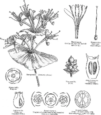
Figure 2. Diagram of Caprifoliaceae flower part ... 5
Figure 3. Weigela x “Bristol Ruby” ... 6
Figure 4. Weigela florida “Jean’s Gold” ... 6
Figure 5. Weigela florida “Pink Poppet” ... 7
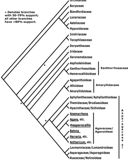
Figure 6. Phylogenetic classification of order Asparagales ... 8
Figure 7. Dracaena braunii ... 9
Figure 8. Cordyline fruticosa “Fairchild red” ... 10
Figure 9. Structure of spirostane aglycon and furostane aglycon ... 12
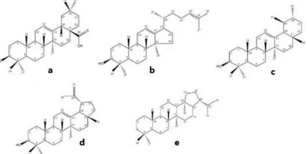
Figure 10. Structure of oleanane- (a), dammarane- (b), ursane- (c), lupane- (d) and hopane-type aglycon (e) ... 14
Figure 11. Available different sugars in saponins ... 15
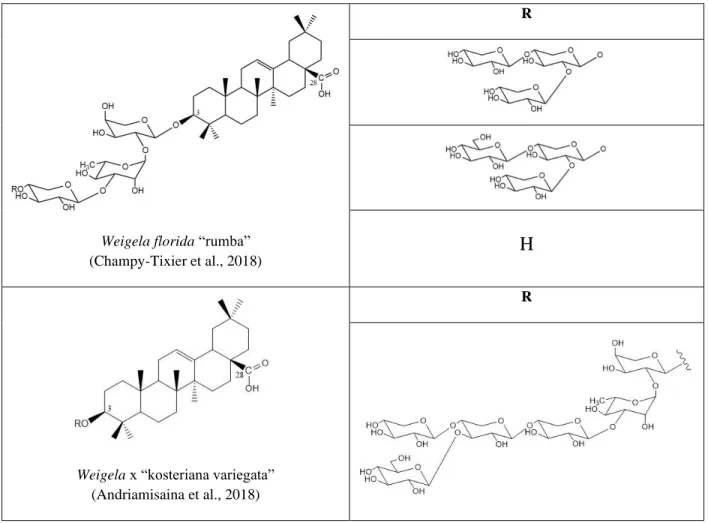
Figure 12. Some triterpenoid saponins elucidated from species of Weigela genus ... 17
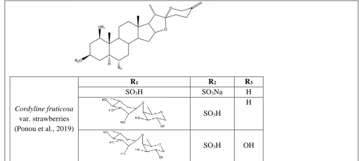
Figure 13. Steroidal saponins elucidated from species of Cordyline genus ... 20
Figure 14. Steroidal saponins elucidated from species of Dracaena genus ... 24
Figure 15. Mechanism of electrospray ionization ... 32
Figure 16. Correlations of HSQC and HMBC between α-L-rhamnose and α-L-arabinose .... 34
Figure 17. Correlations of COSY, TOCSY and ROESY ... 34
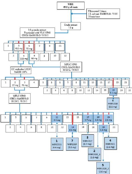
Figure 18. Purification scheme of compounds in roots of Weigela x “Bristol Ruby” ... 36
Figure 19. Structure of compound 1 ... 39
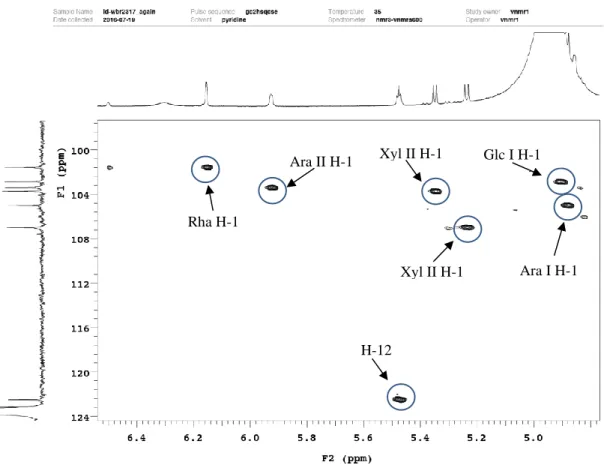
Figure 20. HSQC spectrum of compound 1 ... 40
Figure 21. HSQC spectrum of sugar anomers of compound 1 ... 40
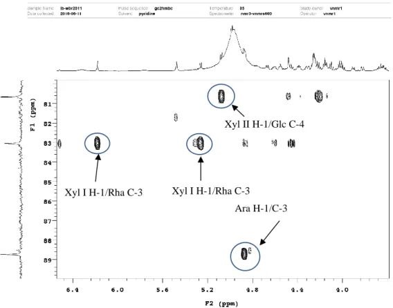
Figure 22. HMBC spectrum of sugar moieties of compound 1 ... 41
Figure 23. HMBC spectrum of sugar moieties of compound 1 ... 41
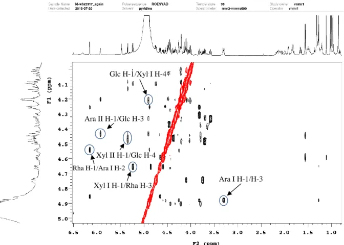
Figure 24. ROESY spectrum of sugar moieties of compound 1 ... 42
Figure 25. ROESY spectrum of sugar moieties of compound 1 ... 42
Figure 26. Mass spectrum of compound 1 ... 43
Figure 28. HSQC spectrum of compound 2 ... 46
Figure 29. HSQC spectrum of sugar anomers of compound 2 ... 46
Figure 30. HMBC spectrum of sugar moieties of compound 2 ... 47
Figure 31. ROESY spectrum of sugar moieties of compound 2 ... 47
Figure 32. ROESY spectrum of sugar moieties of compound 2 ... 48
Figure 33. Mass spectrum of compound 2 ... 48
Figure 34. Structure of compound 3 ... 51
Figure 35. HSQC spectrum of compound 3 ... 51
Figure 36. HSQC spectrum of sugar anomers of compound 3 ... 52
Figure 37. HMBC spectrum of sugar moieties of compound 3 ... 52
Figure 38. HMBC spectrum of sugar moieties of compound 3 ... 53
Figure 39. HMBC spectrum of sugar moieties of compound 3 ... 53
Figure 40. ROESY spectrum of sugar moieties of compound 3 ... 54
Figure 41. Mass spectrum of compound 3 ... 54
Figure 42. Structure of compound 4 ... 57
Figure 43. HSQC spectrum of compound 4 ... 57
Figure 44. HSQC spectrum of sugar anomers of compound 4 ... 58
Figure 45. HMBC spectrum of sugar moieties of compound 4 ... 58
Figure 46. ROESY spectrum of sugar moieties of compound 4 ... 59
Figure 47. Mass spectrum of compound 4 ... 59
Figure 48. Structure of compound 5 ... 61
Figure 49. HSQC spectrum of compound 5 ... 62
Figure 50. HSQC spectrum of sugar anomers of compound 5 ... 62
Figure 51. HMBC spectrum of sugar moieties of compound 5 ... 63
Figure 52. ROESY spectrum of sugar moieties of compound 5 ... 63
Figure 53. ROESY spectrum of sugar moieties of compound 5 ... 64
Figure 54. Mass spectrum of compound 5 ... 64
Figure 55. Structure of compound 6 ... 66
Figure 56. HSQC spectrum of compound 6 ... 67
Figure 58. HMBC spectrum of sugar moieties of compound 6 ... 68
Figure 59. ROESY spectrum of sugar moieties of compound 6 ... 68
Figure 60. Mass spectrum of compound 6 ... 69
Figure 61. Structure of compound 7 ... 69
Figure 62. Purification of compounds in roots of Weigela florida “Pink Poppet” ... 78
Figure 63. Purification of compounds in aerial parts of Weigela florida “Pink Poppet” ... 78
Figure 64. Structure of compound 8 ... 81
Figure 65. HSQC spectrum of compound 8 ... 82
Figure 66. HSQC spectrum of sugar anomers of compound 8 ... 82
Figure 67. HMBC spectrum of sugar moieties of compound 8 ... 83
Figure 68. ROESY spectrum of sugar moieties of compound 8 ... 83
Figure 69. Mass spectrum of compound 8 ... 84
Figure 70. Structure of compound 9 ... 84
Figure 71. Structure of compound 10 ... 85
Figure 72. Structure of compound 11 ... 86
Figure 73. Purification of compounds in leaves of W. florida “Jean’s Gold” ... 89
Figure 74. Structure of compound 12 ... 92
Figure 75. HSQC spectrum of compound 12 ... 92
Figure 76. HSQC spectrum of sugar anomers of compound 12 ... 93
Figure 77. HMBC spectrum of sugar moieties of compound 12 ... 93
Figure 78. ROESY spectrum of sugar moieties of compound 12 ... 94
Figure 79. Mass spectrum of compound 12 ... 94
Figure 80. Structure of compound 13 ... 96
Figure 81. HSQC spectrum of compound 13 ... 97
Figure 82. HSQC spectrum of sugar anomers of compound 13 ... 97
Figure 83. HMBC spectrum of sugar moieties of compound 13 ... 98
Figure 84. ROESY spectrum of sugar moieties of compound 13 ... 98
Figure 85. Mass spectrum of compound 13 ... 99
Figure 86. Structure of compound 14 ... 99
Figure 88. Structure of compound 16 ... 101
Figure 89. Structure of compound 17 ... 101
Figure 90. Purification of compounds in roots of Cordyline fruticosa “Fairchild red” ... 104
Figure 91. Structure of compound 18 ... 107
Figure 92. HSQC spectrum of compound 18 ... 107
Figure 93. HSQC spectrum of compound 18 ... 108
Figure 94. HMBC spectrum of sugar moieties of compound 18 ... 108
Figure 95. ROESY spectrum of aglycon of compound 18... 109
Figure 96. ROESY spectrum of sugar moieties of compound 18 ... 109
Figure 97. Mass spectrum of compound 18 ... 110
Figure 98. Structure of compound 19 ... 111
Figure 99. HSQC spectrum of compound 19 ... 112
Figure 100. HSQC spectrum of sugar moieties of compound 19 ... 112
Figure 101. HMBC spectrum of sugar moieties of compound 19 ... 113
Figure 102. ROESY spectrum of sugar moieties of compound 19 ... 113
Figure 103. Mass spectrum of compound 19 ... 114
Figure 104. Structure of compound 20 ... 115
Figure 105. HSQC spectrum of compound 20 ... 116
Figure 106. HSQC spectrum of sugar moieties of compound 20 ... 116
Figure 107. HMBC spectrum of sugar moieties of compound 20 ... 117
Figure 108. HMBC spectrum of sugar moieties of compound 20 ... 117
Figure 109. ROESY spectrum of sugar moieties of compound 20 ... 118
Figure 110. Mass spectrum of compound 20 ... 118
Figure 111. Structure of compound 21 ... 120
Figure 112. HSQC spectrum of compound 21 ... 120
Figure 113. HSQC spectrum of sugar moieties of compound 21 ... 121
Figure 114. HMBC spectrum of sugar moieties of compound 21 ... 121
Figure 115. ROESY spectrum of sugar moieties of compound 22 ... 122
Figure 116. Mass spectrum of compound 22 ... 122
Figure 118. HSQC spectrum of compound 22 ... 124
Figure 119. HSQC spectrum of sugar moieties of compound 22 ... 125
Figure 120. HMBC spectrum of sugar moieties of compound 22 ... 125
Figure 121. ROESY spectrum of sugar moieties of compound 22 ... 126
Figure 122. ROESY spectrum of sugar moieties of compound 22 ... 126
Figure 123. Mass spectrum of compound 22 ... 127
Figure 124. Structure of compound 23 ... 128
Figure 125. HSQC spectrum of compound 23 ... 129
Figure 126. HSQC spectrum of sugar moieties of compound 23 ... 129
Figure 127. HSQC spectrum of sugar moieties of compound 23 ... 130
Figure 128. ROESY spectrum of sugar moieties of compound 23 ... 130
Figure 129. Mass spectrum of compound 23 ... 131
Figure 130. Structure of compound 24 ... 133
Figure 131. HSQC spectrum of compound 24 ... 133
Figure 132. HSQC spectrum of sugar moieties of compound 24 ... 134
Figure 133. HMBC spectrum of sugar moieties of compound 24 ... 134
Figure 134. ROESY spectrum of sugar moieties of compound 24 ... 135
Figure 135. Mass spectrum of compound 24 ... 135
Figure 136. Structure of compound 25 ... 138
Figure 137. HSQC spectrum of compound 25 ... 138
Figure 138. HSQC spectrum of sugar anomers of compound 25 ... 139
Figure 139. HSQC spectrum of sugar moieties of compound 25 ... 139
Figure 140. HMBC spectrum of sugar moieties of compound 25 ... 140
Figure 141. ROESY spectrum of sugar moieties of compound 25 ... 140
Figure 142. Mass spectrum of compound 25 ... 141
Figure 143. Structure of compound 26 ... 142
Figure 144. HSQC spectrum of compound 26 ... 143
Figure 145. HSQC spectrum of sugar anomers of compound 26 ... 143
Figure 146. HSQC spectrum of sugar moieties of compound 26 ... 144
Figure 148. ROESY spectrum of sugar moieties of compound 26 ... 145
Figure 149. Mass spectrum of compound 26 ... 145
Figure 150. Structure of compound 27 ... 146
Figure 151. Structure of compound 28 ... 146
Figure 152. Purification of compounds in aerial parts of C. fruticosa “Fairchild red” .... 152
Figure 153. Structure of compound 29 ... 154
Figure 154. HSQC spectrum of compound 29 ... 154
Figure 155. HSQC spectrum of sugar anomers of compound 29 ... 155
Figure 156. HSQC spectrum of sugar moieties of compound 29 ... 155
Figure 157. HMBC spectrum of sugar moieties of compound 29 ... 156
Figure 158. ROESY spectrum of sugar moieties of compound 29 ... 156
Figure 159. Mass spectrum of compound 29 ... 157
Figure 160. Structure of compound 30 ... 158
Figure 161. HSQC spectrum of compound 30 ... 159
Figure 162. HSQC spectrum of sugar anomers of compound 30 ... 159
Figure 163. HSQC spectrum of sugar moieties of compound 30 ... 160
Figure 164. HMBC spectrum of sugar moieties of compound 30 ... 160
Figure 165. ROESY spectrum of sugar moieties of compound 30 ... 161
Figure 166. Mass spectrum of compound 30 ... 161
Figure 167. Structure of compound 31 ... 163
Figure 168. HSQC spectrum of compound 31 ... 163
Figure 169. HSQC spectrum of sugar anomers of compound 31 ... 164
Figure 170. HSQC spectrum of sugar moieties of compound 31 ... 164
Figure 171. HMBC spectrum of compound 31 ... 165
Figure 172. ROESY spectrum of compound 31 ... 165
Figure 173. Mass spectrum of compound 31 ... 166
Figure 174. Structure of compound 32 ... 167
Figure 175. HSQC spectrum of compound 32 ... 167
Figure 176. HSQC spectrum of sugar anomers of compound 32 ... 168
Figure 178. HMBC spectrum of sugar moieties of compound 32 ... 169 Figure 179. ROESY spectrum of sugar moieties of compound 32 ... 169 Figure 180. Mass spectrum of compound 32 ... 170 Figure 181. Purification of compounds in roots of Dracaena braunii ... 174 Figure 182. Structure of compound 33 ... 175 Figure 183. Structure of compound 34 ... 175 Figure 184. Structure of compound 35 ... 176 Figure 185. MTS assay summary ... 186 Figure 187. Structure compounds 1-3, 6 and 7 ... 189
Figure 188. Evaluation of the cytotoxic activity of compounds 8, 10, 11, 13, 15, 17 against CT26, B16 and HepG2, in concentrations ranging from 1 to 50 µM... 191
List of Tables
Table 1. Structural features of spirostane derivatives for saponins ... 13 Table 2. Types of aglycons substituted at carbon positions ... 14 Table 3. 13C and 1H NMR spectroscopic data of the aglycon moieties of 1-6 in pyridine-d5 ... 71 Table 4. 13C and 1H NMR spectroscopic data of the sugar moieties of 1-6 in pyridine-d5 ... 73
Table 5. 13C and 1H NMR spectroscopic data of the aglycon and sugar moieties of 8 in
pyridine-d5... 87
Table 6. 13C and 1H NMR spectroscopic data of the aglycon moieties of compounds 12 and 13 in pyridine-d5 ... 77
Table 7. 13C and 1H NMR spectroscopic data of the sugar moieties of compounds 12 and 13 in pyridine-d5 ... 78
Table 8. 13C and 1H NMR spectroscopic data of the aglycon moieties of 18-26 in pyridine-d
5 . 147
Table 9. 13C and 1H NMR spectroscopic data of the sugar moieties of 18-26 in pyridine-d
5 ... 149 Table 10. Mechanisms of cytotoxic activity of saponins ... 184 Table 11. IC50 of compounds 1-3, 6 and 7 against colon cancer cells (CT-26) ... 187
Abbreviations and Symbols
Ara Arabinose Fuc Fucose Glc Glucose Xyl Xylose Rha Rhamnose Agly Aglycon CHCl3 Chloroform MeOH Methanol EtOH Ethanol H2O WaterAcOH Acetic acid UV Ultraviolet
CC Column chromatography
GC Gas chromatography
VLC Vacuum liquid chromatography
MPLC Medium pressure liquid chromatography HPLC High performance liquid chromatography TLC Thin layer chromatography
RPC Reversed phase chromatography MS Mass spectrometry
RP Reversed phase
NP Normal phase
ESI Electro spray ionization NMR Nuclear magnetic resonance IR Infrared spectroscopy
1H NMR Nuclear magnetic resonance of proton
13C NMR Nuclear magnetic resonance of carbon
COSY Correlated spectroscopy
HSQC Heteronuclear single quantum correlation HMBC Heteronuclear multiple bond correlation TOCSY Total correlation spectroscopy
ROESY Rotating frame overhauser effect spectroscopy NOESY Nuclear overhauser effect spectroscopy
ppm Part per million
s Singulet
d Duplet
t Triplet
dt Doublet of triplets td Triplet of doublets m Multiplet br Broad nd Not determined Chemical shift in ppm J Coupling constant
IC50 The half maximal inhibitory concentration
MTS (3-(4,5-dimethylthiazol-2-yl)-5-(3-carboxymethoxyphenyl)-2-(4-sulfophenyl)-2H-tetrazolium)
1
INTRODUCTION
Saponins are a class of phytochemical compounds which are found mainly but not exclusively in plants and normally foam when exposed in aqueous solution. In their structure, saponins are amphiphilic compounds composed of sugar chain(s) attached with an aglycon and as the result, this characteristic is cause of the foaming ability of saponins. The sugar chain(s) contains one or more linear oligosaccharides (L-arabinose, D-galactose,
D-glucose, D-galactose, L-rhamnose and D-fructose) while aglycon (or sapogenin) is either
a steroid (C27) or a triterpenoid (C30).When a single sugar chain normally attached at C-3 of aglycon, it is a mono-desmosidic saponin, while the other has two sugar chains which the one linked to C-3 and another one linked to C-22 of aglycon defined as bi-desmosidic saponins (Chaieb, 2010). Tri-desmosidic saponins have three sugar chains and are usually rare among others (Majinda, 2012).
In plants, saponins can be found in different parts of a number of plants, such as root, trunk, leaf, flower and seed, in both wild plants and cultivated crops. Their concentration, quantification and qualification depend on not only the cultivar, age and distribution, but also the part of plant species. Van et al. (2015) compared the content of saponin in different underground parts of Panax vietnamensis including radix, rhizome and roots.
Saponins possess strong pharmaceutical properties as anti-inflammatory, antimicrobial, anticancer, anti-cardiovascular, adjuvants and absorption enhancer (Moghimipour and Handali, 2015). Saponins are also known to possess mineral complexes of iron, zinc, and calcium. The beneficial effect of absorption activity is a significant consideration to sort of saponins (Milgate and Roberts, 1995). Not only is saponins’ ability to extend the anticancer activity (Boutaghane et al., 2013; Pertuit et al., 2017; Wen et al., 2015), but these compounds also have important contributions in antibacterial (Fang et al., 2015; Mostafa et al., 2013, 2016), anti-inflammatory (Borges et al., 2013; Fu et al., 2017; Xiang et al., 2016), anti-oxydant (Chan et al., 2014) and immunological adjuvant activity (Cruz et al., 2016; Silva and Parente, 2013; Yendo et al., 2016).
2
During this thesis, three genera have been chosen, besed on chemotaxonomic data, for phytochemical investigations: one genus rich in triterpene glycosides, Weigela, and two rich in steroidal saponins, Cordyline and Dracaena.
The Weigela genus contains 10 species but over 200 cultivar names are recorded and still in cultivation. Phytochemical studies on W. hortensis (Murayama et al., 2003), W.
subsessilis (Won et al., 2015), W. stelzneri (Rezgui et al., 2016), W. florida “rumba”
(Champy-Tixier et al., 2018) and Weigela x “kosteriana variegata” (Andriamisaina et al., 2018) reported the presence of saponins.
Cordyline fruticosa “Fairchild red” is a cultivated dwarf evergreen shrub, belongs to a
genus rich in saponins according to literature data (Mimaki et al., 1997, 1998a).
Dragon’s blood, refers to a deep red resin, involve four genera including Dracaena (Asparagaceae), Daemonorops (Arecaceae), Croton (Euphorbiaceae) and Pterocarpus (Fabaceae). These plants have been used a traditional medicine for different diseases in many countries. Plants of genus Dracaena comprises approximately 60 species which are found nearly in tropical and subtropical of Africa (Marrero, 1998). Saponins extracted from species of genus Dracaena, i.e. D. draco L, D. thalioides, D. cambodiana and D.
angustifolia Roxb., exhibit a cytostatic capacity against the cancer cells (González et al.,
2003; Shen et al., 2014; Tang et al., 2014). Dracaena braunii, synonym Dracaena
sanderiana, is an ornamental plant which can be found widely in Asia and Africa. In
Vietnam, this plant is a popular indoor plant known as “Lucky bamboo” in common names. As a part of the Dracaena genus, this plant may contain saponins playing an interesting role in bioactivities.
In our continuing research for new biologically active saponins and completing the chemotaxonomic data about saponins from the genus Weigela (Caprifoliaceae), three species including W. x “Bristol ruby”, W. florida “Pink poppet” and W. florida “Jean’s gold” were phytochemically investigated, together with two species of the Asparagaceae family, Cordyline fruticosa “Fairchild red” and Dracaena braunii.
3
CHAPTER 1
4
1. Botanical study
1.1. Order Dipsacales
Dipsacales, an order of dicotyledon plants, includes only two families, Adoxaceae and Caprifoliaceae, containing 46 genera and 1090 species (The Angiosperm Phylogeny Group, 2009). They are distributed worldwide but the origin was probably in the Northern Hemisphere. Plants in Dipsacales have opposite, often gland-toothed leaves, petals fused into a corolla tube and inferior ovaries. Most members of the order are shrubby, but there are a few herbaceous members as well (Britanica, 1999).
Figure 1. Phylogenetic classification of order Dipsacales (Missouri Botanical Garden) 1.1.1. Family Caprifoliaceae
Caprifoliaceae, one of two families of order Dipsacales, contains 42 genera and over 890 species contributed largely in warm temperate areas in the Northern Hemisphere (Britanica). They are trees or herbs that can be recognized by their opposite leaves and often rather weakly monosymmetric flowers with a more or less radially symmetric calyx; the ovary is inferior and the fruits are often few or one seeded. The bark in the woody taxa often comes off in thin flakes (Missouri Botanical Garden).
5
Figure 2. Diagram of Caprifoliaceae flower part (Watson, L., and Dallwitz, M.Z, 1992) 1.1.2. Genus Weigela
The genus Weigela is named after Christian Ehrenfried Weige (1748-1831), a Germanist Professor of Chemistry, Pharmacy, Botany, and Mineralogy at the University of Greifswald. The first species of the genus, Weigela florida, was introduced by Robert Fortune in 1845 (Sheffield Botanical Garden, 2019).
Weigela genus includes 52 species of deciduous shrubs in the family Caprifoliaceae which
7 of them are accepted as species names containing Weigela decora (Nakai) Nakai, Weigela
florida (Bunge) A. DC., Weigela fujisanensis (Makino) Nakai, Weigela grandiflora
(Siebold & Zucc.) Fortune, Weigela japonica Thunb., Weigela praecox (Lemoine) Bailey and Weigela sanguinea (Nakai) Nakai (The Plan list, 2019).
The species Weigela florida has ovate-oblong in leaves with 5-15 cm in length which an acuminate tip and a serrated margin are included. The flowers have a length of 2-4 cm, with a five-lobed white, pink, or red corolla which are produced in small corymbs of several together in early summer. The fruits appear like dry capsules containing small winged seeds (Lamarck, 1808).
6
1.1.3. Some horticultural species of genus Weigela
a. Weigela x “Bristol Ruby”
Weigela x “Bristol Ruby” is a hybrid deciduous shrub of upright habit with narrowly ovate,
dark green leaves. Flowers are ruby-red produced and cover the foliage. The bell-shaped flowers attract hummingbirds. This plants can grow up to 140cm in height and 120-180 cm in width and may flower during the summer into fall (Gardenia, 2019).
Figure 3. Weigela x “Bristol Ruby” b. Weigela florida “Jean’s Gold”
Weigela florida “Jean’s Gold” is a cultivar deciduous shrub which are highly appreciated
and cultivated for the diversity of shapes and colors of flower. This plant has red flower and can grow up to 2-3 m in height and may flower from May to September (Jardinland, 2019).
7
c. Weigela florida “Pink Poppet”
The cultivar Weigela florida “Pink Poppet” was selected in 1991 as a shrub noted for its dwarf growth habit, early-blooming pink flowers and winter hardiness. Funnel-shaped, light pink flowers bloom profusely in late spring, with a sparse and scattered repeat bloom often occurring in mid to late summer. This plant can grow up to 2-3 m in height (Missouri Botanical Garden, 2019a).
Figure 5. Weigela florida “Pink Poppet”
1.2. Order Asparagales
Asparagales, an order of monocot plants, has been classified by Angiosperm Phylogeny Group. It was introduced firstly by Huber in 1977 (Huber, 1977) and later by Krikorian (1986) and then by the APG 1998, 2003, 2009 and 2016. In the past, many families of the order were distributed in old order Liliales which contains almost of monocot plants. But, it has then redistributed by DNA sequence analysis to three orders including Liliales, Asparagales and Dioscoreales that Asparagales becomes the largest order of monocots with 14 families, 1122 genera and 36205 species (Missouri Botanical Garden, 2019b).
8
Figure 6. Phylogenetic classification of order Asparagales
(Missouri Botanical Garden, 2019b)
1.2.1. Family Asparagaceae
Placed in the order Asparagales, Asparagaceae is a family of flowering plants which includes 114 genera with 2900 known species (Christenhusz and Byng, 2016). Species in this family are herbaceous plants, with rhizomatous roots, fibrous, or thickened in tuber. They develop branched, erect or climbing stems, thorny and sometimes high as in
Asparagus umbellatus. Their leaves are small, thorny and completely devoid of stipules.
The flowers are small, occur cyclically which have 6 tepals, 2 whorls of 3 stamens and a gynoecium with 3 carpels. These plants have superior ovaries and three compartments that mature into dry capsules. This family posscess phytomelanin which create black colored seed coats. The fruits are small berries (Willamette Botany, 2001).
9
1.2.2. Genus Dracaena
The botanical name of Dracaena comes from ancient Greek “drakaina” which means dragon. It refers to a deep red resin and comprises approximately 60 species which are found nearly in tropical and subtropical of Africa (Marrero, 1998). This genus is placed in the family Asparagaceae, subfamily Nolinoideae (formerly the family Ruscaceae) (Sennikov et al., 2016; W. Chase et al., 2009). These are very common green plants, prized for their variegated foliage. Most are shrubs with erect stems, little branched, with broad or narrow leaves, usually grouped together with stems.
Species Dracaena braunii
Dracaena braunii, synonym Dracaena sanderiana Sander, is an ornamental plant which
can be found widely in Africa and Asia. In Vietnam, this plant also known as “Lucky bamboo” in common names and also a popular indoor plant. Dracaena braunii is a vertical, woody, evergreen shrubby species with slender stems and flexible strap-shaped leaves that grow as understory plants in rainforests. It is an upright shrub growing to 1.5 m in height, with leaves 15-25 cm in length and 1.5-4 cm breadth at the base. Indoor height rarely exceeds 90cm. This species can grow in water and have a bright green, cane-like stem, green leaves and roots with different colors (black, white, orange, red) (PlantsRescue, 2019).
10
1.2.3. Genus Cordyline
Cordyline is derived from the ancient Greek “kordyle”, meaning club which is a reference
to the enlarged underground stems or rhizomes (Ho, 2006). This genus is found natively in Western Pacific Ocean region which consists of about 26 species of woody monocots plants which is accepted by World Checklist of Selected Plant Families (Zonneveld, 2019). These are evergreen shrubs and tree-like, woody perennials. The larger perennials resemble palm trees. Long, leathery leaves are produced in tufts or rosettes. Flowers are cup-shaped and sweet-smelling, sometimes produced in large terminal panicles. Round white, red, blue, or purple berries follow. Cabbage palms are good as houseplants or grown in a greenhouse. In warmer areas, they can be used as specimen plants, in a border, or in a courtyard garden (Finegardening, 2019).
Species Cordyline fruticosa “Fairchild Red”
Cordyline fruticosa “Fairchild Red” is a dwarf evergreen shrub that averages 60-90 cm in
height with a spread of 45-60 cm. It has an upright, fountain-shaped growth habit. Leaves are evergreen, linear in shape, narrow, spiral in arrangement, simple, somewhat glossy and medium green with pink margins. Some older foliage can become very dark green with pink or red margins. As plants get older and depending on the amount of light, newer foliage can be predominantly red. It has parallel veins. This plant has pink flower and blooms from spring to fall. Fruit are small, round berries (Looking at plant, 2014).
11
2. Previous phytochemical analysis
2.1. Saponin generalities
Saponins, which is derived from Latin word “sapo” which means “soap” and have a form soap-like foams upon shaking, are known as a non-volatile and surfactant compound. Due to this property, saponin dissolve easily in water and form soap-like. Some of plants which have this ability are named with a word “sapo”, such examples soapwort (Saponaria
officinalis L.) (Bruneton, 2016; Sparg et al., 2004), soapberry (Naidu, 2000). Most saponins
have haemolytic properties and are toxic to most cold-blooded animals. (Herlt et al., 2002)
reported that saponins extracted from seeds of Barringtonia asiatica Kurz., a “fish killer tree” in tropical Asia and Pacific, exhibit piscicidal activity to fish when thrown into water. In structure, saponin contain sugar chain(s) attached with an aglycon (or sapogenin). These sugars could be attached to aglycon one, two or three side chains and the term mono-desmosidic, bi-desmosidic and tri-desmosidic are named for these saponins, respectively (in Greek, desmos means chain) (Naidu, 2000). Aglycons (or sapogenins) are divided into two groups based on their skeleton: steroidal and triterpenoid. Steroidal saponins almost present in the monocotyledonous angiosperms, while triterpenoid saponins occur mainly in dicotyledonous angiosperms (Bruneton, 2016).
Lacaille-Dubois and Wagner (1996) reported biological and pharmacological activities of saponins in several reviews. The biological role of saponins are not completely figured. Saponins are considered as a part of plants’ defense systems and often have antifungal and antimicrobial activities in molecules of plants (Morrissey and Osbourn, 1999). Besides that, a wide number of plants containing saponins are used in traditional medicine, in example, root of Panax notoginseng (Burk.) F. H. Chen. which is utilized in treating injury (Zhang et al., 2013). The genus Bupleurum, officially recognized in Chinese and Japanese pharmacopoeia, is utilized widely to treat different diseases. The main anti-inflammatory compounds in dry root of Bupleurum fruticescens L. (Apiaceae) are saikosaponins (J Just et al., 1998). Active constituents of Achyranthes bidentata, a Chinese traditional medicine which used for strengthening bones and muscles and ensuring proper downward flow of blood, have been shown to be saponins named Bidentatoside II and chikusetsusaponin V
12
methyl ester (Mitaine-Offer et al., 2001). Another example, Momordica charantia utilized to reduce blood glucose and lipids, protect β cells, enhance insulin sensitivity and reduce oxidative stress contains saponins (Keller et al., 2011).
Due to the chemical properties and soap-like foaming, saponins are also used in food industry and cosmetics. In example, the saponin of Quillaja saponaria is accepted to be used in food as “Generally Recognized as Safe” (GRAS) in the USA (Zhao et al., 2012). As pointed out above, saponins can be divided into two groups due to the nature of their aglycon skeletons which are either steroidal or triterpenoid. Saponins are classed on the base of number of sugars (saccharide chain). Sugar could be attached one, two or three side chains to aglycon and the term are named mono-desmosidic, bi-desmosidic and tri-desmosidic, respectively.
a. Structure of aglycon (sapogenin) Steroidal aglycon
Steroidal saponins are available in monocotyledonous angiosperms, not only in the families of Asparagaceae, Dioscoreaceae and Amaryllidaceae but also in the dicotyledonous angiosperm Solanaceae. In structure, steroidal saponins are mainly compounds containing 27 carbon atoms forming the core structures, ie, spirostane-type (16β, 22:22α, 26-diepoxy-cholestan) and furostane-type (16β, 22-epoxy26-diepoxy-cholestan) (Fig.9).
a b
Figure 9. Structure of spirostane aglycon (a) and furostane aglycon (b)
(P. Munafo Jr and Gianfagna, 2014)
In nature, the saponins mainly consist of (25S)-spirostane derivatives (neosaponins), (25R)-spirostane derivatives (isosaponins), and (25S)- and (25R)-furostane derivatives. There are
13
rare reports of aglycon derivatives from furostane [spirofuran, (22R)-16β, 22:22, 25-diepoxy-cholestan] and spirostane [(22R)-22, 26-epoxy-cholestan], as well as derivatives of cholestane without a fused O-heterocycle, cholestan-23-on-derivatives or pregnane derivatives. The ring systems A/B/C/D are linked together in the order trans-trans-trans (5α derivatives) or cis/trans/trans (5β derivatives) (Thakur et al., 2011).
Table 1. Structural features of spirostane derivatives for saponins
Type of sapogenin
Substituent at different C-positions
1β 3β 25 Double bonds Configuration at Reference C-5 C-25 Australigenin OH OH =CH2 α (Blunden et al., 1984) Diosgenin CH3 Δ5 R (Tsukamoto and Ueno, 1936) Triterpenoid aglycon
Triterpenoid saponins mainly contain aglycons with 30 carbon atoms. The most commonly occurring core structures are pentacyclic oleananes and tetracyclic dammarans. Other aglycons of triterpenoid saponins are of the ursane-, lupane-, and hopane-type. The ring systems A/B/C/D/E for oleanane and ursane derivatives are linked together in the order trans-trans-trans-trans, for lupane derivatives in the order trans-trans-trans-cis, and for the rings A/B/C/D in dammarane derivatives in the order trans-trans-trans.
14
Figure 10. Structure of oleanane- (a), dammarane- (b), ursane- (c), lupane- (d) and
hopane-type aglycon (e) (Moses et al., 2014)
Substituents on the frequently occurring aglycons derived from the oleanane structure of saponins are showed in Table 2. The hydroxyl group at position C-3 is found in all structures; very often hydroxyl groups are also reported at positions C-16, C-21, and C-22, and less often in positions C-2 and C-15. The methyl groups in positions C-23, C-24, C-28, C-29, and C-30 can be oxidized to CH2OH- or COOH- moieties, and in some cases also to a CHO- group. Epoxide groups, keto functions and double bonds between C-12 and C-13 are also reported. The hydroxyl groups can be acylated, and this leads to the formation of ester saponins. Acidic components in such cases are formic, acetic, n- and butyric, iso-valerianic, α-methyl butyric, angelic, tiglic, benzoic, cinnamic, and ferulic acid, and, in some cases, sulfuric acid.
Table 2. Types of aglycons substituted at carbon positions (Thakur et al., 2011)
Type of sapogenin
Substitution at carbon positions
3β R1 R2 R3 R4 R5 ∆12
Hederagenin OH CH2OH H COOH CH3 H + Oleanolic acid OH CH3 H COOH CH3 H + b. Sugars (carbohydrate chains)
15
Saponins consisting of one or more carbohydrate chains frequently attached to the 1, C-3, C-28 of aglycon are showed in Fig.11 below.
D-galacturonic acid (GalA) D-glucuronic acid (GlcA) D-fucopyranose (Fuc) D-galactopyranose (Gal) D-glucopyranose (Glc) D-xylopyranose (Xyl) L-arabinopyranose (Ara) L-rhamnopyranose (Rha) D-apiofuranose
(Apif) L-arabinofuranose (Araf)
Figure 11. Available different sugars in saponins
2.2. Saponins isolated from genus Weigela
Plants of genus Weigela contains 10 species but over 200 cultivar names are recorded and still in cultivation. Several species of Weigela plants were investigated and structures of compounds isolated were also elucidated which are triterpenoid saponins of oleanane-type and hederagenin-type. Fig.12 shows different triterpenoid saponins elucidated from species of Weigela genus.
16 Weigela hortensis (Murayama et al., 2003) R H Ac subsessilis Weigela (Won et al., 2015) R1 R2 H β-D-Xyl H β-D-Glc H H R1 R2 R3 Weigela stelzneri (Rezgui et al., 2016) H H H H H H H OH
17
Weigela florida “rumba”
(Champy-Tixier et al., 2018)
R
H
Weigela x “kosteriana variegata”
(Andriamisaina et al., 2018)
R
Figure 12. Some triterpenoid saponins elucidated from species of Weigela genus
2.3. Saponins isolated from genus Cordyline
Plants of genus Cordyline are found natively in Western Pacific Ocean region which consists of about 26 species of woody monocots plants which is accepted by World Checklist of Selected Plant Families. Some species of Cordyline plants were investigated and structures of compounds isolated were also elucidated which are steroidal saponins of spirostane-type and furostane-type. Different steroidal saponins elucidated from species of
18 Cordyline cannifolia (Jewers et al., 1974) R1 R2 R3 R4 R5 OH H OH H Me OH OH H Me H Ac Ac H Me H Ac OH H Me H H OH H Me H Cordyline cannifolia (Jewers et al., 1974) R1 R2 Me H H Me Cordyline rubra (Yang et al., 1990) Cordyline rubra (Yang et al., 1990) Cordyline stricta (Mimaki et al., 1997) Cordyline stricta (Mimaki et al., 1997) Cordyline stricta (Mimaki et al., 1997) Cordyline stricta (Mimaki et al., 1997)
19 Cordyline stricta (Mimaki et al., 1997) R H OH Cordyline stricta (Mimaki et al., 1997) Cordyline stricta
(Mimaki et al., 1998a)
Cordyline stricta
(Mimaki et al., 1998a)
Cordyline stricta
(Mimaki et al., 1998a)
Cordyline stricta
(Mimaki et al., 1998a)
Cordyline stricta
(Mimaki et al., 1998a)
Cordyline fruticosa (Fouedjou et al., 2013) R 1 H ∆5(6) 2 SO3H Cordyline fruticosa (Fouedjou et al., 2013)
Cordyline fruticosa var. strawberries
20 Cordyline fruticosa var. strawberries (Ponou et al., 2019) R1 R2 R3 SO3H SO3Na H SO3H H SO3H OH
Figure 13. Steroidal saponins elucidated from species of Cordyline genus
2.4. Saponins isolated from genus Dracaena
The botanical name of Dracaena comes from ancient Greek “drakaina” which means dragon. It refers to a deep red resin and comprises approximately 60 species which are found nearly in tropical and subtropical of Africa (Marrero, 1998). The genus Dracaena contains both spirostane-type and furostane-type steroidal saponins possessing bioactivities. Different steroidal saponins elucidated from species of Cordyline genus are showed in Fig.14. Dracaena concinna (Mimaki et al., 1998b) R1 R2 H α-L-Rha β-D-Glc H Dracaena concinna (Mimaki et al., 1998b) R1 R2 R3 R4 H H H H H H H SO3- OH OH β-D-Xyl H OH O-β-D-fuc β-D-Xyl H
21 Dracaena concinna (Mimaki et al., 1998b) R1 R2 H α-L-Rha β-D-fuc H Dracaena concinna (Mimaki et al., 1998b) R1 R2 R3 H β-D-Xyl Me H H Me H H H OH H Me OH H Me Dracaena concinna (Mimaki et al., 1998b) R1 R2 H Me H H OH Me Dracaena draco L. (González et al., 2003) R1 R2 R3 R4 Ac Ac Ac H Ac Ac H H Ac H H H H H H H H H Ac H Ac Ac Ac β-D-fuc Dracaena draco L. (González et al., 2003) R1 R2 R3 H H α-L-Rha H H α-L-Rha α-L-Rha β-D-Fuc H Dracaena draco L. (González et al., 2003) R1 R2 R3 α-L-Rha β-D-Fuc H α-L-Rha H α-L-Rha Dracaena draco L. (Hernández et al., 2006) Dracaena surculosa (Yokosuka et al., 2000) R H Ac
22 Dracaena surculosa (Yokosuka et al., 2000) R H α-L-Rhap Dracaena surculosa (Yokosuka et al., 2000) Dracaena surculosa (Yokosuka et al., 2000) R H β-D-Glc Dracaena surculosa (Yokosuka et al., 2000) Dracaena surculosa (Yokosuka et al., 2002) R β-D-Fucp β-D-Glcp Dracaena surculosa (Yokosuka et al., 2002) R β-D-Glcp β-D-Fucp Dracaena cambodiana (Shen et al., 2014) Dracaena cambodiana (Shen et al., 2014) R1 R2
23 Dracaena angustifolia Roxb. (Huang et al., 2013) R1 R2 H H H Dracaena cambodiana (Shen et al., 2014) R1 R2 R3 R4 R5 R6 S4 H H OH CH2OH H S4 H H H CH2OH H S4 H H H CH3 H S4 H OH H CH3 H H OS5 H H H CH3 S4 S5 Dracaena cambodiana (Shen et al., 2014) R Dracaena cambodiana (Shen et al., 2014) R Dracaena cambodiana (Shen et al., 2014) R1 R2 R3 SO3H H H H H S2 H S3 S2 SO3H H S2 S2 S3 Dracaena marginata (Rezgui et al., 2013)
24 Dracaena marginata (Rezgui et al., 2013) Dracaena marginata (Rezgui et al., 2013) Dracaena thalioides (Tang et al., 2014) R1 R2 H H Rha Xyl Rha H Dracaena thalioides (Tang et al., 2014) Dracaena thalioides (Tang et al., 2014) R1 R2 H H Rha H Rha Xyl Dracaena thalioides (Tang et al., 2014) Dracaena thalioides (Tang et al., 2014) R1 R2 R3 Glc Ac Ac Fuc Ac Ac Fuc Ac H Fuc H H Glc Ac H H Ac H Dracaena thalioides (Tang et al., 2014) R1 R2 R3 Rha Ac H Rha Ac Ac H H H Rha H H
25
CHAPTER 2
26
1. Materials and methods
1.1. Materials and extraction
Weigela x “Bristol Ruby” was provided in 2016 from Jardiland® (Chenôve, France). A voucher specimen (N° 20160110) was deposited in the herbarium of the Laboratory of Pharmacognosy, Université de Bourgogne Franche-Comté, Dijon, France.
Weigela florida “Pink Poppet” and Weigela florida “Jean’s Gold” were provided in 2018
from Jardiland® (Chenôve, France). The voucher specimens (N° 2018/05/1 for W. florida “Pink Poppet” and N° 2018/05/2 for W. florida “Jean’s Gold”) were deposited in the herbarium of the Laboratory of Pharmacognosy, Université de Bourgogne Franche-Comté, Dijon, France.
Cordyline fruticosa “Fairchild Red” was collected in a flower shop in Thainguyen city,
Vietnam in 2018. The voucher specimen (N° 2018/10/8) was deposited in the herbarium of the Laboratory of Pharmacognosy, Université de Bourgogne Franche-Comté, Dijon, France.
Dracaena braunii (synonym: Dracaena sanderiana hort. ex Mast.) used in this study were
collected in a garden-shop in Can Tho, Vietnam. A small dried specimen of Dracaena
braunii was deposited in our laboratory (N° 2019/03/20).
The dried and powdered roots of Weigela x “Bristol Ruby” (49 g) were submitted to ultrasounds three times with 1 L of the solvent EtOH/H2O (75/35), 30 min/time. The solvent was evaporated to give 7 g of crude extract.
The dried and powdered of aerial parts of Weigela florida “Jean’s Gold” (56.78 g) were submitted to a microwaves extraction three times with the solvent EtOH/H2O (75/35, 400 mL each one, 200 W, 60°C, 45 min). After filtration and evaporation, 21.49 g of crude extract was yielded.
The dried roots (26.52 g) and aerial parts (28.97 g) of Weigela florida “Pink Poppet” were
extracted separately by microwaves three times for each part with 300 mL of solvent EtOH/H2O (60°C, 30 min, 200W, 75/35, 250 mL of solvent x 3 times). After removal of the solvent, 2.25 g of roots’ residue and 7.7 g of aerial parts’ residue were obtained.
27
The dried roots (74.18 g) and aerial parts (103.87 g) of Cordyline fruticosa “Fairchild red” were submitted separately to microwaves (60°C, 30 min, 200W, 300 mL of solvent EtOH/H2O 75/35 x 3 times). After evaporation of the solvents under vacuum, 24.88 g of roots’ residue and 15.34 g of aerial parts’ residue were obtained.
The roots of D. braunii (6.16 g) were extracted by microwaves (60°C, 30 min, 200W, 250 mL of solvent EtOH/H2O 75/35 x 2 times). After filtration and evaporation, 1.22 g dried crude extract was collected.
1.2. Methods of isolation
Several chromatographic techniques are used to identify and separate the crude extract and obtain pure saponins including TLC (Thin Layer Chromatography), HPTLC (High Performance Thin Layer Chromatography), VLC (Vacuum liquid chromatography), MPLC (Medium Pressure Liquid Chromatography) and HPLC (High Pressure Liquid Chromatography).
1.2.1. Chromatographic methods of analysis
a. Thin Layer Chromatography (TLC)
TLC is a simple, quick and inexpensive procedure used to identify how many components are in a mixture. A TLC plate is a sheet of glass coated with a thin layer of a solid absorbent. A small amount of mixture which need to be analysed is spotted near the bottom of this plate. The TLC plate is placed in a rectangular TLC developing tank with a solvent. This solvent is the mobile phase and it slowly rises on the TLC plate by capillary action. The outcome depends upon a balance among three polarities - that of the plate, the development solvent and the spot material. Different components in the original spot, having different polarities, will move different distances from the original spot location and show up as separate spots (Oleszek and Bialy, 2006).
Chromatographic conditions - Stationary phase:
28
Silica plate high performance HPTLC: 60Å F254, Merck. - Mobile phase:
Chloroform (CHCl3)-Methanol (MeOH)-Water (H2O)
CHCl3/MeOH/H2O 85/15/2; 75/25/3; 70/30/5; 65/31/6; 60/32/7
TLC plates are observed on UV light from 254 to 366 nm before revelation. Revelation
Vanillin sulfuric reagent: Solution consisting of 1 g vanillin, 2 mL sulfuric acid and 95% ethanol up to 100 mL.
After soaking the plates in a dip tank containing the reagent, the plates are then dried at 110°C for a few minutes. Several colorations appear depending on the type of compounds. Detection spots of saponin
The saponin spot forms mainly green on TLC plate, but in some cases, it is the spot of sugar.
b. High Performance Thin Layer Chromatography (HPTLC)
High-performance thin-layer chromatography (HPTLC) is an enhanced form of thin-layer chromatography (TLC). Various enhancements can be made to the basic method of thin-layer chromatography to automate the different steps, to increase the resolution achieved and to allow more accurate quantitative measurements. Chromatographic conditions for HPTLC are similar to TLC (Reich and Maire-Widmer, 2019).
1.2.2 Preparative chromatographic methods
a. Column Chromatography (CC)
Column Chromatography is a method used to purify individual chemical compounds from mixtures of compounds depending on molecular mass or special structure. A column is filled by a stationary phase and an organic solvent or a mixture of solvents flows down through the column. Sample is placed on the top of column and the eluent is then added. Components of the sample are separated from each other by partitioning between the
