HAL Id: tel-03153171
https://hal.univ-lorraine.fr/tel-03153171
Submitted on 31 Mar 2021
HAL is a multi-disciplinary open access archive for the deposit and dissemination of sci-entific research documents, whether they are pub-lished or not. The documents may come from teaching and research institutions in France or abroad, or from public or private research centers.
L’archive ouverte pluridisciplinaire HAL, est destinée au dépôt et à la diffusion de documents scientifiques de niveau recherche, publiés ou non, émanant des établissements d’enseignement et de recherche français ou étrangers, des laboratoires publics ou privés.
Investigation of activation and coupling mechanisms of
the voltage-gated potassium channel KV7.1 in
cardiomyocytes using computational methods
Audrey Deyawe Kongmeneck
To cite this version:
Audrey Deyawe Kongmeneck. Investigation of activation and coupling mechanisms of the voltage-gated potassium channel KV7.1 in cardiomyocytes using computational methods. Biochemistry, Molecular Biology. Université de Lorraine, 2020. English. �NNT : 2020LORR0175�. �tel-03153171�
AVERTISSEMENT
Ce document est le fruit d'un long travail approuvé par le jury de
soutenance et mis à disposition de l'ensemble de la
communauté universitaire élargie.
Il est soumis à la propriété intellectuelle de l'auteur. Ceci
implique une obligation de citation et de référencement lors de
l’utilisation de ce document.
D'autre part, toute contrefaçon, plagiat, reproduction illicite
encourt une poursuite pénale.
Contact : [email protected]
LIENS
Code de la Propriété Intellectuelle. articles L 122. 4
Code de la Propriété Intellectuelle. articles L 335.2- L 335.10
http://www.cfcopies.com/V2/leg/leg_droi.php
UMR 7019 CNRS Laboratoire de Physique et Chimie Théoriques – Université de Lorraine
Thèse de Doctorat
Présentée en vue de l’obtention du titre de
Docteur de l’Université de Lorraine
Spécialité Chimie Théorique et Informatique
par
Audrey DEYAWE KONGMENECK
Investigation des mécanismes d’activation et de
couplage du canal potassique voltage-dépendant K
V7.1
dans les cardiomyocytes à l’aide de méthodes
computationnelles
Investigation of activation and coupling mechanisms
of the voltage-gated potassium channel K
V7.1 in
cardiomyocytes using computational methods
Soutenue publiquement le 07 décembre 2020 à l’Université de Lorraine, Nancy, France Rapporteurs :
Pr. Catherine ETCHEBEST – Université Paris VII Diderot, Paris, France
Dr. Jérôme HENIN – Laboratoire de Biochimie Théorique (UPR 9080 CNRS), Institut de Biologie
Physico-Chimique, Paris, France
Examinateurs :
Dr. Sara LIIN - Department of Clinical and Experimental Medicine, Linköping University, Linköping, Suède Dr. Gildas LOUSSOUARN – Institut du thorax (UMR 1087 CNRS), Université de Nantes, Nantes, France Dr. François DEHEZ – LPCT (UMR 7019 CNRS), Université de Lorraine, Nancy, France
Directeur de thèse :
1
Investigation des mécanismes d’activation et de
couplage du canal potassique voltage-dépendant K
V7.1
dans les cardiomyocytes à l’aide de méthodes
computationnelles
Résumé
Le canal KV7.1 est une protéine transmembranaire diffusant des ions K+ de manière
sélective à travers la membrane plasmique lorsque cette dernière se dépolarise. Au sein du myocarde, KV7.1 est co-exprimé avec la sous-unité auxiliaire KCNE1. Plusieurs
expériences ont mis en évidence les effets de KCNE1 sur l’activité de KV7.1, lui conférant
des propriétés permettant au complexe KV7.1-KCNE1 de générer le courant IKS lors du
potentiel d’action cardiaque. Les nombreuses mutations au niveau des séquences de KV7.1
et KCNE1 associées à des arythmies cardiaques sévères font du canal KV7.1 une cible
thérapeutique majeure.
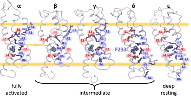
KV7.1 est un tétramère dont chaque sous-unité α compte six hélices transmembranaires
(S1 à S6). Les quatre premières hélices forment le domaine sensible au voltage (VSD), tandis que les deux dernières forment celui du pore (PD). Le mécanisme d’activation de KV7.1 s’effectue par le biais d’une translation de l’hélice S4 vers le feuillet externe de la
membrane en deux étapes, stabilisant le VSD en trois états stables : repos, intermédiaire, et activé. Ces conformations sont capables d’ouvrir ou de fermer le pore via un processus appelé couplage. Ainsi, les états du canal KV7.1 sont Repos/Fermé (RC),
Intermédiaire/Ouvert (IO) et Activé/Ouvert (AO). En présence des sous-unités β KCNE1, le couplage n’a pas lieu à l’état intermédiaire, donc les états du canal IKS sont RC,
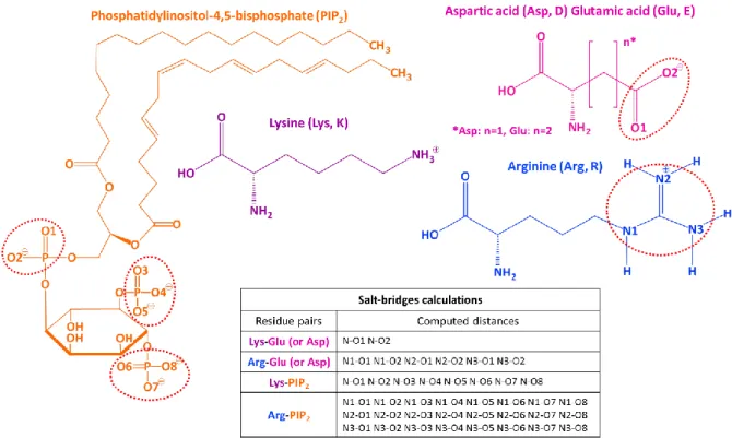
Intermédiaire/Fermé (IC) et AO. En outre, le phospholipide PIP2
(phosphatidylinositol-4,5-bisphosphate) joue un rôle crucial dans le couplage VSD-PD et l’ouverture du pore des canaux KV7.
Malgré les informations apportées par les études fonctionnelles et structurales du canal KV7.1, en absence et en présence de ses modulateurs KCNE1 et PIP2, le mécanisme de son
2
Nous avons abordé ce problème en exploitant des techniques computationnelles telles la dynamique moléculaire (DM). Etant donnée la complexité de la fonction de KV7.1, cette
étude avait trois objectifs, le premier étant d’identifier les interactions protéine-protéine stabilisant chacun de ses états, le deuxième étant de décrire les effets de KCNE1 sur son mécanisme de couplage VSD-PD, le troisième étant de caractériser les effets de KCNE1 sur les interactions des sous-unités α KV7.1 avec PIP2. Nous avons bâti des modèles par
homologie de KV7.1 dans ses trois états, en utilisant la structure cristallographique et des
résultats de modélisations du canal homologue KV1.2 comme patron.
Nous avons effectué des simulations de DM de 500 ns pour chaque modèle, encastré dans une membrane virtuelle entourée de deux couches d’une solution de [KCl] à 150 mM, afin de reproduire une dynamique réaliste du canal dans nos systèmes, et ce en absence et en présence de KCNE1 et de PIP2.
L’analyse des trajectoires DM obtenues visait en premier lieu à valider nos modèles par rapport aux données expérimentales. Par la suite, nous avons identifié les interactions « état-dépendantes » dans les modèles les plus robustes, permettant de prédire les changements de conformation inhérents à l’ouverture de KV7.1, en absence de KCNE1
d’une part, et en sa présence d’autre part. De cette manière, il a été possible d’identifier les interactions protéine-protéine permettant à KCNE1 d’inhiber le mécanisme de couplage VSD-PD de KV7.1 lorsque son VSD est à l’état intermédiaire, ainsi que celles
permettant à KCNE1 d’amplifier les effets de couplage de KV7.1 lorsque son VSD est à l’état
activé. Enfin, pour chaque état stable de KV7.1, nous avons identifié et comparé les
interactions protéine-lipide au sein des modèles KV7.1 et des modèles IKS.
Nos modèles ont été validés par des études fonctionnelles menées par nos collaborateurs (Pr. Jianmin Cui) de l’université Washington de Saint-Louis, Etats-Unis. Cette étude intégrative a révélé un couplage inédit, conceptualisé par un modèle « main-et-coude » ayant probablement lieu dans l’ensemble des familles de canaux KV1 à KV9.
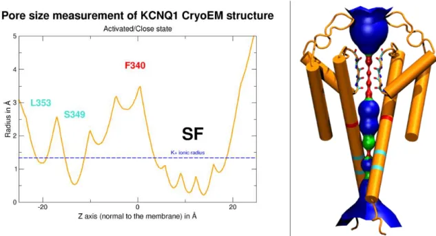
L’hélice S6 de KV7.1 présente un motif SFF (338-340) fortement conservé au sein des
canaux de la famille KV7. L’espace conformationnel des résidus SFF au sein des
trajectoires DM de KV7.1 et d’IKS a révélé l’existence d’une porte hydrophobe inédite,
définie par des orientations « état-dépendantes » de F339 et F340, et validée par des études fonctionnelles.
3
Les analyses effectuées sur les trajectoires DM des modèles IKS indiquent qu’un garrot,
formé par les interactions entre KCNE1 et PIP2 autour de la région intracellulaire des
hélices S6, induisent la fermeture du pore dans les états RC et IC. Enfin, la recherche des déterminants moléculaires de la modulation du couplage de KV7.1 par KCNE1 indique que
4
Investigation of activation and coupling mechanisms
of the voltage-gated potassium channel K
V7.1 in
cardiomyocytes using computational methods
Abstract
KV7.1 channel is a transmembrane protein that opens to selectively diffuse K+ ions across
the plasma membrane upon membrane depolarization. In the myocardium tissue, KV7.1
is co-expressed with the ancillary subunit KCNE1. Several experiments conducted on KV7.1 along with KCNE1 revealed its effects on KV7.1 function, which modify its properties
allowing for the KV7.1-KCNE1 complex to generate the IKS current during the cardiac
action potential. As numerous mutations of KV7.1 and KCNE1 sequences are linked to
severe cardiac arrhythmias, KV7.1 channel constitutes a major therapeutic target.
Each α-subunit of the KV7.1 tetramer counts six transmembrane helices (S1 to S6), the
first four ones forming the voltage-sensor domain (VSD), and the last two forming the pore domain (PD). This channel has a 2-step activation mechanism involving an upward translation of S4 across the membrane towards its outer leaflet and stabilizing the VSD in three stable states: resting, intermediate and activated. These conformations can induce pore opening or closure by a process called VSD-PD coupling. Accordingly, the states for KV7.1 channel are Resting/Closed (RC), Intermediate/Open (IO) and Activated/Open
(AO). In the presence of KCNE1 β subunits, the coupling is suppressed for the intermediate state, thus the states for IKS channel are RC, Intermediate/Closed (IC) and AO.
Furthermore, the PIP2 (phosphatidylinositol-4,5-bisphosphate) phospholipid plays a
crucial role in the VSD-PD coupling and pore opening mechanisms of KV7 channels.
Despite the information drawn from both functional and structural studies of KV7.1 channel
in absence and presence of its modulators KCNE1 and PIP2, its VSD-PD coupling mechanism
5
To address this issue, we harnessed computational techniques, including molecular dynamics (MD) simulations. Given the complexity of the KV7.1 function, the study had
three main objectives. The first one was the characterization of the VSD-PD coupling mechanism underlying the transitions between RC, IO and AO states of KV7.1 channel. The
second one was the characterization of the modulation effects of KCNE1 on the VSD activation and VSD-PD coupling mechanisms of KV7.1 α-subunits. The third one was to
characterize the effects of KCNE1 on the affinity between KV7.1 α-subunits and PIP2 lipids.
We built homology models of KV7.1 in its three states, using the crystallographic structure
and previous models of the homologous KV1.2 channel as a template. We conducted 500
ns MD simulations on each model embedded in a model membrane surrounded by two slabs of a 150 mM [KCl] solution to reproduce a realistic dynamics of the channel, in absence and presence of KCNE1 subunits and PIP2 molecules. At first, we performed
analyses of MD trajectories in order to validate the models with respect to experimental data. Then, we proceeded to identify the state-dependent protein-protein interactions in the most robust models, allowing for the prediction of the conformational changes leading to the open states of KV7.1 in absence of KCNE1 on one hand, and in presence of KCNE1 in
the other hand. The comparison of the obtained results allowed one to determine the molecular determinants of KCNE1 inhibition of KV7.1 VSD-PD coupling when the VSD
reaches its intermediate state, as well as those of KCNE1 amplification of KV7.1 VSD-PD
coupling effects when the VSD is in its activated state. Finally, we identified and compared the state-dependent protein-lipid interactions in KV7.1 models and IKS models.
Our KV7.1 models were validated via functional studies conducted by our collaborators
(Pr. Jianmin Cui) from Washington University of Saint-Louis in the USA. Altogether, the joint study revealed a novel VSD-PD coupling mechanism that we conceptualized by a “hand-and-elbow model” likely to occur in all domain swapped (KV1- KV9) channels.
The S6 helix of KV7.1 has a motif SFF (338-340), highly conserved in KV7 family. The
conformational space of SFF residues in KV7.1 and IKS MD trajectories revealed the
existence of an unidentified yet hydrophobic gate defined by state-dependent orientations of F339 and F340, which was also validated by functional studies.
6
The analyses of IKS MD trajectories suggest that the interactions between KCNE1 and PIP2
form a tourniquet around the cytoplasmic region of S6, leading to pore closure in both RC and IC models. Finally, the investigation of the molecular determinants of the modulation of KV7.1 VSD-PD coupling mechanism by KCNE1 in our IKS models suggests that KCNE1
disrupts the “hand-and-elbow” model of the VSD-PD coupling we previously revealed for the KV7.1 channel.
Related publications
Deyawe A, Kasimova MA, Delemotte L, Loussouarn G, Tarek M (2018) Studying Kv Channels Function using Computational Methods. Methods Mol Biol 1684:321–341 .
https://doi.org/10.1007/978-1-4939-7362-0_24
Hou P, Kang PW, Deyawe Kongmeneck A, Yang N-D, Liu Y, Shi J, Xu X, White KM, Zaydman MA, Kasimova MA, Seebohm G, Zhong L, Zou X, Tarek M, Cui J (2020) Two-stage electro–mechanical coupling of a KV channel in voltage-dependent activation.
Nature Communications 11:1–14 .
7
Acknowledgements
It would be very difficult to thank everyone in only one page since the help of numerous people allowed me to finish the writing of this PhD thesis.
First and foremost, I would like to thank my supervisor Dr Mounir Tarek, for your help, your patience, and the tremendous amount of things I have learn from you, especially your work ethic and the importance to give our best, and to seek for the best. My PhD course has not been an easy road but working under your supervision made me grow up as a scientist, and also as a person, which is priceless. I want to thank my predecessor’s Dr Lucie Delemotte and Dr Marina Kasimova, for the fabulous work they made before, and also after I started my PhD course, which constitute an excellent starting point and a great source of inspiration for my scientific work. I also want to thank Pr Jianmin Cui, for the opportunity he gave me to work with him and his research team, including Dr Panpan Hou and Po-Wei Kang, with which I have also learned a huge number of things. Our collaboration allowed to sublime our respective work, which is amazing.
I also want to thank Pr Catherine Etchebest, Dr François Dehez, Dr Jérôme Hénin, Dr Gildas Loussouarn and Dr Sara Liin, for the honor to save time to take part in my PhD defense as jury members. Specifically, I want to thank Pr Catherine Etchebest and Dr Jérôme Hénin, for accepting to dedicate time to review my PhD thesis.
I also want to warmly thank Dr Antonio Monari and Séverine Bonenberger, for all the insightful discussions and conversations, and of course for all the pep talks you gave me during my entire PhD course. I also want to thank everyone in the LPCT lab for their hospitality, and for the numerous good times I spent with everyone in and out of the lab. I also want to thank all my fellow PhD students, and the interns, for their support and the heartfelt laughs we shared.
My acknowledgments would not be complete without a special thank for my family. To my mother Esther, my father Roger, my step-father Philippe, my sisters Michèle, Joëlle and Gladis, my brothers Bertrand and Gérard, my nieces Téha and Tess, my nephews Ethan and Yaël, as well as my godfather Martin, the rest of my family, and of course my dear friends Anthony and Axelle, for their unconditional love and support, up to this moment when I write these words, with my eyes fogged of tears.
8
Outline
Résumé ... 1 Abstract ... 4 Acknowledgements ... 7 Table of figures ... 10 Introduction ... 14Chapter I. State of the art ... 19
Chapter I.1. From electrophysiology to molecular modeling: A history of ion channels ... 22
Chapter I.2. Implication of cardiac action potential in the heartbeat rate... 39
Chapter I.3. How do the voltage-gated ion channels participate in the progress of the cardiac action potential? 45 Chapter I.3.1. Conductive tissue ... 45
Chapter I.3.2. Contractile tissue ... 47
Chapter I.4. Molecular determinants of IKS current ... 53
Discovery of KV7.1 and KCNE1 through inherited cardiac arrhythmias ... 53
IKS complex (KV7.1+ KCNE1): Structure and function ... 56
Appendix I. A cartoon insight into the cardiac upstroke ... 68
Appendix II. A cartoon insight into the cardiac repolarization ... 69
Chapter II. Methods ... 70
Chapter II.1. Comparative Modeling ... 74
Chapter II.1.1. Principle ... 74
Chapter II.1.2. Template selection ... 74
Chapter II.1.3. Sequence alignment ... 77
Chapter II.1.4. Model building and optimization ... 79
Chapter II.1.5. Evaluation of the obtained molecular model ... 80
Chapter II.2. Classical Molecular Dynamics... 81
Chapter II.2.1. Principle ... 81
Chapter II.2.2. Modeling KV7.1 channels in their membrane environment ... 85
Chapter II.2.3. MD simulations... 89
Chapter II.3. Knowledge-based analysis of MD trajectories ... 93
Chapter II.3.1. Structural mapping of known KV7.1 neighbor residues for structural validation ... 93
Chapter II.3.2. Pore size ... 97
Chapter II.3.3. Knowledge-based determination of weak interactions in MD trajectories ... 99
Chapter III. Results ... 104
9
Chapter III.1.1. Protein-protein sidechain proximities ... 109
Chapter III.1.2. Protein-lipid electrostatic interactions ... 117
Chapter III.1.3. Average pore radii calculations... 121
Chapter III.1.4. Structural comparison with KV7.1 (CryoEM) experimental structure ... 126
Chapter III.2. Characterization of KV7.1 channel state dependent conformations ... 130
Chapter III.2.1. Functional validation of KV7.1 VSD-PD coupling mechanism... 130
Chapter III.2.2. Molecular determinants of the modulation of KV7.1 VSD-PD coupling mechanism by KCNE1 ancillary subunits: Protein-protein interactions involved in VSD-PD coupling ... 150
Chapter III.2.3. A KV7 conserved motif constitutes a hydrophobic gate in the pore of the KV7.1 channel 160 Chapter III.2.4. The protein-lipid interactions that mediate the pore opening mechanism ... 173
Chapter IV. General discussion ... 177
Chapter IV.1. Predicting the overall KV7.1 function... 180
Chapter IV.1.1. VSD activation: PIP2 as a KV7.1 allosteric activator ... 185
Chapter IV.1.2. VSD-PD coupling: KCNE1 as a KV7.1 effector ... 187
Chapter IV.1.3. KV7.1 pore opening mechanism is triggered by a wheelwork of conformational changes related to both VSD activation and VSD-PD coupling mechanisms ... 193
Chapter IV.2. Perspectives: Atomistic scale investigation of the effects of other KV7.1 effectors ... 198
Chapter IV.2.1. Molecular modeling of IKS channel along with small compound ligands ... 200
Chapter IV.2.2. Molecular modeling approaches to study the effects of KV7.1 endogenous ligands ... 214
References ... 219
10
Table of figures
Figure I.1.1: The membrane theory of Julius Bernstein. ... 23
Figure I.1.2: The voltage-clamp technique applied on a squid giant axon. ... 24
Figure I.1.3: Example of current recordings obtained by voltage-clamp experiments. ... 25
Figure I.1. 4: The electrical circuit model of the membrane by Hodgkin and Huxley. ... 26
Figure I.1.5: The patch-clamp technique. ... 28
Figure I.1.6: Phylogenetic tree including the Voltage-Gated Cation Channels (VGCC) superfamily. . 31
Figure I.1.7: Conserved gating charges in the fourth segment of various voltage-gated ion channels. ... 32
Figure I.1.8: The Q/V and G/V curves of a voltage-gated ion channel. ... 33
Figure I.1.9: Cryo-EM structure determination of KCNQ1 subunit. ... 36
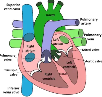
Figure I.2.1: Cartoon picture of the human myocardium and its chambers. ... 39
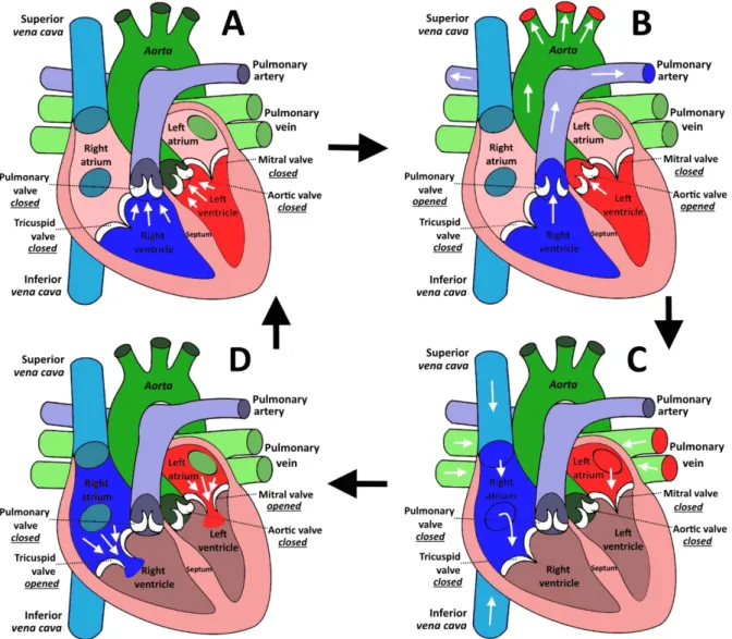
Figure I.2.2: The different phases of blood circulation in the human heart cavities. ... 41
Figure I.2.3: The network of pacemaker cells in the myocardium. ... 42
Figure I.2.4: A normal electrocardiogram (ECG) ... 44
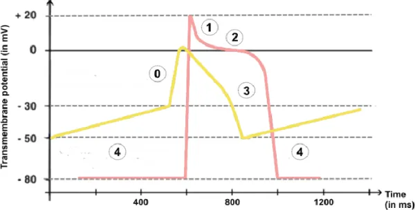
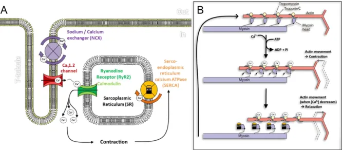
Figure I.3.1.1: Time evolution of the cardiac action potential (in mV) in cardiomyocytes. ... 46
Figure I.3.2.1: The voltage-dependent ionic currents involved in the cardiac action potential. ... 47
Figure I.3.2.2: Diffusion of potassium currents in contractile cardiomyocytes. ... 48
Figure I.3.2.3: The Ca2+ cycle in the cardiomyocytes during cardiac action potential. ... 50
Figure I.4.2.1: CryoEM structure of the Xenopus Laevis KV7.1 subunit. ... 56
Figure I.4.2.2: Mapping KV7.1 selectivity filter in Xenopus Laevis KV7.1 structure. ... 57
Figure I.4.2.3: S4 gating charges in KV7.1 VSD... 58
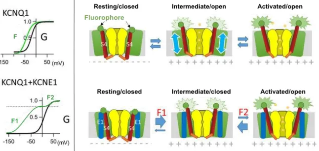
Figure I.4.2.4: Macroscopic properties of KV7.1 channel in the absence and the presence of KCNE1. ... 60
Figure I.4.2.5: Average NMR structure of the ancillary subunit KCNE1. ... 61
11
Figure II.1.2.1: Crystallographic structure of rattus norvegicus KV1.2 channel. ... 74
Figure II.1.2.2: Multiple VSD conformations obtained by MD simulations of KV1.2. ... 74
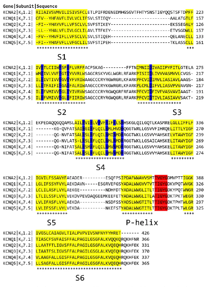
Figure II.1.3.1 : Edited sequence alignment of KV1.2 and KV7 subunit family. ... 78
Figure II.2.1.1: Periodic boundary conditions in MD simulations. ... 82
Figure II.2.1.2: Approximate functions of the Van der Waals (VdW) non-bonded energies. ... 84
Figure II.2.2.1: Membrane lipids used in MD systems. ... 86
Figure II.2.2.2: Onset MD systems of KV7.1 models. ... 87
Figure II.2.2.3: Onset MD systems of IKS models. ... 88
Figure II.3.1.1: Distance calculations of neighbor residues in MD trajectories according to Cysteine cross-linking experiments. ... 94
Figure II.3.1.2: Distance calculations of electrostatic interactions in MD systems. ... 95
Figure II.3.1.3: Example of distances graphs for MD analysis. ... 96
Figure II.3.3.1: Computed distances for assessing electrostatic interactions. ... 100
Figure II.3.3.2: Computed distances for assessing methionine interactions. ... 101
Figure II.3.3.3: Computed distances for assessing hydrophobic interactions. ... 102
Tab III.1.1.1 : State dependent VSD salt bridges in KV7.1 subunits. ... 112
Tab III.1.1.2: State-dependent protein-protein interactions involving KV7.1 residues. ... 112
Tab III.1.1.3: State-dependent protein-protein interactions involving KV7.1 and KCNE1 residues. ... 116
Figure III.1.1.1: State-dependent salt-bridges in the VSD of KV7.1 models. ... 109
Figure III.1.1.2: State-dependent VSD salt-bridges involving E160 (E1) in KV7.1 MD simulations. 110 Figure III.1.1.3: State-dependent pairs of KV7.1 neighbor residues. ... 113
Figure III.1.1.4: Structural mapping of state-dependent protein-protein interactions in IKS models. ... 114
12
Figure III.1.3.1: Pore size comparison of IKS models in their activated state. ... 122
Figure III.1.3.2: Pore size comparison of IKS models in their Intermediate state. ... 123
Figure III.1.3.3: Pore size comparison of IKS models in their resting state. ... 124
Figure III.1.4.1: Voltage sensor domain comparison of AO models with KCNQ1EM structure ... 126
Figure III.1.4.2: Pore domain comparison of RC models with KCNQ1EM structure ... 127
Figure III.1.4.3: Comparison of KV7.1 IO model with NMR structure of human KV7.1 VSD (PDB ID: 6MIE). ... 128
Figure III.2.1.1: Site-directed mutagenesis of KV7.1 residues involved in IO state coupling. ... 131
Figure III.2.1.2: Membrane surface expression of KV7.1 S4-S5L/S6 region mutants. ... 132
Figure III.2.1.3: Structural mapping of KV7.1 residues involved in IO state coupling according to functional study results. ... 132
Figure III.2.1.4: MD simulation results of KV7.1 residues involved in IO state coupling. ... 133
Figure III.2.1.5: Molecular basis of S4-S5L/S6 LQT mutations of KV7.1 revealed by MD simulations. ... 134
Figure III.2.1.6: Residues involved in KV7.1 AO state coupling according to functional studies... 136
Figure III.2.1.7: MD simulation results show the crucial interactions for the KV7.1 AO state coupling. ... 137
Figure III.2.1.8: MD simulation results shows the crucial interactions for KV7.1 IO state coupling. ... 139
Figure III.2.1.9: Double mutant cycle analysis results obtained for KV7.1 channel. ... 142
Figure III.2.1.10: Structural mapping of PD cluster residues in KV7.1 3D model ... 143
Figure III.2.1.11: State-independent interactions of PD cluster residues in KV7.1 models. ... 144
Figure III.2.1.12: State-dependent interactions of PD cluster hook residues in KV7.1 models. ... 145
Figure III.2.1. 13: State dependent VSD-PD coupling interfaces in KV7.1 models. ... 147
Figure III.2.1.14: Hand-and-elbow model for KV7.1 state-dependent VSD-PD coupling mechanism. ... 149
Figure III.2.2.1: Structural mapping of PD cluster residues in IKS AO state model ... 151
Figure III.2.2.2: Interactions of PD cluster outer surface residues in IKS models... 152
Figure III.2.2.3: Interactions of KV7.1 VSD-PD coupling and PD cluster inner surface in IKS models. ... 155
Figure III.2.2.4: State dependent VSD-PD coupling interfaces in IKS models. ... 157
13
Figure III.2.3.1: Conserved motifs in the sixth segment of KV channels. ... 160
Figure III.2.3.2: State-dependent conformations of F340 sidechains in KV7.1 models... 162
Figure III.2.3.3: Conformations of F340 side chains in KCNQ1EM structure and KV7.1 AC model. .... 164
Figure III.2.3.4: Interactions of SFF residues in KV7.1 models. ... 165
Figure III.2.3.5: State-dependent conformations of F340 residues in IKS models. ... 166
Figure III.2.3.6: Interactions of SFF residues in IKS models. ... 168
Figure III.2.3.7: Interactions of PD middle pocket residues in KV7.1 and IKS models. ... 169
Figure III.2.3.8: Intersubunit interactions involving PD middle pocket residues. ... 169
Figure III.2.4.1 Structural mapping of KCNE1 basic residues and PIP2 lipids in the pore domain of IKS models in 8PIP2 systems. ... 175
Figure III.2.4.2: Conserved basic residues in the of KCNE ancillary subunits. ... 176
Figure IV.1.1.1: State-dependent interactions between S4 and PIP2 in IKS models. ... 186
Figure IV.1.2.1: Prediction of KV7.1 VSD-PD coupling mechanism which mediates RC→IO and IO→AO transitions of PD cluster in absence of KCNE1... 188
Figure IV.1.2.2: Prediction of KV7.1 coupling mechanism which mediates RC→IC and IC→AO transitions of PD cluster in presence of KCNE1. ... 190
Figure IV.1.2.3: A S5 binding site for the first S4 gating charge in IKS models. ... 192
Tab IV.1.2.1: PD cluster residues associated with LQTS mutations. 191 Figure IV.1.3.1: The motion of S4 aliphatic residues in KV7.1 models. ... 194
Figure IV.1.3.2: The motion of S4 aliphatic residues in IKS models... 196
Tab IV.1.3.1: LQTS related residues involved in KV7.1 function according to MD simulations. 197 Figure IV.2.1.1: KV7.1 IO model embedded in AutoDock grid box size. ... 206
Figure IV.2.1.2: Primary result of XE991 Molecular Docking on KV7.1 IO model. ... 207
Figure IV.2.1.3: Primary result of XE991 Molecular Docking on IKS AO model. ... 208
Figure IV.2.1.4: Primary result of ML277 Molecular Docking on KV7.1 AO model. ... 210
Figure IV.2.1.5: Mapping of CHARMM force field parameters to optimize for ML277. ... 211
Figure IV.2.1.6: Mapping of CHARMM force field parameters to optimize for XE991. ... 212 Tab IV.2.1.1: New atom types assigned for ML277 in CHARMM FF parametrization. 212
14
Introduction
In living organisms, membranes are made of two layers of amphiphilic lipids which are joined by hydrophobic interactions, named a lipid bilayer [1]. The specific architecture of the latter enables plasma membranes to isolate the cytoplasm from the extracellular media. Similarly, within a cell, other membranes are able to isolate the content of the various organelles from the cytoplasm.
Membranes are permeable to gas and liposoluble compounds, but not to aqueous ones. In this way, plasma membranes allow for the conservation of distinct ion charge imbalance between the cytoplasm and the extra-cellular media, maintained by the function of Na-K pumps, conferring a transmembrane electrical voltage called the resting potential. During the 18th century, it was discovered that plasma membranes are able to generate and
propagate electric currents in muscle cells. Since the last decade of the 19th century,
various experimental methods that belong to electrophysiology have been developed in order to characterize these currents in various cell membranes, leading to the discovery of excitable cells, such as muscular cells, myocardium cells, neuronal cells, or pancreatic β-cells. The plasma membranes of this particular type of cells are characterized by periodic sharp increase/decrease of their transmembrane potential. Named action potential, this phenomenon is generated upon a specific transmembrane voltage called “threshold voltage” and can occur across one cell, or a series of cells that are linked by gap junctions, allowing for the cytoplasms of two distinct cells to communicate.
Typically localized in plasma membranes, the main role of Voltage-Gated Ion Channels (VGICs) consists in passively transporting ions across the lipid bilayer. Triggered by specific transmembrane potentials, each VGIC is able to open in order to selectively diffuse ions (e.g. Na+ K+ Ca2+ Cl-) at a given voltage value, allowing for the fast transport of these
chemical entities that bear an electrical charge along their electrochemical gradient. In the remaining we will discard the Chloride channels (CLCs) as they constitute an evolutionarily family of voltage-gated channels that are structurally unrelated to the other known voltage-gated channels.
15
Thus, the propagation of an action potential relies on the coordination of a set of voltage-gated cation channels (VGCC) which successively open and close with various selective and specific properties. Accordingly, these action potentials are known to orchestrate numerous biological events such as muscle contraction or synaptic signaling. Therefore, VGCCs are crucial for the function of vital organs such as the brain, the heart, muscle, and pancreas. Each of these tissues possess an action potential with specific features.
Despite the major contribution of electrophysiology experiments in the unraveling of VGCC function, this discipline cannot provide information about their structure.
The emergence of DNA sequencing managed to provide a molecular insight into the structure of VGCC. Indeed, their primary sequence determination shed light on VGCC as a super family of 143 transmembrane proteins that are present in most eukaryotic cells and characterized by multiple consensus sequences. This superfamily gathers several families of ion channels which are classified by their ion selectivity. Thus, NaV, CaV, and KV families
are composed of ion channels that are selective to Na+, Ca2+, and K+, respectively. The
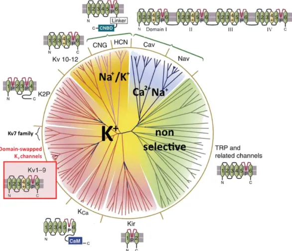
potassium channel family is the largest subfamily among VGCC. It comprises 40 channels, clustered into 12 families, among which 9 (KV1-KV9) are domain-swapped. These proteins
are involved in an important number of biological functions, including homeostasis, secretion, muscle contraction and transmission of the nerve impulse.
DNA sequencing has also allowed for the determination of the mutations that are associated with malfunctions of the VGCC, usually associated with the disruption of action potentials. These functional defects are related to diverse congenital diseases called channelopathies, which include epilepsy, cardiac arrhythmias, and deafness among others.
16
Hence, VGCC constitute numerous therapeutic targets for the treatment of a large number of diseases. To that extent, we focus our entire work on a particular potassium (K+) ions
VGCC called KV7.1. Across the membranes of heart striated muscle cells, the diffusion of
potassium ions through KV7.1 channel generates the slow delayed rectifier current, also
known as the IKS current. It occurs specifically at the end of the action potential, allowing
the membrane to return to its resting potential. This specific action potential controls the heartbeat pace, as it occurs periodically in order to elicit the heart contraction. As a consequence, KV7.1 channel mutations induce channel dysfunction, which are
characterized by changes of cardiac repolarization time, and therefore prolongation or shortening of the cardiac action potential.
The understanding of the modulation mechanisms of KV7.1 leading to the generation of
the IKS current in the heart encompasses various scientific disciplines, including cell
biology, electrophysiology, molecular biology, and computational biochemistry. A large body of experiments allowed to identify the existence of different conformations adopted by the voltage sensor domain (VSD) of KV7.1, which are resting, intermediate and
activated, and the effects of these conformations on the pore domain (PD) conformation, open or closed, through a particular mechanism called VSD-PD coupling. Experiments also highlighted two main effectors of KV7.1 VSD-PD coupling mechanism. The first one is the
ancillary subunit KCNE1, which is co-expressed along with KV7.1 channel, endowing the
latter distinct physical and chemical properties in relation to KV7.1 alone. The second one
is the phosphatidylinositol-4,5-bisphosphate (PIP2) lipid, which has been identified as a
key element of KV7.1 channel function.
Thanks to the emergence of advanced structural biology techniques, a large number of protein structures have been unraveled at a nanoscopic scale [2]. However, owing to their partially hydrophobic surfaces, flexibility, and lack of stability, only a small number of VGCC have managed to be resolved through X-ray crystallography for the last two decades. In the frame of this work, the 3D structure of KV1.2, the first mammalian KV
channel to be resolved by X-ray crystallography, turned out to be essential. Starting from the structure-activity relationship of evolutionarily related proteins, computational methods of molecular modeling such as comparative modeling turned out to be useful and accurate for the prediction of the tridimensional structure of KV7.1 channel, whose none
17
These obtained structures are typically used to provide a molecular insight into experimental findings, with the help of simulations. Thereby, we harnessed these computational techniques to build 3D models of the three stable conformations known for KV7.1 channel. The use of experimental data as structural constraints would maximize
their reliability.
To predict the molecular aspects of the function of KV7.1 channel, computer simulation
methods such as molecular dynamics (MD) allow one to study an isolated chemical system containing a protein alone or along with its specific environment including modulatory subunits, lipids and molecules such as water or ions. The simulation of these chemical systems allows to follow the time evolution of a protein model in its biological environment in order to obtain an atomistic insight into the dynamics of the protein, either at its equilibrium (brute-force or unbiased MD), or in certain conditions in order to induce a specific motion related to its function (under voltage for instance), or in interaction with a small compound. The collection of the time-dependent conformations generated by a simulation is defined as an MD trajectory. Here, we designed several chemical systems for the three 3D models of KV7.1 along with PIP2 lipids, in the absence
and the presence of KCNE1, all mimicking the biological environment of the channel. They were submitted each to ~500 ns unbiased MD simulations. The analyses of the resulting MD trajectories consisted in the investigation of protein interactions, and protein-lipid ones to determine those which stabilize each state of KV7.1. These results allowed
for the prediction of the mechanisms related to KV7.1 function at an atomistic level of
precision, as well as the effects induced by the presence of each of its known modulators, namely KCNE1 and PIP2.
18
In the first chapter of this manuscript, we will present ion channels in the context of the cardiac action potential (AP) to gain a better understanding of the way in which KV7.1 is
implicated in its repolarization phase. The reader would hence be more familiar with the biological mechanisms that link our ion channel of interest, KV7.1 and the various cardiac
arrhythmias caused by its significant number of mutations. The computational methods and strategies we used to design the structure in order to study the function of KV7.1
channel through the knowledge-based metastable states models we have designed will be presented in Chapter II. Results that demonstrate the key modulatory roles of both KCNE1 ancillary subunit and PIP2 lipids on KV7.1 activation and coupling mechanisms will be
detailed in Chapter III, and their coherence in relation with both experimental and computational results obtained for KV7.1 channel alone will be discussed in Chapter IV.
The conclusions and perspectives drawn from this research project will be outlined in Chapter V.
19
Chapter I.
State of the art
Le canal KV7.1 est une protéine transmembranaire qui joue un rôle prépondérant au sein
des membranes des cardiomyocytes, qui assurent la contraction du myocarde qui a lieu à chaque battement cardiaque. KV7.1 assure le transport passif d’ion potassium à travers
ces membranes, c’est -à-dire qu’il diffuse des ions potassium depuis le cytoplasme vers le milieu extracellulaire. La découverte des canaux ioniques à ouverture voltage dépendante tels que KV7.1 est le résultat de plusieurs siècles de recherche scientifique, riches en
avancées cruciales de la part de plusieurs disciplines scientifiques diverses et variées. Parmi ces disciplines, on trouve l’électrophysiologie, la biologie cellulaire, moléculaire et structurale, ainsi que la biophysique, la bioinformatique et la chimie. L’électrophysiologie s’est révélée être celle qui a joué le rôle le plus important dans l’identification des courants électriques au sein des cellules et des tissus à la fin du 18ème siècle.
Dans ce chapitre, nous avons passé en revue les avancées scientifiques clés qui ont permis de découvrir, d’identifier et de caractériser le canal KV7.1 ainsi que son environnement
(
Chapter I.1
)
. Ayant été découvert dans le cadre de plusieurs maladies génétiques à l’origine d’arythmies cardiaques, nous avons pris le parti de fournir une explication détaillée du rôle que ce canal joue dans le déclenchement des battements cardiaques(
Chapter I.2
)
. A partir de la description de la fonction cardiaque à l’échelle macroscopique, nous avons progressivement diminué l’échelle d’observation jusqu’à l’échelle microscopique, là où le courant ionique généré par KV7.1 participe au potentield’action, un processus biologique défini par une série d’événements sous-tendant la contraction musculaire, qui s’effectue de façon automatique et spontanée dans le cœur
(
Chapter I.3
)
. Par la suite, nous sommes placés à l‘échelle moléculaire afin de présenter les recherches ayant permis de déterminer les structures primaire, secondaire, tertiaire et quaternaire du canal KV7.1(
Chapter I.4
)
. Nous nous sommes ensuite intéressés auxfaçons dont un peptide transmembranaire d’une part, et un lipide membranaire d’autre part, peuvent moduler l’activité du canal KV7.1 au sein des cardiomyocytes, par rapport
20
Enfin, nous avons montré comment la combinaison des résultats de biologie moléculaire et de certaines méthodes computationnelles ont permis de mieux expliquer la fonction du canal KV7.1 au sein des cellules musculaires lisses du myocarde. Nous avons mis en
évidence les zones d’ombre qui nécessitent de mener des recherches plus poussées. Dans le cadre de cette thèse, nos hypothèses se sont articulées autour de l’étude des différentes conformations adoptées par le canal KV7.1, ainsi que les potentiels changements
conformationnels inhérents à la présence du peptide transmembranaire et du lipide membranaire connus pour être capables de moduler son activité. A partir de ces hypothèses, nous avons expliqué l’intérêt que peuvent présenter l’utilisation des méthodes computationnelles dans la compréhension des mécanismes d’action et de modulation du canal KV7.1 à l’échelle moléculaire.
21
KV7.1 channel is an integral protein which plays a crucial role in the membranes of the
cardiomyocytes, which are responsible for the contraction of the myocardium necessary for the heartbeat. Specifically, KV7.1 diffuses potassium ions through the membranes,
from the cytoplasm towards the extracellular medium. The discovery of voltage-gated ion channels has been made possible thanks to several centuries of scientific research, full of advances from various scientific fields such as electrophysiology, cellular biology, molecular biology, structural biology, biophysics and computational biology and chemistry. Among these numerous scientific areas, electrophysiology has played the most important role in the identification of these proteins since it allowed for the identification of electrical currents in cells and tissues.
In this chapter, we reviewed the key scientific milestones that allowed for the discovery of the KV7.1 channel and its direct environment
(
Chapter I.1
)
, followed by a thoroughexplanation of its role in the heart rhythm
(
Chapter I.2
)
. Starting from a macroscopic description of the heart and its main function, we progressively decreased the scale of observation toward the microscopic level at which the KV7.1 channel participates in theaction potential, defined by a series of biological processes that are required for the automatic triggering of the heartbeat
(
Chapter I.3
)
. Then, we focused on the molecular level by presenting the primary, secondary, tertiary, and quaternary structures of the KV7.1 channel, and then by explaining how a transmembrane peptide and a membranelipid were both found to be able to modulate the function of the KV7.1 channel in the heart
striated muscle cells
(
Chapter I.4
)
.We showed how the combination of molecular biology and computational methods allowed for a better understanding of the function of KV7.1 in the heart muscle cells.
Finally, we presented the shaded areas that still remain further research. The hypotheses we made about the modulation roles of both the transmembrane peptide and the membrane lipid on the function of the KV7.1 channel lie on the study of its different
conformations and the potential conformational modifications of the KV7.1 channel
structure in the presence of these effectors. From these hypotheses, we presented the ways in which the use computational methods can increase the level of understanding of the various mechanisms of action and modulation of the KV7.1channel at the molecular
22
Chapter I.1. From electrophysiology to molecular modeling: A
history of ion channels
Electric currents were discovered to occur through muscle and brain tissues of various species. The first known breakthrough about these currents was led by the physiologist Luigi Galvani in 1791, when he and his wife Lucia noticed that an isolated frog muscle was able to move when connected with conductive matter. Based on this discovery, they hypothesize that the frog muscle contained an electric fluid able to generate an electric current, which Galvani called “animal current” [3]. One year later, the physicist Alessandro Volta started to be interested in Galvani’s work, but did not believe in his “animal current” hypothesis, convinced that the frog muscle motion was due to the connection of this electric fluid with the metals used in Galvani’s experiments. Nevertheless, Volta was convinced of the existence of organic electric fluids and conducted several experiments in order to reproduce an artificial fluid able to conduct electric currents, which lead him to the invention of his famous voltaic pile in 1800 [4] in which electric currents were generated by aqueous solutions. Thirty years later, the chemist Michael Faraday showed that the currents in voltaic piles were actually the products of redox reactions. Since the 19th century, the knowledge of the electric signals
occurring within the living world had led to the development of experimental techniques allowing for the tracking of the electricity across cell membranes.
This quest, which had initiated since Galvani’s discovery, was marked by major scientific successes that were awarded by several Nobel prizes during the 20th century. In 1889,
based on Volta’s invention and Faraday’s conclusions, the chemist Walther Nernst published an equation that relates the electric potential produced by a redox reaction occurring in electrochemical batteries such as the voltaic one with the energetic contribution of each chemical species involved in the redox reaction. Then he applied this equation on biological membranes to relate the equilibrium potential 𝐸𝑖𝑜𝑛 of a given ion across a membrane (also called reversal potential, it refers to the membrane potential value in which there is no flux of the ion across the membrane) with its concentration gradient [5]: 𝐸𝑖𝑜𝑛 = 𝑅𝑇 𝑧𝐹 × ln ( [𝑖𝑜𝑛]𝑜𝑢𝑡 [𝑖𝑜𝑛]𝑖𝑛 )
23
Where 𝐸𝑖𝑜𝑛 is expressed in millivolts (mV), R is the ideal gas constant in Joules.Kelvin -1.mol-1 (J.K-1.mol-1), T is the temperature in K, z is the net charge of the ion, F is the Faraday
constant in Coulomb.mol-1 (C. mol-1), while [𝑖𝑜𝑛]
𝑜𝑢𝑡 and [𝑖𝑜𝑛]𝑖𝑛 are the concentrations of
the ion, expressed in mol.L-1, outside and inside the cell, respectively. For this
breakthrough, Nernst obtained a Nobel Prize in 1920.
In early 1900s, the biophysicist Julius Bernstein, who invented the rheotome, an innovative electrophysiology tool allowing for the recording of action potential velocities in excitable cells, applied Nernst equation in order to express the electrochemical potential of a given ion across a given plasma membrane. This application lead Bernstein to establish his membrane theory which stated that the living cells are surrounded by a double-layered membrane with a selective permeability to potassium [6], inducing a charge imbalance between each side of the double-layered membrane (Figure I.1.1). During action potential, the potassium permeability of the membrane increases, and its potential is reduced to a lower value. Meanwhile, the molecular structure of plasma membranes was unknown at this time.
The lipid bilayer was first discovered in 1925 thanks the study of Gorter and Grendel on red blood cells [7]. The experiment consisted in the centrifugation of a known number of red blood cells of several mammals, followed by the dissolution of the liposoluble sediments into benzene.
Figure I.1.1: The membrane theory of Julius Bernstein.
According to Bernstein, the ionic distribution around the double-layered cell membrane induces a charge imbalance induced by the asymmetric distribution of positive ions (in purple) and negative ions (in green) around the plasma membrane (in gray) in its resting state. The selective permeability to potassium ions (black arrow), represented by a pore (depicted in blue) allows to maintain the resting potential. Despite the membrane being depicted here as a lipid bilayer, the chemical structure of plasma membranes was unknown at the age of Bernstein theory.
24
The dissolution product was then spread on a Langmuir-Blodgett trough [8] in order to measure the surface area of the red blood cells plasma membranes. This molecular architecture is mainly composed of amphiphilic phospholipids whose polar heads are facing the aqueous intra and extra cellular media, while the hydrophobic tails of the phospholipids are facing each other at the core of the membrane. This arrangement allows for the isolation of the cytoplasm from the extracellular matrix, confirming Bernstein theory. While Gorter and Grendel assumed these lipids as constituting most of the membrane surface area of red blood cells, several studies conducted afterwards proved their assumption wrong [9]. Indeed, considering the known physical properties of the membrane allowing for the cell to perform exchanges with the extracellular matrix, cell biologists tended to predict that plasma membranes were also containing proteins, without knowing how those proteins are interacting with the lipid bilayer [10, 11, 1]. With this scientific knowledge, the voltage-dependent ion selectivity of plasma membranes was partly explained a few decades later. Indeed, during the 50s, Hodgkin and Huxley managed to identify distinct ion selective current through a squid giant axon. To do that, they conducted an experiment called voltage-clamp, which consists in the application of a given electric potential on a plasma membrane using two electrodes that are connected to an electrical circuit designed to record the intensity I of the ionic currents in picoAmpères (µA) that are flowing through the membrane (Figure I.1.2).
Figure I.1.2: The voltage-clamp technique applied on a squid giant axon.
This picture represents the squid axon in which both the recording electrode (in green) and the current-passing electrode (in brown) are inserted. These electrodes are plugged in an electric circuit that allows to record the current intensity I across the membrane of the squid giant axon upon an applied transmembrane voltage Vm.
25
Hodgkin and Huxley identified two distinct ionic currents (Figure I.1.3): an inward sodium current that occur in depolarized conditions, when the applied voltages were increasing towards positive values; and a potassium outward current in hyperpolarized conditions, when the applied voltages were decreasing towards negative values [12].
Based on these results, they manage to conceptualize a mathematical model allowing for the characterization of action potentials in excitable cells [15]. The model is based upon an electric circuit (Figure I.1.4) that involves the lipid bilayer as a capacitance 𝐶𝑀, while ion transporters that generate the currents 𝐼𝑁𝑎, 𝐼𝐾 and 𝐼𝑙 are assimilated as electrical resistances 𝑅𝑁𝑎 = 1 𝑔⁄ 𝑁𝑎, 𝑅𝐾 = 1 𝑔⁄ 𝐾 and 𝑅𝑙 = 1 𝑔⁄ 𝑙, respectively, each of them being
branched in derivation. From this model they developed a differential equation allowing to express the current intensity across a given membrane at a given time, as a function of the transmembrane voltage. The equation is expressed as follows:
𝐼 = 𝐶𝑀𝑑𝑉
𝑑𝑡 + 𝐼𝑁𝑎+ 𝐼𝐾 + 𝐼𝑙
where 𝐼 is the total membrane current, 𝐶𝑀 is the capacitance, 𝑉 the difference of
membrane potential with respect to the resting potential, 𝑡 is the time.
Figure I.1.3: Example of current recordings obtained by voltage-clamp experiments.
On the left panel the graphs project the time evolution of potassium current amplitudes (top panel) and gating current intensities (bottom panel) of the voltage-gated “Shaker” K+ channel in microAmpères (µA)
upon different transmembrane voltages (circled in red dots) in millivolts (mV), applied successively from -120 mV to 0 mV. The right panel depicts the time evolution of sodium current amplitudes of the voltage-gated sodium channel NaV1.5 in µA. The direction of the curves indicates the direction of the ion flux, which
26
𝐼𝑁𝑎, 𝐼𝐾 and 𝐼𝑙 are the components of the ionic current, carried by sodium ions, potassium ions, and chloride ions (along with other ions), respectively (Figure I.1.4). As these components can also be expressed as a function of their equilibrium potentials, the previous equation can also be noted as follows:
𝐼 = 𝐶𝑀
𝑑𝑉
𝑑𝑡 + 𝑔𝑁𝑎(𝑉 − 𝑉𝑁𝑎) + 𝑔𝐾(𝑉 − 𝑉𝐾) + 𝑔𝑙(𝑉 − 𝑉𝑙)
where 𝑔𝑁𝑎, 𝑔𝐾 and 𝑔𝑙 are the conductances of sodium, potassium, and chloride ions,
and 𝑉𝑁𝑎, 𝑉𝐾 and 𝑉𝑙 their reversal potentials, respectively.
The scientific work of Hodgkin and Huxley was awarded by a Nobel Prize in medicine in 1963, and officialized electrophysiology as a new scientific discipline. Later in that decade, they used the Nernst equation to determine the equilibrium potential of a membrane permeable to two or more distinct ions. Hence, for the membrane of squid giant axon, the Goldman-Hodgkin-Katz equation would be expressed as follows:
𝐸𝑚 = 𝑅𝑇 𝐹 ln ( 𝑃𝑁𝑎[𝑁𝑎+] 𝑜𝑢𝑡 + 𝑃𝐾[𝐾+]𝑜𝑢𝑡 + 𝑃𝐶𝑙[𝐶𝑙+]𝑜𝑢𝑡 𝑃𝑁𝑎[𝑁𝑎+]𝑖𝑛+ 𝑃𝐾[𝐾+]𝑖𝑛+ 𝑃𝐶𝑙[𝐶𝑙+]𝑖𝑛 )
Where the membrane potential 𝐸𝑚 is expressed in V, the selectivity 𝑃𝑖𝑜𝑛 for a given ion is expressed in m/s, the extracellular and intracellular concentrations [𝑖𝑜𝑛]𝑜𝑢𝑡 and [𝑖𝑜𝑛]𝑖𝑛, respectively, are expressed in mol/m3.
Figure I.1. 4: The electrical circuit model of the membrane by Hodgkin and Huxley.
The circuit (in the foreground) shows how the membrane current I, divided into different ion currents in the lipid bilayer (in background) are passing through the membrane upon a given voltage E. The resistances RN, RK and Rl are varying with both time and membrane potential.
27
Moreover, Hodgkin and Huxley also identified transient currents that appeared to be triggered by a change in transmembrane potential [15]. They predicted these currents to be generated by a part of ion channels which might act as a “voltage sensor”.
Their prediction was confirmed a couple of decades later, when transient currents were recorded on isolated squid giant axons in such conditions that ionic currents were suppressed so it could not be related to the transport of ions though the membrane. In addition, the direction of these currents appeared to be voltage-dependent: outward in depolarized conditions, and inward in hyperpolarized conditions. Consequently, these currents were identified as gating currents (Figure I.1.3).
In parallel, cell biologists made significant progress in the knowledge of the chemical structure of plasma membranes during the 60’s [1], leading to Singer’s well-known membrane fluid mosaic model [16].
As lipids have been previously shown to constitute half of the membrane surface of erythrocytes [17], this model assumes that the rest of the membrane surface was occupied by integral proteins with hydrophilic intracellular and extracellular regions, and hydrophobic regions allowing for their stability in the low dielectric environment induced by the lipid tails of amphiphilic membrane lipids. Logically, the proteins that span the lipid bilayer allow for the cells to perform exchanges with the extracellular matrix such as ion transport, as shown by Hodgkin and Huxley’s experiments despite of their lack of stability in low dielectric environments [18]. Numerous experiments conducted later confirmed Singer’s fluid mosaic model, which is still assumed to be correct nowadays [19].
28
In the early 70s, a new advanced experimental technique called patch-clamp (Figure I.1.5), which allowed for the direct measure of transmembrane current upon an applied potential in one cell (through “whole-cell” configurations), and then in one or a few ion channels (through “cell-attached”, “inside-out” and/or “outside-out” configurations), has started to be developed. This technique was basically designed for the direct observation of the open and the closed states of a single VGCC and was further made possible by Erwin Neher and Bert Sakmann [20], who obtained a Nobel Prize in medicine in 1991. By realizing a tight seal between a glass pipette filled with a saline solution and a membrane patch from a frog muscle fiber, they were able to record transitions of a single ion channel between its conducting and its non-conducting states through discrete oscillations in the recorded ionic current.
By assimilating ion channels as resistors with respect to the theory of Hodgkin and Huxley, it became possible to record the conductance of a single channel with the use of Ohm’s law:
𝑉𝑚− 𝑉𝑖𝑜𝑛 = 𝑅𝑐ℎ𝑎𝑛𝑛𝑒𝑙× 𝐼
in which 𝑉𝑚 is the applied voltage during patch-clamp measurement, 𝑉𝑖𝑜𝑛 is the reversal potential of the diffused ion, 𝑅𝑐ℎ𝑎𝑛𝑛𝑒𝑙 is the resistance of the ion channel of interest, and 𝐼 the recorded current. As the resistance of a channel is the inverse of its conductance, 𝐼 can be expressed as follows:
Figure I.1.5: The patch-clamp technique.
The top panel shows the distinct configurations that can be used to record ion currents of a cell membrane. The saline solution of the glass pipette is colored in gray, while the cytoplasm of the cell is colored in yellow. The lower left panel depicts the usual patch clamp circuit, in which the electrodes (in black) are put into the glass pipette and the water bath containing the cell and plugged to the device that records the current and displays the measures on an oscilloscope (in green). The “inside-out” configuration, is circled in red as it is the most common one used for the recordings of voltage-gated ion channels currents.
29 𝐼 = (𝑉𝑚− 𝑉𝑖𝑜𝑛) ×
1 𝑅𝑐ℎ𝑎𝑛𝑛𝑒𝑙
= (𝑉𝑚− 𝑉𝑖𝑜𝑛) × 𝑔𝑐ℎ𝑎𝑛𝑛𝑒𝑙
And the conductance of the ion channel of interest can be expressed as: 𝑔𝑐ℎ𝑎𝑛𝑛𝑒𝑙 =
𝐼 (𝑉𝑚− 𝑉𝑖𝑜𝑛)
Furthermore, several studies reported that ionic currents can be specifically abolished in presence of toxins [21] or small compounds [22]. With the use of the patch clamp technique in “whole-cell” configuration, it became possible to study the effects of these compounds on the ionic currents. Despite the advances in the comprehension of ion channels function, the chemistry of these macromolecules remained unknown at the end of the 70s [23].
During the 80s, the emergence of DNA sequencing allowed for the determination of primary sequences of voltage-gated ion channels, starting from the gene coding of the eel voltage-gated sodium channel [24], followed by the Shaker gene of Drosophila Melanogaster, known to code for a voltage-gated potassium channel [25]. The sequences of these two genes turned out to be relatively similar. Indeed, these genes appeared to code for protein sequences which present cytoplasmic NTER and CTER regions, separated by
a tetrameric transmembrane region composed of four domains of six α-helices, separated from each other with intra or extra-cellular loops of various lengths. Within each domain, the first four helical segments S1 to S4 form the voltage-sensor domain (VSD), while the last two helices called segments S5 and S6 are forming the pore domain (PD).
30
These patterns of helices result from the oligomerization of four tetramers in KV channels,
while in NaV channels, the four domains are joined in one single sequence (Figure I.1.6).
The particular primary sequence of the voltage-gated sodium channel turned out to be found later in voltage-gated calcium channels [26]. VGCC family members are sharing a common transmembrane pore domain presenting a four-fold topology of two domains [26], composed of two transmembrane helices separated by a loop which is embedded in the hydrophobic core of the membrane, forming the tetrameric pore through which the ions are translocated. Despite of the shared transmembrane topology and consensus sequences of the primary sequences of VGCC, some members of this superfamily of ion channels are not voltage-gated only. The channels of the cyclic nucleotide-gated (CNG) family [27, 28], are ligand-gated and merely sensitive to transmembrane potential. In addition, the ion channels of the KCa, and HCN families, despite their sensitivity to
voltage, can also be activated by Ca2+ ions [29] and cAMP [30], respectively, as shown by
their respective ligand binding sites. Despite sharing the transmembrane topology of the other VGCC [26], the nonselective cation channels of the transient receptor proteins family (TRP) are usually diffusing ion fluxes upon various stimuli [31]. Nevertheless, these channels are sharing a common function, which is the facilitated diffusion of ions across the membrane down their concentration gradient [32]. The main families of voltage-dependent channels are actively participating in action potentials, such as the NaV, CaV,
and KV families which are composed of ion channels that are selective to Na+, Ca2+, and K+,
31
Figure I.1.6: Phylogenetic tree including the Voltage-Gated Cation Channels (VGCC) superfamily.
This schematic view depicts the evolutionary related 143 members of the VGCC superfamily, which are clustered in four subfamilies. The TRP non-selective cation channels are represented in green lines, the calcium and sodium selective channels in blue lines, the CNG/HCN sodium-potassium channels in pink lines, and potassium selective channels in red lines. For each subfamily, a topology of their transmembrane segments is displayed around the circle. The binding sites of the CNG, HCN and KCa channels are represented
in blue squares. The topology of the subunits of KV channel family, which includes KV7.1 channel, is framed
32
In the fourth membrane spanning sequence of voltage-gated ion channels, numerous basic residues such as arginine or lysine were positioned every three amino acids (Figure I.1.7). Called gating charges, these residues were identified later as responsible for the gating currents [33, 34] which were discovered earlier. All together, these results constitute a second evidence that confirm the predictions of Hodgkin and Huxley.
Later, new experiments that associate electrophysiology experiments with fluorometry assays were developed in order to monitor the conformational change in relation with VSD activation as well as the conductance of a single channel, both according to the transmembrane potential. This technique, called patch-clamp fluorometry [35] is commonly used nowadays and consists in attaching a fluorescent tag to the extracellular NTER region of the S4 segment of the VGCC of interest. In the same manner, voltage-clamp
fluorometry [36] (VCF) allows for the independent recording of both VSD movement (optical fluorescence) and pore conductance (ionic current) in an entire cell. This method has been regularly used to study the function of KV7.1 channel, in which a fluorophore is
attached to the S3-S4 linker and tracked to record the changes of fluorescence emission during voltage-dependent activation. Before the democratization of this method, gating currents used to be monitored on wild-type VGCC in presence of channel blockers [37] or non-conductive VGCC mutants [12, 38, 39]. However, due to their small amplitudes, these currents are most often quite difficult to record.
Figure I.1.7: Conserved gating charges in the fourth segment of various voltage-gated ion channels.
The figure shows a sequence alignment of the S4 segment in the selective VGCC involved in the cardiac action potential. The first and second columns report the gene name and the subunit name of the channel. The strongly conserved basic residues R1 to R6, also known a gating charges, are highlighted in blue. The sequence of the channel KV7.1, which is the object of this thesis, is framed in red.
33
By comparing the gating current/voltage relationship with the ionic current relationship (Figure I.1.8), using normalized gating current and ionic current values, one was able to determine the stable VSD conformations of a VGCC according to the direction of the gating current along the membrane, as well as the conductive state of each stable conformation of the channel. With this hybrid method, it is now possible to determine the voltage-dependence of VSD activation (F/V curve or Q/V curve) of a given wild-type VGCC, when the S4 segment moves across the membrane upon depolarization, along with the voltage-dependence of the conductance (I/V or G/V curve) when the channel starts to conduct ions [40]. In this way, these experiments can be performed on malfunctioning mutants or wild-type VGCC in presence of channel effectors in order to assess their effect on the channel function. The use of Xenopus oocytes to perform both gene expression and electrophysiology had a significant impact on the development of these electrophysiology techniques [41, 42]. However, these numerous methods, while being extremely enriching, cannot provide an accurate molecular insight into the tridimensional (3D) structure of VGCC, nor into their mechanism of function at a molecular scale.
Figure I.1.8: The Q/V and G/V curves of a voltage-gated ion channel.
The graph shows the voltage-dependence (V) of the conductance (G) and the gating charge displacement (Q) for a voltage-gated ion channel. Two pictures are depicting a schematic view of voltage-gated ion channel in its closed state at hyperpolarized voltages, and in its open state at depolarized voltages. The yellow area accounts for the lipid bilayer, while the gray area represents the channel, with the S4 segment in blue, with its gating charges in a white circle, and the linker is depicted as a black line joining S4 and the gate. The direction of both gating charges and ion flux at depolarized potentials is represented by white arrows. Adapted from [40].