Publisher’s version / Version de l'éditeur:
Proceedings of SPIE, 7897, 789708, 2011-03-01
READ THESE TERMS AND CONDITIONS CAREFULLY BEFORE USING THIS WEBSITE.
https://nrc-publications.canada.ca/eng/copyright
Vous avez des questions? Nous pouvons vous aider. Pour communiquer directement avec un auteur, consultez la première page de la revue dans laquelle son article a été publié afin de trouver ses coordonnées. Si vous n’arrivez pas à les repérer, communiquez avec nous à [email protected].
Questions? Contact the NRC Publications Archive team at
[email protected]. If you wish to email the authors directly, please see the first page of the publication for their contact information.
NRC Publications Archive
Archives des publications du CNRC
This publication could be one of several versions: author’s original, accepted manuscript or the publisher’s version. / La version de cette publication peut être l’une des suivantes : la version prépublication de l’auteur, la version acceptée du manuscrit ou la version de l’éditeur.
For the publisher’s version, please access the DOI link below./ Pour consulter la version de l’éditeur, utilisez le lien DOI ci-dessous.
https://doi.org/10.1117/12.875532
Access and use of this website and the material on it are subject to the Terms and Conditions set forth at
Assessing mechanical properties with intravascular or endoscopic
optical coherence tomography
Lamouche, G.; Azarnoush, H.; Vergnole, S.; Pazos, V.; Bisaillon, C.-E.;
Debergue, P.; Boulet, B.; Diraddo, R.
https://publications-cnrc.canada.ca/fra/droits
L’accès à ce site Web et l’utilisation de son contenu sont assujettis aux conditions présentées dans le site LISEZ CES CONDITIONS ATTENTIVEMENT AVANT D’UTILISER CE SITE WEB.
NRC Publications Record / Notice d'Archives des publications de CNRC:
https://nrc-publications.canada.ca/eng/view/object/?id=ffae30b9-92ef-4876-8fe7-abd908f91dfd https://publications-cnrc.canada.ca/fra/voir/objet/?id=ffae30b9-92ef-4876-8fe7-abd908f91dfdAssessing mechanical properties with intravascular or endoscopic
optical coherence tomography
G. Lamouche
a, H. Azarnoush
a,b, S. Vergnole
a, V. Pazos
a, C.-É. Bisaillon
a, P. Debergue
a, B. Boulet
b,
and R. Diraddo
aa
National Research Council Canada, 75 de Mortagne, Boucherville, QC, J4B 6Y4, Canada;
bCentre for Intelligent Machines, McGill University, Montréal, QC, H3A 2A7, Canada.
ABSTRACT
We explore the potential of intravascular or endoscopic optical coherence tomography (OCT) to extract relevant mechanical properties of a tissue deformed by an inflating balloon. Tubular OCT phantoms with different mechanical properties are fabricated. The phantoms are deformed by an inflating balloon, and the deformation is monitored with OCT. A quantitative description of the phantom deformation is obtained by segmenting the OCT images. Two strategies to extract the mechanical properties from this quantitative data are presented: by comparing to a finite-element simulation and by performing a mechanical analysis.
Keywords: optical coherence tomography, mechanical properties, angioplasty, balloon, phantom.
1. INTRODUCTION
The question underlying this paper is: “Can we extract relevant mechanical properties from an intravascular optical coherence tomography (IVOCT) measurement like that of Video 1? Video 1 shows the IVOCT monitoring of the inflation of an angioplasty balloon within a coronary artery obtained in a beating heart setup [1]. After the balloon is sufficiently inflated to fill the lumen, any further inflation cause a deformation of the artery wall. The response of the system thus includes the mechanical response of the tissue surrounding the balloon. Consequently, mechanical properties can indeed be extracted. The true question is then: “Considering the multiple sources of uncertainty in such a measurement, can mechanical properties be measured with an accuracy high enough to be relevant for diagnosis or to provide significant patient specific information?” These sources of uncertainty include, for example, the accuracy of OCT imaging, the complexity of the tissue under investigation, and the assumptions underlying the mechanical analysis. The question is not only relevant for balloon inflation monitored by IVOCT, but for any other OCT measurement where a balloon is inflated that may deform the surrounding tissue, like in some endoscopic OCT applications.
To address this question, we are building a framework of tools. Since some of these tools are still in development, no definite answer to the initial question is yet obtained. This paper describes the work in progress, presenting the current state of the various tools, with the only goal of showing how we intend to find an answer to the question.
The first tool is an OCT system to monitor balloon inflation within a tubular tissue. In this work, an IVOCT system is
used, but an endoscopic OCT system could also be used. We want to perform our study on a variety of samples with well defined mechanical properties over a wide range of values. This could be done with excised tissues, but it is more easily performed with phantoms. Our second tool is thus a collection of artery phantoms with specific mechanical properties. Our third tool is an ensemble of algorithms to perform the segmentation of the OCT images and extract quantitative information. The fourth and fifth tools are two methods to obtain mechanical properties from the quantitative data extracted from the OCT images. In the first method, mechanical properties are obtained by optimizing the parameters of a finite-element simulation to match the measured balloon and tissue deformation. The second method relies on a mechanical analysis of the system to achieve the same goal.
The sections of the paper present these tools: IVOCT system, phantoms with specific mechanical properties, algorithms to analyze OCT images, mechanical properties from comparison with finite-element simulation, and mechanical properties from mechanical analysis. To illustrate the use of the various tools, they are applied throughout the paper to three single-layer tubular phantoms with different mechanical properties.
2. IVOCT SYSTEM
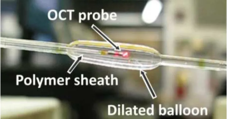
The IVOCT system that monitors balloon inflation and tissue deformation is a custom-built system that can accommodate any commercial source. It is based on a Mach-Zehnder configuration and it includes a balanced detection. The results in this paper were obtained with a Santec swept-source operating at a wavelength of 1.31 µm and sweeping over 100 nm with a 30 kHz sweep rate. An OCT catheter allows inflation monitoring from within a balloon. The OCT probe is 0.8 mm in diameter and rotates within a polymer sheath that is 1.0 mm in diameter. A pullback unit insures the rotation and translation of the OCT probe. To monitor balloon inflation, only the rotation capability is used. Figure 1 illustrates the OCT probe inserted in a partially inflated balloon. A cross-sectional image is usually composed of 1000 radial depth scans, thus imaging is typically performed at 30 frames per second. Details about the IVOCT system and about the OCT catheter can be found in Ref. [1], while the use of IVOCT to monitor balloon inflation is described in Ref. [2].
3. PHANTOMS WITH SPECIFIC MECHANICAL PROPERTIES
Phantoms with specific mechanical properties are the best candidates to test our proposed methods to extract mechanical properties. Our group has developed a strong expertise in fabricating phantoms of sound and diseased arteries. These are silicone tubular structures with multiple layers to mimic the intima, the media, and the adventitia layers of the artery wall [3]. Not only can our phantoms provide the OCT optical signature of artery layers, they can also provide similar mechanical properties in the linear deformation regime [3]. The same fabrication method can be used to model other tubular tissues like those of interest for endoscopic OCT. The fabrication method also allows fabricating complex structures with inclusions like diseased artery structures [4]. Figure 2 presents an example of such a phantom imaged with IVOCT: a diseased coronary artery with a calcification.
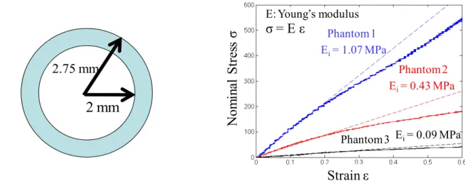
In this early phase of our study, we restricted ourselves to the simplest phantoms. We fabricated three single-layer phantoms with similar dimensions but with different mechanical properties. Figure 3 provides a schematic representation of the single-layer phantoms. Figure 3 also presents stress-strain curves that were measured for each phantom with an Enduratec Electroforce 3100 (Bose Corp., Minnesota, USA). The phantoms thus had very different mechanical
properties. Limiting our analysis to the linear deformation regime, phantoms 1, 2 and 3 had elastic moduli of 1.07 MPa, 0.43 MPa, and 0.09 MPa, respectively.
4. ALGORITHMS TO ANALYZE OCT IMAGES
Contour detection of the balloon surfaces or tissue structures in an IVOCT image provides quantitative information from which mechanical properties can be obtained. Different tissues or tissue structures may ask for specific algorithms. Limiting ourselves to the single-layer phantoms considered in this work, a simple algorithm can perform the task in a robust and efficient way. For a given angular portion of OCT image, a matched filter is used to locate the region where the balloon and tissue are located. Then a convolution is performed with a kernel that spans over a few radial scans. The balloon and tissue contours lead to defined peaks as a result of the convolution, which provide the contour nodes’ locations. The “convolution - peak detection” process is then continuously repeated on adjacent radial scans, the convolution process being limited to a region surrounding the previously found nodes. All this is performed in an automated way.
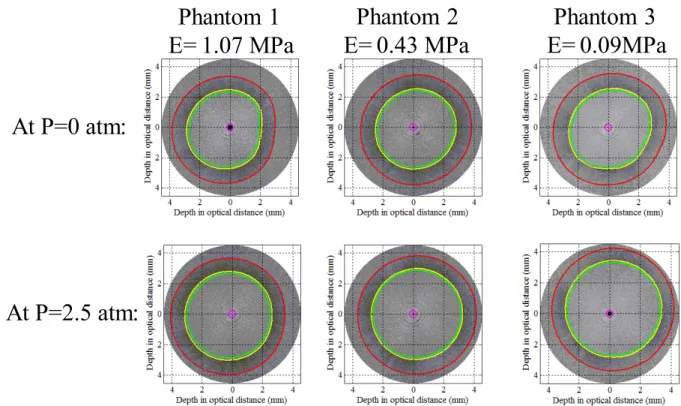
Figure 4 presents IVOCT images of the balloon and phantoms prior to inflation and at an inflation pressure of 2.5 atm. The contours were detected with the algorithm described above. The resulting contours are smooth and follow very closely the balloon and phantom true contours. The IVOCT images are recorded in optical thicknesses (optical refractive index times the geometrical length). All the images were converted to true geometric dimensions to evaluate the average diameter of each contour. Figure 5 presents the average diameter of the inner and outer contours of each phantom as a
Figure 2. Phantom of a diseased artery with a calcification imaged with IVOCT.
Figure 3. Schematic of a phantom (left) and nominal stress as a function of the strain for each phantom used in our experiment (right).
2 mm
2.75 mm
σ = E ε
E: Young’s modulus Ei= 1.07 MPa Ei= 0.43 MPa Ei= 0.09 MPa Phantom 1 Phantom 2 Phantom 3Nom
in
al S
tr
es
s
σ
Strain ε
function of the inflation pressure. Since the three phantoms have very different mechanical properties, the variation of the diameters with the inflation pressure is quite different for each case. The goal is now to convert these variations to values of mechanical properties for each phantom.
5. MECHANICAL PROPERTIES FROM COMPARISON WITH FINITE-ELEMENT SIMULATION A finite-element model to simulate percutaneous coronary interventions was developed within the scope of a previous project. The simulation includes various constitutive models: linear elastic, hyperelastic, and viscoelastic. Both three-dimensional (3D) and two-three-dimensional (2D) versions of the software exist. For the current project, the 2D version is used since it provides fast results, on a time scale of a minute, with accuracy very close to that of the 3D version. With this model, we intend to extract mechanical properties of tissues or phantoms through an iterative process. In this process, the material parameters used in the model will be optimized to provide a simulated deformation similar to that observed experimentally with IVOCT. This is an inverse problem where the optimized material parameters values would then be those of the tissue or the phantom. This optimization process will be eased by using a balloon with previously determined mechanical properties.
We have yet to implement the method to solve the above inverse problem. In the early stage of the project, we are still interested in the direct problem. We use the measured mechanical properties of the phantom to feed the simulation and compare the results with the IVOCT measurements. It is an important step since it allows us to determine the degree of complexity of the simulation that is needed to achieve our goal. This degree of complexity relates to which constitutive models are to be used in the simulation.
Figure 5 presents the results for the inner and outer phantom diameters resulting from the finite-element simulation along with those obtained experimentally with IVOCT. The simulated values were evaluated in the linear deformation regime using the elastic modulus values from Figure 3 for each phantom. Based on Figure 5, there is a very good agreement between simulated and experimental diameter values. Better agreement could be obtained by using a more complex model than the Hookean model. This is currently being investigated, but it will of course introduce additional
Figure 4. IVOCT images of phantoms and balloon contour detection, before and during inflation.
Phantom 1
E= 1.07 MPa
Phantom 2
E= 0.43 MPa
Phantom 3
E= 0.09MPa
At P=0 atm:
At P=2.5 atm:
parameters. The results obtained with the linear regime are satisfying enough for us to attempt the inverse optimization problem with this level of approximation. This will be reported in a future publication.
6. MECHANICAL PROPERTIES FROM MECHANICAL ANALYSIS
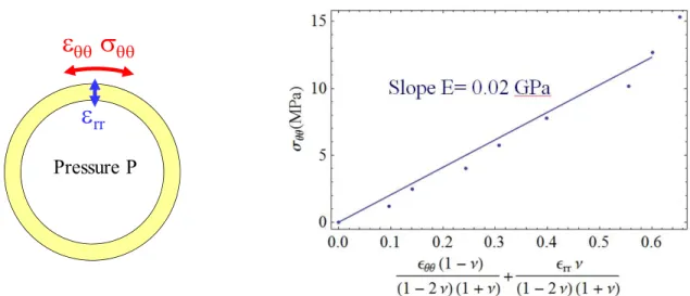
A balloon surrounded by a tissue or a phantom is a layered structure whose deformation can be described through a mechanical analysis. Depending upon the complexity of the ensemble, this description may or may not lead to a tractable problem. This part of our project is at a very early phase. In this section, we only illustrate the type of processing we intend to perform by considering a very simple case: the inflation of the balloon alone.
We thus want to determine the mechanical properties of the balloon from its deformation during inflation. A schematic representation of the balloon under inflation is provided in Figure 6. The segmented IVOCT images provide the circumferential strain value εθθ and the radial strain value εrr. The axial strain, along the balloon axis, can be assumed
negligible in comparison to the other strain values. To obtain the mechanical properties of the balloon, we want to relate these strain values to the circumferential stress σθθ. Since the balloon is essentially a thin-wall vessel, the circumferential
stress σθθ , also called Hoop stress, is given by σθθ=P r/t, where P is the inflation pressure, r is the radius of the balloon
and t is the thickness of the balloon. The Hoop stress can be related to a combination of the circumferential strain εθθ and the radial strain value εrr. This relation is illustrated by the graphic in Figure 6 where the combination of strains is on the
horizontal axis and the Hoop stress is on the vertical axis. The relation is linear and its slope gives the elastic modulus E. The above analysis only provides an example on how we intend to extract mechanical properties from IVOCT measurements using a mechanical analysis. Results for the system composed of a balloon and the various single-layer phantoms were not available for this publication. These results will be included in a future publication.
7. CONCLUSION
We have presented a framework of tools that include an IVOCT system, phantoms with specific mechanical properties, algorithms to analyze OCT images, a method to obtain mechanical properties from comparison with finite-element simulation, and a method to obtain mechanical properties from mechanical analysis. This framework is assembled to
Figure 5. Inner and outer diameters of each phantom as a function of the balloon inflation pressure. Comparison between experimental results and finite-element simulation results.
evaluate if we can extract relevant mechanical properties from IVOCT monitoring or endoscopic OCT monitoring of a tissue deformed by an inflating balloon. The tools related to the IVOCT system, the phantoms, and the analysis of OCT images are all completed. We are currently pursuing the development of the methods to extract mechanical properties for the case of the three single-layer phantoms. Experimental work is also under way for more complex phantoms with an inclusion having different mechanical properties.
REFERENCES
[1] Lamouche, G., Dufour, M., Hewko, M., Vergnole, S., Gauthier, B. Bisaillon, C.-E., Monchalin, J.-P. and Sowa, M.G., "Intravascular optical coherence tomography on a beating heart model," Journal of Biomedical Optics 15(4), 046023-7 (2010).
[2] Azarnoush, H., Vergnole, S., Bourezak, R., Boulet, B. and Lamouche, G., "Optical coherence tomography monitoring of angioplasty balloon inflation in a deployment tester," Review of Scientific Instruments 81(8), 083101-8 (2010).
[3] Bisaillon, C.-E., Lanthier, M.-M., Dufour, M.L. and Lamouche, G., “Durable coronary artery phantoms for optical coherence tomography,” Proc. SPIE 7161, 71612E1-10 (2010).
[4] Bisaillon, C.-E., Dufour, M.L. and Lamouche, G., “Durable phantoms of atherosclerotic arteries for optical coherence tomography,” Proc. SPIE 7548, 75483G1-4 (2010).
Figure 6. Schematic representation of an inflating balloon (left) and linear relationship between the Hoop stress σθθ and a combination of the strain values εθθ and εrr to provide the Young’s modulus E (right). The Poisson ratio ν is 0.46.