HAL Id: hal-02529997
https://hal.archives-ouvertes.fr/hal-02529997
Submitted on 2 Apr 2020
HAL is a multi-disciplinary open access
archive for the deposit and dissemination of
sci-entific research documents, whether they are
pub-lished or not. The documents may come from
teaching and research institutions in France or
abroad, or from public or private research centers.
L’archive ouverte pluridisciplinaire HAL, est
destinée au dépôt et à la diffusion de documents
scientifiques de niveau recherche, publiés ou non,
émanant des établissements d’enseignement et de
recherche français ou étrangers, des laboratoires
publics ou privés.
Distributed under a Creative Commons Attribution| 4.0 International License
The association between accelerometer-assessed physical
activity and respiratory function in older adults differs
between smokers and non-smokers
M.A. Benadjaoud, M. Menai, V.T. van Hees, V. Zipunnikov, Jean-Philippe
Regnaux, M. Kivimäki, A. Singh-Manoux, S. Sabia
To cite this version:
M.A. Benadjaoud, M. Menai, V.T. van Hees, V. Zipunnikov, Jean-Philippe Regnaux, et al.. The
association between accelerometer-assessed physical activity and respiratory function in older adults
differs between smokers and non-smokers. Scientific Reports, Nature Publishing Group, 2019, 9,
pp.10270. �10.1038/s41598-019-46771-y�. �hal-02529997�
the association between
accelerometer-assessed physical
activity and respiratory function
in older adults differs between
smokers and non-smokers
Mohamed Amine Benadjaoud
1, Mehdi Menai
2, Vincent t. van Hees
3, Vadim Zipunnikov
4,
Jean-philippe Regnaux
5, Mika Kivimäki
6, Archana singh-Manoux
2,6& séverine sabia
2,6the association between physical activity and lung function is thought to depend on smoking history but most previous research uses self-reported measures of physical activity. this cross-sectional study investigates whether the association between accelerometer-derived physical activity and lung function in older adults differs by smoking history. The sample comprised 3063 participants (age = 60–83 years) who wore an accelerometer during 9 days and undertook respiratory function tests. Forced vital capacity (FVC) was associated with moderate-to-vigorous physical activity (MVPA; acceleration ≥0.1 g (gravity)) in smokers but not in never smokers: FVC differences for 10 min increase in MVPA were 58.6 (95% Confidence interval: 21.1, 96.1), 27.8 (4.9, 50.7), 16.6 (7.9, 25.4), 2.8 (−5.2, 10.7) ml in current, recent ex-, long-term ex-, and never-smokers, respectively. A similar trend was observed for forced expiratory volume in 1 second. Functional data analysis, a threshold-free approach using the entire accelerometry distribution, showed an association between physical activity and lung function in all smoking groups, with stronger association in current and recent ex-smokers than in long-term ex- and never-smokers; the associations were evident in never smokers only at activity levels above the conventional 0.1 g MVPA threshold. These findings suggest that the association between lung function and physical activity in older adults is more pronounced in smokers than non-smokers.
Poor lung function, characterized by low forced expiratory volume in 1 second (FEV1) and low forced vital
capac-ity (FVC), is associated with an increased risk of death1,2 and chronic conditions, such as lung cancer3 and
cardi-ovascular diseases.4,5 Lung capacity is largely determined by endogenous factors such as age, sex, and body size
and early life exposures6,7. However, there is growing interest in modifiable risk factors for poor lung function and
so far only the role of smoking is widely recognized8. The beneficial impact of physical activity on progression
of chronic obstructive pulmonary disease (COPD) or asthma9,10 has led researchers to assess the role of physical
activity11–18 and sedentary behavior14,19 as determinants of poor lung function in the general population. The
results from these studies suggest that the association of physical activity with lung function might depend on smoking history11–13,15,17,18 such that physical activity potentially mitigates the adverse impact of smoking on lung
function in smokers, possibly via anti-inflammatory and vascular mechanisms20.
Most studies on the association between physical activity and lung function rely on self-reported physical activity data which are prone to reporting bias. Physical activity assessed objectively by accelerometers is more strongly associated with health outcomes21–23. Typically, these studies categorize the duration of physical activities
1Institute for Radiological Protection and Nuclear Safety (IRSN), Fontenay-Aux-Roses, France. 2Inserm U1153,
CRESS, Epidemiology of Ageing and Neurodegenerative diseases, Université de Paris, Paris, France. 3netherlands
eScience Center, Amsterdam, The Netherlands. 4Department of Biostatistics, Johns Hopkins Bloomberg School of
Public Health, Baltimore, 21205, USA. 5EHESP, Center of Research in Epidemiology and Statistics - UMR 1153,
F-35000, Rennes, France. 6Department of Epidemiology and Public Health, University College London, London, United
Kingdom. Mohamed Amine Benadjaoud and Mehdi Menai contributed equally. Correspondence and requests for materials should be addressed to S.S. (email: [email protected])
Received: 7 February 2019 Accepted: 3 July 2019 Published: xx xx xxxx
www.nature.com/scientificreports
www.nature.com/scientificreports/
at different intensities (from sedentary behaviour to moderate-and-vigorous physical activity, MVPA) although this method represents important loss of information as the physical activity scale is a continuum. To address this issue, functional data analysis is useful in order to model the entire distribution of intensities of accelerometer data24–26.
The present study aims to examine whether the cross-sectional association between accelerometer-assessed physical activity and lung function differs by smoking history in a large population-based study of older adults aged 60 to 83 years. To address some of the limitations in current evidence, we use (1) usual categories of activity intensities and indicators of lung function, FEV1 and FVC, and (2) functional data analysis27 to identify activity
intensity ranges associated with performance on spirometry test.
Methods
study population.
Data are drawn from the Whitehall II cohort study that was established in 1985/88 on 10308 British civil servants (67% men) aged 35–55 years28. The study design consists of a clinical examinationevery 4–5 years since inclusion. Accelerometer measurement was added to the study at the 2012/13 wave of data collection (age range = 60–83 years) for participants seen at the central London clinic and for those living in the South-Eastern regions of England who had their clinical assessment at home.
Participants gave informed, written consent to participate, and the University College London Hospital Committee on the Ethics of Human Research approved the study, reference number 85/0938. All experiments were performed in accordance with relevant guidelines and regulations.
physical activity.
During the 2012/13 clinical examination, participants were asked to wear a validated29triaxial accelerometer (GENEActiv; Activinsights Ltd, Kimbolton, Cambs, UK) on their non-dominant wrist for 9 consecutive, 24-hour, days. The data processing has been described elsewhere23. In brief, the accelerometer
sampled data at 85.7 Hz rate, acceleration was expressed relative to gravity (g units; 1 g = 9.81 m.s−2)30, averaged
over 5-second epochs31–33, and corrected for calibration error34.
Accelerometer data were processed in R using the GGIR package version 1.2–11 (https://cran.r-project.org/ src/contrib/Archive/GGIR/). Sleep periods were detected using a validated algorithm aided by a sleep log35. Data
from the first waking up (day 2) to waking up on the day before last day (day 8) were used, corresponding to 7 full days. Only waking periods were retained in the analysis, that is periods between waking and sleep onset (as opposed to the night period). Participants were included in the analysis if they had valid data, defined as daily wear time ≥2/3 of waking hours, for at least 2 weekdays and 2 week-end days. In those with valid data, nonwear time was corrected for using a previously reported algorithm30,33,36.
In order for the activity undertaken to be classified as MVPA, mean acceleration over 5s-epoch needed to be ≥0.10g, between 0.03g and 0.10g for light activity, and <0.03g for sedentary behavior21,23,32,37. The daily time in
different activity level was calculated as the mean of measures over 7 days. For participants with <7 valid days (N = 117), data from weekend and week-days were weighted to represent a 7-day week21–23.
Lung function.
Lung function was measured at the clinical examination in 2012/13 without inhalation of bronchodilators using a portable flow spirometer (ndd Easy on-PC Spirometer, Zurich, Switzerland) adminis-tered by a trained nurse. Participants with health contraindications were not allowed to perform the lung function tests (details in Supplementary Methods 1). Several parameters were recorded: FEV1, peak expiratory flow, the25th, 50th, and 75th percentile forced expiratory flow, and FVC. FVC measures the volume of air that can forcibly
be blown out after full inspiration, measured in milliliters. FEV1 measures the volume of air expelled in the first
second during the FVC maneuver, again measured in milliliters38. Among the 5 attempted tests, we retained the
one with the largest FEV1.
smoking history.
Smoking status was assessed by questionnaire every 4–5 years since inclusion. Participants were classified based on their smoking history in 2012/13: current smokers, recent ex-smokers (smoking cessa-tion within 10 years), long-term ex-smokers (smoking cessacessa-tion more than 10 years before 2012/13) and never smokers.Covariates.
Height and weight were assessed by a trained nurse during the clinical examination. Height wasmeasured in bare feet to the nearest millimetre using a stadiometer, while the participant stood completely erect with the head in the Frankfort plane. Weight was measured in underwear to the nearest 0.1 kg using an electronic Soehnle scale with a digital readout (Leifheit AS, Nassau, Germany). Sociodemographic variables were assessed by questionnaire and included age, sex, ethnicity (Caucasians, non-Caucasians), marital status (married/cohabiting, other), education (5-level variable) and occupational position at age 50 years (high, intermediate or low, represent-ing income and status at work). Health behaviours were assessed by questionnaire and included alcohol consump-tion (number of alcoholic drinks consumed in the last seven days, converted to units of alcohol consumed in a week and categorized as “no/occasional alcohol consumption”, “moderate alcohol consumption” (1–14 units/week in women, 1–21 units/week in men), and “heavy alcohol consumption” (≥14 units in women, ≥21 units in men)), and frequency of fruit and vegetables consumption. Among current and recent ex-smokers, the daily number of cigarettes smoked was self-reported. Respiratory diseases, including COPD and asthma, were identified using link-age to national hospital records over the follow-up (1985–2013) and self-reported information on long-standing illness in 2012/13. The number of chronic diseases was estimated based on records of coronary heart disease, stroke, cancer, depression, diabetes, arthritis, Parkinson’s disease, and dementia identified using linkage to national hospi-tal records and self-reported information on long-standing illness over the follow-up (1985 to 2013).
statistical analyses.
Two sets of analyses were conducted, described below. For all analyses, the significance level was 0.05 and all tests were two-sided.Association of time spent in activity levels with FEV1 and FVC. We first assessed the association between physical
activity and lung function in the total study population using linear regressions adjusted for age, sex, ethnicity, height, weight, smoking status, time spent in physical activity level under consideration, and waking duration (corresponding to the total time spent in sedentary, light and moderate-to-vigorous activities), and then addi-tionally for socio-demographic and behavioural factors, respiratory disease and the number of chronic diseases. Adjustment for height and weight was preferred to adjustment for body mass index as the model fit was better for the former (Δ Akaike Information Criteria = 489 for FEV1 and 735 for FVC). Then, to assess whether the
asso-ciation differs by smoking history, we included interaction terms between time spent in activity level under con-sideration and smoking history (see equation in Supplementary Methods 2). No interactions were found between other covariates and smoking history (all p for interaction >0.22) leading us to not include interactions terms with other covariates in the model. In order to assess whether the associations observed were driven by presence of respiratory diseases, analyses were repeated excluding participants with respiratory disease.
Association between accelerometry distribution and expired air volume curve using functional data analysis. For
each participant indexed by i, the distribution density function of the 5s-epoch acceleration intensities of the waking periods during the observation, designed by fi, was determined using a kernel density estimation39 with a
Gaussian kernel and a plug-in bandwidth selector method40 implemented in the package “ks” of the R software
(version 3.5.1 The R Foundation for Statistical Computing http://www.r-project.org/). As acceleration data are skewed, the transformation log(1 + acceleration) was applied prior to the kernel smoothing. The complete diurnal activity distribution for this subject was then represented by a single function A xi( )=Ti×f xi( ) where Ti
repre-sents the daily waking time and x is the magnitude of acceleration variable which takes its values over the range of the recorded data measured on g units (see interpretation in Supplementary Methods 3).
For each participant, the flow-volume curve noted F, was reconstituted using the peak expiratory flow, the 25th,
50th, and 75th percentile forced expiratory flow, and FVC using penalized spline regression. The expired air
vol-ume curve was then deduced as the solution of the autonomous differential equation dtdy t( )=F y t( ( )) with the condition y second(1 )=FEV1 (see interpretation in Supplementary Methods 4). The volume-time function y(t)
expresses the volume of air exhaled as a function of time (second).
The association between accelerometry distribution, Ai(x), and expired air volume-time function y(t) was
assessed using a function-on-function regression adjusted for covariates and expressed using regression coeffi-cient surfaces (see equation in Supplementary Methods 5). This approach allows the identification of the range of accelerometry associated with the air volume expired at specific times of the spirometry test. Due to the com-plexity of the method, the number of covariates included was limited to age, sex, ethnicity, height, weight, respira-tory disease, and number of chronic diseases6,7. The function-on-function regression was undertaken using the
REFUND package in R (https://cran.r-project.org/web/packages/refund/refund.pdf). This method allowed us to identify accelerometry intensity threshold above which physical activity is associated with better lung function in each smoking history group. Then the association of time spent above this threshold with FEV1 and FVC was
estimated (see details in Supplementary Methods 6).
Results
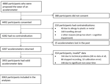
Among the 4880 participants to whom the accelerometer assessment was proposed, 388 did not consent, 210 had contraindications, 15 had their accelerometer lost in the post, 314 did not have valid accelerometer data, and 890 did not have all data points from the spirometry test to rebuild their volume-time curve (flow chart in Fig. 1). Compared to participants not included in the analysis (N = 1817), the analytic sample (N = 3063) did not differ by age (69.2 (standard deviation (SD)=5.6) vs. 69.0 y (SD=5.6), P=0.20), but was composed of more men (73.9% vs. 65.3%, P < 0.0001) and fewer participants from the lowest occupational position (11.0% vs. 12.4%, P = 0.03). Among the 3,063 participants included in the analysis, 2,946 (96.2%) had valid accelerometer data for 7 days, 76 (2.5%) for 6 days, and 41 (1.3%) for 4–5 days. In total missing data were replaced for 1–2 hours over the full obser-vational period for 24.5% of the participants, 2–5 hours for 1.0% of the participants, 5–10 hours for 0.6% of the participants, and 10–25 hours for 0.1% of the participants. Table 1 presents characteristics of the study population. In models adjusted for age, sex, ethnicity, height and weight, compared to never smokers, current, recent, and long-term ex-smokers had respectively 360.0 (95%CI = 254.1, 466.0), 305.6 (95%CI = 221.8, 387.5) and 55.0 (18.5, 91.5) ml lower FEV1, and 278.6 (95%CI = 157.2, 400.0), 238.3 (95%CI = 143.3, 333.2) and 47.6
(95%CI = 5.7, 89.4) ml lower FVC (data not tabulated).
Association of time spent in activity levels with FEV
1and FVC.
In analyses adjusted for age, sex,ethnicity, height, weight, and waking duration, the interaction terms between time spent in MVPA and smoking history (entered as an ordinal variable) were significant for both FEV1 (p = 0.0002) and FVC (p < 0.0001). In
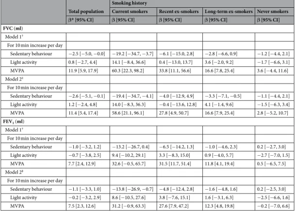
fully adjusted analyses, 10 minutes greater MVPA was associated with 58.6 (95%CI = 21.1, 96.1) ml higher in FVC in current smokers, 27.8 (95%CI = 4.9, 50.7) ml increase in recent ex-smokers, 16.6 (95%CI = 7.9, 25.4) ml in long-term ex-smokers, and only 2.8 (95%CI = −5.2, 10.7) ml in never smokers (Table 2). Similarly, the association between sedentary time and lung function was evident in current but not in never smokers (p for interaction = 0.02 for FEV1 and 0.03 for FVC). There was no evidence of an association of FEV1 and FVC with
light physical activity in all smoking groups, the p for interaction did not reach significance for FEV1 (p = 0.06)
and FVC (p = 0.09). After exclusion of participants with respiratory disease, the association of MVPA with both FEV1 and FVC was slightly attenuated whereas the association with sedentary time remained only for FVC among
current smokers (Supplementary Table 1).
Association between diurnal accelerometry distribution and expired air-volume curve using
functional data analysis.
The accelerometry distribution Ai(x) and the expired air volume-time curve yi(t)www.nature.com/scientificreports
www.nature.com/scientificreports/
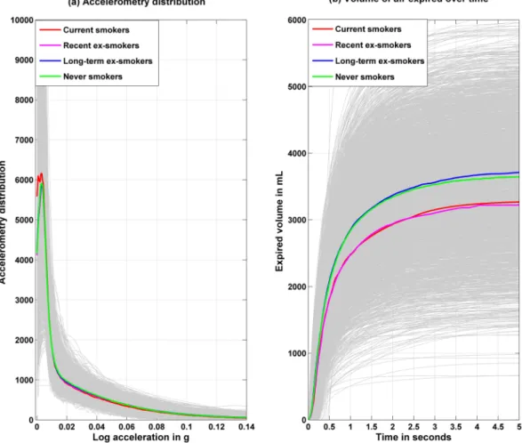
are plotted in Fig. 2; current smokers spent more time in the lowest accelerometry range. Differences in spirome-try test were evident all along the distribution, with never and long-term ex-smokers having better lung function profiles. The plot of the expired air volume-time curve are displayed in the top row of Supplementary Fig. 1 for all the participants, with median functions separately in men and women (left panel), age quartiles (middle panel) and in Caucasians and non-Caucasians (right panel). The regression coefficients for the association of sex, age (per 1 year) and ethnicity with the expired air volume are shown with their 95% confidence interval in the bot-tom row. These results show the association of these covariates along the continuum of spirometry performance
Figure 1. Flow Chart of the Study. * valid data defined as daily wear time ≥2/3 of waking hours, for at least 2 weekdays and 2 week-end days.
All (n = 3063) Smoking history p Current smokers (n = 86) Recent ex-smokers (n=146) Long-term ex-smokers (n = 1251) Never smokers (n = 1580) N = 3063 N = 86 N = 146 N = 1251 N = 1580 Age (years), M(SD) 69.2 (5.6) 67.3 (4.8) 68.2 (5.2) 69.7 (5.6) 69.0 (5.7) <0.001 Men (%) 73.9 69.8 75.3 77.9 70.8 <0.001 Caucasian (%) 93.4 93.0 94.5 95.5 91.7 0.003 Height (cm), M(SD) 170.8 (9.1) 170.3 (8.4) 170.7 (8.6) 171.3 (8.7) 170.5 (9.4) 0.08 Weight (kg), M(SD) 77.7 (14.1) 78.4 (16.2) 80.0 (14.5) 79.0 (14.3) 76.4 (13.8) <0.001 High occupational position at age 50y (%) 45.5 27.9 39.7 45.1 47.3 0.002
Higher education than university (%) 31.4 19.8 21.9 27.3 36.1 <0.001
Married/cohabitating (%) 75.7 58.1 69.9 79.0 74.7 <0.001
High alcohol consumption (%) 14.4 24.4 25.3 18.6 8.7 <0.001
Daily fruit and vegetable consumption (%) 80.3 54.7 75.9 79.1 83.1 <0.001
Cigarettes smoked (per day), M(SD) 12.9 (7.7) 5.6 (11.1)* <0.001
Respiratory diseases (%) 8.0 7.0 15.1 7.9 7.5 0.01
One or more chronic diseases (%) 46.7 48.8 54.1 51.1 42.5 <0.001
Time spent per day (min), M(SD)
Sedentary behaviour 673.9 (88.1) 698.4 (76.8) 674.4 (105.6) 671.9 (86.1) 674.1 (88.4) 0.03 Light physical activity 240.5 (58.1) 220.8 (52.9) 237.3 (70.8) 240.8 (56.6) 241.7 (58.2) 0.009
MVPA 69.3 (36.7) 58.0 (31.3) 69.2 (41.2) 70.1 (37.0) 69.3 (36.2) 0.05
FVC (ml), M(SD) 3639.4 (925.4) 3407.9 (989.9) 3462.4 (973.9) 3667.2 (887.6) 3646.5 (943.7) 0.008 FEV1 (ml), M(SD) 2790.5 (742.2) 2501.1 (767.4) 2565.9 (816.0) 2809.5 (713.1) 2811.8 (750.2) <0.001
Table 1. Characteristics of the study population. Abbreviations: FEV1, Forced Expiratory Volume in 1 sec; FVC,
Forced Vital Capacity; MVPA, Moderate to vigorous physical activity. p: Chi-Square or Anova. *Number of
and not confined to 1 second (FEV1) or at 5 seconds (FVC) as used in the classical multivariate approach. The
direction and strength of association using functional coefficients are consistent with results from the multivar-iate regression for FEV1 (beta for women vs men = −474.7 (−527.8, −421.5) ml) and FVC (beta for women vs
men = −576.3 (−638.0, −514.7) ml) at 1 second and 5 second respectively.
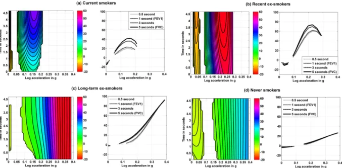
Results from function-on-function regression model are presented in Fig. 3, showing the significant coef-ficient surfaces in each smoking history group and their slices at times 0.5, 1 (FEV1), 3, and 5 (FVC) seconds.
The acceleration levels above which association with spirometry measure was evident were higher in a graded fashion from current to never-smoker groups. A positive (protective) association between physical activity and FVC (time = 5 seconds) was observed for accelerometry values above 0.054g among current smokers, 0.094g among recent ex-smokers, 0.074g among long-term ex-smokers and 0.161g among never smokers (Table 3). More time spent in activity intensities above these smoking-specific accelerometry thresholds was associated with higher FVC, with stronger associations observed in current and recent ex-smokers (Table 3). A similar trend was observed for FEV1. Results in the lower range of accelerometry were less consistent but overall lower lung
function was observed for more time spent at lower accelerometry level among current smokers (<0.03g).
Discussion
Principal findings.
This study of 3063 adults aged 60 to 83 years presents three key findings. First, results based on conventional categorization of physical activity showed that the association between MVPA and lung function is dependent on smoking history, with associations being evident only in current and ex-smokers. Second, function-on-function regression, which takes the continuum of measurement into account, shows an association between physical activity and lung function, both FEV1 and FVC, in all smoking groups but only atvery high levels of physical activity in never smokers. Third, lung function is less consistently associated with time spent in sedentary activity, the association being only evident for FVC among current smokers.
Comparison with others studies.
Longitudinal studies based on self-reported physical activity measures have suggested that physical activity in midlife and early old age is associated with slower decline in lung func-tion16,17,41. In studies that have investigated the modifying role of smoking, physical activity was found to beassociated with higher FEV1 and FVC12, slower decline in lung function13 and lower risk of incident chronic
obstructive pulmonary disease11, only in current or ever-smokers. One study using accelerometer-assessed
Total population
Smoking history
Current smokers Recent ex-smokers Long-term ex-smokers Never smokers β* [95% CI] β [95% CI] β [95% CI] β [95% CI] β [95% CI] FVC (ml)
Model 1†
For 10 min increase per day
Sedentary behaviour −2.5 [−5.0, −0.0] −19.2 [−34.7, −3.7] −6.1 [−15.0, 2.8] −2.8 [−6.6, 0.9] −1.2 [−4.4, 2.1] Light activity 0.8 [−2.7, 4.4] 14.1 [−8.4, 36.6] 0.4 [−13.0, 13.7] 3.6 [−2.0, 9.2] −1.7 [−6.6, 3.1] MVPA 11.9 [5.9, 17.9] 60.3 [22.3, 98.2] 33.8 [11.1, 56.6] 16.6 [7.8, 25.4] 3.6 [−4.4, 11.6] Model 2‡
For 10 min increase per day
Sedentary behaviour −2.6 [−5.1, −0.1] −19.4 [−34.7, −4.1] −4.0 [−12.9, 4.9] −3.3 [−7.1, −0.5] −1.1 [−4.4, 2.1] Light activity 1.2 [−2.4, 4.8] 14.0 [−8.3, 36.3] −0.4 [−13.6, 12.8] 4.1 [−1.4, 9.6] −1.5 [−6.3, 3.4] MVPA 11.4 [5.4, 17.4] 58.6 [21.1, 96.1] 27.8 [4.9, 50.7] 16.6 [7.9, 25.4] 2.8 [−5.2, 10.7]
FEV1 (ml) Model 1†
For 10 min increase per day
Sedentary behaviour −1.0 [−3.2, 1.2] −13.2 [−26.7, 0.4] −6.5 [−14.2, 1.3] −1.0 [−4.6, 2.3] 0.2 [−2.7, 3.0] Light activity −0.7 [−3.8, 2.5] 9.4 [−10.2, 29.1] 3.3 [−8.3, 15.0] 0.9 [−4.0, 5.7] −2.7 [−7.0, 1.5] MVPA 7.7 [2.4, 12.9] 32.6 [−0.5, 65.7] 31.5 [11.7, 51.4] 11.8 [4.1, 19.4] 0.5 [−6.5, 7.5] Model 2‡
For 10 min increase per day
Sedentary behaviour −1.1 [−3.3, 1.0] −13.8 [−26.9, −0.7] −4.8 [−12.4, 2.8] −1.6 [−4.8, 1.6] 0.2 [−2.5, 3.0] Light activity −0.2 [−3.2, 2.9] 8.6 [−10.5, 27.6] 3.8 [−7.6, 15.1] 1.6 [−3.1, 6.3] −2.5 [−6.6, 1.6] MVPA 7.5 [2.3, 12.6] 31.2 [−0.9, 63.3] 27.6 [7.9, 47.2] 12.3 [4.8, 19.8] −0.2 [−7.0, 6.6]
Table 2. Association between physical activity and respiratory function. Abbreviations: CI, confidence interval;
FEV1, Forced Expiratory Volume in 1 sec; FVC, Forced Vital Capacity; MVPA, Moderate to vigorous physical
activity. *Additional adjustment for smoking history. †Model 1: adjusted for age, sex, ethnicity, height, weight,
and waking duration. ‡Model 2: Model 1 additionally adjusted for occupational position at age 50y, education, marital status, alcohol consumption, fruit and vegetable consumption, respiratory disease, and number of chronic diseases. Additionally adjusted for number of cigarettes smoked per day among the current and recent ex-smokers (corresponding to cigarettes smoked before they quitted smoking) groups.
www.nature.com/scientificreports
www.nature.com/scientificreports/
physical activity among 341 adults assessed the cross-sectional association of MVPA with lung function, without accounting for time spent in sedentary behavior and light activities, and also reported the association with MVPA to be evident only in smokers15. Our study using objective physical activity data on a large sample of older adults
adds to the previous findings by showing that the activity intensity threshold at which there is an association between physical activity and lung function depends on smoking history. The association between spirometry measure and physical activity was evident starting from activities in the higher range of light intensities (accel-eration ≥0.06g) in smokers, from physical activities of moderate-to-vigorous intensities (accel(accel-eration ≥0.10g) in ex-smokers and from even more intense MVPA in never smokers. The associations were found all along the spirometry measure, including both FEV1 and FVC. Several mechanisms could underlie the association between
physical activity and lung function. They may involve the anti-inflammatory and vascular benefits of physical activity20. These mechanisms might play a more pronounced role among smokers by compensating for the
delete-rious effect of smoking on the lung13,20. As the study is cross-sectional, the observed association could also reflect
the inability of those with poor lung function to perform physical activity at higher intensity and this would be more pronounced among smokers who might carry multiple health conditions affecting their ability to undertake more intense physical activity42.
Sedentary behaviour is thought to be deleterious for health43. However, it is unclear whether time spent
seden-tary is important because of the adverse effects of inactivity or because it reduces the time available for activities at more intense levels43, given that a day is constrained naturally to 24 hours. Few studies have assessed the impact
of sedentary behaviour on lung function and no robust association has been reported14,19. In the present study, an
association between spirometry measure and sedentary time was found only in current smokers. In other smok-ing groups, the association was small and inconsistent across the various analytic approaches. Both the classical and functional approaches accounted for the diurnal duration constraint so that increase in sedentary time corre-sponds to decrease in active time. Our findings of an association between sedentary behaviour and lung function in current smokers could reflect an aggravating effect of sedentary behaviour on lung function in smokers or the likelihood of smokers with poor lung function to be more sedentary.
Figure 2. Accelerometry distribution during waking time and volume of air expired over time as a function
of smoking status. Grey curves represent individual daily accelerometry distribution over waking time (left panel, number of 5s-epoch over diurnal time) and expired air volume over the time of the spirometry test (right panel). Colored curves represent the median curves among the different smoking history groups.
strengths and limitations.
Strengths of this study include the large sample size, use of accelerometer- assessed physical activity, and functional data analysis approach that is free from laboratory-based thresholds definition of sedentary, light and moderate-to-vigorous activity and accounts for the entire spirometry and diurnal activity intensity distribution. We were able to identify intensities at which physical activity was asso-ciated with respiratory function in groups defined by smoking history. The limitations of this study include its cross-sectional design, although sensitivity analysis excluding COPD and asthma cases were conducted to assess whether the association observed between physical activity and spirometry measure was not driven by difficul-ties in undertaking physical activity among those with respiratory diseases, and the results were broadly similar. Further longitudinal studies are needed to assess the direction of the association between physical activity and respiratory function. Second, although the number of current smokers was small, the trend observed across the smoking groups suggested that the results in smokers were not likely to be by chance. However, future studies are needed to confirm effect size in this group. Finally, although wrist-mounted accelerometer are not designed to distinguish between sitting and inactive standing position, the 0.03 g threshold we used has been reported to accurately separate sedentary behaviours from common motion-based light-intensity activities37.Figure 3. Coefficient surfaces representing the association between accelerometry distribution (X axis) and
volume of expired air over time (Y axis) and their slices at times 0.5, 1 (FEV1), 3, and 5 (FEC) seconds by
smoking history*. *Coefficient surfaces and their slices at times 0.5, 1 (FEV1), 3, and 5 (FVC) seconds (only
significant coefficients at P < 0.05 are shown), from a function-to-function regression model to assess the association between expired air volume-time curve (Y axis) and accelerometry distribution (X axis) adjusted for age, sex, ethnicity, height, weight, respiratory disease, number of chronic diseases, and accelerometry distribution by smoking history groups. Positive values indicate that more time spent in a given accelerometry range is associated with higher air expired volume over the spirometry time scale whereas negative values indicate that more time spent in a given accelerometry range is associated with lower air expired volume over the spirometry time scale.
Current smokers Recent ex-smokers Long-term ex-smokers Never smokers FVC (ml)
Accelerometry threshold 0.054 0.094 0.074 0.161
ΔFVC *[95% CI] for 10 min increase per day in
activity intensity above the threshold 34.7 [23.3, 49.1] 36.5 [29.4, 47.4] 15.1 [13.1, 22.4] 13.2 [7.5, 20.1]
FEV1 (ml)
Accelerometry threshold 0.060 0.093 0.122 0.150
ΔFEV1 *[95% CI] for 10 min increase per day in
activity intensity above the threshold 21.5 [15.1, 28.7] 30.2 [23.7, 35.9] 11.0 [8.5, 13.7] 8.9 [5.3, 12.8]
Table 3. Association between physical activity and respiratory function using threshold for benefits identified
using functional data analysis. Abbreviations: CI, confidence interval; FEV1, Forced Expiratory Volume in 1 sec;
FVC, Forced Vital Capacity; CI, confidence interval. *Estimated change from a function-to-function regression model adjusted for age, sex, ethnicity, height, weight, respiratory disease, and number of chronic diseases.
www.nature.com/scientificreports
www.nature.com/scientificreports/
Conclusion
This study on older adults showed an association between physical activity and better lung function, particularly in recent ex- and current smokers. Among never smokers, the intensity of physical activity required to create an association with lung function was well above that used to define MVPA. These findings suggest that the associ-ation between physical activity and lung function might have been underestimated in non-smokers in previous studies that did not differentiate between intensities of MVPA.
Data Availability
Whitehall II data, protocols, and other metadata are available to the scientific community. Please refer to the Whitehall II data sharing policy at https://www.ucl.ac.uk/whitehallII/data-sharing.
References
1. Sabia, S. et al. Why does lung function predict mortality? Results from the Whitehall II Cohort Study. American journal of epidemiology 172, 1415–1423, https://doi.org/10.1093/aje/kwq294 (2010).
2. Higgins, M. W. & Keller, J. B. Predictors of mortality in the adult population of Tecumseh. Arch Environ Health 21, 418–424 (1970). 3. Wasswa-Kintu, S., Gan, W. Q., Man, S. F., Pare, P. D. & Sin, D. D. Relationship between reduced forced expiratory volume in one second and the risk of lung cancer: a systematic review and meta-analysis. Thorax 60, 570–575, https://doi.org/10.1136/ thx.2004.037135 (2005).
4. Cook, N. R. et al. Height, lung function, and mortality from cardiovascular disease among the elderly. American journal of epidemiology 139, 1066–1076 (1994).
5. Truelsen, T., Prescott, E., Lange, P., Schnohr, P. & Boysen, G. Lung function and risk of fatal and non-fatal stroke. The Copenhagen City Heart Study. International journal of epidemiology 30, 145–151 (2001).
6. Weiss, S. T. Lung function and airway diseases. Nature genetics 42, 14–16, https://doi.org/10.1038/ng0110-14 (2010).
7. Bui, D. S. et al. Childhood predictors of lung function trajectories and future COPD risk: a prospective cohort study from the first to the sixth decade of life. Lancet Respir Med, https://doi.org/10.1016/S2213-2600(18)30100-0 (2018).
8. Postma, D. S., Bush, A. & van den Berge, M. Risk factors and early origins of chronic obstructive pulmonary disease. Lancet 385, 899–909, https://doi.org/10.1016/S0140-6736(14)60446-3 (2015).
9. Garcia-Aymerich, J., Varraso, R., Anto, J. M. & Camargo, C. A. Jr. Prospective study of physical activity and risk of asthma exacerbations in older women. American journal of respiratory and critical care medicine 179, 999–1003, https://doi.org/10.1164/ rccm.200812-1929OC (2009).
10. Mantoani, L. C., Rubio, N., McKinstry, B., MacNee, W. & Rabinovich, R. A. Interventions to modify physical activity in patients with COPD: a systematic review. The European respiratory journal 48, 69–81, https://doi.org/10.1183/13993003.01744-2015 (2016). 11. Behrens, G., Matthews, C. E., Moore, S. C., Hollenbeck, A. R. & Leitzmann, M. F. Body size and physical activity in relation to
incidence of chronic obstructive pulmonary disease. CMAJ: Canadian Medical Association journal = journal de l’Association medicale canadienne 186, E457–469, https://doi.org/10.1503/cmaj.140025 (2014).
12. Burchfiel, C. M. et al. Factors associated with variations in pulmonary function among elderly Japanese-American men. Chest 112, 87–97 (1997).
13. Garcia-Aymerich, J., Lange, P., Benet, M., Schnohr, P. & Anto, J. M. Regular physical activity modifies smoking-related lung function decline and reduces risk of chronic obstructive pulmonary disease: a population-based cohort study. American journal of respiratory and critical care medicine 175, 458–463, https://doi.org/10.1164/rccm.200607-896OC (2007).
14. Jakes, R. W. et al. Physical inactivity is associated with lower forced expiratory volume in 1 second: European Prospective Investigation into Cancer-Norfolk Prospective Population Study. American journal of epidemiology 156, 139–147 (2002). 15. Luzak, A. et al. Association of physical activity with lung function in lung-healthy German adults: results from the KORA FF4 study.
BMC Pulm Med 17, 215, https://doi.org/10.1186/s12890-017-0562-8 (2017).
16. Nystad, W., Samuelsen, S. O., Nafstad, P. & Langhammer, A. Association between level of physical activity and lung function among Norwegian men and women: the HUNT study. The international journal of tuberculosis and lung disease: the official journal of the International Union against Tuberculosis and Lung Disease 10, 1399–1405 (2006).
17. Pelkonen, M. et al. Delaying decline in pulmonary function with physical activity: a 25-year follow-up. American journal of respiratory and critical care medicine 168, 494–499, https://doi.org/10.1164/rccm.200208-954OC (2003).
18. Sin, D. D., Jones, R. L., Mannino, D. M. & Paul Man, S. F. Forced expiratory volume in 1 second and physical activity in the general population. The American journal of medicine 117, 270–273, https://doi.org/10.1016/j.amjmed.2004.01.029 (2004).
19. Vaz Fragoso, C. A. et al. Respiratory impairment and dyspnea and their associations with physical inactivity and mobility in sedentary community-dwelling older persons. Journal of the American Geriatrics Society 62, 622–628, https://doi.org/10.1111/ jgs.12738 (2014).
20. Hopkinson, N. S. & Polkey, M. I. Does physical inactivity cause chronic obstructive pulmonary disease? Clinical science (London, England: 1979) 118, 565–572, https://doi.org/10.1042/cs20090458 (2010).
21. Bell, J. A. et al. Healthy obesity and objective physical activity. The American journal of clinical nutrition 102, 268–275, https://doi. org/10.3945/ajcn.115.110924 (2015).
22. Menai, M. et al. Accelerometer assessed moderate-to-vigorous physical activity and successful ageing: results from the Whitehall II study. Sci Rep 8, 45772, https://doi.org/10.1038/srep45772 (2017).
23. Sabia, S. et al. Physical Activity and Adiposity Markers at Older Ages: Accelerometer Vs Questionnaire Data. Journal of the American Medical Directors Association 16(438), e437–438. e413 (2015).
24. Goldsmith, J., Liu, X., Jacobson, J. S. & Rundle, A. New Insights into Activity Patterns in Children, Found Using Functional Data Analyses. Medicine and science in sports and exercise 48, 1723–1729, https://doi.org/10.1249/MSS.0000000000000968 (2016). 25. Xiao, L. et al. Quantifying the lifetime circadian rhythm of physical activity: a covariate-dependent functional approach. Biostatistics
16, 352–367, https://doi.org/10.1093/biostatistics/kxu045 (2015).
26. Augustin, N. H., Mattocks, C., Faraway, J. J., Greven, S. & Ness, A. R. Modelling a response as a function of high-frequency count data: The association between physical activity and fat mass. Stat Methods Med Res 26, 2210–2226, https://doi. org/10.1177/0962280215595832 (2017).
27. Ramsay, J. O. S., B.W. Functional data analysis. (2006).
28. Marmot, M. & Brunner, E. Cohort Profile: the Whitehall II study. International journal of epidemiology 34, 251–256, https://doi. org/10.1093/ije/dyh372 (2005).
29. Esliger, D. W. et al. Validation of the GENEA Accelerometer. Med. Sci. Sports Exerc 43, 1085–1093, 10.1249/MSS.0b013e31820513be [doi] (2011).
30. van Hees, V. T. et al. Separating movement and gravity components in an acceleration signal and implications for the assessment of human daily physical activity. PloS one 8, e61691, https://doi.org/10.1371/journal.pone.0061691 (2013).
31. da Silva, I. C. et al. Physical activity levels in three Brazilian birth cohorts as assessed with raw triaxial wrist accelerometry. International journal of epidemiology 43, 1959–1968, https://doi.org/10.1093/ije/dyu203 (2014).
32. Hildebrand, M., VT, V. A. N. H., Hansen, B. H. & Ekelund, U. Age group comparability of raw accelerometer output from wrist- and hip-worn monitors. Medicine and science in sports and exercise 46, 1816–1824, https://doi.org/10.1249/mss.0000000000000289
(2014).
33. Sabia, S. et al. Association between questionnaire- and accelerometer-assessed physical activity: the role of sociodemographic factors. American journal of epidemiology 179, 781–790, https://doi.org/10.1093/aje/kwt330 (2014).
34. van Hees, V. T. et al. Autocalibration of accelerometer data for free-living physical activity assessment using local gravity and temperature: an evaluation on four continents. Journal of applied physiology (Bethesda, Md.: 1985) 117, 738–744, https://doi. org/10.1152/japplphysiol.00421.2014 (2014).
35. van Hees, V. T. et al. A novel, open access method to assess sleep duration using a wrist-worn accelerometer. PloS one 10, e0142533 (2015).
36. Catellier, D. J. et al. Imputation of missing data when measuring physical activity by accelerometry. Medicine and science in sports and exercise 37, S555–562 (2005).
37. Bakrania, K. et al. Intensity Thresholds on Raw Acceleration Data: Euclidean Norm Minus One (ENMO) and Mean Amplitude Deviation (MAD) Approaches. PloS one 11, e0164045, https://doi.org/10.1371/journal.pone.0164045 (2016).
38. Hayes, D. Jr. & Kraman, S. S. The physiologic basis of spirometry. Respiratory care 54, 1717–1726 (2009). 39. Wand, M. P. & Jones, M. C. Kernel smoothing. Vol. 60 (Chapman and Hall/CRC 1995).
40. Chacon, J. E. & Duong, T. Multivariate plug-in in bandwidth selection with unconstrained pilot bandwidth matrices. Test 19, 375–398 (2010).
41. Cheng, Y. J. et al. Effects of physical activity on exercise tests and respiratory function. British journal of sports medicine 37, 521–528 (2003).
42. Mantoani, L. C., Dell’Era, S., MacNee, W. & Rabinovich, R. A. Physical activity in patients with COPD: the impact of comorbidities. Expert Rev Respir Med 11, 685–698, https://doi.org/10.1080/17476348.2017.1354699 (2017).
43. van der Ploeg, H. P. & Hillsdon, M. Is sedentary behaviour just physical inactivity by another name? Int J Behav Nutr Phys Act 14, 142, https://doi.org/10.1186/s12966-017-0601-0 (2017).
Acknowledgements
The Whitehall II study has been supported by grants from the UK Medical Research Council (K013351, R024227, S011676); the British Heart Foundation (PG/11/63/29011 and RG/13/2/30098); the British Health and Safety Executive; the British Department of Health; the National Heart, Lung, and Blood Institute (R01HL036310); the National Institute on Aging, National Institute of Health (R01AG056477, R01AG034454); the Economic and Social Research Council (ES/J023299/1). Mika Kivimaki is supported by the Medical Research Council (K013351, R024227, S011676), UK, NordForsk, the Nordic Programme on Health and Welfare, and the Academy of Finland (311492).
Author Contributions
M.A.B., M.M. and S.S. developed the hypothesis and study design. M.A.B., M.M., V.v.H. and S.S. performed statistical analysis. M.A.B. and M.M. wrote the first draft of the manuscript. All authors contributed to interpretation of data and drafting or critical revision of the manuscript for important intellectual content, or in addition, data acquisition. A.S.M. and M.K. obtained funding for the Whitehall II Study.
Additional Information
Supplementary information accompanies this paper at https://doi.org/10.1038/s41598-019-46771-y.
Competing Interests: The authors declare no competing interests.
Publisher’s note: Springer Nature remains neutral with regard to jurisdictional claims in published maps and
institutional affiliations.
Open Access This article is licensed under a Creative Commons Attribution 4.0 International
License, which permits use, sharing, adaptation, distribution and reproduction in any medium or format, as long as you give appropriate credit to the original author(s) and the source, provide a link to the Cre-ative Commons license, and indicate if changes were made. The images or other third party material in this article are included in the article’s Creative Commons license, unless indicated otherwise in a credit line to the material. If material is not included in the article’s Creative Commons license and your intended use is not per-mitted by statutory regulation or exceeds the perper-mitted use, you will need to obtain permission directly from the copyright holder. To view a copy of this license, visit http://creativecommons.org/licenses/by/4.0/.