HAL Id: tel-01674204
https://tel.archives-ouvertes.fr/tel-01674204
Submitted on 2 Jan 2018
HAL is a multi-disciplinary open access
archive for the deposit and dissemination of
sci-entific research documents, whether they are
pub-lished or not. The documents may come from
teaching and research institutions in France or
L’archive ouverte pluridisciplinaire HAL, est
destinée au dépôt et à la diffusion de documents
scientifiques de niveau recherche, publiés ou non,
émanant des établissements d’enseignement et de
recherche français ou étrangers, des laboratoires
Genomewide analysis of road-block termination
Tito Candelli
To cite this version:
Tito Candelli. Genomewide analysis of road-block termination. Genetics. Université Paris-Saclay,
2016. English. �NNT : 2016SACLS587�. �tel-01674204�
NNT : 2016SACLS587
Th`
ese de doctorat
de l’Universit´
e Paris-Saclay
pr´
epar´
ee `
a l’Universite Paris-Sud
´
Ecole doctorale n
◦577
Structure et dynamique des syst`
emes vivants
Sp´
ecialit´
e de doctorat: Sciences de la Vie et de la Sant´
e
par
Tito Candelli
Genomewide analysis of road-block termination
Th`
ese pr´
esent´
ee et soutenue `
a l’Institut Jacques Monod, le 12 D´
ecembre 2016.
Composition du Jury :
M.
Domenico Libri
Directeur de recherche
(Directeur de th`
ese)
Institut Jacques Monod
M.
Alain Jacquier
Directeur de recherche
(Rapporteur)
Institut Pasteur
M.
Vincent Geli
Directeur de Recherche
(Rapporteur)
Genomewide analysis of road-block
termination
Abstract
Transcription of DNA into RNA intermediates constitutes the first step in gene expression. During the last decade, several studies showed that about 80-90% of the genome is transcribed, and that transcription can initiate almost anywhere. This process—known as pervasive transcription—represents a serious threat to proper gene expression as it has the potential to interfere with not only other transcription events, but any DNA-based process. Selective transcription termination is therefore a mechanism of paramount importance for genome transcriptome stability and correct regulation of gene expression. Here we describe road-block termination, a novel termination mechanism for RNA polymerase II that functions to limit pervasive transcription and buffer the consequences of readthrough transcription at canonical terminators in S.cerevisiae. We show that several transcription factors can elicit this termination and that a number of unexpected genomic loci are associated with it. Additionally, we explore the possibility that road-block termination might contribute to specification of replication origins.
R´esum´e
La transcription de l’ADN en ARN constitue la premi`ere ´etape de l’expression d’un g`ene. Durant les dix derni`eres ann´ees, plusieurs ´etudes ont montr´e qu’environ 80-90% du g´enome est transcrit et que la transcription peut d´emarrer presque partout. Ce ph´enom`ene, connu sous le nom de transcription envahissante, repr´esente une menace s´erieuse contre l’expression correcte du g´enome car il peut interf´erer non seulement avec d’autres ´ev`enements de transcription mais ´egalement avec n’importe quel proc´ed´e impliquant l’ADN. Une terminaison s´elective est donc un m´ecanisme de la plus haute importance pour la stabilit´e du g´enome et la correcte r´egulation de l’expression des g`enes. Ici nous d´ecrivons la terminaison road-block, un nouveau m´ecanisme de la terminaison par l’ARN polymerase II, qui a pour fonction de limiter la transcription envahissante et de limiter les cons´equences d’une translecture au niveau des sites de terminaison canoniques de S.cerevisiae. Nous d´emontrons ´egalement que plusieurs facteurs de transcription peuvent entrainer cette terminaison et que certains sites g´enomiques y sont associ´es. De plus, nous explorons ´egalement la possibilit´e que ces terminaisons road-block puissent contribuer `a rendre sp´ecifiques les origines de r´eplication.
Contents
Table of Contents . . . iv
List of Figures . . . viii
Preface . . . x
I
Introduction
1
1 Transcription Initiation . . . 21.1 Spatial Definition: Chromatin Structure and Core Promoter Elements 2 1.2 Temporal Definition: Gene-Specific Transcription Factors . . . 4
1.3 PIC Assembly and Promoter Clearance . . . 5
2 Transcription Elongation . . . 8
2.1 Elongation Through Chromatin . . . 9
2.2 Transcriptional Pausing . . . 11 2.3 The CTD . . . 12 2.3.1 CTD Phosphorylation Dynamics . . . 12 2.3.2 Functional Interactions . . . 14 3 Transcription Termination . . . 16 3.1 The CPF-CF Pathway . . . 17
3.1.1 Recruitment and Assembly . . . 17
3.1.2 The Allosteric Model . . . 18
3.1.3 The Torpedo Model . . . 19
3.2 The NNS Pathway . . . 22
3.2.1 The NNS Complex . . . 22
3.2.2 The Mechanism of Transcription Termination . . . 24
3.2.3 Processing Products of the NNS Pathway . . . 27
3.3 Non-Canonical Termination Pathways . . . 28
3.3.1 Rnt1-Dependent Termination . . . 29
3.3.2 Road-Block Termination . . . 30
4 The Transcriptional Landscape of S.cerevisiae . . . 32
4.1 Control of pervasive Transcription . . . 33
4.2 Classes of Pervasive Transcript . . . 33
4.3 Quality Control Pathways . . . 36
4.4 Functional Role of Pervasive Transcription . . . 37
5 General Regulatory Factors . . . 38
5.1 Rap1 . . . 38
5.2 Abf1 . . . 40
5.3 Reb1 . . . 41
5.4 Cbf1 . . . 42
5.5 Chromatin Remodeling . . . 43
5.5.1 Genome-Wide Effect on Chromatin Structure . . . 44
6 Transcription and Replication . . . 46
6.1 Replication Origin and Their Specification . . . 47
6.1.1 Origin DNA Elements . . . 47
6.1.2 Nucleosome Positioning in Origins . . . 48
6.1.3 Transcription in Origins . . . 49
II
Results and Discussion
53
7 Termination of RNA Polymerase II Through a Road-Block
Mech-anism . . . 54
7.1 Road-Block Termination by Reb1 Restricts Cryptic and Readthrough Transcription . . . 55
7.2 Genomewide Analysis of Road-Block Termination . . . 87
7.2.1 Introduction . . . 88 7.2.2 Results . . . 91 7.2.3 Discussion . . . 111 7.2.4 Supplementary Figures . . . 118 7.3 General Discussion . . . 124 7.3.1 Fail-Safe Termination . . . 124
7.3.2 Road-Block Termination Promotes Genome Stability . . . . 125
8 The Effect of Endogenous Transcription on Origin Specification 128 8.1 Global Visualization of Transcription Around Replication Origins . 129 8.2 Transcriptional Pausing and Termination Are Associated With Repli-cation Origins . . . 130
8.3 Transcription Levels Asymmetrically Affect Origin Efficiency . . . . 133
8.3.1 Licensing Efficiency . . . 134
8.3.2 Firing Efficiency . . . 136
8.3.3 Timing of Firing . . . 138
8.4 Discussion . . . 140
8.4.1 Transcription Termination Is a Feature of Replication Origins141 8.4.2 ACS Orientation Determines the Impact of Transcription on Replication Efficiency . . . 141
9 Dynamics of Nrd1-Nab3 RNA Binding in vitro and in vivo . . . 145
9.1 In vitro Selection of RNA Sequences With High Affinity for the Nrd1-Nab3 Heterodimer . . . 146 9.2 Arrangement of Binding Sites Influences Heterodimer Affinity in vitro 148
9.3 Comparison of SELEX and in vivo Artificial CUT Selection Unveils
Unexpected Dynamics of Nrd1 Binding . . . 152
9.4 Nrd1 Binding Sites GUAG and GUAA Possess Different Extended Consensuses . . . 154
9.4.1 Confirming the Differences in vivo . . . 155
9.5 Discussion . . . 157
9.5.1 Site Arrangement and Spacing Influence Binding Affinity and Termination Efficiency . . . 158
9.5.2 Comparison of SELEX and in vivo CUT Selection Reveals Clusters of Differentially Enriched Sequences . . . 159
Methods . . . 160
Glossary . . . 173
Figures
1.1 Stepwise PIC assembly . . . 6
2.1 Mechanism of transcription through chromatin. . . 9
2.2 CTD phosphorylation states throughout the transcription cycle. . . 13
3.1 Mechanism of CPF-CF termination . . . 20
3.2 Mechanism of NNS termination . . . 25
4.1 Classes of transcripts and their fates. . . 34
5.1 GRFs binding sites and S.cerevisiae centromere structure . . . 39
6.1 ACS consensus and arrangement relative to other origin DNA elements 48 6.2 Stepwise mechanism of DNA replication . . . 51
7.1 Rap1-dependent transcripts are isolated from in vivo selection . . . 92
7.2 Northen blot analysis of Rap1 and Reb1 terminated species . . . 95
7.3 Examples of Rap1-dependent termination detected in vivo . . . 97
7.4 Metagene analysis around sites of Rap1 and Reb1. . . 99
7.5 Metagene analysis around poly(A) sites that are within 300bp up-stream of Rap1 sites. . . 102
7.6 Examples of CPF-terminated transcripts followed by sites of Rap1 . 103 7.7 Analysis of 4 snoRNAs followed by road-block by CRAC and RNA-seq in several mutant strains. . . 104
7.8 RNA-seq analysis of genes followed by Rap1 sites. . . 107
7.9 Metagene analysis around several putative road-blocking factors . . 109
7.11 metagene analysis of RNAPII occupancy around tRNAs . . . 111
8.1 ACS consensus and arrangement relative to other origin DNA elements129 8.2 Metagene analyses showing polymerase occupancy and termination
around replication origins . . . 131 8.3 Northern blot analysis of transcription through ARS305 . . . 132 8.4 Cartoon of sense and antisense transcription relative to the ACS . . 133 8.5 Boxplots comparing licensing efficiencies in high- and low-transcription
populations . . . 135 8.6 Boxplots comparing transcription levels in high- and low-firing
pop-ulations . . . 137 8.7 Boxplots comparing median replication times in high- and
low-transcription populations . . . 138 8.8 A model of how transcription can affect replication efficiency . . . . 142 8.9 Correlations between transcription and replication efficiency in a
NNS defective strain . . . 143
9.1 SELEX procedure . . . 147 9.2 Enrichment of Nrd1 and Nab3 sites with different arrangements and
spacings in the SELEX experiment . . . 149 9.3 Schematic of the selection process in the artificial CUT selection . . 150 9.4 Enrichment of Nrd1 and Nab3 sites with different arrangements and
spacings in the artificial CUT selection . . . 151 9.5 Direct enrichment comparison between SELEX and artificial CUT
selection for all 4 nucleotides motifs . . . 153 9.6 Comparison of flanking nucleotides between GUAG and GUAA in
Preface
Transcription of DNA into RNA intermediates constitutes the first step in gene expression. Even minute changes in transcription patterns can upset the balance of many essential cellular constituents, generating a cascade of responses with signifi-cant repercussions on every biological process. Because of this massive potential, transcription is one of the most finely regulated events in the cell and according to the Saccharomyces Genome Database (SGD) [27] Gene Ontology annotation, 1231 out of 6691 genes in S.cerevisiae (18%) can influence or directly take part in the transcriptional process.
In eukaryotes, three distinct RNA polymerases exist. RNA Polymerase I (RNAPI): responsible for the transcription of Ribosomal RNA (rRNA); RNA Polymerase II (RNAPII): responsible for the transcription of both protein coding genes and many non-coding RNAs; and RNA Polymerase III (RNAPIII): responsible mainly for the transcription of tRNAs and some rRNA. Although products of RNAPI and RNAPIII are by far the most abundant in the cell, RNAPII is tasked with the production of an extremely varied set of transcripts and it is estimated that 80% of the genome is actively transcribed by it [34]. Because of this pervasiveness, transcription by RNAPII must be tightly regulated to ensure its products are viable, as well as to prevent interference with other processes. In this dissertation i will focus on how transcrition by RNAPII is controlled—especially through transcription termination—and what its effects are on other DNA-based biological processes.
The first three chapters of the introduction to this work will describe the transcrip-tional process along its three main steps: initiation, elongation and termination. I will highlight the main molecular determinants that give rise to each phase, as
well as mechanistically characterize the process when appropriate. Because of the relevance for the results that will be presented, I have devoted particular attention to transcription termination and described it in detail. In chapter 4, i will talk about the transcriptional landscape of S.cerevisiae; a look into the world of perva-sive transcription, along with the mechanisms that control it. I will highlight the different classes of non-coding RNAs transcribed by RNAPII as well as the quality control pathways that ensure their degradation. In connection with the results of this dissertation, chapter 5 will discuss a particular class of transcription factors known as General Regulatory Factors. I will describe these factors in the context of their multiple functions, focusing on their chromatin remodeling capabilities and their function at gene promoters. Finally, in chapter 6 I will consider the process of DNA replication and its interaction with transcription. I will first put replication in its appropriate context by describing the structure of replication origins and the mechanics of the process itself. I will then discuss the available literature in regard to the effect of transcription on replication initiation and origin specification.
In the results part, i will outline three different projects. The first consists of the characterization of road-block termination, a novel termination mechanism for RNAPII. Second, i will explore the interaction between transcription and DNA replication, with particular attention to the effect of transcription on origin usage. Finally, the last chapter will focus on NNS termination and how the components of the NNS complex contact their cognate binding sites in different contexts.
The results presented here were obtained using S.cerevisiae as a model organism. Therefore, the ensemble of data cited in this manuscript refers to this organism unless otherwise stated.
“
Mathematics, rightly viewed, possesses not only truth, but supreme beauty —a beauty cold and austere, without the gorgeous trappings of painting or music.”
— Bertrand Russell
“
The ending isn’t more important than any of the moments that led to it.”
— Dr. Rosalene, To the Moon
“
Sometimes it is more important to take in the spectacular than to worry about the pressing business of staying alive.”
Part I
1
Transcription Initiation
Initiation is the first step in any transcription event. It therefore needs to be accurate in when and where it occurs. Transcription initiation fundamentally relies on the assembly of the Pre-Initiation Complex (PIC) (a super-complex 1.5 megadaltons in size containing RNAPII [47]) on DNA. The assembly of such complex is spatially defined by two elements: chromatin structure and core promoter elements. Both contribute to limit the amount of spurious transcription by ensuring robust assembly of the PIC only in promoter regions. In addition to spatial regulation, timing and intensity of transcription initiation must also be controlled. Specific promoters can be finely tuned by the binding of gene-specific transcription factors, that can act as either enhancers or repressors; modulating initiation efficiency either constitutively or in response to environmental effects. Finally, when the PIC is fully assembled, it eventually escapes the promoter and enter productive elongation.
1.1
Spatial Definition: Chromatin Structure and
Core Promoter Elements
Chromatin is a higher order structure that forms when DNA wraps around histones, proteins that can efficiently arrange loose DNA into compact structures. The
simplest unit of chromatin consists of 140 nucleotides of DNA tightly wrapped around a histone, forming a nucleosome. The organization of the genome around nucleosome units has a multitude of consequences, not least of which is to sterically prevent DNA binding proteins from accessing their substrate. As transcription relies on assembly of RNAPII and the PIC on DNA to complete its initial phase, nucleosomes pose a considerable barrier to efficient initiation [52,81]. The insulation of DNA by nucleosomes has been harnessed by the cell and made into a regulatory mechanism that can spatially define where transcription initiates, as transcription factors must bind DNA for it to occur. To favor transcription factor binding to DNA in promoter regions, the latter are always associated with an Nucleosome Free Regions (NFR), an area of the genome where nucleosomes are depleted, leaving naked DNA available for binding. Although certain sequence elements can passively discourage nucleosome association, several complexes actively mediate depletion of nucleosomes from promoter regions, such as SWI1/SNF and the closely related RSC complex. These complexes can be recruited in two ways: through sequence specificity [8,90] or through recruitment by gene-specific transcription factors such as Reb1p, Abf1p, and Rap1p to promoter regions [8,53,66,172].
While chromatin defines the position of transcription initiation, core promoter elements provide specificity for many early-acting general transcription factors. A number of promoter elements were identified in metazoans, where they have been shown to regulate position and intensity of transcription initiation; but although a Transcription Start Site (TSS) consensus was recently defined [112], no sequence was found to be universally required for transcription initiation [19]. In S.cerevisiae promoter elements remain poorly characterized and seem to lack the majority of the sequence elements found in their metazoan counterparts. The major element known to bring about the assembly of the PIC in S.cerevisiae is the TATA box. This very short consensus sequence, TATAWAWR [9], is present in about 15% of yeast genes [83] and is recognized by the TATA Binding Protein (TBP), an essential factor for PIC assembly. At these promoters, TBP binds DNA as part of the Spt-Ada-Gcn5-Acetyl transferase (SAGA) complex, changing the conformation of DNA and
priming the promoter for assembly of other general transcription factors. TATA-dependent promoters, however, are not the only type of promoter in S.cerevisiae. The majority of yeast promoters (85-90% ) are known as TATA-less and require binding of the TFIID complex in lieu of SAGA [155]. Curiously, TBP, along with a number of other shared subunits and co-factors, is also contained in the TFIID complex, but it was recently shown that, in this context, its binding activity is not required for gene activation [83]. TFIID and SAGA have largely overlapping roles in activating gene expression, however, the predominant activity of the two complexes can be associated with functional differences. While TFIID generally dominates over house-keeping genes that do not require regulation, SAGA—and as a consequence TBP binding—has a larger effect over highly regulated and stress-inducible genes [76]. The binding of either complex represents the first step towards assembly of other general transcription factors into the PIC.
1.2
Temporal Definition: Gene-Specific
Transcrip-tion Factors
While nucleosome positioning and core promoter elements define where transcription should initiate, they do not generally actively regulate it on their own. In the cell, many genes need to be activated in response to specific conditions or external stimuli. These regulated genes are generally inactive and become actively transcribed only when the conditions of their activation are met. The main mechanism that enables these transcriptional switches is the presence of gene-specific transcription factors. These DNA-binding proteins specifically target promoter regions, modulating their activity in response to a large number of conditions. Gene-specific transcription factors can activate—or repress—transcription in a variety of ways: activation can occur by binding DNA and recruiting NFR-generating complexes, or otherwise facilitating PIC assembly, and even by relocating chromatin to the nuclear periphery [21]. Alternatively, transcription factors can constitutively repress their target genes and selectively lose the DNA-binding capability under certain conditions, such as
the presence of a ligand.
Genome-wide studies on transcription factor organization highlighted the combinato-rial potential that emerges when several transcription factors interact with the same promoters [65]. Regulation of a single promoter by several distinct transcription factors can exploit their different requirements—qualitative or quantitative— to force the emergence of complex regulatory logic.
1.3
PIC Assembly and Promoter Clearance
Assembly of the Pre-Initiation Complex starts with the binding of either TFIID or SAGA to promoter DNA. The presence of TBP in these complexes modifies the structure of DNA, allowing the step-wise recruitment of several general transcription factors and of RNAPII [For review see162].
TFIIA and TFIIB are the first factors to make contact with TBP, stabilizing its interaction with DNA. However, while TFIIA simply acts as an auxiliary factor and is dispensable [78], TFIIB is required for RNAPII recruitment [17]. The presence of TFIIB acts as a platform for TFIIF docking. The addition of TFIIF has the double effect of recruiting RNAPII (RNAPII is bound to TFIIF when in free form [147]) and of further stabilizing the whole PIC. Despite the inclusion of RNAPII in the forming PIC, at this stage promoter DNA is firmly wound-up in a double helix and therefore the ternary complex1 required for transcription cannot yet form. TFIIE and TFIIH are recruited to the PIC to solve this problem. TFIIE acts as a bridge between RNAPII and TFIIH, who contains an ATPase module and is able to unwind promoter DNA [72]. This will eventually contribute to DNA melting and the formation of the open PIC, a structural variant that precedes the shift into elongation.
1
The ternary complex is defined as the three-way interaction between DNA, RNA, and RNAPII that forms within transcribing polymerases
Promoter DNA TBP TAFs TAFs TFIIH core Pol II Pol II +1 TFIIH kinase ATP NTPs Transcription bubble Nascent RNA Elongation factors Elongation factors Initiation factors Downstream DNA Upstream DNA Unbound promoter Upstream promoter complex Core PIC Closed PIC Open PIC Initially transcribing complex Elongation complex TFIIF TFIIF TFIIE TFIIE TFIIH TFIIA TFIIA TFIIB TFIIB 5 cap
Figure 1.1: Stepwise assem-bly of general transcription fac-tors and RNAPII on a promoter. adapted from [162].
The order of stepwise assembly of general tran-scription factors into a functional PIC was first discovered in vitro [15]. In vivo, however, there is evidence for the activity of the mediator com-plex in providing additional assembly pathways [46]. Mediator is a large and flexible protein complex that can interact with virtually every general transcription factor and with RNAPII. It is known for its fundamental role in transducing regulatory signals from gene-specific transcrip-tion factors to the polymerase. Without Media-tor, the PIC can drive basal transcription levels, but its activity cannot be modulated in response to external factors. Studies have implicated me-diator in the recruitment of TFIIE and TFIIH independently of RNAPII, providing alternative ways to assemble the complete PIC. Addition-ally, interactions between RNAPII and mediator were found to be required for transcription in vivo [171].
After the assembly of the PIC and the Medi-ator complex on the promoter, RNAPII relies on TFIIH to relax DNA and physically separate the two strands, creating what is referred to as the transcription bubble. Studies in human re-port that once the bubble first opens, it spans about 7 nucleotides. It then extends forward, allowing the process of transcription to begin. Polymerases at this stage, however, have to con-tend with the fact that the RNA-DNA hybrid is
too short to be stable. According to in vitro studies, forming a sufficiently long—and therefore stable–hybrid requires several rounds of abortive initiation, where the small RNA is displaced from the template and released. When the RNA-DNA hybrid reaches a length of about 10 nucleotides, the upstream half of the bubble, which now spans 17-18 nucleotides, collapses, suddenly closing [71]. This event marks the detachment of what will eventually become the elongation complex from the scaffold of general transcription factors that is going to be retained at the promoter [134].
2
Transcription Elongation
After escaping the PIC, RNAPII enters the phase of productive elongation. During this phase, the polymerase travels along DNA, catalyzing the addition of nucleotides to the growing RNA molecule that is being synthesized. The simple synthesis of RNA, however, is not enough to qualify a mature transcript. Several essential processing steps take place during transcription elongation and contribute to the production of fully formed transcripts. Among these, the addition of the 5’ cap, addition of a poly(A) tail, and formation of an export-competent transcript all rely on the presence of RNAPII and the Transcription Elongation Complex (TEC) in order to be carried out properly. The precise composition of the TEC is poorly understood. However, as RNAPII progresses through the transcription unit, several complexes and co-factors are known to dynamically associate with it in order to enact the various maturation steps. Transcription elongation is therefore a highly regulated activity that coordinates several different processes to produce mature transcripts. This dynamic regulation is enacted by the cell through several distinct mechanisms, such as the phosphorylation of the C-Terminal Domain (CTD) and the modification of histones. These very same regulation mechanisms—along with important regulatory sequences—will eventually mark the end of transcription elongation and the transition to transcription termination.
2.1
Elongation Through Chromatin
Chromatin represents an extremely repressive barrier to any kind of DNA based process. As I briefly touched upon in previous sections, chromatin components— histones—need to be actively dislodged from promoter regions in order to allow the Pre-Initiation Complex to assemble. Elongating RNAPII faces very similar problems, as in order to synthesize the RNA, it has to move through an array of nucleosomes without losing contact with DNA. Although in vitro evidence has shown that RNAPII can effectively elongate through a single nucleosome [104]—possibly due to spontaneous disassembly and reassembly of nucleosomes, a process that was recently shown to happen every few seconds [86]—the elongation complex alone is not enough to mediate transcription through multiple nucleosomes.
RPD3s
Met
Deacetylation
of deposited
octamers
Methylation
ac
ac
ac
ac
ac
Displaced
octamer
Hyperacetylation
of octamer to be
disrupted
ac
ac
ac
ac
ac
ac
ac
ac
ac
ac
Met
FACT
RNAPII
HAT
Set2
IIIIIIIIIIII
PI
PI
PI
PI
PI
PPP
RNAP
RNAP
RNAP
RNAP
RNAP
RNAP
RNAP
RNAP
RNA
RNA
RNA
RNA
FACT
Figure 2.1: Overview of the main actors in the mechanism of transcription through chromatin. Nucleosomes are destabilized through acetylation and chap-eroned away—either partially or completely—by FACT and other complexes. Addition of methyl groups to histone tails allows the recruitment of Histone De-Acetylases (HDACs) and the restoration of chromatin structure. adapted from [168].
The TEC can overcome this problem by enlisting the help of several histone chaper-ones and chromatin remodeling complexes, as well as by exploiting post translational
modifications of histones (Fig: 2.1). The current model for transcription through nucleosomes posits that, depending on the intensity of transcription, histones can either be completely removed from DNA, or be partially destabilized as to allow RNAPII to more easily transcribe through them [94]. The most notable actors in this phase are Histone Acetyl-Transferases (HATs) such as Gcn5 and the FACT (Facilitates chromatin transcription) complex [For review see150]. HATs are posited to travel with the polymerase, depositing an acetyl group on histone tails. This has the consequence of destabilizing inter-nucleosome interactions as well as low-ering the affinity for DNA by increasing negative charges, resulting in a more relaxed chromatin structure and more unstable nucleosomes. Once histones are acetylated, FACT—also traveling with the polymerase—destabilizes the H2A-H2B dimer1, removing it and facilitating transcription through the remaining incomplete
nucleosome structure.
We saw how, in order to efficiently elongate, RNAPII needs to destabilize the chromatin structure. However, in the long run, this destabilization can have negative effects, as it results in a more relaxed structure that can potentially give rise to intragenic transcription initiation. In order to prevent this phenomenon, the composition, modifications, and overall structure of nucleosomes must be reset after the passage of RNAPII. Specific histone chaperones such as Spt6, together with methyl-transferases and HDACs, are involved in this process. First, Spt6 and other histone chaperones reconstruct a complete histone in the wake of transcribing RNAPII. Subsequently, methyl-transferases such as Set2 methylate lysine 36 on histone H3. Although this modification—unlike acetylation—has no structural consequences on the organization of nucleosomes, it can act as a platform for recruitment of HDACs. The RPD3 complex has high affinity for H3K36 methylation and is recruited immediately after the passage of RNAPII in order to remove the acetyl groups from histones and thus reset the structure of chromatin.
1
Two of the four core components of a histone. Histones are composed of two H2A-H2B dimers and one H3-H4 tetramer arranged in a symmetrical structure.
2.2
Transcriptional Pausing
Nucleosomes do not represent the only obstacle to productive elongation. A number of events can potentially prevent RNAPII from elongating forward, such as DNA damage, misincorporation of a nucleotide, or collision with another DNA-bound protein. This causes RNAPII to temporarily stop, a phenomenon known as tran-scriptional pausing. One of the mechanisms used by the polymerase to resolve pausing is called backtracking.
During backtracking, RNAPII moves backwards, retracing its steps. how much the polymerase is able to backtrack in vivo is a matter of debate, early in vitro studies reported up to 100 nucleotides, while more recent crystal structures concluded that this figure can range from 4-5 up to 12-15 nucleotides [28]. This backwards movement causes part of the already synthesized RNA to slide forward into a channel connected to the outside of the complex. Presence of RNA into the channel promotes the binding of TFIIS2 to the complex [28]. This stimulates the intrinsic endonucleolytic activity of RNAPII, which results in cleavage of the extruding RNA and realignment of the 3’ end of the nascent transcript with the catalytic site of the polymerase. At this point, RNAPII has effectively reset its position, having moved back and gotten rid of the extra segment of RNA. It can therefore restart its forward translocation and resume the normal catalytic activity.
In some cases, the backtracking process cannot resolve pausing. As transcriptional pausing becomes more prolonged, it progressively evolves towards another state called transcriptional arrest. An arrested polymerase necessitates the intervention of specific factors in order to restart elongation or be removed from the DNA template. For example, when DNA damage causes RNAPII to arrest, a number of sequentially acting E3 ubiquitin ligases are required to attach ubiquitin chains on the polymerase and lead to its degradation, allowing DNA repair factors to access the damage. The current model posits that Rsp5 acts to monoubiquitinylate RNAPII, while
2
the Elc1/Cul3 complex elongates that chain, resulting in the recruitment of the proteasome and disassembly of the elongation complex through degradation of the polymerase [10].
2.3
The CTD
RNAPII and the elongation complex are fundamental elements in coordinating many of the co-transcriptional processes that contribute to the maturation of the nascent RNA. The main subunit of the polymerase, Rpb13, possesses a structure that allows it to dynamically recruit all the necessary factors and complexes in a timely fashion: The CTD. The CTD is an unstructured C-terminal domain composed, in S.cerevisiae, of 26 repeats of the heptapeptide YSPTSPS4. This cluster of repeats can be differentially phosphorylated in different phases of transcription elongation, acting as a dynamically changing interaction surface for different co-factors.
2.3.1
CTD Phosphorylation Dynamics
The CTD heptapeptide contains a high number of phosphorylatable residues. Out of the 7 amminoacids, 5 can support the addition of a phosphate group: Tyr1,
Ser2, Thr4, Ser5, and Ser7. The combinatorial phosphorylation of Ser2 and
Ser5, however, provides the majority of the better known functional contribution
to transcription elongation and it was recently shown that phospho-groups at these two residues are more abundant than on any of the other residues [176].
Unlike Ser2and Ser5, our understanding of the consequences of Tyr1, Thr4, and
Ser7 phosphorylation is still limited. In vertebrates, Thr4 has been implicated
in the processing—but not transcription—of histone genes [74], while Ser7 was shown to recruit the CTD phosphatase Rpap2 specifically to Small Nuclear RNAs
3
Also known as Rpo21
4
(snRNAs) genes [44]. Recent studies in S.cerevisiae found that phosphorylation of Tyr1 impairs recruitment of specific termination factors [119, 165] , however no
role is known for Tyr1 in transcription elongation.
In light of this, in the following paragraphs I will focus mainly on the mechanisms and effects of Ser2and Ser5phosphorylation.
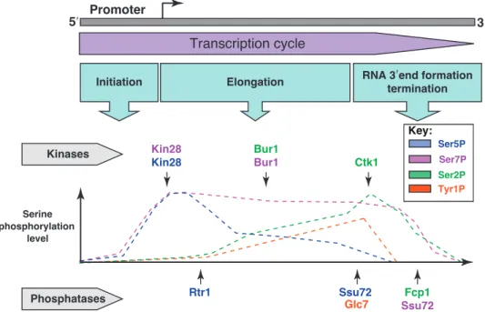
Key:
Initiation RNA 3terminationʹend formation
Transcription cycle Promoter Ser5P Ser7P Ser2P Kin28 Kin28 Phosphatases Ctk1 Bur1 Bur1 Rtr1 Ssu72 Fcp1 Ssu72 5ʹ 3ʹ Kinases Serine phosphorylation level Elongation Tyr1P Glc7
Figure 2.2: General view of Ser2, Ser5, Ser7, and Tyr1 phosphorylation
along the transcription cycle, kinases and phosphatases involved in CTD mod-ification are represented immediately above and below the graph. The two main phosphorylation states, Ser2 and Ser5, are dominant at the 3’ and
5’ respectively, reflecting their functional roles in the termination and early elongation phases of transcription. Ser7 is consistently present throughout the
transcription cycle, but its functional impact in yeast remains elusive. Adapted from [45].
During the initiation phase of transcription, the CTD of RNAPII starts off unphos-phorylated (Fig: 2.2). When the PIC is fully assembled, Kin28, a catalytic subunit of the general transcription factor TFIIH, phosphorylates the CTD heptapeptide on Ser5. In S.cerevisiae, the CTD remains mostly Ser5 phosphorylated for the
first 450 nucleotides of transcription elongation [120]. After this point the combined action of the Ser5-phosphatase Rtr1 [77, 126] and the Ser2-kinases Ctk1 [144]
make Ser2 the most prominent mark 5
. Despite phosphorylation of Ser2reaching
saturation about 600 nucleotides from the TSS [120], Ser5 phosphorylation is still present on many repeats, resulting in the presence of a double phosphorylation pattern with important functional consequences (see below). Only Towards the 3’ end of the gene the action of CTD phosphatase Ssu72 completely abrogates the Ser5-P mark, leaving Ser2-P as the only active mark. Finally, additional activity
of the Fcp1 phosphatase results in the removal of most phospho-marks from the CTD, readying the polymerase for another round of transcription.
2.3.2
Functional Interactions
As I outlined above, the transcription cycle follows specific patterns of CTD phos-phorylation: unphosphorylated CTD is recruited to promoter regions, Ser5-P
dominates during early elongation and gradually makes way for Ser2-P, which is
the dominant mark in the later stages of transcription. Each of these stages comes with the potential to interact with numerous co-factors and provides modularity to the elongation complex.
The unphosphorylated state of free-form RNAPII CTD allows the polymerase to interact with the mediator complex; an interaction that is thought to contribute to the recruitment of RNAPII to active promoters. Once the PIC is assembled, the polymerase needs to escape the promoter and leave the Pre-Initiation Complex be-hind. The modifications that take place at this stage, namely Ser5phosphorylation,
are thought to disrupt the interaction between RNAPII and mediator—thereby allowing promoter clearance—although evidence remains inconclusive [35,170].
The presence of Ser5 mark during early elongation has two direct consequences:
it stimulates capping of the nascent transcript through recruitment of the capping enzymes [160], and it has the potential to promote early transcription termination
5It is interesting to note that the phosphorylation state of RNAPII CTD is independent
of transcript length, but exclusively depends on the amount of nucleotides from the TSS. This will have important implications for the termination of non-coding transcripts.
through the recruitment of the Nrd1-Nab3-Sen1 (NNS) complex [186]. While capping is ubiquitous and required to prevent premature degradation of the transcript, early termination is a quality control mechanism that requires (in addition to Ser5-P)
the presence of specific sequence elements on the nascent transcript and will be described in detail in chapter 3.
Studies in mammals have reported that the CTD is required for splicing to occur properly [161]. In particular heptapeptides containing Ser2 phopshorylation are known to recruit several splicing factors [62]. Recent studies in S.cerevisiae show differential phosphorylation patterns in intronless and intron-containing genes, hinting at a possible fuctional interaction between splicing and CTD phopshorylation also in yeast [122].
Towards the end of the transcription cycle, Ser2-P becomes the most prominent
mark. This phase sees the recruitment of a number of different actors. Chromatin remodelers and histone modifying complexes such as Set2 and Spt6 are recruited through the CTD, making sure that the structure of nucleosomes is maintained [16].
Finally, 3’ end processing, termination, and export are all affected by the CTD. binding of components of the cleavage and polyadenylation complex such as Pcf11 and Rtt103 stimulates the termination of transcription and the processing of the transcript 3’ end (such as poly(A) tail addition), while recruitment of export factors such as Yra1 direct a rapid and efficient export to the cytoplasm [96].
3
Transcription Termination
After its synthesis and maturation are complete, the nascent RNA molecule must be released from the DNA template, and the elongation complex must be disassembled and its components recycled. In S.cerevisiae, transcription termination is enacted by several widely different mechanisms. Two predominating pathways terminate the vast majority of transcripts generated by RNA Polymerase II: the Cleavage and Polyadenylation Factor/Cleavage Factor I (CPF-CF) pathway and the Nrd1-Nab3-Sen1 (NNS) pathway. Both these mechanisms rely on short sequences on the nascent RNA—coupled with specific modifications on the CTD of RNAPII— to recruit specific factors and enact the disassembly of the elongation complex and the release of the transcript in the nucleus. Moreover, both transcription termination mechanisms are strictly intertwined with some steps of 3’ end processing and maturation, influencing the fate of the transcript after termination.
In addition to the two main pathways cited above, several non-canonical termination mechanism will be described. These mechanisms are dedicated to the termination of specific RNA species, or can act as backups when the main pathways fail.
3.1
The CPF-CF Pathway
The CPF-CF pathway was the first termination mechanism described in S.cerevisiae because of its association with the termination of protein-coding genes1. CPF-CF
termination is unique as it results in cleavage of the nascent RNA before termination occurs. The site of cleavage is specified through sequence elements present on the nascent RNA and plays an important role in kickstarting the termination reaction.
The main actor of this termination mechanism is the CPF-CF complex, a large assembly of modular sub-complexes that act in concert to execute all the required steps. This complexity makes CPF-CF the most reliable, efficient, and precise termination mechanism in S.cerevisiae.
3.1.1
Recruitment and Assembly
Recruitment and initial assembly of the CPF-CF complex onto the nascent RNA is promoted by two mechanisms: interaction with specific sequences elements, and interaction with the polymerase CTD.
A key component of the CPF-CF complex, Pcf11, contains a peptide sequence able to recognize the CTD. This CTD Interaction Domain (CID) is able to specifically recognize the Ser2-phosphorylated version of the heptapeptide. Given the nature
of this CTD modification—which is confined to the later stages of transcription— density of the CPF-CF complex around the polymerase is selectively increased where the complex is more likely to be needed for termination (i.e. at the 3’ end of transcription units), facilitating the eventual binding of CPF-CF to the sequence elements on the nascent RNA.
Unlike in human, where the cleavage site is defined by a single highly conserved hexanucleotide sequence on the nascent RNA, Yeast CPF-CF complex recognizes a
1Its activity can extend to certain kinds of non-coding transcripts as well (see section
number of degenerate short sequences. Two sub-complexes of CPF-CF, Cleavage Factor 1A (CF1A) and Cleavage Factor 1B (CF1B), are responsible for the recogni-tion of these sequences. In particular, Rna15 and Hrp1 (components of CF1A and CF1B respectively) directly bind the nascent RNA. Associated factors Rna14 and Pcf11 contribute to the assembly of the whole complex by interacting with RNAPII and forming a scaffold that serves to tether the catalytic portion of the CPF-CF complex to the cleavage site.
The bulk of the catalytic activity of the CPF-CF complex is contained in the Cleavage and Polyadenylation Factor (CPF) sub-complex. CPF directly contacts the cleavage site with its Ysh1 subunit and is responsible for the cleavage of the nascent RNA, one of the events that is thought to kickstart the termination reaction. CPF also coordinates the polyadenylation reaction through the subunits Yth1 and Fip1. These factors recruit and tether the poly(A) polymerase Pap1 to the complex, which will begin catalyzing the addition of a poly(A) tail after the transcript has been cleaved.
Despite the wealth of knowledge available on the mechanics of CPF-CF recruitment and assembly, some controversy still surrounds the actual termination mechanism. Two main models describing the termination reaction exist in the literature, the allosteric model and the torpedo model.
3.1.2
The Allosteric Model
After cleavage and release of the RNA, the elongation complex has successfully accomplished its job in the transcriptional process and is ready to be disassembled. The allosteric model is one of the two main mechanistic models that describes the process by which the TEC is removed from the DNA template.
The allosteric model argues that cleavage of the RNA is a dispensable signal, and that termination can happen independently of this step. It posits that after transcription of the cleavage site, RNAPII loses a lot of factors that qualify the
elongation complex as such. Loss of these “anti-termination” factors—components of the elongation complex that would prevent termination from occurring—would trigger conformational changes, destabilize the polymerase, and allow components of the CPF-CF complex itself to elicit the disassembly of RNAPII from the template.
Several studies support this model. RNAPII was shown to lose a number of associated elongation factors after reaching the 3’ end [87]. In addition, the component of the CPF-CF complex Pcf11 was shown to be able to terminate the polymerase in vitro by binding the nascent RNA and the Ser2-phosphorylated moiety of RNAPII [205].
Ulterior support to this last study was provided by the same authors two years later, when they discovered that Pcf11 is able to perform the same feat in drosophila [206]. Finally, a very recent study was able to reconstitute transcription termination in an in vitro system in the absence of cleavage [204].
3.1.3
The Torpedo Model
According to the torpedo model, cleavage represents the main termination signal for the CPF-CF complex, as it leaves an uncapped 5’-P on the transcript associated with the still transcribing elongation complex. These unprotected 5’ is the substrate of 5’→3’ exonucleases, a class of enzymes that are known to progressively degrade RNA polypeptides. The 5’→3’ exonuclease Rat1 was discovered to be associated with the CPF-CF complex and is thought to attack the 5’ moiety of the RNAPII-associated transcript, starting a processivity race with RNAPII. Upon winning the race, Rat1 would destabilize the structure of the ternary complex within the polymerase, causing it to break apart and detach from the DNA template.
There are several lines of evidence that support this model for CPF-CF transcription termination. Both Rat1 and its human homologue Xrn2 exhibit termination defects in model cases when mutated [88,190]. Furthermore, Rat1 and its co-factor Rtt103 were found to be strongly associated with the 3’ end of genes and in physical association with the CPF-CF complex [88,107], supporting the idea of a functional
recruitment to zones of active transcription termination. Homology studies found that homologues of Rtt103 in both humans and C.elegans have roles in transcription termination [31,125]. Finally, recent mechanistic studies in vivo have demonstrated the kinetic competition between Rat1 and the elongation complex. By employing mutant polymerases that elongate faster or slower than the wild type version, the authors were able to show that slower polymerases result in earlier termination, consistent with the notion that Rat1 needs to physically catch up with the polymerase in order to elicit termination [54].
CTD
RNAPII Ser2-P
RNA
ORF PolyA site
TSS Pcf11 Rna15 Hrp1 Pap1 CID CPF/CF complex Recruitment of CPF/CF-complex Ser2-P Ser2-P 5’ RNAPII Ser2-P RNA ORF TSS
RNA cleavage and polyadenylation Ser2-P Ser2-P 5’ 3’ 5’ DNA RNAPII Ser2-P Ser2-P Ser2-P 3’ 5’ RNAPII Ser2-P ORF Ser2-P Ser2-P RNAPII Rat1 Rtt103 ! CID! Rai1 Torpedo model Allosteric model Dissociation of elongation complex
Figure 3.1: Overview of the main mechanistic step that lead to CPF-CF termination. The complex is recruited thanks to CTD phosphorylation and binding sites on the RNA. The transcript is then cleaved and the elongation complex terminated in accordance with the torpedo or allosteric model.
At the same time, several reports argue against the torpedo model as sole effector of transcription termination. In vitro studies were unable to reproduce the termination effect observed in vivo using only Rat1 [37]. More recent ventures re-attempted the in vitro approach with limited success [136], but managed to demonstrate that Rat1 is able to terminate polymerases that are destabilized by nucleotide misincorporation.
Several additional mechanistic studies showed that the exonucleolytic activity of Rat1 is unable to mediate the release of the polymerase from the template [107,138]. Moreover, termination defects caused by Rat1 mutants were not associated with stabilization of the RNAPII-associated transcript, arguing against the model.
3.1.4
A Unified View of CPF-CF Transcription Termination
As evidence for and against the two models piles up, a unified view that combines elements of both torpedo and allosteric model is taking shape. While the effect of Rat1 on transcription termination (of at least some transcripts) is established, its role as main effector of CPF-CF termination has been repeatedly called into question. Several studies have now described interdependencies between Rat1 and other subunits of the CPF-CF complex—notably Pcf11—and the perceived nature of Rat1 is shifting towards that of a molecular effector that is integrated into a larger system. The proof of principle that termination is possible without cleavage has been recently provided—albeit in vitro [204]—and presence of Rat1 has been convincingly shown to facilitate termination [54], arguing for a model that integrates these two mechanisms.
3.2
The NNS Pathway
NNS dependent transcription termination is the second of the main termination mechanisms in S.cerevisiae. It is involved in the termination of Small Nuclear RNAs (snRNAs), Small Nucleolar RNAs (snoRNAs) and a number of other non-functional non-coding RNAs. It sets itself apart from CPF-CF termination in a number of ways. First and foremost, it relies on a completely different—and much smaller—set of proteins: the two RNA binding proteins Nrd1 and Nab3 [30], together with the helicase Sen1. Because of the different molecular effectors, the termination mechanism—although still not fully elucidated—is appreciably different.
The NNS complex also distinguishes itself because of the different fate imposed on the RNA released: instead of being exported to the cytoplasm after polyadenylation, the transcripts released are subjected to the activity of degradation enzymes [185]. To this end the NNS complex recruits both the nuclear exosome and a specific set of 3’ end processing factors known as TRAMP (Trf4/Air2/Mtr4p Polyadenylation), which drives polyadenylation and stimulates degradation [82,185].
NNS termination operates mainly on non-coding RNAs and is generally restricted to the early stages of transcription elongation. Despite not being directly involved in the termination of protein-coding genes, it can play a role in the regulation of gene expression by acting as an attenuator (i.e. terminating some transcription events, preventing them from producing functional RNAs) [5]. Examples of this phenomenon include the IMD2 or URA2 genes [79]. Alternatively, NNS was shown to terminate transcription of non-coding RNAs whose transcription is involved in regulation [180].
3.2.1
The NNS Complex
The main molecular effectors of the NNS complex are the three protein Nab3, Nrd1, and Sen1.
Nab3
This factor was originally identified as a polyadenylated RNA binding protein. Nab3 contains several structural domains: a conserved RNA Recognition Motif (RRM) that can contact specific sequence elements on the nascent RNA, a region necessary for the interaction with Nrd1, and an essential Glutammine/Proline region at the C-terminus.Biochemical experiments have shown that Nab3 forms a stable heterodimer with Nrd1 and contacts the RNA as such [30]. In addition, the structure of the RRM has been solved, revealing the structural basis for the preference of the sequence UCUUG [106]. Finally, its Glutammine/Proline region—despite being generally unstructured—can assemble into amyloid structures [187].
Nrd1
Identified as part of the “nuclear pre-mRNA downregulation” family of proteins, Nrd1 is the most abundant of the three members of the complex. Its main features consist of an RRM structure that allows it to contact the nascent RNA, a CTD interaction domain (CID) that mediates the interaction with RNAPII (see below) and a Nab3 interaction motif that allows it to form a stable heterodimer.Nrd1’s RRM was shown in vivo to contact the consensus sequence GTA[A/G] [174]. Recent in vitro studies, however, have shown that several other G-rich and A-rich sequences could be bound equally well [7], although the in vivo relevance of these studies remains to be demonstrated.
In addition to the RNA, Nrd1 can contact RNAPII through its CID [92, 186]. Although dispensable for cell viability, the CTD-CID interaction is required for efficient termination.
Curiously, Nrd1 also contains a Glutammine/Proline region at the C-terminus, similarly to Nab3. Deletion of this region shows no growth or termination defects, but is synthetic lethal if combined with other aphenotypic mutations on Nab3 [our unpublished data]. The functional implications of these genetic interactions are still unknown.
Sen1
This extremely large (253kDa) and very low abundance (125 molecules per cell) protein is the only member of the NNS complex to have enzymatic activity [173]. Sen1 was characterized as a helicase of the SFI superfamily and is very closely related to Upf1, a member of the Non-sense Mediated mRNA Decay (NMD) pathway in the cytoplasm. Unlike its close relative, Sen1 possesses a nuclear localization signal and acts in the nucleus, where it can physically interact with the other members of the NNS complex Nrd1 and Nab3.Structurally, Sen1 contains a helicase domain able to hydrolyze ATP and a large N terminal domain. The helicase domain was recently purified in E.coli and biochemically analyzed, revealing binding affinity for both DNA and RNA, but a slower translocation rate on RNA [175]. Moreover, its ATPase activity was shown to be necessary for termination in vitro [143]. The N-terminal region of Sen1 was implicated in the interaction with the RNAPII, as well as other factors such as Rnt1 and Rad2, but the implications of the latter interactions remain obscure.
3.2.2
The Mechanism of Transcription Termination
As in the case of CPF-CF, the NNS complex is recruited to the region of termination through two distinct mechanisms that cooperate to maximize efficiency: the CTD of the polymerase [186] and specific sequence elements on the nascent RNA [30]. Within the NNS complex, Nrd1 and Nab3 are the major interactors of these elements, providing specificity and ensuring that Sen1—believed to be the molecular effector of NNS termination—is recruited only in the appropriate circumstances [143].
The CTD of RNAPII is contacted by the CID domain of Nrd1. This domain preferentially recognizes the Ser5-phosphorylated variant, which is the prevalent
CTD phosphorylation state in the first 500-600 nucleotides of transcription. This preference confers to the NNS complex a high degree of specificity for terminating transcription in the early stages of elongation. According to the current model for NNS termination, the interaction with the CTD occurs prior to RNA binding, and
facilitates recognition of sequence elements on the nascent transcript. Presence of Ser5-P CTD was shown to be a pre-requisite for efficient termination, as placing
high efficiency NNS binding sites at the end of long transcription units—where the levels of Ser5-P would be completely supplanted by Ser2-P—does not result in
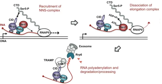
termination [63]. RNAPII Ser5-P Sen1 Dissociation of elongation complex Rrp6 Exosome Trf4 TRAMP Nab3 Nrd1 CID A A A A A A
RNA polyadenylation and degradation/processing CTD Nab3 Nrd1 RNAPII Ser5-P Sen1 CID DNA RNA Recruitment of NNS-complex CTD Nab3 Nrd1 RNAPII Ser5-P Sen1 CID ATP ADP+Pi Dissociation of elongation complex
Figure 3.2: Main stages of NNS-dependent termination. the NNS complex is recruited thanks to Ser5-phosphorylated CTD and sequence elements on
the transcript. Termination is elicited by Sen1, presumably by translocating along the transcript. Finally, the exosome is recruited to the transcript and the transcript is either trimmed or completely degraded.
Recruitment of Nrd1 to the CTD, however necessary, is not sufficient to trigger termination. The Nrd1-Nab3 heterodimer must also contact the nascent RNA through the RRM domains of the two subunits. Original studies have investigated the sequence elements that drive NNS termination, pinpointing two core consensuses: UCUU as the main binding site for Nab3, and GUA[A/G] as the main site for Nrd1 [23]. More recent investigations redefined these consensuses and identified new sequence elements that can increase termination efficiency when in proximity of canonical binding sites. Use of an in vivo SELEX (Systematic Evolution of Ligands by Exponential enrichment) strategy allowed to extend the core consensus sequences for both Nrd1 and Nab3 with nucleotides that proved critical for binding [142]. In addition, AU-rich sequences found downstream of Nrd1 sites were shown to play a
role in increasing both termination efficiency and recruitment of Nrd1 [142]. Similar conclusions have been reached by in vivo crosslinking studies [195].
Despite the efforts expended in identifying sequence elements that could univocally lead to NNS termination, a lot of ambiguity remains on what constitutes an NNS terminator in vivo. While presence of Nrd1-Nab3 binding sites is required, no consistent pattern emerges in number, spacing, or quality of Nrd1/Nab3 sites at known NNS termination sites. In vitro studies on model cases have identified some features of heterodimer binding. For example, mutation of Nab3 binding sites proved to be more deleterious to heterodimer recruitment than mutation of Nrd1 sites [22]. Moreover, multiple heterodimers were found to bind the same RNA sequence, possibly cooperatively [22]. It remains impossible, however, to generalize these results beyond the few sequences tested. While the NNS complex could simply rely on a high number of low affinity sites to reach an occupancy threshold, it remains possible that several unseen elements play a role in qualifying NNS terminators, influencing the quantity and quality of Nrd1 and Nab3 binding sites necessary for an efficient termination.
When the Nrd1-Nab3 heterodimer is bound to the nascent RNA, the molecular effec-tor of NNS termination, the helicase Sen1, is recruited to the complex. Studies have shown that Sen1 is strictly required to terminate transcription, but the mechanism through which this happens is not clear. Significant advances in the understanding of this phenomenon came from use of an in vitro transcription termination system [143]. In this context, Sen1 alone was found to be sufficient to disassemble the elongation complex. Termination was shown to occur preferentially at sites of pausing and to require both the interaction of Sen1 with the nascent transcript and ATPase activity. It is unclear whether ATP-dependent translocation of Sen1 on the nascent RNA is required for termination. However, results from an in vivo study suggest the existence of a kinetic competition between transcription elongation and Sen1 translocation on the RNA. The authors investigated the effect of the speed of transcription on NNS termination, showing that faster transcription results in longer
NNS-terminated transcripts, while slower transcription produces shorter transcripts and is able to suppress mutations on Sen1 [69]. Taken together, these results support a model where, akin to the bacterial termination factor Rho, Sen1 would contact the nascent transcript and translocate in a 5’ to 3’ direction, eliciting termination upon catching up with the polymerase.
3.2.3
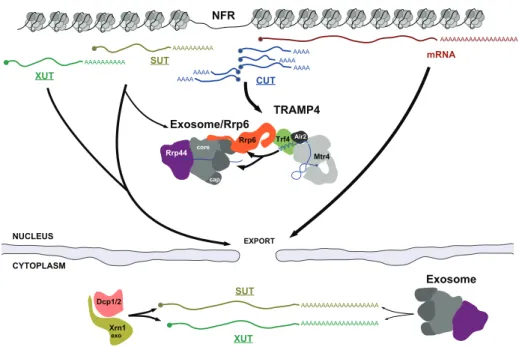
Processing Products of the NNS Pathway
The process of NNS termination is strictly connected with 3’ end processing or degradation mediated by the nuclear exosome, a multiprotein complex endowed with exonuclease activity [185]. The exosome plays a major role in nuclear RNA quality control, degrading aberrant transcripts, a number of non-functional non-coding RNAs, and trimming the precursors of functional small non-coding RNAs such as sn/snoRNA [for review see85]. The exosome is composed of six non-catalytic subunits arranged in a ring-like structure, together with three cap subunits that can bind RNA. The catalytic activity of this complex is dependent on two active 3’→5’ exonuclease, Dis3 and Rrp6. Dis3 associates with the ring on the opposite side of the three cap subunits, and degrades RNAs that are threaded through the cap proteins and into the ring [111]. The exosome is present throughout the nucleus and in the cytoplasm. However, only the nuclear version can associate with the other exonuclease, Rrp6, whose activity is known to regulate the levels of many NNS targets.
Recruitment of the exosome to NNS targets takes place via one of the exosome’s co-factors: the TRAMP complex. TRAMP (for Trf4/Air2/Mtr4p Polyadenylation) is a nuclear complex composed of the poly(A) polymerase Trf4, the RNA-binding protein Air2 and the helicase Mtr4. Trf4 is the core subunit of the complex, to which both Air2 and Mtr4 bind independently. It possesses poly(A) polymerase activity, but unlike Pap1—the canonical poly(A) polymerase associated with the CPF-CF complex—it can only add tails in a distributive manner. Trf4 is also the factor responsible for the coordination between the NNS complex and the nuclear
exosome. A recent study showed that Trf4 contacts Nrd1 through a small motif called Nrd1 Interaction Motif (NIM). The NIM on Trf4 mimics Ser5-P CTD and
can therefore compete with the CTD of RNAPII for the interaction with the CID (CTD interaction domain) on Nrd1. The interaction of Nrd1’s CID with the CTD and Trf4 are mutually exclusive. These findings have suggested a model whereby TRAMP is recruited to the RNA when the CID of Nrd1 is freed from the CTD of the polymerase [182], allowing the coordination of events going from termination to the handover of the transcript to TRAMP and the exosome.
As a co-factor of the exosome, TRAMP is able to both recruit and stimulate its activity. Addition of a poly(A) tail to the terminated transcript is thought to provide an unstructured platform that can be easily be threaded through the non-catalytic subunits of the exosome. However, TRAMP has been known to stimulate exosome activity even indipendently of poly(A) polymerase activity [182].
By virtue of the tight connection between NNS and TRAMP, NNS-terminated transcripts are usually subject to rapid degradation. SnoRNAs and snRNAs consti-tute notable exceptions, in that they are heavily structured functional non-coding transcripts that are recruited to the exosome, but undergo only trimming of their 3’ ends instead of complete degradation. This is thought to occur thanks to the presence of secondary structure and additional proteins binding the RNA, preventing the transcript from being entirely threaded through the exosome [123].
3.3
Non-Canonical Termination Pathways
CPF-CF- and NNS-dependent termination seemingly account for the vast majority of RNAPII transcription termination events in the cell. Several additional mecha-nisms, however, can terminate transcription in S.cerevisiae. These non-canonical termination pathways are generally thought to elicit termination of particular RNA species, but can also act as fail-safe pathways in restricting readthrough transcription [60].
3.3.1
Rnt1-Dependent Termination
The yeast Rnase III homologue Rnt1 is an enzyme that binds and cleaves double-stranded RNA stem-loops at a defined recognition site. Rnt1’s known function in the cell is that of cleaving polycistronic rRNAs and snoRNAs transcripts, promoting their subsequent trimming and processing by the exosome [60]. Recently, Rnt1 binding sites have been identified downstream of a number of genes and its cleavage activity has been implicated in transcription termination.
Studies on the model gene NPL3 have shown that deletion of Rnt1 leads to transcrip-tional readthrough and can even mediate the production of dicistronic transcripts [59]. Rat1, the mediator of the CPF-CF termination according to the torpedo model, was found to be also required for proper termination by Rnt1. This led to a model where Rnt1 cleaves a stem-loop that forms downstream of the CPF-CF cleavage site, generating a non-polyadenylated transcript, and leaving an uncapped 5’ on the nascent transcript. This free 5’-OH is a substrate for exonuclease Rat1, and transcription termination is thought to occur with a mechanism akin to the CPF-CF torpedo model, with Rnt1 as the cleaving agent instead of the CPF complex [59,157].
The termination mechanism is usually very intimately connected with 3’ end pro-cessing and with the fate of the transcripts it produces. The case of Rnt1-dependent termination, however, is peculiar in this respect. Use of in vivo reporter systems showed that, in the absence of a polyadenylation site, Rnt1-dependent transcripts are unstable and supposedly targeted by TRAMP and the exosome [59]. However, addition of a cryptic polyadenylation site close to the Rnt1 binding site in the same system results in increased transcript stability that is Pap1-dependent. This suggests that depending on its environment, Rnt1 can either stimulate the usage of a nearby Polyadenylation site or produce transcripts that are targeted for degradation [157].
3.3.2
Road-Block Termination
Road-block termination represents another non-canonical mechanism that can mediate transcription termination. Road-block was first observed as a termination mechanism for RNAPI, where a DNA binding factor acts as a physical obstacle for the polymerase. The polymerase is thought to stall at the DNA binding site and eventually dissociate from the template through unclear mechanisms [98,99].
When the mechanism was first described, in vitro work had shown that transcription factor Reb1 was able to pause all three yeast RNA polymerases [98]. Later studies from the same authors confirmed that the DNA binding site for Reb1 was coincident with sites of RNAPI transcription termination in vivo [149]. Combination of these experiences led to a model where Reb1 is binding DNA and terminating RNA polymerase I at specific rDNA loci. It was only in 2012 that a Reb1 paralogue— Nsi1, who binds the same consensus sequence as Reb1—was implicated as the true in vivo effector of RNAPI termination, while Reb1 was proven to not have a role [151].
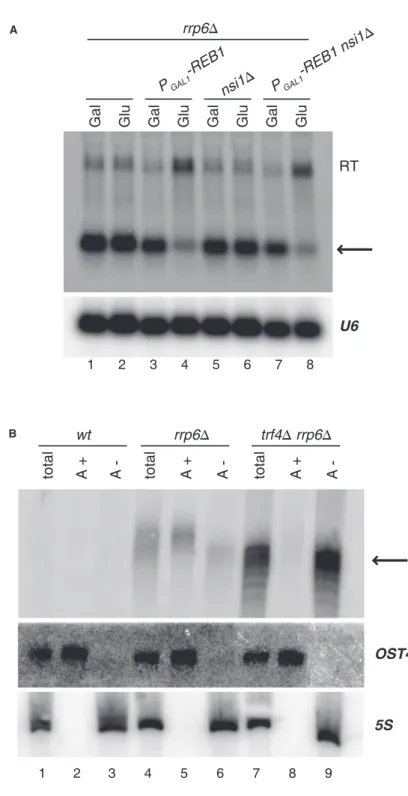
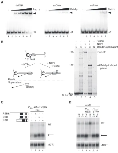
I have participated to a study of the laboratory showing that Reb1 is the effector of roadblock transcription termination for RNA polymerase II in vivo. This study will be described in the results section.
4
The Transcriptional Landscape of
S.cerevisiae
The rise of microarrays and next generation sequencing techniques has made the exploration of the transcriptome possible. Early application of tiling arrays to the transcriptome of S.cerevisiae showed that, in addition to protein coding genes and a multitude of functional non-coding RNAs, the genome is pervasively transcribed and RNA molecules can arise from many unannotated regions [34,128,197]. There are multiple possible reasons for this phenomenon. Studies have shown that yeast promoters, despite showing directionality, can fire bidirectionally and give rise to non-functional RNAs [128, 197]. Additionally, transcription usually arises in poorly chromatinized areas of the genome, pointing to the possibility that the genome might provide a low barrier to transcription initiation outside regions of high nucleosome occupancy. These factors contribute to the widespread occurrence of transcription outside of annotated regions, which is usually referred to as pervasive transcription and contributes to the generation of large quantities of non-coding (mostly non-functional) RNAs.
4.1
Control of pervasive Transcription
Pervasive transcription represents a non-negligible fraction of all RNAPII transcrip-tion. Therefore, it has the potential to interfere with other physiological events and needs to be carefully regulated. Control of pervasive transcription occurs on two levels: First, RNAPII that initiates spuriously need to be rapidly terminated, in order to avoid interference with other processes on DNA; second, the resulting transcripts need to be efficiently degraded, to prevent accumulation of toxic species.
The NNS complex is the main termination pathway involved in control of pervasive transcription [5, 179]. Binding sites for Nrd1 and Nab3 are frequently enriched in areas where pervasive transcription occurs, such as antisense to coding RNAs and in intergenic regions [179]. Acting early in the transcription cycle, NNS is an effective tool to block such transcription events before they can do damage. Despite the major role of NNS, CPF-CF, as well as some non-canonical termination pathways, have been implicated in termination of pervasive transcription [29,114,183].
Once termination has occurred, transcripts are released into the nucleus. These RNA species do not possess coding potential and might be deleterious to the cell if accumulated in sufficient quantities. In order to prevent such accumulation, the cell evolved RNA quality control systems that can degrade spurious and aberrant transcripts. These decay pathways can be directly connected to termination and 3’ processing, as in the case of NNS and the TRAMP-Exosome [179], or recognize specific features that mark non-functional transcripts, such as poor coding potential.
4.2
Classes of Pervasive Transcript
Because of their rapid turnover, the majority of pervasive transcripts are difficult to detect in wild type cells. Several studies found that deletion of certain elements of RNA quality control would affect the stability of only a subset of pervasive transcripts, making them appear in transcriptome analyses [183,196]. Over time, it