Plant Survival Graduate School
PhD Thesis
The Role of Palmitoylation in the
Secretory Pathway of Plants
Ph.D. student: Stigliano Egidio
Faculté des sciences Secrétariat-décanat de Faculté Rue Emile-Argand 11 2000 Neuchâtel - Suisse Tél: + 41 (0)32 718 2100 E-mail: [email protected]
IMPRIMATUR POUR THESE DE DOCTORAT
La Faculté des sciences de l'Université de Neuchâtel
autorise l'impression de la présente thèse soutenue par
Monsieur Egidio STIGLIANO
Titre:
“The role of palmitoylation in the secretory
pathway of plants”
sur le rapport des membres du jury composé comme suit:
- Prof. Jean-Marc Neuhaus, Université de Neuchâtel, directeur de thèse - Dr Giselbert Hinz, Université de Heidelberg, D
- Dr Guillaume Gouzerh, Université de Neuchâtel - Dr Matthias Erb, Max Planck Institute, Iena, D
Acknowledgments
During these 4 years I was able to substantially increase my skills. But this long trip was primarily a human journey where I met people from all over the world. Languages and cultures that have changed me for the better ... I hope!
There are many people who would like to thank. Firstly I would like to thank Prof. Jean-Marc Neuhaus that gave me the chance to do Ph.D. in his laboratory.
Here I met two fantastic fellow travellers: Sanaa and Alessandro, with whom a fruitful collaboration and clear friendship was born.
A Special thanks to Guillaume and Sophie that through their wise counsel have made less arduous the path of the doctorate.
A warm thanks to Dr. Marc Creus with whom I shared office and apartment for a year. He is a person always full of advices and encouragements and to whom I will always be infinitely grateful.
Finally, one last thank to Dr. Di Sansebastiano: without him I would never
have started this career. Big hugs to all friends of these years: Barbara, Chris, Erica, Giuseppe, Nicola, Giuseppe, Max, Fred, Anne-Flore (la petite Typhenn) ... and all those that I have forgotten in this short page, but I will never forget for the rest of my life.
TABLE OF CONTENTS
TABLE OF CONTENTS ... 7 ABBREVIATIONS ... 11 SUMMARY ... 13 1. INTRODUCTION ... 15 1.1 ENDOPLASMIC RETICULUM ... 151.1.1 HSPs (Heat Shock Proteins) ... 18
1.2 GOLGI APPARATUS ... 18
1.2.1 Retromer complex ... 21
1.3 ESCRT COMPLEX ... 24
1.4 VACUOLE STRUCTURE AND FUNCTIONS ... 25
1.4.1 Sequence- specific Vacuolar sorting Determinants (ssVSD) ... 28
1.4.2 C-terminal Vacuolar Sorting Determinants (CtVSD) ... 29
1.5 BRIEF AND GENERAL DESCRIPTION OF THE ANIMAL AND YEAST ENDOMEMBRANE SYSTEMS ... 30
1.6 ACTIN MICROFILAMENTS AND MICROTUBULES ... 32
1.7 BRIEF OVERVIEW OF THE MOST COMMON TRAFFIC INHIBITORS IN PLANT CELLS ... 34
1.7.1 Brefeldin A (BFA) ... 34
1.7.2 Wortmannin ... 35
1.7.3 Tyrphostins ... 36
1.7.4 Bafilomycins and Concanamycins ... 37
1.7.5 2Bromopalmitate (2BP) ... 40
2. PALMITOYLATION OF ATRMR1 ... 43
2.1 VACUOLAR SORTING RECEPTORS (VSRS) ... 43
2.2 RMRS ... 46
2.3 PRELIMINARY ANALYSIS OF RMR1 PALMITOYLATION ... 48
2.4 INVESTIGATION OF A ROLE OF PALMITOYLATION IN ANCHORING TO MEMBRANE MICRODOMAINS ... 48
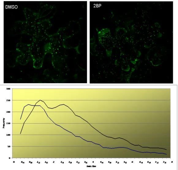
2.5 LOCALISATION OF RMR1 IS AFFECTED BY 2BP ... 50
2.6 BIOTIN SWITCH ASSAY OF PALMITOYLATION ... 52
2.7 STABILITY OF THE TRIPLE CYSTEINE MUTANT ... 55
... 55
2.8 PHOSPHORYLATION OF RMR1 ... 56
2.9 DISCUSSION ... 59
3. ROLE OF PALMITOYLATION IN THE SECRETORY PATHWAY ... 63
3.1 INTRODUCTION ... 63
3.2 S-PALMITOYLATION ... 63
3.2.1 Mechanism of protein palmitoylation by Palmitoyl Acyl Transferases (PATs) ... 64
3.2.2 Palmitoylation and raft localization ... 67
3.3 ABOUT OTHER LIPID POST-TRANSLATION MODIFICATIONS:N-MYRISTOYLATION,S-ISOPRENYLATION AND ADDITION OF GPI(GLYCOSYL PHOSPHATIDYLINOSITOL) ANCHORS ... 69
3.4 SMALL GTPASES AND POLAR GROWTH ... 73
3.5 THE CYTOSKELETON IS NOT AFFECTED BY 2BP ... 82
3.6 INHIBITION OF APICAL GROWTH ... 86
3.7 EFFECTS OF 2BP ON THE LOCALISATION OR MARKERS OF THE SECRETORY SYSTEM ... 92
3.8 INTERFERENCE OF 2BP WITH THE LOCALIZATION OF RAB GTPASES ... 94
3.9 ULTRASTRUCTURAL ANALYSIS IN A.THALIANA ROOTS ... 102
4.2 Blocking the ER-Golgi trafficking does not retain the vacuolar reporter GFP-Chi in the ER……...110
4.3 GFP-Chi aggregates and reaches the vacuole as a dimer………...115
4.4 Immunogold labelling in GFP-Chi expressing plants ... 116
4.5 Discussion ... 117
5. MATERIAL &METHODS ... 121
5.0 Bacterial strains ... 121
5.0.1 Preparation of heat-shock competent E.coli cells ... 121
5.0.2 Transformation of E.coli by heat-shock ... 121
5.0.3 Preparation of electroporation competent A.tumefaciens cells ... 122
5.0.4 Transformation of A.tumefaciens by electroporation ... 122
5.1 PLANT MATERIAL AND PLANT TRANSFORMATION TECHNIQUES ... 122
5.1.1 Growth conditions ... 123
5.1.2 Soils and mediums for growth ... 123
5.1.3 Seed sterilization ... 123
5.1.4 Preparation of A.thaliana leaf protoplasts and PEG-mediated transformation ... 123
5.2 AGRO-INFILTRATION OF N.BENTHAMIANA LEAVES ... 124
5.3 PHYSCOMITRELLA PATENS GROWTH CONDITIONS ... 125
5.3.1 Strain conservation ... 125 5.3.2 Culture media ... 126 PP NO3 ... 126 PP NH4 ... 127 Supplements ... 127 5.4 PROTOPLASTS MEDIA ... 128
5.4.1 Protoplast solid culture medium. ... 128
5.4.2 Protoplast liquid culture medium. ... 128
5.4.3 Protoplast top layer ... 128
5.4.4 Protoplast isolation and regeneration ... 128
5.4.5 Transformation of Physcomitrella patens ... 129
Notes ... 130 Solutions. ... 130 5.5 MOLECULARBIOLOGY ... 131 5.5.1 PCR ... 131 5.5.2 DNA digestion ... 131 5.5.3 DNA ligase ... 132
5.5.4 Total RNA extraction from A.thaliana leaves ... 132
5.5.5 cDNA synthesis ... 132
5.5.6 Genomic DNA extraction from A.thaliana leaves ... 133
5.5.7 DNA precipitation ... 133
5.5.8 DNA extraction from agarose gel ... 133
5.5.9 Isolation of plasmid DNA from E.coli in a small-scale ... 133
5.5.10 Isolation of plasmid DNA from E.coli in a big-scale ... 134
5.5.11 DNA electrophoresis ... 134
5.6 PROTEINTECHNIQUES ... 134
5.6.1 Chloroform/methanol precipitation ... 134
5.6.2 Protein extraction for solubilisation assay ... 135
5.6.3 Sucrose gradient fractionation ... 135
5.6.4 SDS-PAGE ... 136
5.6.5 Western Blot ... 137
5.6.6 Membrane Stripping ... 137
5.6.7 BSA (Biotin Switch assay) ... 138
5.6.8 Membrane fractionation from N.benthamiana leaves ... 140
5.7 MICROSCOPY ... 140
5.7.1 Transmission electro microscopy (TEM) ... 140
5.7.2 Preparation of the samples ... 140
5.7.3 Immunogold labelling ... 141
5.7.4 Post-staining with uranyl acetate/lead citrate ... 141
5.8 SCANNING ELECTRON MICROSCOPY ... 142
5.8.1 METHOD ... 142
5.9 CONFOCAL MICROSCOPY ... 143
5.10 PLASMIDSANDCONSTRUCTS ... 143
5.10.2 pSOUP contains: ... 143
5.10.3 List of Constructs and stable expressing plant lines ... 144
5.10.4 pGREEN_35S ... 144 5.10.5 pGREEN_YFP ... 144 5.10.6 pGREEN_SpYFP ... 144 5.10.7 pGREEN_RFP ... 144 5.10.8 pGREEN SpYFP_RMR1 ... 145 5.10.9 pGREEN RMR1_YFP ... 145 5.10.10 pGREEN RMR1_RFP ... 145 5.10.11 pGREEN p6_RFP ... 145 5.10.12 pGREEN GONST1-RFP ... 145 5.10.13 pGREEN GFP-Chi ... 145
5.10.14 pGREEN Venus SYP61 ... 146
5.10.15 pGREEN GFP-Talin ... 146
5.10.16 MBD-RFP ... 146
5.10.17 pCAMBIA EGFP-Rac8 ... 146
5.10.18 pGREEN p6RFP ... 146
5.10.19 Sar1H74L ... 146
5.10.20 Constructs to generate point mutants ... 146
5.11 USE OF DRUGS AND DYES ... 147
5.11.1 N.benthamiana pollen germination medium ... 147
5.11.2 Physcomitrella patens transgenic lines ... 148
5.11.2.1 Pp αTub-GFP ... 148
5.11.2.2 Pp GFP-Talin ... 148
6. OUTLOOKS ... 153
Abbreviations
ADP: Adenosine Diphosphate AP: Adaptin Protein
ARF: ADP Ribosylation Factor Bip: Binding immunoglobulin protein BFA: Brefeldin A
BP-80: Binding Protein of 80 kDa BSA: Biotin Switch Assay CCV: Clathrin-Coated Vesicle COPI: Coat Protein I
COPII: Coat Protein II
Ct-VSD: C-terminal Vacuolar Sorting Determinant DIP: Dark-Induced Protein
DNA: Deoxyribonucleic Acid DV: Dense Vesicle
EYFP: Enhanced Yellow Fluorescent Protein
ERAD: Endoplasmic Reticulum Associated Degradation ERES: Endoplasmic Reticulum Export Site
ESCRT: Endosomal Sorting Complex Required for Transport GDP: Guanosine Diphosphate
GEF: GTP Exchange Factor GFP: Green Fluorescent Protein
GFP-Chi: Green Fluorescent Protein-chitinase vacuolar sorting determinant Hsp: Heat Shock Protein
LV: Lytic Vacuole
MVB: Multivesicular Body
PA domain: Protein Associated domain PAC: Precursor Accumulating Vesicle Palm: palmitoylation
PM: Plasma Membrane PSV: Protein Storage Vacuole
psVSD: Protein Structure dependent Vacuolar Sorting Determinant PVC: Prevacuolar Compartment
Rab: Ras-related in Brain
RING: Really Interesting New Gene RFP: Red Fluorescent Protein
RMR: Receptor-like Membrane Ring-H2 RNF13: RING Finger Protein 13
SNAP: Soluble NSF Attachment protein RNA: Ribonucleic Acid
SNARE: Soluble N-ethylmaleimide-sensitive Protein Attachment Protein Receptor SP: Signal Peptide
SRP: Signal Recognition Particle
ssVSD: Sequence-specific Vacuolar Sorting Determinant 2BP: 2-Bromopalmitate
UPR: Unfolded Protein Response
Summary
Keywords: RMR1, palmitoylation, Biotin Switch Assay, GFP-‐Chi, ERV pathway, GFP-‐Chi aggregation.
This study aimed to study an important eukaryotic post-translational modification, the S-palmitoylation. Until now, there was no study of palmitoylation in plant cell biology. In the first part of this study, we wanted to study the palmitoylation of the vacuolar receptor AtRMR1, as predicted in
silico. We used a very innovative technique, the Biotin Switch Assay, which does not use radioactive
palmitate, is much less time-consuming as it is possible to obtain results after three to four day. Another advantage for cell biology is that it allows the characterization of entire palmitoyl-proteomes. Yeast and neuronal palmitoyl-proteomes have indeed been recently characterized. The study of RMR1’s palmitoylation revealed the first palmitoylated plant transmembrane protein. The palmitoylation of a small fraction of RMR1 at a higher molecular weight deserves further discussion. In the second part of this thesis, I addressed the more general role of palmitoylation in the secretory pathway through the use of a potent palmitoylation inhibitor: 2BP. This study showed a specific action of the drug in TGN/post-TGN compartments. The drug affected the structural maintenance of macrovesicles of secretion. The macrovesicles of secretion have recently been characterized by cryo-fixation and by electron tomography. They are structures reminiscent of a bunch of grapes, where each grape is a secretory vesicle. They are associated with the building with TGN-rich secretory vesicles with a diameter of few tens of nanometers. These vesicles are particularly visible in tissues with a high growth rate, such as pollen tubes. 2BP drastically changed the state of aggregation of macrovesicles of secretion. In an imaged way, each grape was released and a diffuse fluorescence was observed. Palmitoylation is therefore important in the formation or stability of this important secretory structure. A possible extension of this work would be the isolation of palmitoylated proteins involved in this stabilization. In addition, palmitoylated Rabs were detected for the first time in a plant. The three plant Rabs for which I detected the effect of 2BP are located in post-Golgi compartments.
In a last part of the thesis, I decided to investigate the route of secretion of GFP-Chi, a vacuolar marker widely used in the lab that can be followed along the route of secretion. Apparent contradictions have been reported: a vacuolar sorting of this marker by the Golgi-TGN-PVC pathway or an independent-COPII trafficking that can bypass the classic route. Unexpectedly, when GFP-Chi was co-expressed with NtSar1H74L (a dominant-negative mutant blocking the ER-Golgi trafficking by preventing the formation of COPII vesicles), the reporter reached the vacuole. This suggests that the alternative pathway bypassing the Golgi can take place. I also detected the presence of a possible GFP-Chi dimer associated with the membrane fraction upon ultracentrifugation. The nature of this dimer of a soluble protein remains to be investigated although several cases of aggregation of soluble proteins have been reported in the literature (like β-amyloids presents in the of Alzheimer disease).
1. Introduction
A notable function in all eukaryotic cells is to control the protein trafficking among different intracellular compartments and the plasma membrane. The efficiency of this trafficking is guaranteed by several classes of proteins and compartments, which cooperate among them in order to ensure the correct working of a global and widely conserved process which is named "trafficking".
It will highlight the different roles of proteins involved in cellular traffic with special emphasis on vacuolar trafficking and discuss the role of the ER in protein synthesis and above all the beginning of the route of secretion.
Then, I will continue with a structural description of the Golgi and proteins involved in ensuring the proper sorting to numerous post-Golgi organelles such as the Trans-Golgi Network (TGN), Prevacuolar compartment (PVC), and finally, with special emphasis, to the recycling endosome (RE) (and Rabs proteins involved therein), and vacuole (as one of the final station, with the plasma membrane, in protein and membrane trafficking).
A special mention will be given to describing two classes of vacuolar sorting receptors, RMRs and VSRs. Part of this research work will focus on RMR1 and its palmitoylation. Precise attention will also be given to soluble vacuolar markers and signals needed to address them to the vacuole.
We will then discuss the process of palmitoylation: mechanisms, functions and possible roles in polarized growth as pollen tubes and Arabidopsis roots.
Finally we will describe the use of various drugs that have become customary in the domain of the cell biology of plants as brefeldin A, wortmannin, concanamycin A, bafilomycin A, tyrphostins and, for the first time in plant cells, the 2-bromopalmitate (2BP), as a specific inhibitor of palmitoylation. I will highlight the possible use of 2BP as a specific inhibitor of Rab localized in the system for recycling and not others located in other compartment.
1.1 Endoplasmic Reticulum
In animal cells Endoplasmic Reticulum (ER) membranes typically constitute more than half of the total membranes. The ER is organized in a kind of net-like labyrinth of tubules and
space called the ER lumen. The ER has a main role in lipid and protein synthesis and it is also an intracellular Ca2+ store, which is used for many intracellular processes such as signal transduction. Finally the ER is also the synthesis site of all transmembrane and soluble proteins for almost all cellular organelles.
As the ER has so many different functions, distinct regions have developed specialized structures. One of the most notable functions resides in rough ER. In fact, most proteins are inserted into the ER membrane before that their synthesis is complete. This process, called co-translation import, is different from the post-co-translational import of proteins by nuclei, peroxisomes, chloroplasts and mitochondria (Blobel and Dobberstein, 1975).
The rough ER has been named because of the presence of ribosomes coating the membrane and it differs from the smooth ER. Many cells have little smooth ER, but in certain specialized cells it is abundant and has very specific functions such as to synthesize steroids using cholesterol as precursor (Shibata, et al., 2006). Another function of the ER in most eukaryotic cells is to sequester Ca2+ from the cytoplasm. The release of Ca2+ into the cytosol is a response to many extracellular stimuli. Ca2+ is then captured by active pump transporters and stored in the ER lumen by a plethora of Ca2+ binding-proteins. Many studies have focused on the role of fatty acids, and more specifically palmitate, in initiating ER stress in
vitro (Karaskov, et al., 2006; Laybutt, et al., 2007; Cunha, et al., 2008; Eizirik, et al., 2008)
and in vivo (Laybutt, et al., 2007). For instance, long time treatment with palmitate, but not oleate, causes a morphological stretching of the ER, which could lead, in turn, to a stress response (Karaskov, et al., 2006). Because almost all eukaryotic proteins are synthesized by cytosolic ribosomes, those that have to be transported in other compartments have to pass by the membranes in an unfolded state and then refolded in the membranes of the final compartments. The ER membrane takes care of a single event of membrane insertion.
The ER has unique features among all the compartments of eukaryotic cells: it deals with folding and assembling of not only resident proteins but also proteins destined to other compartments.
Proteins synthesized in the ER are of two types: transmembrane proteins, which are only partially inserted into the ER membrane and water-soluble proteins, which are totally translocated and released into the ER lumen. These proteins, regardless of their final destination, are directed to the ER membrane by an N-terminal signal peptide, which initiates their translocation by a common mechanism. This mechanism uses two main components: a signal-recognition particle (SRP), which cycles between cytosol and ER membrane and
specifically recognizes the signal peptide and a SRP receptor localized in the ER. The N-terminal signal peptide is cleaved by a peptidase co-translationally while the polypeptide is entering into ER lumen (Vitale, et al., 1993). Studies carried out at the end of the 90's using fluorescent tagged proteins have demonstrated that the nascent polypeptide remains in an aqueous environment (Crowley, et al., 1993).
Therefore the current model for protein translocation and insertion into ER membrane includes a multi-protein complex, the translocon pore (Hamman, et al., 1998; Vitale and Denecke, 1999).
The Endoplasmic Reticulum is first station in the secretory pathway. Proteins travel starting from the ER, passing through the Golgi, to be delivered either to the plasma membrane or to the vacuole. This biosynthetic protein movement (or anterograde traffic) is counterbalanced by retrograde traffic. Pathways bypassing the Golgi also exist (Levanony, et al., 1992; Vitale and Raikhel, 1999).
The co-translational removal of the N-terminal signal peptide is essential for the correct folding of the nascent polypeptide. This function is taken by a class of chaperones globally termed signal peptidases (Figure 1-1, (Jackson and Blobel, 1980)).
Figure 1-1: How proteins enter into the secretory pathway
N-terminal signal sequences allow targeting of nascent secretory and membrane proteins to the endoplasmic reticulum in a signal recognition particle (SRP)-dependent manner. Signal sequences have a tripartite structure, being composed of a hydrophobic core region (h-region) flanked by an n- and c-region. The latter contains the signal peptidase consensus cleavage site. Normally, signal sequences are cleaved off co-translationally; the resulting cleaved signal sequences are termed signal peptides. Signal sequences are extremely variable, both in their length and in their amino acid composition.
1.1.1 HSPs (Heat Shock Proteins)
Loss of native and correct conformation leads to a deficiency of functional proteins but also to another problem for the cells: protein aggregation. In fact, under physiological conditions proteins might aggregate since the very first moment of their synthesis.
The crucial role of guaranteeing the correct folding and assembly has been elucidated in the last three decades with the discovery of molecular chaperones. These proteins are responsible for protein “quality control” (Hurtley and Helenius, 1989).
Chaperones are a large class of unrelated proteins, which take care correctly of non-covalent folding, and/or assembly of proteins, without being stable members of the final structure/s (Ellis, 1997; Liberek, et al., 2008). They are either constitutively expressed or induced under stress conditions, are indispensable for the cells and have been found in all living organisms. Many of these proteins are classified on the basis of their sequence homology and molecular weight (Gething, 1997): HSP (Heat- Shock Protein)110, HSP100, HSP90, HSP70, HSP60, HSP40, HSP10 and small HSP families.
Although these proteins are classified as inducible proteins not all of them are induced upon stress. Interactions with HSPs are responsible for a) maintaining chaperone partners in a folding-competent, folded or unfolded structures b) compartment localization, trafficking c) reducing the risk of protein aggregation and d) targeting unfolded or aggregated proteins to degradation compartments (vacuoles or proteasomes) for their removal from the cell.
1.2 Golgi apparatus
Due to its regular structure the Golgi apparatus (GA) was one of the first cell organelles to be discovered by the first light microscopists. Camillo Golgi described it for the first time in 1898, working in nerve cells of cats. It consists of flattened stacks and membrane-enclosed compartments, called cisternae. Generally each GA consists of four to six cisternae, but in some unicellular organism they can reach up to 60 cisternae. Each Golgi stack has a cis and a trans-face. The Golgi apparatus is the major site for the glycan synthesis, and also a site for the maturation and sorting of proteins previously synthesized in the ER. Proteins coming from the ER enter the cis-Golgi network (a tubular and cisternal structure) and then leave from the trans-Golgi Network (see later in the text for a more complete description), which might be described as a delivering station for vacuoles,
plasma membrane and chloroplasts (Villarejo, et al., 2005; Rose and Lee, 2010). Proteins destined to the Golgi, or beyond, are initially packaged into small COPII-coated vesicles. Membrane proteins are selectively and actively incorporated into these small vesicles (~50 nm). Many of these proteins display ER-exit signals in their cytosolic domain that are recognized by components of COPII complex. These components may also be cargo receptors selecting proteins for the GA, which are then recycled back to the ER. On the contrary, soluble proteins have exit signals that recognize portion of transmembrane proteins (Bannykh, et al., 1998; Lee, et al., 2004).
COPII coats have only four protein components: two internal cargo-receptors SEC23 and SEC23, and two external proteins, forming a dimer, SEC 31 and SEC13 (Stagg, et al., 2006; Stagg, et al., 2007; Stagg, et al., 2008). In Arabidopsis each of these COPII coat proteins has different isoforms: two for SEC13 and SEC31, five for SEC23 and four for SEC24 (Bassham Diane, et al., 2009). An important component of the COPII machinery is Sar1. The role of Sar1 was initially found in yeast as a suppressor of the mutation sec12-1, which blocks the trafficking between ER-Golgi (Nakano, et al., 1988; Andreeva, et al., 2000). Unlike other GTPases (Arfs, Rabs), Sar1 has no lipid moiety but it directly interacts with Sec12 for GTP/GDP exchange on Sar1. Sar1-GTP is responsible for the recruitment of all COPII components from the cytosol onto the nascent vesicles. Sec23 is responsible for the GTP hydrolysis, which is followed by the disassembly of the COPII coat before the vesicle fusion with the target membrane. Therefore, mutations in the GTP/GDP exchange domain of Sar1 would be predicted to block vesicles in the ER, while mutations in the GTPase activity domain would prevent the fusion of the vesicles (Oka and Nakano, 1994). Arabidopsis possesses three Sar1 isoforms, (Biermann, et al., 1996). Generation of mutants in functional domains of Sar1 were very useful tools to study the trafficking between ER and Golgi. In this study, I used the well-known dominant negative mutant, NtSar1H74L, with a mutation of the histidine 74, which is important for the coordination of one water molecule in the GTP-binding site. Co-expression of Sar1H74L with ERD2-GFP (a cis-Golgi marker), drastically blocks the marker in the ER (Andreeva, et al., 2000). The AtSar1H74L also works as a dominant-negative mutant, blocking the vacuolar marker sporamin-GFP in the ER (Takeuchi, et al., 2000).
In contrast to COPII vesicles, COPI vesicles are responsible for the retrograde traffic to the ER and within the Golgi (Lee, et al., 2004).
COP. They interact with specific domains in the cytosolic tail of receptor or cargo proteins. B-COP is composed of three subunits: α-COP, β'-COP, ε-COP. The latter group of proteins might be considered analogous, at least structurally to the clathrin triskelion (see below) and they are assembled onto the outer surface of the vesicles. Exception for γ-COP and δ-COP, plants encode different isoforms of the COP subunits, suggesting that different COPI vesicles might co-exist in plant cells. A first study (Donohoe, et al., 2007) postulated the presence of two different pools of COPI vesicles, COPI-a (derived from cis-cisternae) and COPI-b (derived from trans-cisternae), based only on a difference in coat thickness. Although the co-existence of two different pools of COPI vesicles is fascinating more studies are needed to confirm the initial observation.
Another class of vesicles ensures trafficking in post-Golgi compartments: clathrin-coated vesicles.
Clathrin consists of three heavy chains of 192Kda each, bound to three 30Kda light chains. This complex is called triskelion based on its three-legged appearance when observed by negative staining or rotary shadowing (Ungewickell and Branton, 1981; Kirchhausen, et al., 1986; Schmid, 1997). Each molecule of triskelion represents the unit of the coating budding vesicle made of, essentially, pentagon and hexagon. The second major constitutive element are the adaptins (APs), firstly identified because of their ability to favour clathrin assembling in physiological conditions (Keen, 1990). In animals, as in plants, we may identify four AP complexes, AP1, AP2, AP3 and AP4. They are structurally similar, being constituted of two distinct large 100kDa subunits, two medium size subunits (47-50kDa) and one small subunit (17-19kDa).
Clathrin-coated vesicles, in plants, have been mainly localized at the PM and at the cell plate and over-expression of dominant-negative mutant subunits interferes with the recycling of the auxin carriers PIN1 and PIN2 (Dhonukshe, et al., 2007; Hinz, et al., 2007; Richter, et al., 2009).
The Trans-Golgi Network (TGN) represents a necessary step for proteins, destined to the vacuole. For example, CLV3 is principally accumulated in the vacuole rather than in the extracellular space when it is fused to vacuolar sorting signal (VSS, see later in the text) (Rojo, et al., 2002). The VSS of soluble cargo proteins is recognized by specific vacuolar sorting receptors (VSRs or RMRs: see next in the text) and carried to prevacuolar compartment (PVC)/multi vesicular body (MVB) to be released.
1.2.1 Retromer complex
Retrograde traffic from MVB/PVC to TGN also exists and is mediated by the retromer
complex (Oliviusson, et al., 2006; Collins, 2008; Collins, et al., 2008). In animals and yeasts,
the retromer is composed of two complexes: a larger one made of Vps26p, Vps29p and Vps35p and a smaller one made of two members of the sorting nexin family (Seaman, et al., 1998; Haft, et al., 2000; Schellmann and Pimpl, 2009). The larger retromer complex is involved in cargo recognition, as Vps35 interacts with the cytosolic domains of Vps10p, MPR and VSR1 (Nothwehr, et al., 2000; Arighi, et al., 2004; Oliviusson, et al., 2006; Collins, 2008) (Figure 1-3).
Sorting nexins can curve the membrane via their BAR (Bin, amphiphysin, Rvs) domain In plants the exact composition of the retromer has not been fully understood. Arabidopsis genome contains three genes for VPS35, two for VPS26, only one for VPS29 and three for VPS5 (AtSNX1, 2a and 2b) (Vanoosthuyse, et al., 2003; Oliviusson, et al., 2006; Jaillais, et al., 2007). VPS35 and VPS26 (Oliviusson, et al., 2006) have been positively identified in 90nm vesicles. Additionally, in KO mutants for VPS29 or in double KO mutant for VPS35 (vps35b-1/vps35c-1), missorting of storage proteins toward apoplast was shown in
Arabidopsis seeds maybe due to an impairment in recycling of the receptors (Shimada, et al.,
2006; Yamazaki, et al., 2008).
Regarding SNX1, it was shown that is required to maintain intracellular level of auxin carrier PIN2, which it is drastically reduced in snx1 mutant (Kleine-Vehn, et al., 2008). All these findings taken together, support the involvement of plant retromer in processes of endosomal transport (Schellmann and Pimpl, 2009).
VPS35, VPS29 and VPS26 were found at the PVC/ late endosome compartment (LEC) (Oliviusson, et al., 2006), while SNX1 were identified in different endosomal compartments: BFA-induced compartment (Robinson, et al., 2008a), wortmannin-sensitive organelles (PVC/LEC, (Tse, et al., 2004)). SNX1 co-localizes with VPS35 and PI(3)P-sensor FYVE-YFP (a typical PI(3)P binding domain) (Vermeer, et al., 2006; Kleine-Vehn, et al., 2008). SNX2a is associated, by its PX domain, to the TGN and PVC, strongly suggesting that it might cycle between these compartments (Phan, et al., 2008). This model assumes that the interaction between receptor (all these studies were performed using the elective VSR1) and cargo occurs in the TGN. However, a convincing example of an interaction receptor-cargo
Supporting the idea that receptor-cargo interaction might occur in the ER, it was convincingly shown that the interaction between aleurain and BP80 occurs in the ER (Niemes, et al., 2010a). At the TGN the interaction is lost and the receptor can recycle back to the ER or to an early Golgi compartment by the retromer complex (Niemes, et al., 2010b). The authors also discuss on previous localization results for GFP-BP80, widely used as PVC marker. In fact its presence at the PVC might reflect an aberrant accumulation of the reporter due to the deletion of whole ligand-binding domain, and is probably destined to the vacuole for degradation. Ultimately the movement of vacuolar cargo, previously released at the TGN, continue to the lytic vacuole in a receptor-independent manner, maybe by maturation of TGN into PVC and finally fusion with the tonoplast. The current model is proposed in Figure1-2.
Furthermore, the plant TGN has been recently described as functional equivalent of the animal early endosomes, as indicated by its rapid labelling of the FM4-64 (Dettmer, et al., 2006).
Figure 1-2: Receptor mediated sorting and recycling in plants.
Secretory pathway begins in the ER where VSR recognizes the cargo for exit from ER (it is not known what type of vesicles used) and reaches the TGN where they dissociate. The receptor can recycle back to the ER while the cargo continues to PVC and then, by organelle fusion, vacuole (Niemes, et al., 2010a).
BP80 ? Retromer BP-80 COPI COPII
Figure 1-3: Assembling of Retromer Complex
Retromer is a multi-subunit complex that mediates the retrograde transport of acidic hydrolase receptors between endosomes and the trans-Golgi network (TGN). The subunits of Saccharomyces cerevisiae retromer are named vacuolar protein sorting-5 (Vps5), Vps17, Vps26, Vps29 and Vps35, whereas those of the human retromer are named sorting nexin-1 (SNX1), SNX2, VPS26, VPS29 and VPS35.
Genetic and biochemical analyses have shown that both the S. cerevisiae Vps35 and human VPS35 proteins interact with the cytosolic domains of retrograde cargo proteins (Vps10) (Nothwehr, et al., 2000) and the cation-independent mannose 6-phosphate receptor (Nothwehr, et al., 2000; Arighi, et al., 2004). Vps26, Vps29 and Vps35 in S. cerevisiae (or VPS26, VPS29 and VPS35 in mammals) form a subcomplex (Seaman, et al., 1998; Haft, et al., 2000) that might be responsible for cargo recognition and regulatory functions.
The figure shows a working model for the assembly and function of mammalian retromer. In this model, SNX1/2 subcomplexes are recruited onto endosomal membranes. The VPS26–VPS29–VPS35 subcomplex is then recruited through interactions with the N termini of SNX1 and SNX2. Once in place, VPS35 captures retrograde cargo proteins in ‘retromer-coated’ membrane domains.(picture taken from (Bonifacino and Rojas, 2006))
1.3 ESCRT complex
The ESCRT complex is involved in responsible for sorting of transmembrane proteins tagged with an ubiquitin moiety into the intraluminal vesicles of MVB (Figure1-4). These vesicles will be released into the vacuolar lumen for degradation. ESCRT is divided in 4 complexes (ESCRT0-III) that are transiently associated with the endosomes and act sequentially for the inward pushing of membrane into the MVB (Babst, 2005; Babst, 2006). Disassembly of the ESCRT complex is an energy-dependent mechanism involving the AAA-ATPase Vps4, belonging to the ESCRTIII complex (Lata, et al., 2008). In plants the best characterized Vps4
homolog is SKD1 (Haas, et al., 2007). It has an ATPase activity regulated by LIP5, homolog of Vta1p (which is always associated with ESCRTIII).
Figure 1-4: Assembling of the ESCRT complex
The four ESCRTS complexes are recruited to endosomes by their interactions with membranes, clathrin, and ubiquitin and with each other. Features of both yeast and mammalian pathways are included. Lipid recognition of either phosphatidylinositol-3-phosphate (PtdIns3P) by the FYVE domain of Vps27 (ESCRT-0) or the GLUE domain of Vps36 (ESCRT-II), or PtdIns(3,5)P2 by Vps24 (ESCRT-III) might contribute to the early or late endosomal localization of the components (BOX 3). All of the ESCRTs except ESCRT-III recognize and bind the ubiquitinated cargo, either through an ubiquitin-interacting motif (UIM) (ESCRT-0), an ubiquitin E2 variant (UEV) domain (ESCRT-I) or the GLUE domain of Vps36 (ESCRT-II). ESCRT-III orchestrates the last steps in the pathway in which ubiquitin is removed by a de-ubiquitinase (degradation of alpha-4 (Doa4)), and the complexes are disassembled by the AAA+ ATPase Vps4. Budding away from the cytosol is depicted as being facilitated by a curvature-inducing factor that could flex the membrane by being localized to the neck of the budding vesicle. The ESCRT components might facilitate the recruitment of such curvature-inducing factors or the concentration of inverted cone shaped components in the endosomal membrane, such as lysobisphosphatidic acid (LBPA) (with the caveat that yeast does not produce LBPA), and non-cargo transmembrane proteins that have bulky glycans on the luminal side of the membrane, such as tetraspannins. Did2, Vps2 and Vps60 are shown interacting with the Vps4–Vta1 complex. The bottom panels list ESCRT subunits and accessory proteins from Saccharomyces cerevisiae and their mammalian homologues. AMSH, associated molecule with the SH3 domain of STAM; CHMP, charged multivesicular body (MVB) proteins; SH3, Src-homology-3; STAM, signal transducing adaptor molecule; UBPY, ubiquitin-specific protease Y (Williams and Urbe, 2007).
1.4 Vacuole structure and functions
A peculiar intracellular structure in plant cells is the vacuole. Essentially it is a large endomembrane compartment delimited by a specific membrane called tonoplast.
It acts as a buffering compartment containing many different inorganic and organic compounds but it serves also as a protein storage compartment to be used during germination.
Plant cells may contain at least two vacuoles, (Frigerio, Hinz et al. 2008): a lytic vacuole (LV) and a protein storage vacuole (PSV). In addition to PSV and LV other examples of co-existing vacuoles have been reported. In fact, during leaf senescence there may be de novo formation of a senescence-associated vacuole (SAV). This vacuole is characterized by a higher cysteine-protease activity and a lower pH than a LV (Otegui, et al., 2005) .
The tonoplast lipid composition has been characterized in different species: Acer
pseudoplatanus cell cultured cells (Tavernier, et al., 1993), Mesembryanthemum crystallinum
leaf (Duperon, P., J. P. Allais, et al. (1992). Duperon, Allais, et al. 1992), red beet root (Marty and Branton, 1980), mung bean seedling (Yoshida, et al., 1986), oat primary leaf (Verhoek, Haas et al. 1983). It shows a very different composition in lipids and sterols and in any case the lipids/sterols ratio is different from that of the plasma membrane.
A special mention to plant aquaporins. Aquaporins facilitate the transport of water across a lipid bilayer in an osmotic-dependent manner and they accomplish an indispensable physiological role in all the organisms (Maurel, 1997; Chrispeels, et al., 1999; Kjellbom, et al., 1999; Tyerman, et al., 1999).
Plant aquaporins have unique characteristics. They show a high multiplicity of homologs being represented with 35 isoforms in Arabidopsis and in rice (Johanson, et al., 2001; Quigley, et al., 2002; Sakurai, et al., 2005; Maurel, et al., 2008).
They are distinguished in four sub-groups: PIPs (Plasmalemma Intrinsic Proteins, with two distinct phylogenetic subgroups PIP1 and PIP2 and 13 isoforms in Arabidopsis), TIPs (Tonoplast Intrinsic Proteins, 10 homologs in Arabidopsis) (Johanson, et al., 2001; Quigley, et al., 2002). The third group includes the nodulin-26-like intrinsic proteins (NIPs), which are localized in the peribacteroid membrane of N2-fixing symbiotic root nodules. Arabidopsis encodes 9 isoforms (Wallace, et al., 2006). The fourth group comprises SIPs proteins (Small basic Intrinsic Proteins) with 3 homologs in Arabidopsis (Johanson, et al., 2001; Ishikawa, et al., 2005) and residing in the ER. This classification is obviously based on the localization of plant aquaporins, which is unique. Firstly, TIPs participate at rapid osmotic equilibrium between cytosol and vacuolar lumen (Coury, et al., 1999) and although they enter into the secretory pathway, it is thought that TIPs acquire their function only at their final destination. Secondly, TIPs are highly expressed in land plants (A.thaliana, mung bean, radish) (Johnson, et al., 1989; Maeshima, 1992; Marty-Mazars, et al., 1995), with few exceptions. The TIP content is relatively low in tobacco suspension cells (Matsuoka, et al., 1997) while it is extremely low in crassulacean acid metabolism plants (Maeshima, et al., 1994).
possible cooperation of the two subgroups in keeping the osmotic pressure. A.thaliana contains 23 isoforms of aquaporins (Weig, et al., 1997). The sequence identity of amino acids between TIPs and PIPs is less than 40%. TIPs are 23-26 kDa proteins and are smaller than PIPs, which are 30kDa.
The vacuole might be considered as one of the final compartments (along with the plasma membrane) along the secretory pathway. Most soluble proteins are incorporated into the lumen of the vacuole in an inactive form and are then processed to an active form by Vacuolar Processing Enzymes (VPEs) (Hara-Nishimura, et al., 1991).
It has been demonstrated that the delivery of soluble protein precursors to the PSV is mediated by dense vesicles, with a density of 1,24g/cm3 in maturing pumpkin and castor been seeds (Hara-Nishimura, et al., 1985; Fukasawa, et al., 1988). Due to this unique feature, these vesicles have been labelled as precursors-accumulating (PAC) vesicles. Isolation of these vesicles has revealed the presence of protein precursors of 11S globulin and 2S albumin. PACs are 300-400 nm vesicles forming directly from the ER and are labelled by a type I membrane protein, PV72 (Hara-Nishimura, et al., 1998; Shimada, et al., 2002; Watanabe, et al., 2002; Shimada, et al., 2003a). PV72 is related to Arabidopsis homologue VSR1 (Shimada, et al., 2003a). Trafficking to the PSV is also ensured by smaller vesicles, ~ 150nm, called dense vesicles (DVs). These vesicles formed onto the cis-Golgi, are carried through the Golgi and released at TGN and they do not have any protein coat (Hohl, et al., 1996; Hillmer, et al., 2001). Isolation of DVs demonstrated the absence of γ-COP and of BP-80/VSR1 (Hinz, et al., 1999).
In tobacco seeds of DIP (Dark Intrinsic Protein) organelles has been detected, which are labelled by RMR1 (Jiang, et al., 2000).
Processing of vacuolar proteins sorted to the lytic vacuole in green tissues has been largely investigated in Arabidopsis, which encodes three VPEs: βVPE is specific for seeds (and therefore, the promoter is silent in vegetative tissues); αVPE and γVPE are specific for vegetative organs. These enzymes are responsible for the processing of soluble proteins in the lytic vacuole and are transported by AP1/clathrin coated vesicles (Jürgens, 2004).
The sorting of soluble proteins to vacuoles is ensured by three different so-called Vacuolar Sorting Determinants (VSD).
One type was identified in the N-terminal propeptides of prosporamin and proaleurain, and were initially termed NTPP; the second type was found in the C-terminal propeptide of barley
Hayashi, 1993; Neuhaus and Rogers, 1998). The NTPP propeptide requires a conserved sequence, recognized by a sorting receptor, and can work for other proteins, which are normally not sorted, to the vacuole.
Therefore, I will refer to the NTPPs as “sequence -specific VSD” (ssVSD); similarly, CTPPs will be reported as “C-terminal VSD”. Internal determinants of storage proteins have been very difficult to identify and no amino-acid sequence has been reported to be important for a correct sorting. Probably, a main role for correct targeting is accomplished by the three-dimensional structure of the proteins. Therefore, this third class of proteins will be designated as “physical structure VSD” (psVSD) (Di Sansebastiano, et al., 1998).
1.4.1 Sequence- specific Vacuolar sorting Determinants (ssVSD)
One of the first ssVSD to be characterized was found in the sporamin of sweet potato tubers (Maeshima, et al., 1985). After cleavage of the signal peptide, as indeed happens for all proteins entering the secretory pathway, prosporamin has a 16 amino acids propeptide (HSRFNPIRLPTTHEPA) at the N-terminus which determines the correct sorting to the vacuole where it is removed and sporamin assumes its active conformation (Matsuoka, Matsumoto et al. 1990). Deleting the propeptide led to a full secretion of sporamin in the apoplast, demonstrating that these amino acids are essential for a correct vacuolar sorting (Matsuoka and Nakamura, 1991; Neuhaus and Rogers, 1998).
A single point mutation, Asn-26 to Gly, reduced by about 40% the delivery to the central vacuole, while another mutation, Ile-28 to Gly, caused a total secretion of the vacuolar protease (Matsuoka and Nakamura, 1992). The sporamin ssVSD works also efficiently when placed at the C-terminus, demonstrating that it is the specific sequence that is important for a correct and efficient delivering to the vacuole and not the position of the propeptide (Koide, et al., 1997).
Barley aleurain, a cysteine protease, closely related to mammalian cathepsin L, is efficiently sorted to the central vacuole. It is synthesized as pro-protein and sorted in a post-Golgi compartment. It has been localized in vacuoles by immunoelectron microscopy in aleurone cells, a compartment physically different form PSV (Holwerda, et al., 1990; Neuhaus and Rogers, 1998). Barley aleurain has an N-terminal propeptide, which contains the vacuolar sorting determinant. If this part was also partially removed, the mutated form of aleurain was secreted, while an exchange with the C-terminal pro-peptide of a secreted protease it caused its efficient vacuolar retention. These data clearly showed that this N-terminal extension of proaleurain, SSSSFADSNPIRPVTDRAAST, is an efficient VSD (Holwerda, et al.,
1992).This N-terminal extension is highly conserved among all the aleurains found in monocotyledons and dicotyledons. Furthermore, comparison between sporamin VSD and aleurain VSD demonstrated the presence of a conserved and central NPIR motif that was critical for correct protein sorting (Ahmed, et al., 2000).
1.4.2 C-terminal Vacuolar Sorting Determinants (CtVSD)
The presence of a C-terminal pro-peptide in soluble vacuolar protein has been a first clue for Ct-VSD. Accordingly to the literature only few C-terminal amino acids of few proteins have been identified as involved in vacuolar sorting. These are: barley lectin (Dombrovski, et al., 1993) a chitinase , a glucanase and an osmotin from tobacco (Neuhaus, et al., 1991; Sticher, et al., 1992; Melchers, et al., 1993), 2S albumin storage protein from Brazil nut (Saalbach, et al., 1991) and pea (Higgins, et al., 1986).
Fusion of the C-terminal pro-peptide of tobacco chitinase to several reporters (glucuronidase and GFP) demonstrated that the propeptide GLLVDTM is necessary and sufficient for vacuolar targeting. Contrarily to ssVSD, no motif is indispensable for vacuolar sorting and different amino-acid substitution led to a different efficiency of delivering to the vacuole. However, there is a way to abolish totally vacuolar sorting of barley lectin: addition of 1 or 2 Gly caused a total secretion of the reporter to medium (Dombrowski, et al., 1993). For few other proteins it has been possible to describe a blockage of sorting by adding two extra Gly residues: an ER retention signal H/KDEL and peroxisomal targeting for PTS1. A possible explanation for a possible interactions between Ct-VSD proteins and their vacuolar receptor involves the last 20-30 amino-acids that confers a three-dimensional structure for a correct folding of the full length which is then recognized by the receptors (Fedorov and Baldwin, 1997).
Interestingly, use of Brefeldin A (see later in the text) on suspension of tobacco protoplasts transfected with the reporter RGUS-Chi (Sansebastiano, et al., 2007) caused about 50% of the retention and not almost 100% of secretion such as for the wortmannin (see later in the text). This might suggest the existence of two different pathways for the chitinase: a classical one passing through the Golgi and an escape pathway in which soluble cargoes appear to exit the ER and to be delivered to the lytic vacuole in green tissues, although little is known about proteins involved in this pathway.
1.4.3 Physical-structure Vacuolar sorting determinant (psVSD)
This is a third class of heterogeneous proteins sorted to the vacuole for which it is not possible to assert that a pro-peptide, albeit present, is a sufficient signal for the sorting.
In this new class of proteins we may include vicilin-like proteins and legumin-like proteins that accumulate into dense vesicles at trans-side of Golgi and are then sorted to the vacuole without the help of CCVs (Hinz, et al., 1999). Although phytohemagglutinin (PHA) has been one the first vacuolar markers, it is still unknown where its sorting determinant resides.
A study on legumin indicated that the sorting information might involve several elements of sequences, indicating a role of the structure (Saalbach, et al., 1991).
Another possible sorting mechanism would be protein aggregation (Vitale and Chrispeels, 1992). The existence of this mechanism has already been shown for animal cell proteins, where it is caused by a lowered pH (Castle, et al., 1997).
In plant cells it is very interesting to note that the precursor of pea legumin isolated from ER and Golgi showed a higher affinity to membranes than the mature form. Determinants for aggregation would involve hydrophobic regions on the surface of the regions formed by the folding of the structure (Hinz, et al., 1997).
1.5 Brief and general description of the animal and yeast endomembrane
systems
Many differences can be highlighted between sorting of hydrolases in mammalian cells and in plants.
In mammalian cells the major pathway for diverting soluble hydrolases, to the lysosome (corresponding to the lytic vacuole for plant cells), rather than to the plasma membrane, implicates that they are marked by the addition of mannose 6-phosphate (Traub and Kornfeld, 1997; Lemmon and Traub, 2000).
So far, two different mannose 6- phosphate receptors (MPR) have been identified by their ability to bind M6P: the 46kDa cation dependent MPR, and the 300 kDa calcium independent receptor (Kornfeld, 1992). Including the delivery to the lysosome, the cation-independent receptors have been involved in also in recycling of insulin-like growth factor receptor (Ghosh, et al., 2003a; Ghosh, et al., 2003b; Ghosh and Kornfeld, 2003). MPR exit from the TGN by clathrin-coated vesicles containing the AP-1 adaptor (Figure 1-5).
vacuolar hydrolases, such as carboxypeptidase Y (CPY) and membrane proteins such as carboxypeptidase S (Conibear and Stevens, 1998). Sorting of CPY is mediated by the sorting receptor Vps10p, which gathers cargoes at the TGN for delivery to the PVC/endosome and then recycles back to the TGN. In this case clathrin and AP-1 are not important for sorting of CPY In fact, yeast with multiple deletions of APs gene is still able to efficiently sort CPY to the vacuole (Huang, et al., 1999; Yeung, et al., 1999). It is proposed that other protein might compensate their role: GGA (Golgi-localizing, gamma adaptin ear homology domain, Arf binding) proteins (Boman, et al., 2000; Dell'Angelica, et al., 2000). In yeast, gga1and
gga2knock-outs partially abolish sorting of CPY and show a high fragmented vacuole
(Dell'Angelica, et al., 2000; Hirst, et al., 2000).
In yeast protein delivery by TGN vesicles to the endosome/prevacuole is defined by different classes of proteins, which include Ypt51p/Vps21p Rab like-GTPase, Vac1p/Pep7p containing a FYVE domain that binds to phosphatidylinositol 3 -phosphate, Vps45p, Pep12p (Conibear and Stevens, 1998; Corvera, et al., 1999; Waters and Pfeffer, 1999; Wurmser, et al., 1999). Similarly, but in an opposite direction, a retrograde traffic to the TGN takes place. In both yeast and animals, Shiga toxin fragment or chimeric protein fused with the cytosolic tail of the TGN resident protein TGN38, are both transported straight to the TGN via early/recycling endosomes (Ghosh, et al., 1998; Mallard, et al., 1998), while furin and MPR are recycled back to the Golgi from the late endosomes (Hirst, et al., 1998b; Mallet and Maxfield, 1999). However MPR and furin can recycle to the TGN from the plasma membrane following a pathway, which involves early sorting endosomes and then late endosomes (Mallet and Maxfield, 1999). MPR needs Rab7 to traffic from early to late endosome before arriving to the TGN (Hirst, et al., 1998a; Press, et al., 1998). Subsequently, a set of retrieval phenotypes were isolated in yeast, identifying a new complex that coats vesicles is termed retromer (Seaman, et al., 1998). This complex is composed of two sub-complexes: the first including Vps35p, Vps29p, Vps26p and Pep8p and the second one including Vps5p and Vps17p. Vps5p is homologs to the mammalian proteins sorting nexin1 and 2 (SNX1 and SNX2). SNX1 and SNX2 bind to the luminal domain of different receptor tyrosine kinase for internalization them (Kurten, et al., 1996; Haft, et al., 1998).
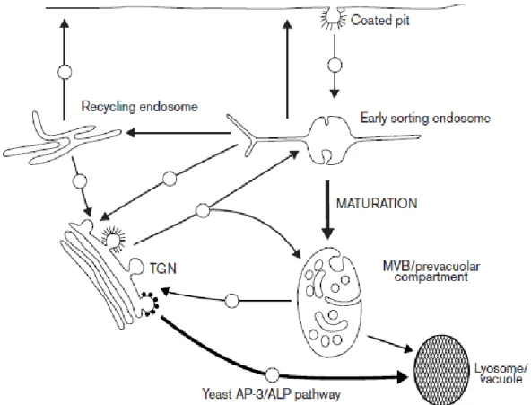
Figure 1-5: Scheme of the endocytic system in yeast and non-polarized cells
The arrows indicate the major trafficking routes through the endocytic system, with spheres indicating transport steps involving known or surmised vesicular intermediates. The bifurcation in traffic flow from the TGN to both the early sorting endosome and to the MVB reflects the maturation process that underlies MVB formation. Initially, multiple fusion events involving vesicles from both the cell surface and the TGN deliver material to a newly forming early sorting endosome. Recycling components (e.g. transferrin receptors) are sorted into the tubular extensions of the structure, which contain the bulk of the membrane of the early sorting endosome, en route to the tubular recycling endosome or to the cell surface for direct recycling. Over time, the sorting endosome loses the capacity to fuse with PM-derived vesicles, but not with vesicles from the TGN. The radiating tubules are replaced by extensive internal membrane invaginations as the maturing endosome moves toward the centre of the cell along microtubules. The available data indicate that in yeast, CPY (not shown) is delivered to a more mature prevacuolar compartment, whereas in animal cells, MPRs may preferentially enter the endocytic pathway at the early sorting endosome. Nevertheless, recent evidence suggests there is a pathway from the TGN to an early endosome in yeast as well and that recycling to the TGN may take place from either the early endosome or prevacuole, similar to animal cells. In yeast, current information supports the direct delivery of ALP to the vacuole in an AP-3- dependent process, although there is presently no evidence for a similar direct route to the lysosome in animal cells (Lemmon and Traub, 2000) .
1.6 Actin microfilaments and microtubules
Actin polymerizes into filaments that offers myriads of advantages to the cells. Actin is necessary for the survival of the cells: it guarantees internal mechanical support, tracks for the for delivering of intracellular components and driving force for cell movements (Martin and Chang, 2006). Under physiological conditions actin polymerizes into long and stable filaments. Beginning of actin polymerization is slow because small oligomers are unstable,
but once the filaments have been synthesized, polymerization is almost completed (Thomas, et al., 2009). Actin polymerization is polar because all the subunits are added in the same directions.
All eukaryotic cells have more than 100 accessory proteins involved in maintaining a pool of actin monomers, initiating the polymerization, and regulating the assembly and the turnover of the monomers and filaments and cross-linking filaments into a very dense network or bundles. Genes for many of these accessory proteins were inserted 1 billion years ago on the phylogenetic tree: amoebas, fungi and animals share many of the molecular mechanism which regulate actin formation and functions (Vidali, et al., 2009).
Plants lost almost 200 genes required for the assembly of cilia and flagella which are responsible for the cell movement in animals (Vitha, et al., 2000). In budding yeast cells actin filaments are responsible for transferring almost all organelles and secretory vesicles to the daughter cell before cell division. Filaments assemble at sites of plasma membrane internalization in budding and fission yeast (Jackson and Casanova, 2000). In these sides “actin patches” assemble de novo and provide force to internalize endocytic vesicles from the outer environment, and then disassemble in a process self-limited by space and time. Many, maybe all, eukaryotic cells use actin filaments to transport organelles.
Fission yeast (Mathur and Hulskamp, 2002) and plant cells (Marc, 1997; Mathur and Hulskamp, 2002) mainly depend on formin to assemble polarized actin cables for transport in polarized cells. In the last years a critical step towards the comprehension of plant actin cytoskeleton was an overview of the highly organization of actin filaments, and above all of the extreme dynamics of actin filaments (Nebenfuhr, et al., 2002). Imaging of plant actin cytoskeletons has not been an easy challenge since all classical fixation and embedding methods result in a poor preservation of actin filaments (Geldner, 2004). Moreover, the slow penetration of chemical fixatives, which is enhanced in higher plants by a rigid cell wall, was suspected to provoke artefacts of actin rearrangements (Richter, et al., 2007). Therefore, cryofixation methods have been developed in order to improve chemical fixation procedures. These techniques guarantee a better preservation of structures (Teh and Moore, 2007).
Elevated calcium concentration leads to fragmentation of F-actin microfilaments (MFs) and to depolymerisation of microtubules (MTs).
MFs are involved in vesicle delivering to the target membranes. Basically, they are distributed along the longitudinal axis of the pollen tubes and are almost absent in the apical zone,
MTs are the next component of cytoskeleton. They are involved in several cellular processes such as mitosis, cytokinesis and vesicular transport. They are polymers composed of α and β heterodimers. Studies in animals and yeast have already shed light on their fast dynamics, their association with other factors and biogenesis. It is the organization between all these events which regulates the spatial organization of microtubules and their ability to respond to specific cellular requirements (Tse, et al., 2006).
Genes involved in microtubules biogenesis have been identified in a group of embryo-lethal mutants: TFC-A (KIESEL), TFC-C (PORCINO), TFC-D (CHAMPIGNON), TFC-E, ARL2
(TITAN5) (Dettmer, et al., 2006). All these mutants show a defective cortical microtubule
array.
Extension of a given MT is finely tuned by polymerization and depolymerisation rates at its ends. In animals, this is ensured by the presence of microtubule-organizing centres, which it is not well defined in plants. Therefore, rapid and efficient spatial reorganization of MTs still remains one of the fascinating mechanism areas in plant cell research.
Stabilization of microtubules is another important mechanism. A putative microtubule stabilizer has been identified in the gene microtubules organization1 (mor1). This gene encodes a homologue of the TOGp-XMAP215 class of very conserved proteins. This mutant is characterized by a very high disorder in the cortical microtubules at high temperature.
1.7 Brief overview of the most common traffic inhibitors in plant cells
1.7.1 Brefeldin A (BFA)
BFA is a macrocyclic lactone that inhibits the activity of ARF GTPases through the interaction with their associated GEFs (Tse, et al., 2004). BFA has been described as an inhibitor of secretion (Satiat-Jeunemaitre and Hawes, 1992; Driouich, et al., 1993) or endocytosis (Grebe, et al., 2003). This depends upon where different BFA-sensitive Arf-GEFs are located, whether early Golgi (Osherov, et al., 1993; Posner, et al., 1994) or post-Golgi (Geldner, et al., 2003).
For instance in BY-2 cells, ER-to-Golgi traffic is inhibited at 10 µ/ml (Jaillais, et al., 2006; Jaillais, et al., 2008) even if it is not possible to detect any morphological changes in Golgi structures (Tse, et al., 2004). In other cells, at same concentration, it is already possible to observe morphological changes in Golgi apparatus (Geldner, et al., 2001; Grebe, et al., 2002; Boonsirichai, et al., 2003; Geldner, et al., 2003; Grebe, et al., 2003; Takano, et al., 2005;
Geldner, et al., 2007). One of the preferred systems in plant cells to study the role of the BFA is the Arabidopsis root. Here, the TGN marker VHA-a1 is present in the core of BFA compartments while the remaining of the Golgi stacks are located on the surface of the structure, as monitored by the heterologous Golgi marker sialyl transferase (Satiat-Jeunemaitre and Hawes, 1992).
The proton-pump is required is not only required for the formation of BFA compartments but also for the transport of FM4-64 to TGN/LE and in the end to the tonoplast (Baluska, et al., 2002).
Also the post-Golgi Arf-GNOM accumulates in the core of the structure (Vanhaesebroeck, et al., 2001; Knight and Shokat, 2007), while PVC/MVB markers (ARA7 and BP80) are not observed in the core of BFA compartments, as verified by different technical approaches. However, it has also been asserted (Welters, et al., 1994) that MVB enter into the BFA compartments as seen by the formation of BP80 and AtSNX-1 containing aggregates, although it has been also been demonstrated that the formation of PVC/MVB aggregates at high BFA concentrations (50-100µgmL-1) do not represent BFA compartments (Baggiolini, et al., 1987), meant as PM proteins that recycle between PM and TGN (Arcaro and Wymann, 1993).
Indeed, BFA compartments are mainly filled by cell wall polysaccharides (Wymann, et al., 1996) suggesting an accumulation of secretory vesicles rather than endocytic compartments(Vlahos, et al., 1994).
1.7.2 Wortmannin
Wortmannin is a fungal metabolite that was originally described as a potent inhibitor of the respiratory burst in neutrophils and monocytes (Das, et al., 2005).
Phosphatidylinositol 3-kinase (PI3K) is the target of wortmannin (Welters, et al., 1994; Dasilva, et al., 2005). PI3K is activated by receptor tyrosine kinase at PM to produce the lipid second messenger phosphatydilinositol-3,4,5-trisphosphate (PIP3) (Sadhu, et al., 2003). PIP3, in turn, recruits and activate downstream different proteins containing lipid-binding domains. These effectors are: I) protein kinases that promote cell growth, survival and proliferation, such as Akt1, PDK1 (phosphoinositide dependent kinase1) and Tec family kinase; II) GAPs (GTPase-Activating Protein, see earlier in the text) and GEFs (Guanine nucleotide-Exchange
In plants, PI3K is also important for development and signalling (Knight, et al., 2006), although less information is available.
PI3K binds wortmannin and the interaction involves the Lys833 within the ATP binding site of PI3K (Tse, et al., 2004; Jaillais, et al., 2006; Oliviusson, et al., 2006; Jaillais, et al., 2008; Silady, et al., 2008; Wang, et al., 2009). Synthesis of another of PI3K inhibitor LY294002 was later reported (Lam, et al., 2007; Wang, et al., 2009).
Both inhibitors show little selectivity within the PI3Ks family members and their structures give little information how to design more selective drugs, which would have a big therapeutic potential.
IC87114 was the first selective inhibitor of one of the PI3K members (Lam, et al., 2007). This inhibitor inhibits p110δ with very high affinity (mid-nanomolar concentrations) and shows between 100-1000 fold selectivity between p110δ and the other members of the family (p110α, p110β, p110γ) (Tse, et al., 2004; Miao, et al., 2006; Delhaize, et al., 2007; Miao, et al., 2008).
No data is available about the use of IC87114 in plant cells.
In plants PI3K homologs have been described (Jaillais, et al., 2006; Miao, et al., 2006; Jaillais, et al., 2008; Robinson, et al., 2008a; Robinson, et al., 2008b) and wortmannin has been demonstrated to be an efficient inhibitor of protein trafficking to the plant vacuole (Wang, et al., 2007). In cells treated with wortmannin the PVC dilates as it can be seen with VSR or ARA7 tagged fluorescent proteins resulting in fluorescent ring-structures (Gazit, et al., 1993; Osherov, et al., 1993). This effect, which can be used as a tool for the identification of PVCs (Posner, et al., 1994), is rapid as it can be visualized 15 minutes after the application of the drug. It can be interpreted as a TGN fusion with the PVC, contributing to the enlargement of the PVC (Kovalenko, et al., 1994). It seems that the wortmannin-induced enlargement of the PVC is a general observation since it has been highlighted in BY-2 cells, Arabidopsis and rice (Yaish, et al., 1988; Shechter, et al., 1989), root cells of Arabidopsis, tobacco, pea and mung bean (Umezawa, et al., 1986) germinating mung bean seeds (Yaish, et al., 1988; Gazit, et al., 1989; Levitzki and Mishani, 2006).
1.7.3 Tyrphostins
Research on the development of PTKs (Protein Tyrosine Kinase) started after finding, in the early 80s, that natural compounds, such as quercetin, erbstatin, genistein and lavendustin inhibit the activities of PTKs such as pp60src and EGFR. Even if these substances have
showed low affinity, selectivity or poor potency, they have been very useful in order to develop new synthetic compounds with a more potent inhibitory activity: tyrphostins.
The activity of itaconic acid (Holen, et al., 1995; Austin and Shields, 1996) along with the erbstatin (Ortiz-Zapater, et al., 2006) triggered the research for the synthesis of these new compounds. The structure of these new compounds has been used as base for a vast number of compounds, many of which showed a very high specificity for PTKs and insignificant inhibition of Ser/Thr kinase (Dhonukshe, et al., 2007).
Tyrphostins are classified in 1) competitive for the substrate and non-competitive for the ATP (ATP competitive, (Robinson, et al., 2004; Robinson, et al., 2008a)), 2) bisubstrate-competitive (Robinson, et al., 2008a) and 3) mixed bisubstrate-competitive (Reichardt, et al., 2007). Further investigations led to synthesis’ of cyclic tyrphostins (Drose and Altendorf, 1997). In mammalian cells it has been highlighted that they can inhibit endocytosis towards the TGN (Dettmer, et al., 2006). Two articles supported tyrphostins A23 as useful tool to study endocytosis in plants: I) the internalization of the hTfR expressed in Arabidopsis protoplast is inhibited by tyrphostin A23 (Dettmer, et al., 2006); II) tyrphostin A23 was reported to inhibit the internalization of PIN2, but not of FM4-64 in Arabidopsis roots, especially preventing its accumulation in BFA compartments (Dettmer, et al., 2005).
Tyrphostin A23 seems to have different effects on different subcellular compartments:
Arabidopsis roots pre-treated with tyrphostin A23 are unable to form BFA compartments, and
do not form cell plate formation (Robinson, et al., 2008a). This demonstrates that tyrphostin A23 acts at level of the Golgi, as Golgi based secretion is required for the formation of BFA-compartments and cytokinesis (Higgins, 1992).
1.7.4 Bafilomycins and Concanamycins
ATPases can be distinguished on the base of their transport mechanism, their sensitivity to specific inhibitors and their quaternary structures (Bowman, et al., 1988). An initial classification has been made by Pedersen and Carafoli (Pedersen and Carafoli, 1987), who introduced the terms P-type, V-type and F-type ATPases.
P-type ATPases pass through a phosphorylated transitional stage; F-type ATPases are mostly involved in ATP synthesis (they are also named F-ATP synthase); V-type ATPases are genetically related to F-types but function only in proton pumping. Later, it came also clear