Apatite-Polymer Composites for the Controlled Delivery of Bone Morphogenetic Proteins
by
Tseh-Hwan Yong S.B. Chemical Engineering
Massachusetts Institute of Technology, 1998
Submitted to the Department of Materials Science and Engineering in Partial Fulfillment of the Requirements for the Degree of Doctor of Philosophy in Materials Science and Engineering
at the
MASSACHUSETTS INSTITUTE OF TECHNOLOGY June 2005
©Massachusetts Institute of Technology. All rights reserved.
Author:
Department of Materials*Science and Engineering March 16, 2005
Certified by:
-- Professor Ja'cki-Y. Ying Adjunct Professor of Chemical Engineering Thesis Supervisor
Certified by:
Professor Michael Rubner Professor of Materials Science and Engineering Thesis Reader
Accepted by:
~- ---- Fecssor Geibtaft
Ceder
Professor of Materials Science and Engineering Chairman, Departmental Committee on Graduate Students
M H INSIIIIJ- __ MASSACHUSETTS NSTTTE OF TECHNOLOGY JUL 2 2 2005 LIBRARIES _ _ _~~~~~~~~~~ - - --- -- -- - - - I _ . II I
J-Apatite-Polymer Composites for the Controlled Delivery of Bone Morphogenetic Proteins
by
Tseh-Hwan Yong S.B. Chemical Engineering
Massachusetts Institute of Technology, 1998
Submitted to the Department of Materials Science and Engineering on March 16, 2005 in Partial Fulfillment of the Requirements for the Degree of
Doctor of Philosophy in Materials Science and Engineering
Abstract
Current treatment of bone defects due to trauma, cancer, or degenerative spine diseases involves the implantation of a bone graft. Autografts, which are harvested from the patient's own body, are associated with problems of limited availability and surgical morbidity. The use of allografts obtained from donors is also not desirable due to the risks of disease transmission and the costs of maintaining bone banks. The ideal solution would be to regenerate native bone to fill the defects. A group of potent growth factors known as bone morphogenetic proteins (BMPs) have been hailed as alternatives to bone grafts due to their ability to elicit new bone formation. Clinical use of BMPs involves loading the protein solution onto collagen sponges and subsequent implantation. However, these conventional collagen carriers show rapid clearance of BMPs within - 2 weeks, whereas bone healing is a longer process, especially in higher mammals. The poor BMP retention in collagen sponges may explain the greater response variability in higher mammals, ranging from full bone bridging within weeks to no bone union. These sponges are also not capable of tunable or multifactor release that could benefit healing in certain anatomic sites, e.g. avascular sites and prolonged non-unions. Hence, the motivation of this thesis is to develop new carriers that allow more efficacious and flexible delivery of BMPs to achieve bone healing.
The carrier should ideally exhibit (i) sustained release to maintain the response and activity of bone-forming cells, (ii) low initial burst to avoid adverse effects of a bolus administration anmd to conserve the expensive growth factor, and (iii) tunable release to meter out BMPs at the desired rate. In particular, tunable release and low burst release have long been challenges in controlled delivery systems. A carrier that can offer such temporal control will be highly valuable to the delivery of other therapeutic proteins, drugs and genes as well.
To this end, we have devised a novel composite of two biomaterials with proven track records: poly(lactic-acid-co-glycolic acid) (PLGA) and apatite. The controlled release strategy was based on the use of a biodegradable polymer with acidic degradation products to manipulate the dissolution of the basic apatitic component. Proteins were pre-adsorbed onto the apatitic component such that as the apatite dissolved, proteins were released.
Apatite-PLGA composites were formed into microparticles by a solid-in-oil-in-water emulsion process. In contrast to polymeric microparticles prepared by the conventional water-in-oil-in-water emulsion process, these composite microparticles exhibited zero-order, low burst release. Low burst release was attributed to the affinity of the apatite for the protein; until the apatite was dissolved, the protein was sequestered and prevented from premature release. Accordingly, the use of apatite singly as a carrier would have led to extremely slow release.
A model protein, bovine serum albumin (BSA), and a therapeutic protein, recombinant human BMP-2 (rhBMP-2), were encapsulated in these apatite-PLGA composite particles. The release profile was modified systematically by changing variables that affected polymer degradation and apatite dissolution, such as polymer molecular weight, polymer hydrophobicity, apatite loading, and apatite particle size. An increase in polymer molecular weight, apatite loading or apatite particle size reduced the release rate of both BSA and rhBMP-2. Interestingly, increasing polymer hydrophobicity diminished the release of BSA, but enhanced the release of rhBMP-2. Slower polymer degradation associated with greater polymer hydrophobicity might have decreased the total amount of protein released, but preserved a larger bioactive fraction due to milder pH conditions within the particles. A suitable particle formulation for sustained rhBMP-2 delivery was identified as protein-sCAP-59 kD PLGA.
When rhBMP-2 was encapsulated in these composite microparticles, it was released in a sustained fashion over 100 days. More importantly, the bioactivity of the protein was retained, as evaluated by its ability to induce the differentiation of mesenchymal stem cells toward the osteoblast lineage. Specifically, the levels of osteoblastic phenotype markers such as alkaline phosphatase (ALP) and osteocalcin were found to be significantly elevated compared to the controls. In contrast, rhBMP-2 released from conventional collagen sponges after 2 weeks did not increase the ALP expression over the controls.
Protein-loaded composite microparticles were dispersed in secondary matrices, either gelatin or collagen sponges, for bone tissue engineering. Multifactor release from these scaffolds was possible through the incorporation of different sets of composite microparticles containing different proteins and exhibiting distinct release profiles. Collagen sponges injected with rhBMP-2-loaded composite microparticles were implanted in subcutaneous sites in rats. These composite collagen sponges stimulated a much higher degree of cellularity and vascularity than the controls without BMPs. The increased vascularity might be evidence of the angiogenic activity of rhBMP-2 at low concentrations in vivo.
Thesis Supervisor: Jackie Y. Ying
Acknowledgments
This thesis would not have been possible without the kind support of my thesis advisor, Professor Jackie Ying. I would like to thank her for her guidance, mentorship and for providing the opportunity to pursue a new research direction. I gratefully acknowledge my thesis committee members, Professors Anne Mayes, Michael Rubner and Myron Spector for their support, informative discussions, and valuable suggestions. I also thank Professor Paul Laibinis for his guidance and for providing me with a good grounding in research.
I have truly enjoyed the camaraderie and stimulating environment of the Nanostructured Materials Research Laboratory. I thank Pemakorn Pitukmanorom, Yee San Su, Noreen Zaman, and Cindy Ren for their wonderful friendship and support both inside and outside of the laboratory. I also thank Dr. Suniti Moudgil, Dr. Todd Zion, Dr. Edward Ahn, Dr. Su Seong Lee, Dr. Jason Sweeney, Dr. Justin McCue, Dr. Neeraj Sangar, Dr. Thomas Lancaster, Steven Weiss, Jianyi Cui, Dr. Xiaohua Huang, Hong He, and Linda Mousseau for helpful discussions and many instances of kind assistance. I also acknowledge Pemakorn Pitukmanorom for help with the nitrogen adsorption analysis and mercury porosimetry.
I was very fortunate to have two bright, capable and enthusiastic UROP students on the project, Elizabeth (Lizzie) Hager and Anita Kris. Lizzie was involved in the synthesis and testing of BSA-loaded composite particles. Much of our understanding of gelatin scaffolds was established through her senior thesis research. Anita was involved in the in vitro and in
vivo testing of BMP-loaded composite collagen sponges.
I thank Professor Clark Colton and Daryl Powers for the use of the cell culture facilities, Professor Hammond, Kris Stokes and LaRuth McAfee for the use of the GPC, Dr. Anthony Garratt-Reed and Patrick Boisvert for assistance with electron microscopy at the CMSE, and the Institute for Soldier Nanotechnologies for the use of the mercury porosimeter and the Zwick/Roell machine.
I would like to thank the staff at the Institute of Bioengineering and Nanotechnology in Singapore who showed me much generosity with their time and expertise during my stay in summer 2004. In particular, I would like to thank Shujun Gao for his guidance and help with the animal studies.
My heartfelt gratitude goes to my father, mother and sister for their unwavering love, support and understanding. I also thank my father-, mother-, brother-, and sister-in-law for their encouragement and constant support. Finally, I thank my wonderful husband, Steve, for his immense patience, love, emotional (and financial) support, and belief in me. Thank you,
Steve. This thesis is as much your accomplishment as it is mine.
This research was supported by the Singapore-MIT Alliance, and the Institute of ]3ioengineering and Nanotechnology (Agency for Science, Technology and Research, Singapore).
Table of Contents
Chapter 1 - Background and Motivation 15
1.1 Bone Regeneration 15
1.2 Bone Morphogenetic Proteins as Alternatives to Bone Grafts 15
1.3 Delivery Systems for Bone Morphogenetic Proteins 17
1.4 Research Objective 19
1.5 Apatite-Polymer Composites for Delivery of BMPs 20
1.6 References 22
Chapter 2 - Synthesis, Characterization, and In Vitro Release Profiles of
Apatite-Polymer Composite Microparticles for the Controlled Delivery of a Model Protein 31
2.1 Introduction 31
2.1.1 Conventional Double Emulsion Processing 31
2.1.2 Solid-in-Oil-in-Water Processing 32
2.1.3 Adsorption of Proteins onto Apatite 32
2.2 Experimental 33
2.2.1 Synthesis of Hydroxyapatite and Carbonated Apatite 33
2.2.2 Characterization of Apatite 34
2.2.3 Adsorption of Proteins onto Apatite 34
2.2.4 Effect of pH on the Dissolution of Apatite and the Release of Adsorbed
Proteins 35
2.2.5 Preparation of Composite Microparticles 36
2.2.6 Preparation of PLGA Microspheres by Double Emulsion Processing 37 2.2.7 Characterization of Microparticle Morphology and Size 38 2.2.8 Evaluation of Encapsulation Efficiency of Composite Microparticles 38
2.2.9 Evaluation of In Vitro Protein Release 39
2.2.10 Evaluation of Polymer Degradation 39
2.2.11 Evaluation of Calcium Release from Composite Microparticles 39
2.3 Results and Discussion 40
2.3.2 Protein Adsorption onto Apatite 42 2.3.3 Effect of pH on Protein Release from Protein-Apatite Complexes 45 2.3.4 Effect of Processing Parameters on Composite Particle Size 47 2.3.5 Encapsulation Efficiency of Protein in Composite Microparticles 49 2.3.6 Effect of Material and Processing Parameters on the In Vitro Release from
Composite Microparticles 50
2.3.6.. 1 Polymer Molecular Weight 50
2.3.6.2 Polymer Hydrophobicity 51
2.3.6.3 Apatite Particle Size 53
2.3.6.4 Addition of Buffering Apatite 54
2.3.6.5 Protein Loading on Apatite 55
2.3.6.6 BSA-Apatite Complex Loading in Particles 56
2.3.7 Comparison of Protein Release from Polymeric Microspheres Prepared by
Double Emulsion Method 57
2.3.8 Degradation of the Polymeric Phase of Composite Microparticles 58
2.3.9 Calcium Release from Composite Microparticles 59
2.4 Summary 61
2.5 References 63
Chapter 3 - Application of Apatite-Polymer Composite Microparticles to the Controlled
Delivery of BMPs 68
3.1 Introduction 68
3.1.1 Osteoinductive Effect of BMPs 68
3.2 Experimental 69
3.2.1 Adsorption of BMPs onto Apatite 69
3.2.2 Aseptic Preparation of rhBMP-2-Loaded Composite Microparticles 70 3.2.3 Estimation of the Magnitude of Initial Burst from Composite Microparticles 71
3.2.4 Evaluation of In Vitro Release 71
3.2.5 Cellular Response 72
3.2.5.1 Effect of Release Medium Collected at Each Time Point 72
3.2.5.3 Statistical Analysis 74
3.3 Results; and Discussion 74
3.3.1 Adsorption of rhBMPs onto Apatite 74
3.3.2 Estimation of Proportion of Unbound rhBMPs in Composite Microparticles 75 3.3.3 Effect of Material and Processing Parameters on the In Vitro Release of
rhBMP-2 from Composite Microparticles 75
3.3.3.1 Polymer Molecular Weight 76
3.3.3.2 Apatite Particle Size 76
3.3.3.3 Apatite Loading 77
3.3.3.4 Polymer Hydrophobicity 78
3.3.3.5 Release Medium 79
3.3.4 Effect of Conditioned Medium Collected at Each Time Point 81
3.3.5 Effect of Prolonged Exposure to Conditioned Medium 85
3.3.5.1 Measurement of Induced Alkaline Phosphatase Activity 85
3.3.5.2 Measurement of Osteocalcin Expression 87
3.3.5.3 Evaluation of Cell Proliferation 88
3.4 Summary 90
3.5 References 92
Chapter 4 - Incorporation of Apatite-Polymer Composite Microparticles into Bulk
Matrices 95
4.1 Introduction 95
4.1.1 Tissue Engineering Scaffolds 96
4.1.1.1 Gelatin Scaffolds 97
4.1.1.2 Collagen Sponges 97
4.2 Experimental 97
4.2.1 Preparation of Composite Gelatin Scaffolds 97
4.2.2 Preparation of Composite Collagen Sponges 99
4.2.3 Characterization of Scaffolds and Sponges 99
4.2.3.2 Porosity Measurements 100
4.2.4 Evaluation of Release Kinetics 100
4.2.4.1 Release of BSA 100
4.2.4.2 Release of rhBMP-2 100
4.2.5 Cellular Response 100
4.2.5.. 1 Evaluation of Cell Viability by Microscopy 101
4.2.5.2 Evaluation of Cell Proliferation 102
4.2.5.3 Assessment of Osteoblastic Markers 102
4.2.5.4 Statistical Analysis 103
4.2.6 Preliminary In Vivo Testing in a Rat Ectopic Model 103
4.3 Results and Discussion 104
4.3.1 Mechanical Properties of Scaffolds 104
4.3.2 Porosity and Pore Size Distribution of Scaffolds 106 4.3.3 In Vitro Release of Proteins from Scaffolds 110
4.3.3.1 Composite Gelatin Scaffolds 110
4.3.3.2 Composite Collagen Sponges 112
4.3.4 Cellular Response 114
4.3.4. 1 Evaluation of Cell Viability by Microscopy 114
4.3.4.2 Evaluation of Cell Proliferation 115
4.3.4.3 Measurement of Induced Alkaline Phosphatase Activity 116
4.3.4.4 Measurement of Osteocalcin Expression 118
4.3.5 Preliminary In Vivo Testing 119
4.3.5.1 Evaluation of Dosage Required for Ectopic Bone Formation 124
4.4 Summary 127
4.5 References 129
Chapter 5 - Recommendations for Future Work 134
5.1 Further Investigation of Release Mechanism 134
5.3 Incorporation of Composite Microparticles into Tissue Engineering Scaffolds 135
5.4 In Vivo Studies 136
5.5 Use of Composites in Other Shapes and Sizes 136
5.6 Application to Other Therapeutic Agents 136
5.7 References 137
List of Figures
1.1. A local pharmacokinetic curve showing release rate of radiolabeled BMP-2 from the
rabbit ulna osteotomy site 18
1.2. Protein is released from the composite microparticle as a result of polymer hydrolysis
leading to dissolution of the apatite substrate 21
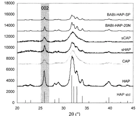
2.1. Schematic of the S/O/W process for preparing composite microparticles. 36 2.2. Schematic of the W/O/W process for preparing polymeric microparticles. 37 2.3. XRD patterns of apatites prepared with surfactant (sCAP, sHAP) and without surfactant (CAP, HAP), and commercial BABI-HAP-SP and BABI-HAP-20N
powders 41
2.4. FITC-BSA adsorption isotherms of (a) HAP and (b) CAP at room temperature, and (c)
HAP and (b) CAP at 40C 43
2.5. Adsorption of FITC-BSA onto HAP with time at room temperature 44 2.6. Release: of BSA from BSA-HAP and BSA-CAP complexes in buffers of pH 3, 4, 5
and 7.4 47
2.7. ESEM micrographs of composite microparticles prepared with 59 kD PLGA and (a) submicron-sized sCAP particles and (b) micron-sized CAP particles 48 2.8. ESEM micrographs of composite microparticles prepared with CAP and (a) 13 kD PLGA and (b) 50/50 blend of 13 kD and 24 kD PLGA. 48 2.9. Protein release from composite particles loaded with 15 itg of FITC-BSA per mg of
carrier 51
2.10. Effect of PLA incorporation on protein release 52
2.11. Effect of PEG incorporation on protein release 53
2.12. Effect of carbonated apatite particle size on protein release 54 2.13. Effect of buffering apatite on protein release, expressed as (a) cumulative mass of BSA released per mg of carrier and (b) cumulative percentage of BSA released 55
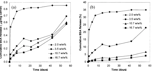
2.15. Effect of BSA-CAP complex loading on protein release, expressed as (a) cumulative mass of BSA released per mg of carrier and (b) cumulative percentage of BSA
released 57
2.16. Release of (a) BSA from PLGA and PLGA-PLA (2:3) microparticles prepared in-house by the double emulsion method, and (b) FITC-IgG from PLGA microparticles
blended with 0, 1, 2 and 5 w/w% of PEG 58
2.17. Release of acid from the degradation of PLGA of different molecular weights in blank
CAP-PLGA composite particles 59
2.18. Calcium release from microparticles prepared with CAP-13 kD PLGA, CAP-59 kD
PLGA, BSA-CAP-59 kD PLGA, and bare CAP 60
3.1. Effect of PLGA molecular weight on rhBMP-2 release from composite microparticles 76 3.2. Release of rhBMP-2 from composite particles fabricated from 59 kD PLGA, and
1-jtm sCAP and 7-tm CAP 77
3.3. Effect of sCAP loading on rhBMP-2 release 78
3.4. Effect of polymer hydrophobicity on rhBMP-2 release 79
3.5. RhBMP-2 release in BES buffer, complete BME, and serum-free BME, all at pH 7.4 80 3.6. Change in PLGA molecular weight with time in BES buffer and complete BME, both
at pH 7.4 80
3.7. Effect of rhBMP-2 concentration on induced ALP activity in C3H1OT1/2 cells 81 3.8. ALP activity induced by release medium collected at each time point from rhBMP-2-loaded composite microparticles fabricated from 59 kD PLGA with a sCAP loading of
0.08 mg/mg of PLGA 82
13.9. ALP activity induced by release medium collected at each time point from rhBMP-2-loaded composite microparticles fabricated from 59 kD PLGA with a sCAP loading of
0.1 8 mg/mg PLGA 83
13.10. ALP activity induced by release medium collected at each time point from rhBMP-2-loaded composite microparticles fabricated from a 3:2 blend of 59 kD PLGA and 25
kD PLA 83
3.11. ALP activity induced by release medium collected at each time point from
3.12. Effect of length of exposure to rhBMP-2 on ALP activity 86 3.13. Effect on ALP activity of prolonged exposure to release media collected from rhBMP-2-loaded composite microparticles fabricated from 59 kD PLGA 86 3.14. Effect of length of exposure to rhBMP-2 on osteocalcin levels 87
3.15. Effect on osteocalcin levels of prolonged exposure to release media collected from
rhBMP.-2-loaded composite microparticles fabricated from 59 kD PLGA 88
3.16. Effect of length of exposure to rhBMP-2 on cell number 89
3.17. Effect on cell number of prolonged exposure to release media collected from rhBMP-2-loaded composite microparticles fabricated from 59 kD PLGA 89
4.1. Typical stress-strain curve for composite gelatin scaffolds 105 4.2. Compressive moduli of composite gelatin scaffolds with different composite particle
loadings 106
4.3. Pore size distributions of composite gelatin scaffolds loaded with (a) 0 w/w%, (b) 10 w/w%, (c) 33 w/w% and (d) 43 w/w% of composite microparticles 107 4.4. ESEM mnicrographs of a composite gelatin scaffold (10 w/w% particles) showing (a) a pore greater than 200 tm, and (b) composite microparticles dispersed in the scaffold. 108 4.5. Pore size distributions of composite Helistat® sponges loaded with (a) 0 w/w%, (b) 10
w/w% and (c) 33 w/w% of composite microparticles 109
4.6. ESEM micrographs of the cross-section of a composite collagen sponge showing (a) pores greater than 200 pm and (b) composite microparticles held in the sponge 110 4.7. BSA release from composite particles and composite gelatin scaffolds 111 4.8. BSA release from composite particles and composite collagen sponge 113 4.9. RhBMP-2 release from composite particles and composite collagen sponge 113 4.10. Fluorescent cell viability staining of cell-seeded composite collagen sponges at (a) 4
days and (b) 7 days 114
4.11. Number of C3H1OT1/2 cells growing on composite collagen sponges or on the bottom
4.12. ALP activity induced by prolonged exposure to composite collagen sponges containing composite particles loaded with rhBMP-2, rhBMP-6, rhBMP-2 and rhBMP-6 and no
BMP (control) 117
4.13. Effects of rhBMP-2 and rhBMP-6 concentrations on induced ALP activity in
C3H10['1/2 cells 118
4.14. Osteocalcin levels induced by prolonged exposure to composite collagen sponges containing composite particles loaded with 2, 6, 2 and
rhBMP-6 and no BMP (control) 119
4.15. H&E staining of excised composite collagen sponges at 1 week and 2 weeks 122 4.16. H&E staining of excised collagen sponges at 1 week and 2 weeks 123 4.17. Radiographs at 2 weeks and 4 weeks of rats implanted with collagen sponges loaded
with rhBMP-2 of 2 fpg, 5 jtg, 10 pig, 20 pig, and 50 Etg 125
4.18. Radiographs at 2 weeks and 4 weeks of rats implanted with collagen sponges loaded with rhBMP-6 of 2 pig, 5 ig, 10 pg, 20 tg, and 50 pg 126
List of Tables
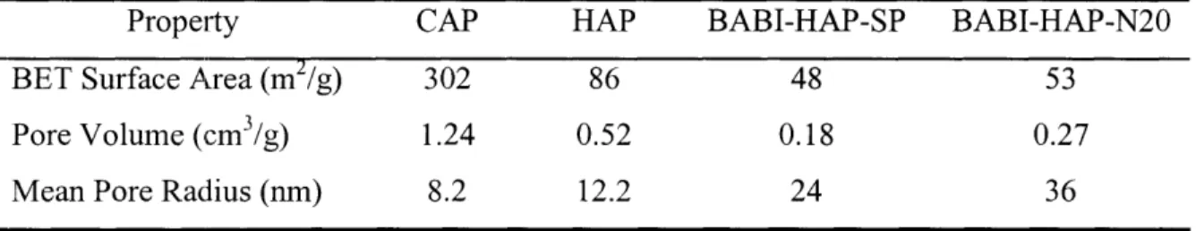
2.1. Grain sizes and particle sizes of apatite powders 41 2.2. BET surface areas, pore volumes and mean pore radii of apatites 42
2.3. Protein adsorption capacities of apatites 42
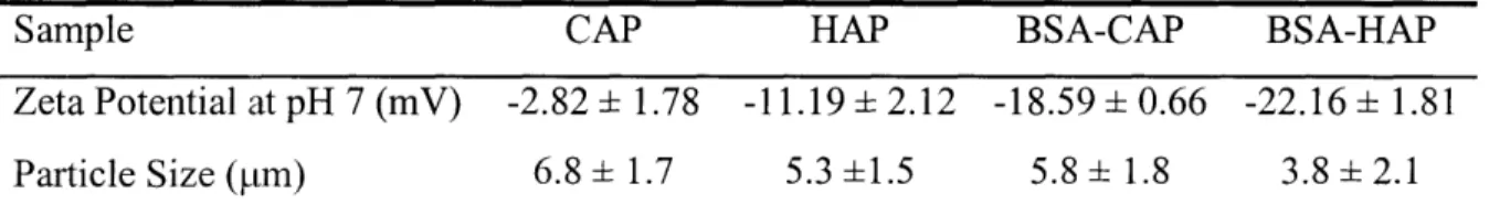
2.4. Adsorption of proteins of different IEP and size onto CAP 44 2.5. Zeta potential and particle size of apatites and BSA-apatite complexes 45 2.6. Effects of processing parameters on composite particle size 49 2.7. Encapsulation efficiency of selected BSA-loaded composite microparticles 50
3.1. Adsorption of rhBMPs onto 5 mg of CAP 75
Chapter 1 - Background and Motivation
1.1 Bone Regeneration
Bone is a naturally regenerative tissue; it is able to heal from fractures and breaks by recapitulating the embryonic skeletal developmental process. However, an estimated 5-10% of fractures fail to recover properly and proceed to delayed union or nonunion [1]. The repair of bone loss associated with trauma and cancer is also typically not observed. Current treatment involves the implantation of autogenic or allogenic bone grafts, a procedure that an estimated 1.5 million patients undergo in the United States each year [2]. Autografts, long considered the gold standard in bone grafting, are plagued with problems of limited supply, morbidity associated with graft harvest, and variability in fusion success rate. The use of allografts is dampened by the risks of disease transmission and the costs of maintaining bone banks. Synthetic grafts constructed of metals and ceramics are also used, but their mechanical and chemical incompatibility with bone tissue often leads to implant failure [3]. The ideal solution would be to regenerate native bone to fill the defects. A group of potent growth factors known as bone morphogenetic proteins (BMPs) has the ability to elicit new bone formation. These proteins provide a promising alternative to bone grafts, and have garnered much excitement and interest.
1.2 Bone Morphogenetic Proteins as Alternatives to Bone Grafts
BMPs are a class of fourteen cytokines belonging to the transforming growth factor-3 (TGF- [3) superfamily. They were discovered by Urist in 1965 as the components in demineralized bone matrix responsible for inducing ectopic bone and cartilage formation in muscle and subcutaneous sites of rodents [4, 5]. BMPs induce bone morphogenesis in a multistep cascade reminiscent of embryonic skeletal formation, beginning with the migration of mesenchymal cells, their differentiation into cartilage-forming cells (chondrocytes) or bone-forming cells (osteoblasts), deposition of bone matrix, establishment of functional marrow within the bone, and culminating in remodeling of the bone consistent with the anatomic site and biomechanic environment [1, 4, 6, 7]. In addition to bone induction (osteoinduction), BMPs also play a role in the development of other tissues and organs such as kidney, gut, lung, teeth, heart, limb, and brain [6, 8].
Among the osteoinductive BMPs, BMP-2, BMP-4 and BMP-7 appear to have greater potency [4, 8], and are being produced with high bioactivity and purity via recombinant DNA technology. In vitro administration of BMP-2 and BMP-7 to embryonic rat calvarial cells, rat osteosarcoma cells or mouse fibroblasts resulted in enhanced osteogenic activity, as evidenced by (differentiation into osteoblasts and elevated expression of bone mineralization proteins. In vivo treatment with BMP-2 or BMP-7 augmented the healing of defects in rodents [9, 10], rabbits [11-15], dogs [16-18], sheep [19], and non-human primates [13, 20, 21]. Critical-size defects, which do not heal spontaneously, were bridged within 3 months in primates [20, 21]. These animal studies validate the safety and efficacy of 2 and BMP-7 in promoting orthopedic repair. However, the results from human trials have shown more variation. Geesink et al. reported that amongst six patients receiving BMP-7 with a collagen carrier for the treatment of high tibial osteotomy, four showed bridging by 6 weeks, one by 10 weeks, and one showed no bone formation during the course of the study [22]. When similar BMP-7 carriers were implanted in the maxillary sinuses of three patients with maxillary atrophy, only one exhibited satisfactory bone formation, whereas the remaining two showed little bone formation after 6 months [4]. More recently, 450 patients with open tibial fractures were randomly assigned to 3 groups receiving (i) the standard of care (tissue irrigation, d6bridement, followed by intramedullary nail fixation), (ii) standard of care and 6 mg of rhBMP-2 on a collagen sponge, or (iii) standard of care and 12 mg of rhBMP-2 on a collagen sponge [23]. One year after treatment, 52% of the patients who received the standard of care had healed, compared to 59% and 72% of patients who were further treated with 6 mg and 12 -mg of rhBMP-'2, respectively. While the value of BMP treatment was undeniable, the results from clinical studies were not as impressive as those observed in animal studies where healing occurred more quickly and more completely. The greater variability and slower response in humans may be attributed to a smaller population of multipotent cells, which are also less responsive than, those in smaller animals. It has been proposed that the therapeutic outcomes may be enhanced by carriers capable of (i) delivering BMPs at a rate that matches the responsiveness of the cells or (ii) delivering a suitable cocktail of growth factors to stimulate the cells [24, 25;].
1.3 Delivery Systems for Bone Morphogenetic Proteins
In order for BMPs to exert an effect on bone healing, they must be delivered to the defect site. Bolus injections have limited effect because of the rapid clearance of exogenous proteins from the body. To increase retention of BMPs at the defect site, a carrier is needed that has desirable release kinetics. Furthermore, if the volume of bone to be regenerated is large, the carrier has to be combined with a scaffold or serve as a scaffold itself to allow for the cell migration and growth, and the deposition of extracellular matrix [25].
Delivery systems for BMPs have been constructed from a variety of materials, which can be categorized as natural polymers, synthetic polymers, inorganic materials, and composites of the above.
Of the natural polymers, collagen is the most widely used [9, 21, 26, 27] and was the first to be employed commercially for BMP delivery. Demineralized bone matrix (DBM), the material from which Urist originally extracted BMPs, is also commonly used [28, 29] as the residual levels of BMPs, matrix proteins and calcium in DBM could enhance osteoinductivity. Other natural polymers such as gelatin [30-33], hyaluronic acid [34], chitosan [35] and alginate [36, 37] have also been tested. Potential issues with naturally derived materials include batch variations in purity and quality, as well as the risk of disease transmission.
Biodegradable and/or biocompatible synthetic polymers, such as poly(oc-hydroxy acids) [38-41], poly(ortho esters) [42], polypropylene fumarate [43] and polyethylene glycol [44], offer benefits of reproducible manufacture and readily tailored functionality. In
particular, poly(cc-hydroxy acids) comprising lactic acid and glycolic acid are approved by the Federal Drug Administration (FDA), and have been used as suture material since the 1970s. Biodegradable polymers are designed to decompose to non-toxic products in physiological environments. However, some degradation products, such as those from poly(u-hydroxy acids) and polyanhydrides, are acidic and may cause tissue inflammation.
Hydroxyapatite (HAP) [19, 45, 46], calcium phosphate [45, 47, 48], silica [49], bioglass [50, 51] and titanium [52, 53] are some of the inorganic materials that have been used as BMP carriers. The most commonly used is HAP because of its similarity to the mineral component of bone and its osteoconductivity. The osteoconductivity of HAP and calcium phosphate is sometimes attributed to their ability to concentrate growth factors, including BMPs, in the body [54]. The release of proteins from ceramics can be very slow
and sustained because of the high affinity of certain ceramics for proteins [54-58]. Drawbacks of ceramics include their brittleness and difficulty in processing.
A fruitful combination of the above three classes of materials as composites would allow the harnessing of the benefits of each component. Furthermore, the drawbacks of one material may be countered by another. For example, HAP and calcium phosphate can act as a buffer against the acidity of the degradation products from poly(oc-hydroxy acids) [59, 60]. Composites have been produced with enhanced mechanical and handling properties [61-66], improved bioactivity [67-69], optimized biodegradation [59, 60, 70], and microstructures that more closely resemble natural tissues [71, 72]. Some composites that have been explored for BMP delivery include HAP-collagen [17, 26, 73, 74], poly(lactic acid-co-glycolic acid) (PLGA)-calcium phosphate [75], PLGA-cellulose [76, 77], and PLGA-gelatin [14].
1U Ll ,nn r 4 Thu
-. (C0
Q 0 50 - 40- 20-- 1-4: + 4.., r -. a~~~~~~~< 0 2 4 !3 8 '10 t 2 14 '16 Days post-treatrentFig. 1.1. A local pharmacokinetic curve showing release rate of radio-labeled BMP-2 from the rabbit ulna osteotomy site [25]. The various carriers shown were either implanted (* hyaluronic acid pad and A collagen sponge) or injected ( Gelfoam paste and * buffer) into the defect. Reprinted from [25] with permission from Elsevier.
Many of these materials are transformed into BMP carriers by simply mixing in the proteins during processing or by soaking pre-fabricated carriers in the protein solution. Variations in release kinetics amongst the carriers are observed due to inherent differences in the affinity of the materials for BMPs and in the carrier dimensions (Fig. 1.1). However, adjustment of BMP release kinetics with such carriers in order to attain the optimal profile is difficult and has not been accomplished. The optimal BMP release profile may vary with
animal species, age of host, anatomic site, wound history and other factors [25]. For example, slower release rates may be required in more fluid environments where BMP clearance may be faster, and in more compromised sites where the healing response is diminished. Tunable carriers would offer greater potential and flexibility in realizing the desired release profile.
Two products containing BMPs have recently been approved by FDA for spinal fusion: Stryker's OP-I comprising BMP-7 and Medtronic Sofamor Danek's INFUSE containing BMP-2. Both products use collagen sponges as the carrier for BMPs; the sponges are loaded by soaking with a BMP solution for 10-20 min. The BMP loading in these sponges is on the order of milligrams per implant, which is several orders of magnitude above the natural occurrence in bone (- 0.002 mg of BMP-2 can be extracted per kg of powdered bone [78]). The release of BMPs from collagen tends to be rapid: 70-90% of the load is depleted by the first week [29., 34, 79]. However, bone healing is often a much longer process requiring weeks or months, especially in higher mammals with less responsive cells. A possible reason for the use of supra-physiological BMP doses in collagen carriers is the need to overcome the low availability of BMPs at later stages of healing [25] since cellular response to BMPs has been found to increase with dosage as well as time of exposure [77, 80]. Therefore, carriers that are capable of sustained release of lower but still therapeutic levels of BMPs would allow greater efficacy and cost-savings by optimizing the use of these expensive proteins. The presence of BMPs over the entire duration of healing in higher mammals may also reduce the variation in response (see Section 1.2), and enhance the therapeutic outcomes. Furthermore, tunable release or multifactor release at different rates, which current collagen sponge carriers cannot accomplish, may also augment the bone healing response.
1.4 Research Objective
Certain anatomic sites and certain indications, e.g. prolonged non-unions, may require BMP release kinetics that current collagen sponge carriers cannot provide. The objective of our research is to improve the efficacy of BMP delivery for bone healing by developing carriers that can retain and meter out BMPs at the appropriate dose and for a sufficient duration to achieve the desired host response. Ideal release characteristics of such carriers include:
1. Sustained release - to make BMPs available over the entire period of healing, possibly lasting for several weeks to several months.
2. Low burst release - to avoid the adverse effects of an overdose, and to conserve the expensive therapeutic proteins, making the formulation more cost-effective.
3. Tunable release - to allow flexibility in designing release rates for different therapeutic proteins, host species, and anatomic sites.
Other desirable properties of the carrier include biocompatibility, biodegradability, bioactivity, ease of manufacture, and surgical practicability. Such a carrier would also have broader applications in the delivery of other therapeutic proteins and drugs.
To achieve the research objective, the following approach was taken:
* Development of a biocompatible, biodegradable carrier capable of tunable, sustained, and low-burst release (Chapter 2)
* Application of the carrier to the delivery of BMPs (Chapter 3)
· In vitro testing to establish bioactivity of released BMPs (Chapter 3)
* Incorporation of BMP carriers into scaffolds capable of supporting in vivo cell migration and growth (Chapter 4)
· Ascertaining the safety and efficacy of these scaffolds in vitro and in vivo (Chapter 4)
1.5 Apatite-Polymer Composites for Delivery of BMPs
Our strategy for a tunable controlled delivery platform is based on the use of a polymer with acidic degradation products to control the dissolution of a basic inorganic substrate on which BMPs have been adsorbed. The release of acid from the hydrolysis of the polymer leads to the dissolution of the inorganic substrate and consequently, the desorption and release of the protein. Hence, protein release is anticipated to be accelerated over passive leaching from the inorganic substrate, yet, more controlled than the release from polymeric microspheres due to the affinity of the substrate for the protein. The release mechanism is depicted in Fig. 1.2.
Systematic modulation of the release profile may be possible by changing variables that affect polymer degradation, subsequent acid generation, and/or inorganic substrate dissolution. These variables include polymer type, polymer molecular weight, polymer composition (including copolymers and blends), inorganic substrate type, inorganic substrate
loading, inorganic substrate particle size, relative proportion of polymer and inorganic substrate, and protein loading on the inorganic substrate. This delivery platform can be applied to any therapeutic agent that can be adsorbed and sequestered on the surface of a basic inorganic material. - O e '
water
°e
O . composite microparticle o proteinO
protein adsorbed on apatiteFig. 1.2. Protein is released from the composite microparticle as a result of polymer hydrolysis that leads to dissolution of the apatite substrate.
Candidates for the polymeric component include polyanhydrides, poly(oc-hydroxy acids) and poly(ortho esters), all of which degrade to produce acids. Poly(c-hydroxy acids) comprising lactic acid (LA) and glycolic acid (GA) - PLGA, PLA, and PGA - are attractive candidates because they are FDA-approved, and are commercially available in a wide range of molecular weights. Furthermore, poly(oc-hydroxy acids) are bulk eroding polymers, which may be more suitable for this controlled release mechanism as they allow the buildup of acid within composite particles necessary for dissolving the apatitic phase. On the other hand, acidic degradation products from surface eroding polymers, e.g. polyanhydrides, may diffuse away too quickly to encounter the apatitic phase. The pH within degrading PLGA microspheres has been found to be less than 4.7 [81] and as low as 1.5 [82]. Such a pH range
should be sufficient to cause the dissolution of a basic material.
A number of basic inorganic materials, such as HAP, carbonated apatite (CAP), calcium phosphate and calcium carbonate, exhibit bioactivity and enhanced integration with host bony tissue [83-88]. These materials are also employed as fillers to enhance the mechanical properties of the softer natural and synthetic polymers [61, 62, 64]. In addition, they have been used to alleviate the acidity created by the degradation of poly(oc-hydroxy
acids) [59, 60, 89, 90], which is relevant to our current application. The high affinity of HAP for proteins (see Section 1.3) suggests that HAP should be capable of holding BMPs on its surface without premature release. Nanocrystalline HAP and CAP, the syntheses of which were previously developed in our laboratory [91-93], were selected as the inorganic phase of the composites. CAP differs from HAP (Cal0(PO4)6(OH)2) in that it has carbonate ions substituted in the hydroxyl or phosphate sites in the crystal lattice [94]. CAP is more amorphous, more resorbable, and closer in composition to natural bone mineral.
The dimensions of the carrier would depend on the method of administration, defect site to fill, and impact on release kinetics. The apatite-polymer composite carriers may be prepared as particles, which can be delivered by injection into the bone defect, compressed into pellets for implantation, or dispersed in a secondary matrix that can be formed into tissue engineering scaffolds. In addition, the composites may be fabricated as films or porous, bulk scaffolds.
1.6 References
[1] Wozney JM. Overview of bone morphogenetic proteins. Spine 2002;27:S2-S8. [2] Deutsche Bank Alex. Brown Report; 2001.
[3] Lane JM, Tomin E, Bostrom MPG. Biosynthetic bone grafting. Clin Orthop Rel Res
1999;367:S1 07-S117.
[4] Groeneveld EHJ, Burger EH. Bone morphogenetic proteins in human bone regeneration. Eur J Endocrinol 2000; 142:9-21.
[5] Urist MR. Bone: Formation by autoinduction. Science 1965;150:893-899.
[6] Balemans W, Hul WV. Extracellular regulation of BMP signaling in vertebrates: a cocktail of modulators. Dev Biol 2002;250:23 1-250.
1[7] Kloen P', Doty SB, Gordon E, Rubel IF, Goumans M-J, Helfet DL. Expression and activation of the BMP-signaling components in human fracture non-unions. J Bone Joint Surg [Am] 2002;84A: 1909-1918.
1-8] Hoffman A, Weich HA, Gross G, Hillmann G. Perspectives in the biological function, the technical and therapeutic application of bone morphogenetic proteins. Appl Microbiol Biotech 2001 ;57:294-308.
[9] Lutolf MP, Weber FE, Schmoekel HG, Schense JC, Kohler T, Muller R, Hubbell JA. Repair of bone defects using synthetic mimetics of collagenous extracellular matrices. Nat Biotechnol 2003;21:513-518.
[10] Saito N, Okada T, Horiuchi H, Narumichi M, Takahashi J, Nawata M, Ota H, Nozaki K, Takaoka K. A biodegradable polymer as a cytokine delivery system for inducing bone formation. Nat Biotechnol 2001;19:332-335.
[11] Koempel JA, Patt BS, O'Grady K, Wozney JM, Toriumi DM. The effect of recombinant human bone morphogenetic protein-2 on the integration of porous hydroxyapatite implants with bone. J Biomed Mater Res 1998;41:359-363.
[12] Kokubo S, Fujimoto R, Yokota S, Fukushima S, Nozaki K, Takahashi K, Miyata K. Bone regeneration by recombinant human bone morphogenetic protein-2 and a novel biodegradable carrier in a rabbit ulnar defect model. Biomaterials 2003;24:1643-1651.
[13] Suh DY, Boden SD, Louis-Ugbo J, Mayr M, Murakami H, Kim H-S, Minamide A, Hutton WC. Delivery of recombinant human bone morphogenetic protein-2 using a compression-resistant matrix in posterolateral spine fusion in the rabbit and in the non-human primate. Spine 2002;27:353-360.
[14] Ueki K, Takazakura D, Marukawa K, Shimada M, Nakagawa K, Takatsuka S, Yamamoto E. The use of polylactic acid/polyglycolic acid copolymer and gelatin sponge complex containing human recombinant bone morphogenetic protein-2 following condylectomy in rabbits. J Crano-Maxillofac Surg 2003;31:107-114.
[15] Zegzula HD, Buck DC, Brekke J, Wozney JM, Hollinger JO. Bone formation with use of rhBMP-2. J Bone Joint Surg [Am] 1997;79A: 1778-1790.
[16] Cullinane DM, Lietman SA, Inoue N, Deitz LW, Chao EYS. The effect of recombinant human osteogenic protein-1 (bone morphogenetic protein-7) impregnation on allografts in a canine intercalary bone defect. J Orthop Res 2002;20:1240-1245.
1-17] Itoh S, Kikuchi M, Takakuda K, Nagaoka K, Koyama Y, Tanaka J, Shinomiya K. Implantation study of a novel hydroxyapatite/collagen (HAp/Col) composite into weight-bearing sites of dogs. J Biomed Mater Res 2002;63:507-515.
[-18] Muraka:ni N, Saito N, Takahashi J, Ota H, Horiuchi H, Nawata M, Okada T, Nozaki K, Takaoka K. Repair of a proximal femoral bone defect in dogs using a porous surfaced
prosthesis in combination with recombinant BMP-2 and a synthetic polymer carrier. Biomaterials 2003;24:2153-2159.
[19] den Boer FC, Wippermann BW, Blokhuis TJ, Patka P, Bakker FC, Haarman HJTM. Healing of segmental bone defects with granular porous hydroxyapatite augmented with recombinant human osteogenic protein-1 or autologous bone marrow. J Orthop Res 2003;21:521-528.
[20] Ripamonti U, Ramoshebi LN, Matsaba T, Tasker J, Crooks J, Teare J. Bone induction by BMPs/OPs and related family members in primates. J Bone Joint Surg [Am] 2001;83-A:SI-116.
[21] Cook SD, Wolfe MW, Salkfeld SL, Rueger DC. Effect of recombinant human osteogenic protein-1 on healing of segmental defects in non-human primates. J Bone Joint Surg [Am] 1995;77A:734-750.
[22] Geesink RGT, Hoefnagels NHM, Bulstra SK. Osteogenic activity of OP-1 bone morphogenetic protein (BMP-7) in a human fibular defect. J Bone Joint Surg [Br]
1999;81B:710-718.
[23] Govender S, Csimma C, Genant HK, Valentin-Opran A. Recombinant human bone morphogenetic protein-2 for treatment of open tibial fractures - A prospective, controlled, randomized study of four hundred and fifty patients. J Bone Joint Surg [Am] 2002;84A:2123-2134.
[241 Einhorn TA. Clinical applications of recombinant human BMPs: Early experience and future development. J Bone Joint Surg [Am] 2003;85A:82-88.
[25-1 Li RH, Wozney JM. Delivering on the promise of bone morphogenetic proteins. 'Trends Biotechnol 2001;19:255-265.
[26] Takaoka K, Nakahara H, Yoshikawa H, Masuhara K, Tsuda T, Ono K. Ectopic bone induction on and in porous hydroxyapatite combined with collagen and bone morphogenetic protein. Clin Orthop Rel Res 1988;234:250-254.
[27] Geiger M, Li RH, Friess W. Collagen sponges for bone regeneration with rhBMP-2. Adv Drug Deliv, Rev 2003;55:1613-1629.
128] Uludag H, D'Augusta D, Palmer R, Timony G, Wozney J. Characterization of rhBMP-2 pharmacokinetics implanted with biomaterial carriers in the rat ectopic model. J Biomed Mater Res 1999;46:193-202.
[29] Uludag H, D'Augusta D, Golden J, Li J, Timony G, Riedel R, Wozney JM. Implantation of recombinant human bone morphogenetic proteins with biomaterial carriers: A correlation between protein pharmacokinetics and osteoinduction in the rat ectopic model. J Biomed Mater Res 2000;50:227-238.
[30] Holland TA, Tabata Y, Mikos AG. In vitro release of transforming growth factor-31 from gelatin microparticles encapsulated in biodegradable, injectable oligo(poly(ethylene glycol) fumarate) hydrogels. J Control Release 2003;91:299-313.
[31] Raiche AT, Puleo DA. In vitro effects of combined and sequential delivery of two bone growth factors. Biomaterials 2004;25:677-685.
[32] Tabata Y, Yamada K, Miyamoto S, Nagata I, Kikuchi H, Aoyama I, Tamura M, Ikada Y. Bone regeneration by basic fibroblast growth factor complexed with biodegradable hydrogels. Biomaterials 1998;19:807-815.
[33] Yamamoto M, Takahashi Y, Tabata Y. Controlled release by biodegradable hydrogels enhances the ectopic bone formation of bone morphogenetic protein. Biomaterials 2003;24:4375-4388.
[34] Kim HI), Valentini RF. Retention and activity of BMP-2 in hyaluronic acid-based scaffolds in vitro. J Biomed Mater Res 2002;59:573-584.
[35] Lee J-Y, Nam S-H, Im S-Y, Park Y-J, Lee Y-M, Seol Y-J, Chung C-P, Lee S-J. Enhanced bone formation by controlled growth factor delivery from chitosan-based biomaterials. J Control Release 2002;78:187-197.
1-36] Saito A, Suzuki Y, Ogata S, Ohtsuki C, Tanihara M. Prolonged ectopic calcification induced by BMP-2-derived synthetic peptide. J Biomed Mater Res 2004;70A: 115-121.
[137] Simmons CA, Alsberg E, Hsiong S, Kim WJ, Mooney DJ. Dual growth factor delivery and controlled scaffold degradation enhance in vivo bone formation by transplanted bone marrow stromal cells. Bone 2004;35:562-569.
[38] Andriano KP, Chandrashekar B, McEnery K, Dunn RL, Moyer K, Balliu CM, Holland KM, Garrett S, Huffer WE. Preliminary in vivo studies on the osteogenic potential of bone morphogenetic proteins delivered from an absorbable puttylike polymer matrix. J Biomed Mater Res 2000.;53:36-43.
[39] Bessho K, Cames DL, Cavin R, Ong JL. Experimental studies on bone induction using low-molecular-weight poly(DL-lactide-co-glycolide) as a carrier for recombinant human bone morphogenetic protein-2. J Biomed Mater Res 2002;61:62-65.
[40] Duggirala SS, Mehta RC, DeLuca PP. Interaction of recombinant human bone morphogenetic protein-2 with poly(d,l lactide-co-glycolide) microspheres. Pharm Dev Technol 1996; 1:11-19.
[41] Meikle MC, Mak W-Y, Papaioannou S, Davies EH, Mordan N, Reynolds JJ. Bone-derived growth factor release from poly(ac-hydroxy acid) implants in vitro. Biomaterials
1993;14:177-183.
[42] Rai B, Teoh SH, Ho KH, Hutmacher DW, Cao T, Chen F, Yacob K. The effect of rhBMP-2 on canine osteoblasts seeded onto 3D bioactive polycaprolactone scaffolds. Biomaterials 2004;25:5499-5506.
[43] Vehof JWM, Fisher JP, Dean D, van der Waerden JCM, Spauwen PHM, Mikos AG, Jansen JA. Bone formation in transforming growth factor 3-1-coated porous poly(propylene fumarate) scaffolds. J Biomed Mater Res 2002;60:241-251.
[44] Burdick. JA, Mason MN, Hinman AD, Thorne K, Anseth KS. Delivery of osteoinductive growth factors from degradable PEG hydrogels influences osteoblast differentiation anmd mineralization. J Control Release 2002;83:53-63.
[45] Alam MI, Asahina I, Ohmamiuda K, Takahashi K, Yokota S, Enomoto S. Evaluation of ceramics composed of different hydroxyapatite to tricalcium phosphate ratios as carriers for rhBMP-2. Biomaterials 2001 ;22:1643-1651.
[46] Yuan fH, de Bruijn JD, Zhang X, van Blitterswijk CA, de Groot K. Use of an osteoinductive biomaterial as a bone morphogenetic protein carrier. J Mater Sci Mater Med 2001 ;12:761-766.
1[47] Niedhart C, Maus U, Redmann E, Schmidt-Rohlfing B, Niethard FU, Siebert CH. Stimulation of' bone formation with an in situ setting tricalcium phosphate/rhBMP-2 composite in rats. J Biomed Mater Res 2003;65A:17-23.
1-48] Ruhe PQ, Kroese-Deutman HC, Wolke JGC, Spauwen PHM, Jansen JA. Bone inductive properties of rhBMP-2 loaded porous calcium phosphate cement implants in cranial defects in rabbits. Biomaterials 2004;25:2123-2132.
[49] El-Ghannam A, Ning CQ, Mehta J. Cyclosilicate nanocomposite: A novel resorbable bioactive tissue engineering scaffold for BMP and bone-marrow cell delivery. J Biomed Mater Res 2004;71A:377-390.
[50] Nicoll SB, Radin S, Santos EM, Tuan RS, Ducheyne P. In vitro release kinetics of biologically active transforming growth factor-31 from a novel porous glass carrier. Biomaterials 1997;18:853-859.
[51] Mahmood J, Takita H, Ojima Y, Kobayashi M, Kohgo T, Kuboki Y. Geometric effect of matrix upon cell differentiation: BMP-induced osteogenesis using a new bioglass with a feasible structure. J Biochem 2001; 129:163-171.
[52] Puleo IDA, Kissling RA, Sheu M-S. A technique to immobilize bioactive proteins, including bone morphogenetic protein-4, on titanium alloy. Biomaterials 2002;23:2079-2087.
[53] Schmidmaier G, Wildemann B, Cromme F, Kandziora F, Haas NP, Raschke M. Bone morphogenetic protein-2 coating of titanium implants increase biomechanical strength and accelerates bone remodeling in fracture treatment: A biomechanical and histological study in rats. Bone 2002;30:816-822.
[54] Yuan H, Zou P, Yang Z, Zhang X, De Bruijn JD, de Groot K. Bone morphogenetic protein and ceramic-induced osteogenesis. J Mater Sci Mater Med 1998;9:717-721.
[55] Barralet JE, Aldred S, Wright AJ, Coombes AGA. In vitro behavior of albumin-loaded carbonate hydroxyapatite gel. J Biomed Mater Res 2002;60:360-367.
[561] Combes C, Rey C. Adsorption of proteins and calcium phosphate materials bioactivity. Biomaterials 2002;23:2817-2823.
[57] Gautier H, Guicheux J, Grimandi G, Faivre-Chauvet A, Daculsi G, Merle C. In vitro influence of apatite-granule-specific area on human growth hormone loading and release. J Biomed Mater Res 1998;40:606-613.
[58] Ziegler J, Mayr-Wohlfart U, Kessler S, Breitig D, Gunther K-P. Adsorption and release properties of growth factors from biodegradable implants. J Biomed Mater Res 2002;59:422-428.
[159] Ara M, Watanabe M, Imai Y. Effect of blending calcium compounds on hydrolytic degradation of poly(DL-lactic acid-co-glycolic acid). Biomaterials 2002;23:2479-2483.
[60] Linhart W, Peters F, Lehmann W, Schwarz K, Schilling AF, Amling M, Rueger JM, Epple M. Biologically and chemically optimized composites of carbonated apatite and polyglycolide as bone substitution materials. J Biomed Mater Res 2001;54:162.
[61] Balac , Uskokovic PS, Aleksic R, Uskokovic D. Predictive modeling of the mechanical properties of particulate hydroxyapatite reinforced polymer composites. J Biomed Mater Res (Appl Biomater) 2002;63:793-799.
[62] Durucan C, Brown PW. Calcium-deficient hydroxyapatite-PLGA composites: Mechanical and microstructural investigation. J Biomed Mater Res 2000;51:726-734.
[63] Kokubo T, Kim H-M, Kawashita M. Novel bioactive materials with different mechanical properties. Biomaterials 2003;24:2161-2175.
[64] Liu Q. de Wijn JR, van Blitterswijk CA. Nano-apatite/polymer composites: mechanical and physicochemical characteristics. Biomaterials 1997; 18:1263-1270.
[65] Liu Q, de Wijn JR, van Blitterswijk CA. Composite biomaterials with chemical bonding between hydroxyapatite filler particles and PEG/PBT copolymer matrix. J Biomed Mater Res 1998;40:490-497.
[661] Takagi S, Chow LC, Hirayama S, Eichmiller FC. Properties of elastomeric calcium phosphate cement-chitosan composites. Dent Mater 2003;19:797-804.
[67] Moursi AM, Winnard AV, Winnard PL, Lannutti JJ, Seghi RR. Enhanced osteoblast response to a polymethylmethacrylate-hydroxyapatite composite. Biomaterials 2002;23:133-144.
[68] Ohtsuki C, Miyazaki T, Tanihara M. Development of bioactive organic-inorganic hybrid for bone substitutes. Mater Sci Eng C 2002;22:27-34.
[69] Rizzi SC, Heath DJ, Coombes AGA, Bock N, Textor M, Downes S. Biodegradable polymer/hydroxyapatite composites: surface analysis and initial attachment of human
osteoblasts. J Biomed Mater Res 2001;55:475-486.
[70] Matsumoto T, Okazaki M, Inoue M, Ode S, Chang-Chien C, Nakao H, Hamada Y, Takahashi J. Biodegradation of carbonate apatite/collagen composite membrane and its controlled release of carbonate apatite. J Biomed Mater Res 2002;60:651-656.
1-71] Estroff LA, Hamilton AD. At the interface of organic and inorganic chemistry: Blioinspired synthesis of composite materials. Chem Mater 2001;13:3227-3235.
[72] Itoh S, Kikuchi M, Koyama Y, Takakuda K, Shinomiya K, Tanaka J. Development of an artificial vertebral body using a novel biomaterial, hydroxyapatite/collagen composite. Biomaterials 21)02;23:3919-3926.
[73] Louis-Ugbo J, Kim H-S, Boden SD, Mayr MT, Li RC, Seeherman H, D'Augusta D, Blake C, Jiao A, Peckham S. Retention of 125I-labeled recombinant human bone morphogenetic protein-2 by biphasic calcium phosphate or a composite sponge in a rabbit posterolateral spine arthrodesis model. J Orthop Res 2002;20:1050-1059.
[74] Wang Y-J, Lin F-H, Sun J-S, Huang Y-C, Chueh S-C, Hsu F-Y. Collagen-hydroxyapatite microspheres as carriers for bone morphogenetic protein-4. Artif Organs 2003;27:162-168.
[75] Ruhe PQ, Hedberg EL, Padron NT, Spauwen PHM, Jansen JA, Mikos AG. rhBMP-2 release from injectable poly(DL-lactic-co-glycolic acid)/calcium-phosphate cement composites. J Bone Joint Surg [Am] 2003;85A:75-81.
[76] Schrier JA, Fink BF, Rodgers JB, Vasconez HC, DeLuca PP. Effect of a freeze-dried CMC/PLGA microsphere matrix of rh-BMP-2 on bone healing. AAPS Pharm Sci Tech 2001;2:article 118.
[77] Woo B-H, Fink BF, Page R, Schrier JA, Jo YW, Jiang G, DeLuca M, Vasconez HC, DeLuca PP. Enhancement of bone growth by sustained delivery of recombinant human bone morphogenetic protein-2 in a polymer matrix. Pharm Res 2001;18:1747-1753.
[78] Seeherman H, Wozney J, Li R. Bone morphogenetic protein delivery systems. Spine 2002;27:S 16-S23.
[79] Uludag H, Gao T, Porter TJ, Friess W, Wozney JM. Delivery systems for BMPs: factors contributing to protein retention at an application site. J Bone Joint Surg [Am] :2001 ;83A: S1-128.
[80] Puleo DA. Dependence of mesenchymal cell responses on duration of exposure to bone morphogenetic protein-2 in vitro. J Cell Physiol 1997;173:93-101.
1[81] Brunner A, Mader K, Gopferich A. pH and osmotic pressure inside biodegradable microspheres during erosion. Pharm Res 1999; 16:847-853.
182] Fu K, Pack DW, Klibanov AM, Langer RS. Visual evidence of acidic environment within degrading poly(lactic-co-glycolic acid) (PLGA) microspheres. Pharm Res 2000; 17: 100-106.
[83] Ben-Nissan B. Natural bioceramics: from coral to bone and beyond. Curr Opin Solid State Mat Sci 2003;7:283-288.
[84] Damien E, Hing K, Saeed S, Revell PA. A preliminary study on the enhancement of the osteointegration of a novel synthetic hydroxyapatite scaffold in vivo. J Biomed Mater Res 2003;66A:241.-.246.
[85] Barralet JE, Grover L, Gaunt T, Wright AJ, Gibson IR. Preparation of macroporous calcium phosphate cement tissue engineering scaffold. Biomaterials 2002;23:3063-3072. [86] Landi , Celotti G, Logroscino G, Tampieri A. Carbonated hydroxyapatite as bone substitute. J European Ceram Soc 2003;23:2931-2937.
[87] Frankenburg EP, Goldstein SA, Bauer TW, Harris SA, Poser RD. Biomechanical and histological evaluation of a calcium phosphate cement. J Bone Joint Surg [Am]
1998;80A: I I 12-1124.
[88] Kim H-.M. Ceramic bioactivity and related biomimetic strategy. Curr Opin Solid State Mat Sci 2003;7:289-299.
[89] Schiller C, Epple M. Carbonated calcium phosphates are suitable pH-stabilising fillers for biodegradable polyesters. Biomaterials 2003;24:2037-2043.
[90] Tsunoda M. Degradation of poly (DL-lactic acid-co-glycolic acid) containing calcium carbonate and hydroxyapatite fillers - Effect of size and shape of the fillers. Dent Mater J 2003;22:371-382.
[91] Ahn ES. Nanostructured Apatites as Orthopedic Biomaterials [Ph.D. Thesis]. Cambridge, MA: Massachusetts Institute of Technology; 2001.
[92] Ahn ES, Gleason NJ, Nakahira A, Ying JY. Nanostructure processing of hydroxyapatite-based bioceramics. Nano Lett 2001; 1:149-153.
[93] Ying JY, Ahn ES, Nakahira A. Nanocrystalline apatites and composites, prostheses incorporating them, and method for their production. U.S. Patent Number 6,013,591, 2000. [94] Fleet ME, Liu X. Carbonate apatite type A synthesized at high pressure: new space group and orientation of channel carbonate ion. J Solid State Chem 2003;174:412-417.
Chapter 2 - Synthesis, Characterization, and In Vitro Release Profiles of Apatite-Polymer Composite Microparticles for the Controlled Delivery of a Model Protein
2.1 Introduction
Proteins experience short half-lives in vivo on the order of minutes or hours due to enzymatic degradation, evisceration through the reticulo-endothelial system, immunological inactivation, and other pathways [1-3]. The encapsulation of proteins in microparticles protects the proteins from their external environment and slows their release [4]. Methods of preparing microparticles loaded with water-soluble drugs and proteins include water-in-oil-in-water emulsions [5-9], coacervation [10-12], and spray drying [10, 13-15]. The microparticles are typically constructed of polymers; some examples are biodegradable synthetic polymers such as poly(lactic-co-glycolic acid) (PLGA) and polycaprolactone, and natural polymers such as chitosan and gelatin.
Formulating the protein delivery vehicle as microparticles allows direct injection of the particles into the bloodstream or a defect site. Through compression or sintering, microparticles can also be formed into other shapes and sizes [16, 17]. A further application of microparticles is in the release of multiple therapeutic proteins, such as a combination of growth factors to encourage tissue regeneration. Different sets of microparticles, each containing a therapeutic protein and each designed with a distinct release profile, can be dispersed in a bulk matrix or scaffold. For versatile multifactor release, it is important that the release profiles of the microparticles be tunable.
2.1.1 Conventional Double Emulsion Processing
Water-in-oil-in-water (W/O/W) emulsion, also known as double emulsion, is arguably the most frequently cited method of producing polymeric microparticles encapsulating water soluble agents due to its relative processing ease and low equipment demand. This process involves adding an aqueous solution of the therapeutic agent to an organic solution of the polymer. The mixture is agitated by vortexing, homogenization or sonication to form a water-in-oil (W/O) emulsion. This emulsion is then added to a large volume of water supplemented with a surfactant, and agitated again to create a W/O/W emulsion. Solid microspheres are obtained after the organic solvent is removed by extraction or evaporation [8].
While the microparticle yield and protein encapsulation efficiency can be relatively high with this process, it has several limitations. These microparticles typically exhibit high initial burst as a large proportion of the protein is only loosely associated with the microparticles on the surface or in the pores. The release rate after the initial burst is also difficult to predict and control. The effects of a wide range of material and processing parameters on release have been studied, including polymer molecular weight [ 18], surfactant type [ 19], ratio of inner and outer water phases [5, 20], addition of hydrophilic or hydrophobic agents [21, 22], and addition of salt to the outer water phase to reduce protein leaching [6]. These studies have highlighted the difficulty in predicting a priori the effect of each parameter on the release profile. In addition, the effect on release is often a change in magnitude, and not a change in rate. A further drawback of the double emulsion process is the exposure of the protein to denaturing organic solvent at the water-oil interface, which may lead to protein unfolding, aggregation and deactivation [23].
2.1.2 Solid-in-Oil-in- Water Processing
To circumvent protein aggregation at the water-oil interface, a semi-aqueous technique has been developed in which the protein is lyophilized and dispersed as a solid powder in the organic polymer solution to create a solid-in-oil (S/O) suspension. This suspension is then emulsified in an aqueous phase to form a solid-in-oil-in-water (S/O/W) suspension [24]. The rationale behind this technique is the observation that dehydrated proteins are less prone to denaturation in anhydrous solvents because conformational changes are kinetically prohibited [25]. Bovine serum albumin (BSA) encapsulated by this technique was found to show little change in secondary conformation from its lyophilized state [24].
2.1.3 Adsorption of Proteins onto Apatite
The S/C)/W emulsion method overcomes the problem of protein denaturation at the W/O interface, but does not address issues of high initial burst and poor tunability of release from polymeric microspheres. To reduce initial burst, the protein can be formulated into less soluble forms such as by precipitation with divalent ions [26-28] or adsorption onto a substrate. In the former case, the concentration of the divalent ion, such as zinc, needed to precipitate each type of protein has to be carefully determined. The diffusion or displacement
of the divalent ions leads to solubilization and release of the protein. In the latter case, the substrate used for adsorption should have a high affinity for the protein and a high surface area for adsorption. Protein release is affected by desorption of the protein and/or dissolution of the substrate. A suitable candidate for adsorbing bone morphogenetic proteins (BMPs) is apatite, which has a natural affinity for BMPs. In fact, the osteoconductivity of apatite and calcium phosphate is sometimes attributed to their ability to concentrate growth factors such as BMPs in the body [29]. As a result of this high protein affinity, the release of proteins from apatite and calcium phosphate is typically very slow and sustained [30].
Our strategy for a tunable, controlled delivery platform utilizes a polymer with acidic degradation products, poly(lactic-co-glycolic acid) (PLGA), to affect the dissolution of a basic apatitic substrate on which a protein has been pre-adsorbed. Such a composite can be formulated into microparticles by the S/O/W method. Protein release is anticipated to be accelerated over passive leaching from apatite, yet, more controlled than release from polymeric microspheres due to apatite's affinity for protein.
2.2 Experimental
2.2.1 Synthesis of Hydroxyapatite and Carbonated Apatite
Nanocrystalline hydroxyapatite (HAP) and carbonated apatite (CAP) were synthesized according to the method developed by Ahn et al. [31-33]. For the synthesis of HAP, 900 ml of 0.167 M Ca(NO3)2 (Fluka) and 900 ml of 0.100 M (NH4)2HPO4 (Fluka) were prepared in deionized (DI) water. The pH of the (NH4)2HPO4 solution was raised to 10.4 with ammonium hydroxide. The Ca(NO3)2solution was added to the (NH4)2HPO4solution at a rate of- 3 ml/min to precipitate HAP. The resulting suspension was stirred at room temperature for 72 h. After this aging step, the white precipitate was washed with solutions of decreasing pH,
followed by two ethanol washes. The gel was air-dried overnight, then oven-dried at 120°C for 24 h. The dried gel was ground in a heated mortar and calcined at 5500C for 2 h (ramp rate = 4C/min). After calcination, the HAP powder was sieved through a 45-tm mesh.
CAP was synthesized by the same method but with the following modifications. The carbonate source, (NH4)HCO3, was added to the (NH4)2HP04 solution at a concentration of 0. 100 M. After oven drying, the gel was ground and sieved. The powder was not calcined to avoid driving off the carbonate groups at elevated temperatures.
For the synthesis of submicron-sized apatite particles, modifications were made to the above protocol to reduce agglomeration. The concentrations of the Ca(NO3) 2 and (NH4)2HP04/(NH4)HCO3 solutions were reduced 10-fold. Tween 80 (Aldrich) was added as a surfactant to constitute 11 v/v% of the (NH4)2HP0 4/(NH4)HCO3 solution. The particles were collected and washed by ultrafiltration instead of centrifugation. Washes with ethanol were not performed due to its incompatibility with the ultrafiltration device. Water was removed by fieeze drying to obtain the final product. Calcination, which leads to grain growth, was not performed on these apatite powders. The hydroxyapatite and carbonated apatite thus prepared are referred to as sHAP and sCAP, respectively.
Two types of HAP powder were purchased from Berkeley Advanced Biomaterials, Inc. (BABI) for comparison with our materials. The two products were BABI-HAP-SP and BABI-HAP-N20, with advertised mean particle sizes of 5 ptm and 20 nm, respectively.
2.2.2 Characterization of Apatite
Powder X-ray diffraction (XRD) patterns of the various apatite powders were obtained with a Siemens D5000 0-0 diffractometer (45 kV, 40 mA, Cu K.). Grain size analyses were performed on the <002> diffraction peaks using Scherrer's method. The BET surface area was determined by nitrogen adsorption on a Micromeritics ASAP 2000/2010 Analyzer. Particle size distribution was evaluated using a Horiba CAPA-300 Particle Size Analyzer.
2.2.3 Adsorption of Proteins onto Apatite
Fluorescein isothiocyanate bovine serum albumin (FITC-BSA, Sigma Aldrich) was used as a model protein. The adsorption of FITC-BSA onto apatite was typically conducted as follows. FITC-BSA was dissolved in DI water at a concentration of 0.25 mg/ml, and added to 10 mg of apatite. The suspension was stirred for 16 h at room temperature to allow the adsorption of protein onto the apatite. The resulting BSA-apatite complex was collected by centrifugation, and the supernatant was filtered and saved. Subsequently, the BSA-apatite complex was washed with DI water and lyophilized. The amount of protein adsorbed was determined as the difference between the protein concentration of the initial stock solution and that of the supernatant after adsorption. Protein concentration was analyzed by Coomassie Plus total protein assay (Pierce).