DOI: 10.1007/s10439-005-8159-4
Mechanobiology in the Third Dimension
J
OHNA. P
EDERSEN1and M
ELODYA. S
WARTZ1,21Biomedical Engineering Department, Northwestern University, Evanston, IL 60208 and2Integrative Biosciences Institute,
´
Ecole Polytechnique F´ed´erale de Lausanne (EPFL), Lausanne, Switzerland
(Received 11 May 2005; accepted 6 July 2005)
Abstract—Cells are mechanically coupled to their extracellular environments, which play critical roles in both communicating the state of the mechanical environment to the cell as well as in mediating cellular response to a variety of stimuli. Along with the molecular composition and mechanical properties of the extra-cellular matrix (ECM), recent work has demonstrated the impor-tance of dimensionality in cell-ECM associations for controlling the sensitive communication between cells and the ECM. Matrix forces are generally transmitted to cells differently when the cells are on two-dimensional (2D) vs. within three-dimensional (3D) matrices, and cells in 3D environments may experience mechan-ical signaling that is unique vis-`a-vis cells in 2D environments, such as the recently described 3D-matrix adhesion assemblies. This review examines how the dimensionality of the extracellular environment can affect in vitro cell mechanobiology, focusing on collagen and fibrin systems.
Keywords—Cell mechanics, Tissue mechanics, Collagen, Fibrin, Tissue engineering, Hydrogel, Fibroblast, Stress shielding, Cell strain.
INTRODUCTION
The development, remodeling, and pathogenesis of tis-sues such as bone,43 tendon,112 lung,50,107 arteries,49,192
cartilage,110 breast,19 skin101 and others all depend in
part on mechanical signals. These phenomena also re-late to such fundamental processes as stem cell differ-entiation, in which biomechanical factors can determine lineage fate.3,60 As a result of their importance, con-siderable effort has been expended over the last sev-eral decades to define the scope of mechanobiological effects on cells and determine their underlying mecha-nisms. The study of these processes encompasses several broad research areas including mechanosensing mecha-nisms, integrin-mediated intracellular signaling pathways, and the mechanics of the cell and its specific cytoskeletal components. In addition, many tools have been developed or modified to explore micromechanical behaviors at the Address correspondence to Melody A. Swartz, Laboratory for Mechanobiology and Morphogenesis, Integrative Biosciences Institute, Swiss Federal Institute of Technology Lausanne (EPFL), Station 15, 1015 Lausanne, Switzerland. Electronic mail: [email protected]
single-cell or single-molecule level such as atomic force microscopy,12magnetic208,210and optical74,172bead
cytom-etry, nanopatterned adhesion surfaces,139,191,211
microma-chined surfaces,48,73particle tracking microrheology198and
tissue force culture monitors.53
With our evolving knowledge of mechanobiology, an appreciation is emerging for the extent to which the three-dimensional (3D) environment of the cell governs the way cells both sense and respond to their in vitro environments, particularly for cells that naturally exist within the 3D in-terstitial space (e.g. fibroblasts). These cells behave very differently in 3D vs. two-dimensional (2D) environments, not only in terms of their morphology and adhesion (see Fig. 1) but also in their biological response to biophysical factors. The modulation of cellular response is due to many interrelated factors, including how the extracellular matrix (ECM) transmits stress and strain to the cell, how the cell transmits stress to the ECM, and how the two are coupled. Therefore, a continuing challenge to the mechanobiologist is to create relevant, mechanically dynamic 3D models for the in vitro study of mechanobiology.
This review examines the role of the ECM dimensional-ity in mediating a cell’s response to its biophysical environ-ment, focusing exclusively on in vitro studies of mechanobi-ology. In Section 2 we discuss the mechanical behavior of commonly used in vitro matrices as compared to those found in native tissue, focusing on the structures of type I collagen and fibrin gels and their differences in behavior in bulk vs. local deformations. We review in Section 3 the major players in cell–matrix coupling, which together with the second section builds a foundation for considering the differences in how cells experience mechanical stress in 3D vs. 2D environments. With this framework, examples of cell behavior in 3D vs. 2D are discussed in Section 4, followed by specific relationships to mechanobiology in Section 5 (cells exerting forces on their ECM) and Sec-tion 6 (ECM transmitting forces to cells in 3D). Finally, we end with a short consideration of confocal imaging of cells in 3D in order to study cell–matrix interactions in the context of 3D mechanobiology. While not exhaustive on any of these individual topics (for these the reader is 1469
FIGURE 1. Fibroblast morphology on 2Dvs.in 3D matrices. 3T3 fibroblasts were transfected with GFP-actin and cultured for 24 h (A) on collagen-treated glass and (B) within a 3D collagen gel (2.1 mg/ml). Stress fibers (i.e. polymerized f-actin) are seen more readily in cells grown on 2Dvs.in 3D gels. Bar= 20 µm.
referred to excellent reviews and articles on collagen and fibrin gel mechanics,34,35,160,167mechanobiology of various
tissues,101,107,110,180 general cell mechanics,90,98–100 and
confocal microscopy techniques184), this review integrates
these themes to evaluate the relevance and importance of dimensionality in mediating cellular responses to the bio-physical environment.
PROPERTIES OF IN VITRO MATRICES Comparison with Tissue Composition and Function
The ECM is a tissue-specific, heterogeneous, and com-plex mixture of various biopolymers and water. In many tissues, type I collagen is the primary structural compo-nent of healthy interstitial ECM, with elastin fibers and other types of collagen (out of more than 20)77 making up the remainder of the structural (fibrous) components. The huge proteoglycan molecules are also important me-chanically because their high fixed charge density imbibes water which regulates hydration and resists compressive forces;110,176they are most abundant in tissues such as the
cornea and articular cartilage. Fibronectin is a well charac-terized cell adhesion substrate; it also binds to other proteins including collagen, heparin, fibrin, and tissue transglutam-inase, making it a uniquely important “universal glue” of matrix proteins.150 While these proteins contribute to the
structural integrity and mechanical properties of the ECM, other matrix proteins instead regulate cell–matrix interac-tions necessary for cells to evolve and function in a 3D environment. These nonstructural proteins, called matri-cellular proteins, specifically support various intermediate states of cell adhesion and de-adhesion to help regulate
cell motility, proliferation, apoptosis, and differentiation, which are the building blocks of tissue development, tumor formation, and many tissue pathologies.23 Thus, in order
for cells to utilize their extracellular environment for many complex functions including intercellular signaling, protein storage and transport, growth and remodeling, and mechan-ical functions, they locally remodel the ECM to create an exquisitely fine-tuned environment in which these functions can be optimized.
Matrices that can orchestrate such complex functions of a natural tissue are not feasible to recreate in vitro. In-stead, the main role of most in vitro matrices is simply to provide a substrate with adhesive properties and, in the case of 3D experiments, structural integrity, with the caveat that many cell functions modulated by other ECM proteins will be missing. Thus, simple (single-component) natural or synthetic matrices are typically used that can provide some degree of structural integrity and basic cell adhesion functions; reconstituted type I collagen gels or fibrin gels are among the most common.
Reconstituted Type I Collagen and Fibrin Matrices
Type I collagen is the most abundant fibrous protein of healthy interstitial tissue (e.g., lung, skin, etc).77
Typi-cally purified from rat tail tendon56,57or bovine cartilage106
by acid digestion, collagen forms a gel when returned to neutral pH in the range of 0.3–30 mg/ml. Reconsti-tuted collagen gels are mechanically weaker and more highly hydrated than natural tissues (see Table 1). These gels are commonly used in many standard in vitro 3D as-says such as fibroblast contraction and migration,16,28,115 angiogenesis invasion,137,222 vasculogenesis,143 epithelial
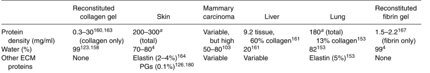
TABLE 1. Typical values for somein vivoandin vitromatrix properties.
Reconstituted Mammary Reconstituted
collagen gel Skin carcinoma Liver Lung fibrin gel
Protein 0.3–30160,163 200–300a Variable, 9.2 tissue, 180a(total) 1.5–2.2167
density (mg/ml) (collagen only) (total) but high 60% collagen161 13% collagen153 (fibrin only)
Water (%) 99123,158 70–804 50–80103 20161 82153 994
Other ECM None Elastin (2–4%)164 Variable Variable Elastin (5%)153 None
proteins PGs (0.1%)126,180
aComputed from percentage of water, assuming total tissue density of 1 g/ml.
ductal formation,19,147tumor cell72,129,174and macrophage migration,27,59,70 and many others. Cells bind to
colla-gen via various integrins that match multiple binding se-quences on the surface of the molecule.201 Pore size and
fiber diameter can be tuned in a modest range by al-tering the collagen concentration or pH during gelation, but large changes in pH are not possible when prepar-ing samples with cells suspended in the soluble collagen solution.160 When the solution gels, the individual
colla-gen monomers condense and are crosslinked laterally to form large fibers, but these larger fibers are not crosslinked into a gel—thus they fall into the class of physical gels217
because the fibers are merely entangled instead of cova-lently bound.84,203 The ability of collagen fibers to slide and slip with respect to each other will be highlighted later. Collagen gels can be crosslinked via glutaraldehyde148and by glycation,79 although glutaraldehyde is toxic to cells
(and thus cannot be used when suspending the cells within the gel) and glycation can take weeks. Reconstituted colla-gen gels, therefore, are mechanically weak but biologically compatible with many cell types, and can serve as an in
vitro environment for short-term studies or an initial
scaf-fold that will be remodeled by cells inside for long-term studies.
Fibrin is also commonly used in 3D cultures. As the primary component of a healing wound and a biologically active growth matrix for remodeling and regeneration, it clots into a quick-forming seal that is the body’s first re-sponse to tissue damage (for a detailed review on fibrin chemistry, see Mosesson et al.).138Thus, it is commonly
used for in vitro studies of various types of cell migration, angiogenesis and gel contraction due to its role in wound healing,200thrombosis,183macrophage migration,40and its importance in tumor angiogenesis.52,181 It has the advan-tage that mechanical properties and network architecture are tunable to a greater extent than those of collagen by varying its composition (i.e., relative amounts of fibrinogen, thrombin, and Ca2+).167,189Furthermore, it forms a useful
matrix into which fusion proteins (such as growth factors) can be attached to the matrix via the clotting transglutam-inase factor XIIIa.168 Cells must proteolytically degrade
the dense fibrin mesh by releasing plasmin activators or MMPs (matrix metalloproteinases) in order to successfully migrate;97,117,162 thus, fibrin is a useful matrix to study
protease-dependent cell migration. The structural architec-tures of typical in vitro gels made of type I collagen and fibrin as seen by confocal reflectance microscopy are shown in Fig. 2.
FIGURE 2. Collagen and fibrin gel morphology as seen via confocal reflectance microscopy. Single slice confocal reflectance images of (A) 2.5 mg/ml collagen gel and (B) fibrin gel with 2.96 mg/ml fibrinogen. The collagen fibers are on average longer, thicker, and not as straight. Bar= 20 µm.
Gels can also be made from elastin, although these are typically used for zymography;8 fibronectin,195 which is used as a coating for 2D cell attachment or as a supplement to collagen gels; and hyaluronan,45which is often used in in vitro studies of cell–cartilage interactions. Alginate, a
nat-urally occurring polysaccharide, forms a gel when divalent cations are added to the aqueous sodium alginate solution and is often used to study chondrocyte behavior;83 since
it is minimally adhesive for cells, it often has RGD bind-ing sequences added to create a scaffold with specifically selected adhesion properties.166Other materials have been
developed in recent years to specifically control mechanical properties, density, porosity, pore size, adhesion site speci-ficity and density for tissue engineering applications, but the effect of these materials on the mechanobiology of cells embedded within them has not yet been studied in detail. This review is limited to results from experiments with type I collagen and fibrin gels, which are the most commonly used 3D in vitro matrices for studying the mechanobiology of many interstitial cells like fibroblasts.
Matrix Architecture of Collagen and Fibrin
A surprisingly broad range of values have been reported for fiber diameter and mesh size, which are the two key determinants of matrix architecture in collagen and fibrin gels (see Table 2). Possible reasons for the discrepancies include (1) the methods used to obtain these measurements can introduce artifacts, and (2) the matrix architecture de-pends on its composition and conditions of gelation (e.g., temperature, pH, ionic strength, etc). Many estimates of fiber diameter and mesh size came from various forms of electron microscopy, which allows for nanometer resolu-tion; however, the fixation and dehydration required for standard electron microscopy techniques can collapse the highly hydrated mesh as well as dehydrate the fibers which can themselves be highly porous.4 Electron microscopy
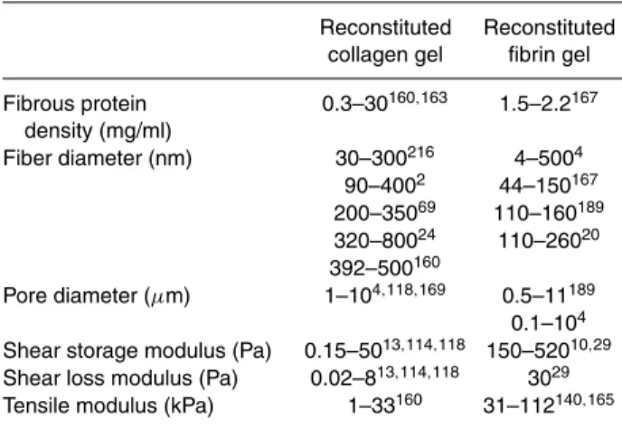
TABLE 2. Typical mechanical properties of simple in vitro
matrices.
Reconstituted Reconstituted collagen gel fibrin gel
Fibrous protein 0.3–30160,163 1.5–2.2167 density (mg/ml) Fiber diameter (nm) 30–300216 4–5004 90–4002 44–150167 200–35069 110–160189 320–80024 110–26020 392–500160 Pore diameter (µm) 1–104,118,169 0.5–11189 0.1–104 Shear storage modulus (Pa) 0.15–5013,114,118 150–52010,29 Shear loss modulus (Pa) 0.02–813,114,118 3029 Tensile modulus (kPa) 1–33160 31–112140,165
techniques which use quick-frozen samples can yield re-sults similar to those found with confocal microscopic ob-servations of fully hydrated gels,205but the sublimation of frozen water can lead to artifacts from the salt left behind (Mark Johnson, personal communication). Confocal reflec-tion microscopy may over-estimate fiber diameter because light reflecting from the fiber edges suffers from in-plane diffraction artifacts,69and may over-estimate the number of
fibers in a given plane due to diffraction along the optical axis of the microscope.109Interfiber spacing or pore
diam-eters of 5µm (see Table 2) may seem large in comparison to cell diameters of approximately 20µm, or compared to the fibers shown in Figs. 2 and 3. However, calculating the number of intersections of a 20µm diameter spherical cell with a 3D cubic lattice of fibers spaced 5µm apart yields the surprising result that the sphere will intersect the fibers at approximately 40 places, depending on fiber diameter (unpublished data).
In reconstituted collagen matrices, the fiber diameter and fiber spacing depends on collagen concentration as well as the pH and ionic strength of the environment in which the gel is forming;220 increasing pH in the range of 6.0–9.0
decreases the average fiber diameter from 500 to 392 nm, while increasing the collagen concentration increases fiber density but does not significantly affect fiber diameter.160
For fibrin matrices, the more complex chemistry of fib-rin yields more variables that regulate fiber diameter dur-ing clottdur-ing, includdur-ing fibrinogen concentration, thrombin concentration, CaCl2concentration, the presence of active
factor XIII, and ionic strength.4,167 Fiber spacing is not typically reported when fiber diameters are obtained using electron microscopy due to the collapse of the mesh during preparation. However, average fiber spacing in collagen gels has been estimated at between 5 and 10µm169using
a density theory developed by Fanti and Glandt,64 which
is within an order of magnitude of the pore size estimated from diffusion studies.169 Saltzman also notes that
aver-age fiber spacing in collaver-agen only depends weakly on the concentration of the gel; it decreases as 1/√c.169 Fibrin
gel spacing may also be larger than previously thought; recent confocal measurements that also put average fiber spacing in a fibrin gel at between 5 and 10µm.189Electron
microscopy measurements on fibrin clots yielded average fiber spacing of between 0.1 and 0.5µm (calculated from Ryan et al.167). Clearly, more work is needed to definitively determine the fiber spacing in 3D meshes without prepa-ration artifacts, but recent evidence suggests that the pore size of most reconstituted collagen and fibrin matrices are on the order of several microns.
Bulk Mechanical Properties
The mechanical properties of fibrous tissues depend on both the strength of the fibers that make up the tis-sue and the organization and architecture of those fibers.
FIGURE 3. Local remodeling of the extracellular matrix near cells. Shown are fibroblasts in a fibrin gel (2 mg/ml fibrinogen); green shows f-actin (phalloidin), red shows the reflectance of the fibrin fibers. Note the close engagement between the f-actin CSK and the ECM fibers, and the higher density of fibers immediately between cells. Image credit: C.P. Ng, Northwestern University.
For many tissues with a dense and well-organized matrix placed under tension, fiber organization governs the low strain response (or “toe region,” as the low-modulus por-tion of the stress–strain curve is commonly named), and when the organizational entropy has been expended from the tissue a higher modulus is seen that reflects the fiber strength (controlled by the enthalpy of the molecular bonds in the fiber) until failure is reached (Fig. 4). In real tissues, fibers are organized to best support physiologic loads and provide specific biomechanical functions. For example, in tendon, collagen fibers are organized in uniaxial bundles parallel to the direction of tension such that the toe re-gion (arising here from the “uncrimping” of the collagen fibers) is quite small and most of the functional range is de-pendent on fiber strength,203whereas in skin the collagen
and elastin fibers are randomly oriented in 2D to facilitate 2D stretch.180
In reconstituted collagen and fibrin matrices, response to mechanical strain primarily arises from water move-ment and the reorganization of the fiber architecture; thus, their mechanical behavior is mostly entropic as opposed to enthalpic.76Since the solid fraction of fibers is very low in
these gels (i.e., they usually consist of 99% or more water by mass), the bulk mechanical properties are generally
in-dependent of the fiber strength (e.g., the gels fail at higher strain than the individual fiber failure strain, but lower stress than the fiber failure stress) because only a fraction of the fibers bear substantial loads even near matrix failure. The fiber density, organization, and crosslink density determine the pore size and porosity of the matrix, and the pores in turn govern the hydraulic conductivity (i.e. relative ease for interstitial fluid movement), which controls the transient response to deformation as water enters or leaves the inter-stitial space. Compressive loads in hydrogels are initially borne by the fluid phase, and since the fluid volume fraction is so high, the hydraulic conductivity is high resulting in weak resistance to compression.
Under tension, the fibers in the matrices align as orga-nizational entropy is removed and the structure becomes more compact as the fibrous network collapses.206 Since
reconstituted collagen matrices are much weaker in ten-sion than their individual collagen fibers—10 kPa vs. 100– 1000 MPa65,160—the low tensile strength of most collagen
gels must be due to fiber rearrangement via bending or sliding.114Quasi-static elasticity tests yield very little
ap-preciable stored energy when samples are loaded in a tensile fashion.148Rheological shear tests can instead be used to
FIGURE 4. Fiber organization regimevs.fiber strength regime in gel deformation. (A) In an unstrained gel, the fibers are re-laxed. (B) Initially as the gel is strained, a high compliance behavior is observed as entropy is removed from the system (“toe region”). (C) As the strain continues to increase, load is transferred to the fibers themselves. Some fibers (thick gray) lose entropy as they begin to bear loads via the strength of their intermolecular bonds (enthalpy) until those bonds fail.
of their higher sensitivity, and can even detect the changes in stiffness as a collagen or fibrin gel forms.67,167 Under
shear loading, the fibers rearrange and do not necessarily come under significant amounts of tension; therefore, they cannot support significant loads. This explains the 1000-fold difference between the tensile and shear moduli of a reconstituted collagen gel (Table 2).
Unlike collagen gels, in which fibers are entangled with weak hydrogen bonds,85,171 fibrin gels are
cova-lently crosslinked by the transglutaminase factor XIII.138
Crosslinking fibers in a hydrogel reduces their entropy, or ability to absorb deflection via fiber rearrangement, by con-straining relative fiber movement more than in entangled physical gels. This crosslinking has a major effect on the bulk elastic properties; for example, fibrin gels made with an inhibitor to the crosslinker factor XIII show a three-fold decrease in the storage modulus compared to those made without the inhibitor.167
The freedom of movement of the fibers in the gel, and the resulting dissipation of stress, implies that mechani-cal stresses imposed on 3D gels do not induce uniform strain fields within the matrix, which is described in the next section and which may have important implication for cell response to mechanical stress in 3D vs. 2D en-vironments. Cells plated onto surfaces do not generally experience this difference between local and global stress because most surfaces used in vitro do not have freedom of relative motion. This effect on the local distribution of strain around a cell may be one important reason why dimension-ality is a key regulator of cellular response to mechanical input.
Non-Affine Mechanical Behavior
Many elastic materials we have familiarity with in daily life deform on the microscale in an affine way with the macroscale deformation—that is, the strain is equal on all scales and deformation is continuous throughout the volume.152However, as introduced earlier, fibrous matrices
with low fiber volume fraction and crosslink density such as those used as in vitro scaffolds can act quite differently. In these materials, the freedom for fibers to bend, buckle and slip when an external mechanical load is applied im-plies that the strain on any given fiber within that material may not match that of the bulk matrix due to the resulting dissipation of stress; the local and bulk deflections will be non-affine, and the strains will not be equal in all locations (Fig. 5).35,76,89The relative contributions of fiber buckling, slippage, and bending to overall stress dissipation depend on the fiber architecture and type of stress applied, although very few examples have been examined in the context of mechanobiology. Fiber slippage has been identified as a possible source of stress dissipation in tissues with bun-dles of aligned fibers in close contact,133 but in random
3D fibrous materials some analyses suggest that slippage will only be important if the fibers are short.39,44Chandran
and Barocas show that fiber bending is more likely than fiber slippage at points where two fibers meet in a colla-gen gel.34 Regardless of the mechanism of stress
dissipa-tion, the local details of non-affine network deformation for a specific hydrogel are difficult to predict or model theoretically, although a recent computational study has highlighted the importance of matrix microstructure in the
FIGURE 5. Non-affine deformation. Three fibers from “before stretch” are shown (in dark gray) in overlay in the “after stretch” panel. Points on individual fibers are tracked from “before” to “after” stretch using red arrows.
mechanical response of fibrous gels.1In short, non-affine mechanical behavior of the ECM implies that the informa-tion the cell receives about its local environment may not necessarily be correlated to its global environment.
One way to remove mechanical freedom and make a loosely crosslinked matrix more likely to deflect in a bulk-affine fashion is to anchor it to a surface and allow the cells to exert tension on the matrix, thereby decreasing the orga-nizational entropy. Afterwards, when a stress is applied to this matrix (either internally or externally), its mechanical response becomes more bulk-affine in nature. This method, commonly referred to as “pre-stressing” the matrix, is fre-quently used in studying fibroblast mechanics,28,81although
stress is not really stored either by the cells or by the fibers.84
Since the local response of pre-stressed gels is more likely to be affine with the bulk response, the mechanical informa-tion that the cell gathers about its local environment more closely reflects the global environment.
Another transition between the two regimes—
continuum-like deformations in a highly crosslinked gel and non-affine network deformations in a sparsely crosslinked gel—has been recently explored using an actin-scruin model by Gardel et al.76, which expands on similar
findings by Tseng and Wirtz.199 Actin polymerizes into
fibers that are crosslinked by scruin (an actin-binding pro-tein) in a manner similar to the crosslinking of fibrin by factor XIII. Gardel and colleagues observe two clearly de-fined types of networks; networks with relatively dense crosslinks that stiffen under strain, and networks with sparse crosslinks that do not stiffen under strain. Furthermore, in the sparse crosslink regime, the elastic modulus depends only weakly on the ratio of crosslinks to fibers, whereas in the regime of strain-stiffening networks, the elastic mod-ulus depends strongly on the crosslink ratio. The sparse crosslink results are interpreted as non-affine network be-havior, which exists below a critical crosslink density ratio. These results suggest that a key parameter for determining the response of a network to a mechanical load is the relative density of crosslinks to fibers.
Recent work on the deformation of semiflexible polymer networks has also explored the transition from affine to non-affine deformation as a function of crosslink density and filament rigidity. Xu et al. showed experimentally,221
followed by Head and colleagues computationally,89 that non-affine network deformation becomes increasingly affine as the crosslink density increases or as the fibers are made more resistant to bending. Together with Gardel’s experimental findings, this demonstrates that the key to non-affine network deformation is the ability of the system of individual fibers to bend, and that non-affine deformation can arise even in the absence of energy-dissipative events (e.g., fiber slippage).
The ability of fibers to bend, and thus yield non-affine network deformation behavior, is typically not duplicated in the 2D surfaces used for mechanobiology experiments.
Surfaces such as collagen-coated silicone87,95or
matrigel-laminated polyacrylamide66 do not deform in a non-affine
fashion, but instead deform in patterns that can be reduced with minimal ambiguity to a smooth vector field;32 i.e.,
they deform as continua. This implies that the detailed in-formation about cell response to mechanical forces gained in these experiments may not necessarily apply to cells embedded in loosely crosslinked gels.
Indeed, one of the major challenges in mechanobiology is to better characterize cell strain vs. bulk strain in various 3D systems undergoing mechanical perturbations so that mechanisms of cell response can be better investigated. The actual cytoskeletal strain profile of a cell embedded in a 3D gel relative to that of the bulk gel has not yet been measured. Computational and theoretical methods can give us insight into the mechanisms that result in non-affine deformation within these matrices, but they cannot yet pre-dict the local strain profile of a cell in a given 3D matrix. In summary, non-affine behavior may lead to inhomogeneous strain and, as we shall discuss next, local stress shielding or strain shielding, but this is a nascent area of experimental investigation and is difficult both to model theoretically and explore computationally.
PHYSICAL COUPLING OF CELLS TO THE ECM Cytoskeleton–ECM Connections
Integrins are transmembrane proteins that couple the intracellular and extracellular structural protein networks: they connect the cytoskeleton to the ECM. They are het-erodimeric receptors that are specialized in both the ex-tracellular ligands with which they interact as well as the cytoskeletal network components with which they interface intracellularly (for a recent review of integrin biochemistry, see Arnaout et al.5). They consist of anα subunit and β sub-unit. Eighteenα subunits are known and 8 β subunits have been identified, but only 24 uniqueαβ integrin pairs have been found. Theβ subunit is thought to be the main effec-tor in signaling and binding to cytoplasmic proteins, while theα subunits modulate the binding reactions, perhaps by changing ligand binding efficiency via intermolecular in-teraction with the cytoplasmic part of theβ subunit.125,175
Specific integrin–substrate interactions depend to varying degrees on the identity of the integrin subunits, the type of binding sites on the substrate or ligand, the presence of various divalent cations, and the integrin activation state,5
highlighting the extent to which the cell–matrix coupling can be tuned to accommodate specific matrix conditions. Integrin activation involves binding of proteins on the cy-toplasmic side of the membrane, allowing the integrin to act not only as an “outside-in” signaling molecule alert-ing the cell of attachment, but also as a selectively ac-tive cell/ECM anchor that can be modulated by binding of cytoplasmic proteins. Integrins appear to be the first
element in the signaling cascade that allows detection of external forces (i.e., mechanotransduction), and they have been shown to play key roles in determining cell shape,113 levels of cytoskeleton tension,210 and other types of cell
response to mechanical stimuli.25,36
Inside the cell membrane, multi-molecule complexes called cell–matrix adhesions connect the cytoskeleton to the activated integrins and are critically important in transmitting mechanical signals from the ECM (reviewed in Cukierman et al.).46 A large array of proteins are
thought to be a part of focal adhesions under various conditions, and still more molecules apparently interact with adhesion complex proteins to transduce signals or alter function of the cell/ECM coupling, including notably focal adhesion kinase (FAK), the G-protein Rho, and extracellular signal-regulated kinase (ERK) (see Friedl and Br¨ocker68 and Ingber101 for reviews). At least four types of cell–matrix adhesions exist that differ in terms of their molecular identities, forces they can exert, and the extracellular substrate required for them to form (reviewed in Cukierman et al.).46 The first two types of adhesions,
focal contacts (which mature into focal adhesions) and focal adhesions, are perhaps the best known and can exist on single component surfaces. They are capable of transmitting forces from a cell to its substrate in both cell contraction and cell migration,9,124,179and focal adhesions
must be maintained in a tensile state to survive.108Fibrillar
adhesions require fibronectin, an additional ECM protein and a mechanically compliant substrate,86,108 and are thought to be important primarily in the organization and coupling of cell-surface fibronectin to the ECM. Finally, 3D-matrix adhesions were recently described as a unique adhesion requiring a mechanically compliant 3D substrate as well as fibronectin and an additional ECM protein.47
However, little is known about the ability of these adhesions to bear load and their relationship to cell migration.
Most of the existing work characterizing the molecu-lar players in focal adhesions and their capacity for trans-mitting force has been done on 2D substrates, but cur-rently it appears that focal adhesions in 2D have largely the same features as focal adhesions on single component 3D matrices.47Focal adhesions also require approximately
10 min to mature and stabilize,15 and they appear to
un-dergo aging and growth that may or may not be related to their ability to transmit forces and modulate adhesion.96 Balaban et al. found that the amount of force exerted by a fibroblast on an elastic substrate scaled linearly with focal adhesion area (5.5 nN/µm2up to 6µm2) as measured by the presence of vinculin.9 Other investigators have noted
the presence of small focal adhesions, less than 1µm2 in
area, that can support forces greater than 50 nN.191It seems
likely that the fibrillar adhesions and their association with thin actin fibers might have some influence on cell shape and cell motility, whereas focal contacts/adhesions primar-ily exist to transduce large loads and remodel the matrix.
Locomotive cells often have smaller focal adhesions than cells in a contractile phenotype, and some cells that mi-grate extremely rapidly have no detectable focal contacts at all.59,70
The cytoskeleton to which these various adhesion com-plexes engage is comprised of three variably active poly-mer meshes: the actin filament network, the microtubule network, and the intermediate filament network. All three networks exist as polymerized fibers in dynamic equilib-rium with monomers or polymerized subcomponents in the cytosol.22,91,142These cytoskeletal networks are
biochemi-cally distinct and seem to have different primary functions, but significant cross-talk and interactions exist between them.61,213
The actin cytoskeleton has been implicated in cell shape maintenance,36 cell migration,151 cell force generation,55 and mechanotransduction.100,130As the actin cytoskeleton is bound to the ECM via focal adhesions and integrins, it can only react to mechanical inputs delivered along those paths with the possible exception of fluid shear stress. This force may be transduced via membrane fluidity-induced G protein activation or stimulation of actin complexes at the cell membrane.31,49 Interestingly, the same non-affine
de-formation behavior seen with the ECM (Section 2) can also be observed in the cytoskeleton itself.94 This implies that
cytoskeletal strain is heterogeneous and non-affine, which may introduce a possible mechanism for localizing cy-toskeletal remodeling or adhesion reinforcement processes only in the areas they are needed. Because mechanical strain on the cytoskeleton has been shown to alter the binding affinity of paxillin,170a focal adhesion protein, it is possible that the non-affine deformation of the cytoskeleton limits binding of paxillin only to areas near the integrins under strain. In an affine network, stressing one area would result in uniform strain across the network, but observations of cytoskeletal reinforcement in response to mechanical force suggest that the reinforcement is not global, but local.38
It is therefore possible that the non-affine network defor-mation behavior is critical for regulating local cytoskeletal remodeling.
Until recently, the intermediate filament network was thought to be kinetically stable in comparison with the actin cytoskeleton or the microtubules, and its role in cel-lular mechanobiology appears to be restricted to spatial separation and stabilization of cellular compartments and intercellular contacts like desmosomes.178However, recent work has demonstrated that the intermediate filament net-work is more dynamic than previously appreciated and ap-pears to be involved in cell migration, although a major role in mechanotransduction or force generation has yet to be found (reviewed in Helfand et al.).91 Microtubules
are extremely dynamic and active in certain phases of cel-lular division, and play an important role in generating and maintaining structural polarity in epithelial cells.141
secondary role in force transduction and maintenance of structures like lamellipodia,7,213and help generate the cell polarity required for migration.61 Other cell-specific cy-toskeletal networks include the cytokeratin network in ep-ithelial cells, whose role in cell force generation or mechan-otransduction is still unclear.149,155
Stress and Strain Shielding
The ECM can shield stress or strain from embedded cells in two ways: it can shield stress by the bulk mechanical properties of the cell–matrix composite, and it can shield strain locally by its non-affine behavior. Stress shielding via bulk mechanical properties refers to the normal sharing of load that occurs in any composite material when one component of that material is weaker than the other.105
For example, if cells with an approximate overall shear stiffness of 7.5 Pa208are embedded in a 3D collagen matrix
of shear stiffness 22 Pa,13 the matrix will determine the
overall shear stiffness of the composite (assuming the cell volume fraction is not too high). For any externally applied stress, the matrix typically bears most of the stress such that the cells are subjected to the same strain as that of the matrix, and since the strain on the cells is reduced, so is the stress they carry—they are “stress shielded” by the stronger bulk material surrounding them. In a well-organized tissue like a tendon, cells are aligned along fibers that are up to seven orders of magnitude stiffer than the cells114,208such that the
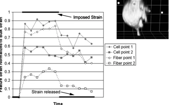
cell bears almost no load at all. These scenarios depend on the different stiffness of cells and their matrix, but also on homogenous material properties throughout both materials. The matrix may also shield strain locally by its non-affine deformation behavior as discussed in Section 2. The degree to which local matrix deformation is non-affine, and the effect this has on a cell embedded within the matrix, depends on the crosslink density of the matrix, the cell– matrix adhesion density and the stiffness of both the cell and fibers. In a matrix with low adhesion site density and small degree of crosslinking, one might speculate that the bulk ECM strain will be greater than that of the cell. This potential non-affine strain coupling is only relevant in 3D systems because of the additional degrees of freedom for relative fiber motion. As mentioned earlier, the actual cou-pling of cell and ECM strains in such non-affine networks has not been explored, although preliminary observations of a cell undergoing solid shear in a 3D collagen gel support the non-affine arguments (Fig. 6). As entropy is expended with increasing levels of tensile strain, the non-affine case is less likely to occur, as discussed in the previous sec-tion. On the other extreme, in a matrix with high adhesion site density and high degree of crosslinking, i.e. a matrix with affine deformation behavior, it is more likely that the cell strain will mimic that of the bulk matrix. Thus, in a loosely crosslinked fibrous matrix, local strain shielding is a function of local fiber organization and the density of
mechanical coupling between the cell and the fibers local to it.
Furthermore, the cell can actively control its local me-chanical environment by remodeling its local matrix, and can do so in a way that either increases or decreases sensi-tivity to local mechanical conditions. Increasing local fiber density, either via matrix contraction or collagen fibrilloge-nesis, and crosslinking surrounding fibers will strengthen the mechanical “cocoon” around a cell (Fig. 3) and shield it from bulk matrix stresses and strains. Alternatively, con-tracting the matrix into an aligned structure will increase the sensitivity of the cell to global stresses.28 The
abil-ity of cells to only sense local mechanical events matches their abilities to only alter the local mechanical milieu, but with large numbers of cells acting in concert these local changes can add up to large scale tissue changes. Thus, the spatially complex mechanical system we have described is dynamic. Such matrix remodeling events typically oc-cur within a time frame of hours,82 requiring that they
be considered in experiments that run for long periods of time.
COMPARISON OF CELL BEHAVIOR IN 3D VERSUS 2D ENVIRONMENTS
Differences between cells in 3D and 2D environments have been noted in overall morphology, matrix adhesion, manifestation of actin stress fibers, mechanisms of stress generation, cellular migration strategies, gene and protein expression, and response to flow, among others. While the mechanisms underlying these differences remain unclear, some investigators speculate that not only the dimension-ality but also the compliance of the matrix are key factors driving the difference in cell response.46
Cell Morphology and Adhesion
Since the early 1970s, investigators have observed mor-phological differences between fibroblasts sparsely plated on glass and those embedded within 3D collagen matri-ces, favoring the spindle or stellate shape when in 3D and a spread cell with prominent cellular extensions in 2D.57
In a recent comprehensive study of the effect of matrix composition and dimensionality on cellular adhesion and morphology, Cukierman et al. found that fibroblasts as-sume different morphologies in relaxed 3D collagen gels and within matrices reconstituted from explanted tissue digests, varying between flattened, spindled, stellate, and dendritic shapes.47Grinnell and co-investigators have noted that only the dendritic phenotype of fibroblasts is expressed in relaxed gels with sparse cell density.190In contrast, cells in high cell density matrices supplied with pro-contractility growth factors move from a dendritic phenotype to a stellate
FIGURE 6. Non-affine deformation of a cell in a strained collagen matrix. GFP-actin transfected fibroblasts were placed in a collagen gel and viewed live under confocal microscopy. Points on the cell (gray) and matrix fibers (white) were tracked while a solid shear of 25% was imposed on the gel using a micromanipulator. Measurements were normalized to the imposed deflection (i.e., the strain that would have existed in an affine system). Spots close to each other on the same cell (see inset) did not move in an affine fashion with the gel or with each other. Moreover, considerable energy (entropy) was lost in the deformation as shown by the lack of elastic recovery after the bulk strain was reversed.
or bipolar morphology within 4 h, as they begin to substan-tially contract their matrix.
Along with cellular morphology, the type of adhe-sions generated by fibroblasts also depends on the type and dimensionality of substrate. For example, 3D-matrix adhesions have only been found on cells within mechani-cally compliant 3D matrices comprised of multiple types of ECM proteins. These 3D-matrix adhesions were much more elongated than focal or fibrillar adhesions from single-component 3D matrices or 2D substrates, and were the only adhesions to include paxillin, vinculin, FAK, phosphotyro-sine, α-actinin, activated β1 integrin and α5 integrin all co-localized.47
The mechanism by which the cell senses the dimension-ality of its substrate, and thus expresses the appropriate adhesions and morphology, is not clear. One possibility is that the cell can integrate global cues around its entire surface and thereby “sense” the spatial organization of ac-tivated adhesions. Another possibility is that the formation of 3D-matrix adhesions requires a molecularly complex substrate with a compliance typically not seen in in vitro 2D surface experiments or in mechanically flattened 3D matrices. In fact, when cell-derived 3D matrices were stiff-ened by crosslinking with glutaraldehyde, cells plated into that matrix did not form 3D-matrix adhesions but instead formed focal adhesion-like structures,47suggesting that
me-chanical cues may be the key input that informs the cell of the dimensional status of the matrix.
Fibroblast, Macrophage, and Tumor Cell Migration
Cell migration is a complex orchestration of events, in-cluding cellular shape change, adhesion and de-adhesion to the extracellular substrate, and exertion of force on the sub-strate via adhesion complexes. Since all of these processes differ in 3D vs. 2D environments, it is not surprising that cell migration through 3D matrices differs greatly from that on 2D substrates. In 2D, adhesion is critical for modulating migration via force transmission to the substrate in hap-tokinetic cell migration.68Briefly, the cell forms adhesions
forward of the main cell body, generates traction forces to move the cell body, and then detaches adhesions at the rear of the cell. Other migration mechanisms must exist, however, as some non-adherent cell types fail to migrate on 2D collagen surfaces at all, but are capable of migration inside a 3D collagen matrix.27
An inverse correlation between adhesion strength and migration speed has been shown in numerous 2D studies. For example, in a recent study of fibroblast migration on 2D surfaces, Katz et al. showed that the cells migrated more slowly and formed focal contacts on immobilized fi-bronectin surfaces, as compared to surfaces with adsorbed, but not covalently linked, fibronectin where fibrillar con-tacts were used for adhesion.108This study also found that
cells plated on immobilized fibronectin formed focal con-tacts while cells plated on mobile (adsorbed but not cova-lently linked) fibronectin formed fibrillar contacts. There is some evidence that adhesion strength and cell migration
speed might have a similar inverse relationship in 3D contexts115as that seen in 2D on fibronectin,108 collagen-coated polyacrylamide,219and decades ago on glass42 sur-faces.
In the absence of other cues, cells tend to migrate in the direction in which they are already aligned, a phenomenon called contact guidance.197Therefore, one consequence of
mechanical stress aligning the fibers of a gel is that the mechanical force can affect the direction of cell migration within that gel. Contact guidance has already been exploited to direct neurite outgrowth in vitro.51Fibroblasts in
partic-ular have long been known to align along aligned collagen fibers,17 but the extent to which aligned fibers direct cell
process extension and migration, instead of the reverse, is still under investigation. Cells may experience competition between mechanical signals and biochemical ones, as seen in a recent study that place a chemotactic gradient in oppo-sition to a contact guidance field.26In this case, the chemo-tactic gradient appeared to dominate the cell response, but the fibers of the 3D environment still influenced cell alignment.
In one of the few studies to directly compare 3D and 2D migration rates, Friedl’s group showed that on hyaluronic acid (HA)-coated 2D surfaces, MV3 melanoma cells dis-played increasing migration rates with increasing concen-trations of HA, but showed no dependence of migration rate with HA concentration in 3D collagen matrices.129
They suggest that this is likely due to the fact that HA forms a low modulus gel in a 3D hydrated lattice, which contrasts with the stiff-branched strands that bind to a 2D surface. This difference in the physical conformation of an ECM molecule (HA, in this case) illustrates the subtle but powerful effect dimensionality can have on the physical environment around a cell.
Another recent study showed that non-muscle myosin heavy chain II-B (NMHC II-B) was required for mi-gration and fiber translocation in 3D collagen matrices, but was not required for migration on 2D surfaces.132
In 2D, this molecule could be shown to participate in a “hand-over-hand” lamellipodial mechanism for locally re-tracting collagen fibers towards the cell. NMHC II-B−/− cells only contracted floating collagen matrices one-third as much as control cells, but gel contraction could be fully restored by transfecting the NMHC II-B−/− cells with GFP NMHC II-B. Furthermore, the internal cellu-lar localization of NMHC II-B depended on whether or not the cell was plated into a 3D matrix or onto a 2D substrate. These findings further demonstrate that cells on 2D surfaces vs. in 3D matrices use different mecha-nisms for exerting force, even when they both appear to be using a haptokinetic strategy for interacting with their substrate.
Some immune cell types including T lymphocytes and dendritic cells migrate through 3D collagen matrices with-out adhesion mediated byβ1integrin, the primary
collagen-binding integrin.72 Indeed, although neutrophils can
mi-grate inside 3D collagen gels in an integrin-independent fashion,70 they are apparently unable to migrate on 2D
collagen-coated surfaces altogether.27 This indicates an
amoeboid mode of migration—that is, movement through the formation of pseudopods in matrix pores and subse-quent “pulling” of the cell body via cell shape changes through the pore—which is clearly irrelevant for migra-tion on 2D substrates. Even some larger tumor cells that normally use integrin- and protease-dependent migration strategies in 3D gels can continue to migrate via this strat-egy when MMP and other protease activity is blocked. In a recent study, fibrosarcoma and carcinoma cells were sub-jected to a protease inhibitor cocktail in an in vitro 3D col-lagen gel migration model, blocking the normal proteolytic migration strategy of these cells.218 However, the cells’
migration speed remained essentially unchanged because they switched to a new migration strategy that was marked by a lack of the normal indicators of proteolytic migra-tion. The investigators observed noβ1 integrin clustering, no association between MT1-MMP andβ1integrin, and a diffuse cortical actin CSK. Similar results were obtained with cells that were pre-treated with the protease inhibitor cocktail and then injected into murine dermal tissue and observed intravitally.218The emergence of this mutability
in migration strategy appears to be specific to the 3D en-vironment, since adhesion-independent migration has not been observed to date in 2D migration studies.
Thus, we see that the haptokinetic cell migration strat-egy, long considered the primary means of mesenchymal cell locomotion, is not the only means available to cells moving through a 3D matrix. Since the 3D matrix is con-siderably more compliant than many of the surfaces used to study fibroblast migration to date, more studies of cell migration in 3D are needed to supplement our understand-ing of adhesion-based 2D cell locomotion. The realm of non-adherent cell migration in 3D matrices is only now emerging, and it raises questions not only about mecha-nisms for generating the shape changes and internal forces required for this kind of cell migration, but also whether the cells undergoing this type of movement are still able to sense the mechanical state of the matrix, or even whether it is important that they do so.
STRESS GENERATION AND THE ROLE OF SUBSTRATE STIFFNESS IN STRESS
FIBER FORMATION
Fibroblasts and other contractile cells compact collagen gels in both 2D and 3D via force generation during cell mi-gration, called traction,88or via the actin-myosin machinery of the cell, called contraction.131Due to its importance in wound healing and fibrosis, the generation of forces by cells within a matrix was one of the first areas of 3D cell mechanobiology. Early experiments showed that fibroblasts
suspended in free-floating collagen compacted the gels to a small fraction of their original size in a cell density-dependent manner.16Further investigation revealed that the fibroblasts were not degrading the matrix significantly but were instead reorganizing existing collagen fibers.84 This
was shown to be a two-step process whereby the cells re-arranged the fibers and then non-covalently stabilized the reorganized state. This process depended on an intact actin cytoskeleton and the ability of the cells to adhere to the collagen fibers, which required serum in the cell media.84
However, another study showed that if the cells were an-chored and allowed to generate tension in the gel (for at least 24 h), then on release the cells could contract the gel more quickly than they had contracted unanchored gels, apparently using actin-myosin machinery.136 For a num-ber of years, investigators attempted to reconcile the two modes of fibroblast-mediated gel compaction—traction and contraction—into a single mechanism. However, in recent years it has become clear that the cells are responding in two distinct fashions determined by the local extracellular compliance,6 and that these two compaction mechanisms
are indeed distinct and, to some extent, independent. Cells exert forces in an anchored gel using the actin-myosin machinery, which manifests visually as stress fibers.30,120,214 Stress fibers are large bundles of
polymer-ized actin filaments heavily crosslinked byα-actinin119and
often containα-smooth muscle actin (α-SMA).182Larger
stress fibers are indicative, all things being equal, of larger forces.30Anchored gel assays have been used to probe the differentiation of fibroblasts into myofibroblasts—a con-tractile cell type important for mid-term wound healing responses and responsible for tissue fibrogenesis (reviewed in Tomasek et al.).194
Traction: Stress Fiber-Independent Force Generation
Cells in floating or unanchored gels exert forces on the ECM via a stress fiber-independent mechanism. In vivo in dermal tissue, fibroblasts behave similarly to those in re-laxed in vitro gels in that they do not exhibit stress fibers.96It
was suggested over 20 years ago that stress fibers required tension for their formation,30 and numerous experiments
have sustained that view by finding that gel compaction in relaxed collagen gels does not involve stress fibers.55,80,114
Recent work is continuing to focus on elucidating the mech-anisms by which cells generate forces without stress fibers. Cells in relaxed 3D collagen gels exert forces to contract that matrix without the presence of stress fibers, and do not require fibronectin to interact with their 3D matrix. Vanni and colleagues showed that cells within 3D collagen gels can contract those gels without visible stress fibers and, using GFP-α-actinin and YFP-β-actin to visualize actin fibers near the cell membrane, showed that forces were in-stead generated by the cortical CSK.202They estimated this force at 60 nN for a single pseudopod cell process which
is relatively small compared to forces generated by stress fibers, but significant as it can clearly reorganize the local collagen matrix. A contracting gel assay was used by an-other group to postulate a per-cell traction force parameter of 2.73 × 10⁻⁴dyn/cm2,13 and to develop the anisotropic
biphasic theory for modeling cell and gel mechanics.14
Fibroblasts also appear to be able to switch between contractile (stress positive) and migratory (stress fiber-negative) phenotypes based on their mechanical environ-ment. Using a contracting rod assay that compacts in the radial dimension but not axially, Shreiber, Barocas, and Tranquillo showed that once fibroblasts had compacted the matrix, they reverted to a migratory phenotype.179This
sug-gests that reversion and cell migration out of a wound might be the reason for the absence of myofibroblasts in a wound after it is closed.194The appearance of this wound–healing-like behavior in a system without inflammatory factors or immune cells demonstrated the importance of mechanical cues in this critical cellular function.
Compaction: Stress Fiber-Dependent Force Generation
Although cells can contract collagen gels without stress fibers, the cell must express the contractile machinery of stress fibers to exert large forces on its substrate, through focal adhesions or 3D-matrix adhesions. Whileα-SMA is not strictly necessary for stress fiber formation, the appear-ance of α-SMA in stress fibers is used as a marker for the emergence of the myofibroblast cell phenotype and is associated with highly contractile cells.96When incubated
in media with exogenous TGF-β1(which promotesα-SMA
expression), fibroblasts contract their collagen matrix more strongly in both floating and anchored matrices, enhanced stress fiber formation in anchored matrices,6and separately
fibroblasts transfected withα-SMA have been shown to contract their matrices to a greater extent than those trans-fected withα-cardiac- or β- or γ -cytoplasmic actin.95These increases in stress fibers were not due to an increase in total actin, but specifically an increase inα-SMA as measured by Western blots. Blocking the adhesion of the cells to the substrate by using an anti-β1integrin antibody blocked the upregulation ofα-SMA, even in the presence of TGF-β1. All these results indicate that TGF-β1is a potent regulator of α-SMA expression and cell contractility, but that this regulation is dependent on the fibroblast being anchored to a matrix that is under tension.
The differentiation of fibroblasts to myofibroblasts is now understood to be dependent on adhesion to the ma-trix, presence of TGF-β (whether exogenous or endoge-nously upregulated by mechanical stress), presence of cel-lular fibronectin,177and tension in the extracellular matrix (reviewed in Hinz and Gabbiani).96Ehrlich and Rajaratnam showed that from an initial population of fibroblasts, the cells differentiate into myofibroblasts in areas of a collagen gel under stress while those in stress-free regions do not
differentiate (i.e., they do not form stress fibers).55Although
the mechanisms by which the fibroblasts sense the tension in the matrix and the exact biochemical mechanisms by which all the steps of differentiation and contraction are carried out remain unknown, it seems clear that stress fiber-mediated matrix contraction is a result of myofibroblast differentiation, and that cells can also contract a relaxed matrix in the absence of stress fibers via a non stress fiber-regulated mechanism.
Cell Response to Substrate Compliance or Stored Stresses
It is now clear that cell behavior is extremely sensitive to the compliance or stiffness of their matrix. A recent study by Yeung et al. on fibroblasts and endothelial cells on collagen-or fibronectin-coated polyacrylamide gels showed a sharp transition between the absence of actin stress fibers for cells on soft gels to expression of actin stress fibers when the 2D substrate stiffness was increased above 3 kPa.223
The differences between the morphology and stress fiber expression vanish if the cells are allowed to make cell– cell contact; under these conditions all cells express stress fibers. Both fibroblasts and endothelial cells spread more fully and quickly on stiffer matrices, but neutrophils proved insensitive to substrate stiffness, spreading with equal effi-cacy on surfaces spanning the range of stiffnesses studied. Another study on the spreading of smooth muscle cells on collagen-coated polyacrylamide showed similar trends, but also demonstrated that the slight over-expression of actin via the expression of a GFP-fusion actin can push a cell into a stress fiber regime even on moderately soft gels (about 1 kPa).58 Taken together, these findings demonstrate that
cells with mechanical functions are sensitive to mechani-cal cues that other cells (such as neutrophils) completely ignore.
Although little is known about the effects of 3D matrix stiffness on embedded cells, there is evidence to suggest that cells will respond in 3D in a similar fashion to that in the 2D surface studies described earlier. Fibroblasts cul-tured in anchored 3D collagen matrices develop a stellate morphology in contrast to the dendritic morphology seen in cells in relaxed 3D collagen matrices,81and they use
dif-ferent signaling pathways to regulate gel contraction after the release of the gel from its anchoring points.82 It seems
likely that the local fiber compliance determines whether or not cells can express stress fibers and focal adhesions, and therefore determines the cells’ ability to remodel the collagen mesh via contraction.190
Fibroblasts can also respond to substrate compliance by altering the mechanical environment via the generation of new matrix components, proteolysis of existing matrix, and communication with neighboring cells. Lack of ma-trix stiffness, for example, leads fibroblasts to downreg-ulate collagen XII mRNA and protein expression.196 In 3D vs. 2D cultures, fibroblasts increase the ratio of
colla-gen degradation to production,146 increase production of
decorin and dermatan sulfate glycosaminoglycan,121 and
express increased levels of VEGF and HGF in 3D vs. 2D culture,154although whether these responses are due to the
dimensionality of the environment or the substrate stiffness remains undetermined.
Although fibroblasts and smooth muscle cells are in-vestigated more frequently in mechanobiology assays due to their known sensitivity to mechanical stimuli, other cells have been shown to respond morphologically to differences in the mechanical stiffness of their environment. In a recent investigation, Flanagan et al. showed that neurons branch more frequently on soft matrigel-coated polyacrylamide gels than stiff ones.66 As noted earlier, the soft gel might
more closely mimic the 3D environment than a stiffer gel if that low stiffness is due to entropy in the matrix architecture.
CELL RESPONSE TO 3D MECHANICAL ENVIRONMENTS
Fibroblast Response to Tension and Compression
As observed by many investigators, stress fibers are rarely seen in fibroblasts in vivo except in tissues which un-dergo significant and consistent mechanical loading, such as tendon.157 Fibroblasts can also express stress fibers
when wound healing or fibrotic pathways are activated which causes them to differentiate into myofibroblasts.81,194
In short-term cultures in relaxed 3D collagen gels, this differentiation pathway can be induced by exogenous TGF-β,86,190but stress fibers can also appear after
fibrob-lasts are allowed to contract the gel for several days95which
decreases organizational entropy and thus allows matrix tension to be sustained.160
The alignment of cells in a gel with imposed stress has been repeatedly investigated, but it remains unclear how much of the cell alignment is a passive process and how much is an active cellular response to the force. Girton, Barocas, and Tranquillo recently showed that both collagen fibers and cells in a 3D collagen gel aligned perpendicular to the applied compression.78 In contrast, cells and fibers
under tension align parallel to the direction of stress.54 It
is possible that the fibers were aligning passively under the load, as seen in acellular collagen samples,160and that the
cells were merely reporting fiber alignment, but the low levels of maximum strain reported during this experiment (0.2%) make that seem unlikely.54 In this case, it seems
more likely that the cells are aligning in order to deposit more collagen along this direction,18and thus shield
them-selves from strain.54 Indeed, when cells are exposed to external stress, they can reinforce their local environment by producing more ECM. This has been seen in 2D, where cyclic strain induced smooth muscle cells to synthesize collagen, hyaluronate, and chondroitin sulfate,122as well as in 3D, where stretch increased collagen XII mRNA and
protein expression in fibroblasts.196The evidence for both
cellular alignment and increased matrix synthesis in fibrob-lasts subjected to imposed 3D matrix stress suggests that the cells are reinforcing their environment in the most effi-cient way possible: by concentrating reinforcement in the principle direction of strain.54,96
Externally applied stretch can also be shown to have a direct and immediate effect on cytoskeletal networks. Sawada and Sheetz prepared cell-free cytoskeletal networks by plating mouse fibroblasts onto collagen-coated silicone and then destroying the cell membrane with a detergent wash. These networks bound exogenously supplied paxillin at the focal adhesions when the networks were stretched,170
and binding of paxillin was inhibited by phenylarsine oxide just as in vivo. When Costa and colleagues grew aortic en-dothelial cells on pre-stretched fibronectin-coated silicone substrates and then allowed those substrates to suddenly contract, they found that the response of the cytoskeleton varied considerably depending on the rate of shortening.41
If shortening occurred very quickly (5% s−1 or greater), the actin cytoskeleton buckled with a very short periodicity (well below its persistence length) and then completely dis-assembled within 5 s, only to re-form 60 s later. Shortening the cells on a slower time scale yielded no such dramatic effects; in fact, no effect could be seen at all if the shorten-ing strain rate was 0.5% s−1or less. Taken together, these findings indicate that the cytoskeleton is a very early link in the mechanotransduction chain that leads to the changes in gene expression, cell differentiation, migration and align-ment discussed earlier. However, it is clear that in non-affine 3D networks, the cytoskeleton may be buffered from such direct and powerful mechanical input.
Finally, compressive stresses in 3D culture systems have been recently explored in a 3D tissue engineered airway wall model, which mimics the airway mucosa with lung fi-broblasts and epithelial cells.37In this model, both static and dynamic compressive stresses upregulated matrix remod-eling proteins and induced myofibroblast differentiation, among other effects.
Cell Response to Interstitial Flow
Interstitial fluid flow, which refers to fluid flow through the 3D matrix (as opposed to flow across the surface of cells, as in endothelial cell response to fluid shear stress), exists between the blood and lymphatic capillaries as lymph forms185as well as in dynamically compressed tissues like
bone and cartilage.83,116 Furthermore, because
inflamma-tion and angiogenesis both involve factors that increase vessel permeability (i.e., vascular endothelial growth fac-tor or VEGF), interstitial flow is locally increased during wound healing and inflammation, and may be enhanced from angiogenic tumors into the peripheral stroma. Cellu-lar response to interstitial flow is an emerging area of 3D mechanobiology research, due to its potential importance
in cartilage remodeling83 and bone development,116
mi-crovascular development and remodeling,92,143tumor drug
delivery,104lymphangiogenesis,21 and in vasoconstriction
responses.212Interstitial flow (through the medial layer of
the blood vessel wall) has also been implicated in the vas-cular remodeling that leads to intimal hyperplasia.188,192
It was recently shown that fibroblasts subjected to in-terstitial flow while embedded in a 3D matrix aligned perpendicularly to the direction of flow145 and differen-tiated into myofibroblasts as indicated by the upregula-tion ofα-smooth muscle actin via autocrine upregulation of TGF-β1.144 Another recent study showed that blood
and lymphatic endothelial cells subjected to interstitial flow responded very differently under 3D vs. 2D fluid shear stress in distinct cell-type-dependent fashions.143
Lymphatic endothelial cells formed large vacuoles and long extensions when subjected to interstitial flow for 6 days, while blood endothelial cells formed extensive multi-cellular structures, many of which contained lumen. Blood endothelial cells also tended to aggregate in static control cultures whereas lymphatic endothelial cells re-mained viable as isolated single cells spread through the 3D collagen gel. These differences in behavior, both be-tween cell types and bebe-tween static vs. flow conditions, may be due to their differing environments and functions
in vivo.143
Tada and Tarbell have shown in a theoretical model that even smooth muscle cells (SMCs) normally considered shielded from blood flow may hypothetically be affected by transmural flow to a surprising degree: the fenestral pore system may focus the small amount of transmural flow onto SMCs in the vessel wall and subject them to appreciable shear stress.187 Indeed, recent in vivo work
by the same group demonstrates a correlation between the myogenic response of SMCs and transmural fluid fil-tration through the arteriolar wall.111Cell culture experi-ments comparing the effects of shear stress on SMCs in 3D collagen gels vs. plated on 2D collagen-coated surfaces showed the SMCs to be much less responsive to flow in 3D than in 2D, but both still significantly increased pro-duction of prostaglandins compared to those under static conditions.212This experiment provides evidence that the
3D environment may either buffer fluid shear stress on cells or increase their tolerance to shear; however, more work is needed to elucidate the mechanisms underlying these differences.
Interstitial flow differs from 2D flow in many ways. First and most obviously, the 2D case involves shear stress on the luminal side with matrix adhesion on the abluminal side; this means that the stress is not necessarily transmitted to the cell through the ECM, e.g. via integrin receptors. However, recent work is revealing the importance of the glycocalyx, a layer of membrane-bound macromolecules on the apical cell surface, in how the cell senses shear stress. It was re-cently suggested that the glycocalyx projects into the fluid