S Y M P O S I U M : A B J S C A R L T . B R I G H T O N W O R K S H O P O N H I P P R E S E R V A T I O N S U R G E R Y
Pelvic Morphology Differs in Rotation and Obliquity Between
Developmental Dysplasia of the Hip and Retroversion
Moritz Tannast MD, Peter Pfannebecker MD, Joseph M. Schwab MD, Christoph E. Albers MD, Klaus A. Siebenrock MD, Lorenz Bu¨chler MD
Published online: 14 July 2012
Ó The Association of Bone and Joint Surgeons1 2012
Abstract
Background Developmental dysplasia of the hip (DDH) and acetabular retroversion represent distinct acetabular pathomorphologies. Both are associated with alterations in pelvic morphology. In cases where direct radiographic assessment of the acetabulum is difficult or impossible or in mixed cases of DDH and retroversion, additional indi-rect pelvimetric parameters would help identify the major underlying structural abnormality.
Questions/Purposes We asked: How does DDH and retroversion differ with respect to rotation and coronal obliquity as measured by the pelvic width index, anterior inferior iliac spine (AIIS) sign, ilioischial angle, and obturator index? And what is the predictive value of each variable in detecting acetabular retroversion?
Methods We reviewed AP pelvis radiographs for 51 dysplastic and 51 retroverted hips. Dysplasia was diagnosed
based on a lateral center-edge angle of less than 20° and an acetabular index of greater than 14°. Retroversion was diagnosed based on a lateral center-edge angle of greater than 25° and concomitant presence of the crossover/ischial spine/posterior wall signs. We calculated sensitivity, spec-ificity, and area under the receiver operating characteristic (ROC) curve for each variable used to diagnose acetabular retroversion.
Results We found a lower pelvic width index, higher prevalence of the AIIS sign, higher ilioischial angle, and lower obturator index in acetabular retroversion. The entire innominate bone is internally rotated in DDH and exter-nally rotated in retroversion. The areas under the ROC curve were 0.969 (pelvic width index), 0.776 (AIIS sign), 0.971 (ilioischial angle), and 0.925 (obturator index). Conclusions Pelvic morphology is associated with ace-tabular pathomorphology. Our measurements, except the AIIS sign, are indirect indicators of acetabular retroversion. The data suggest they can be used when the acetabular rim is not clearly visible and retroversion is not obvious. Level of Evidence Level III, diagnostic study. See Guidelines for Authors for a complete description of levels of evidence.
Introduction
Developmental dysplasia of the hip (DDH) and acetabular retroversion represent distinct acetabular pathomorpholo-gies. In DDH, the acetabulum is undercovered and often excessively anteverted [7,8,16, 21]. In acetabular retro-version, coverage is typically excessive, especially anteriorly [18]. Both conditions lead to distinct pathome-chanical problems: a dysplastic acetabulum leads to static overload of the articular cartilage while a retroverted One or more of the authors (JMS) has received fellowship funding
from the Maurice E. Mu¨ller Foundation of North America. Each author certifies that he or she, or a member of his or her immediate family, has no commercial associations (eg, consultancies, stock ownership, equity interest, patent/licensing arrangements, etc) that might pose a conflict of interest in connection with the submitted article.
All ICMJE Conflict of Interest Forms for authors and Clinical Orthopaedics and Related Research editors and board members are on file with the publication and can be viewed on request. Each author certifies that his or her institution approved the human protocol for this investigation, that all investigations were conducted in conformity with ethical principles of research, and that informed consent for participation in the study was obtained.
M. Tannast (&), P. Pfannebecker, J. M. Schwab,
C. E. Albers, K. A. Siebenrock, L. Bu¨chler Department of Orthopaedic Surgery, Murtenstrasse, Inselspital, University of Bern, 3010 Bern, Switzerland e-mail: [email protected]
Clin Orthop Relat Res (2012) 470:3297–3305
DOI 10.1007/s11999-012-2473-6
and Related Research
®
acetabulum leads to dynamic impingement between the prominent anterosuperior aspect of the acetabulum and the femoral head-neck junction.
Based on a number of reports and consistent with our clinical observation, there are indicators that the pathologic acetabular morphology in DDH and retroversion is asso-ciated with alterations in pelvic morphology [1–7,14,15,
26]. Kojima et al. [14,15], using three-dimensional (3D) CT, showed a decrease in the transverse diameter of the pelvic inlet and outlet in DDH, suggesting a general nar-rowing of the bony pelvis. Fujii et al. [7] used CT to examine rotational deformity of the innominate bone in DDH. They noted internal rotation of the innominate bone in dysplastic hips compared with controls. In addition, hips with acetabular retroversion, both in the control group (n = 4) and in the setting of DDH (n = 9), had externally rotated innominate bones. While this suggests opposing rotational abnormalities of the innominate bone between acetabular retroversion and DDH, these two groups were not compared exclusively.
By directly comparing the pelvic anatomy in DDH and acetabular retroversion, we can establish association between pelvic morphology and acetabular pathomor-phology. This would allow us to diagnose acetabular pathomorphology in cases where direct radiographic assessment of the acetabulum is difficult or impossible. Furthermore, using radiographic measurements on plain AP pelvis radiographs would eliminate the need for addi-tional, expensive, and sometimes radiation-intense imaging studies.
Our goal, therefore, was to devise a method for assess-ing pelvic morphology in the presence of acetabular dysplasia and DDH using plain radiographs. To this end, we developed four radiographic parameters: pelvic width index, radiographic appearance of the anterior inferior iliac spine (AIIS) sign, ilioischial angle, and obturator index. For each of these four key measurement variables, we
asked two questions: (1) How do these variables differ between dysplastic hips and retroverted hips? And (2) what is the predictive value of each variable to detect acetabular retroversion?
Patients and Methods
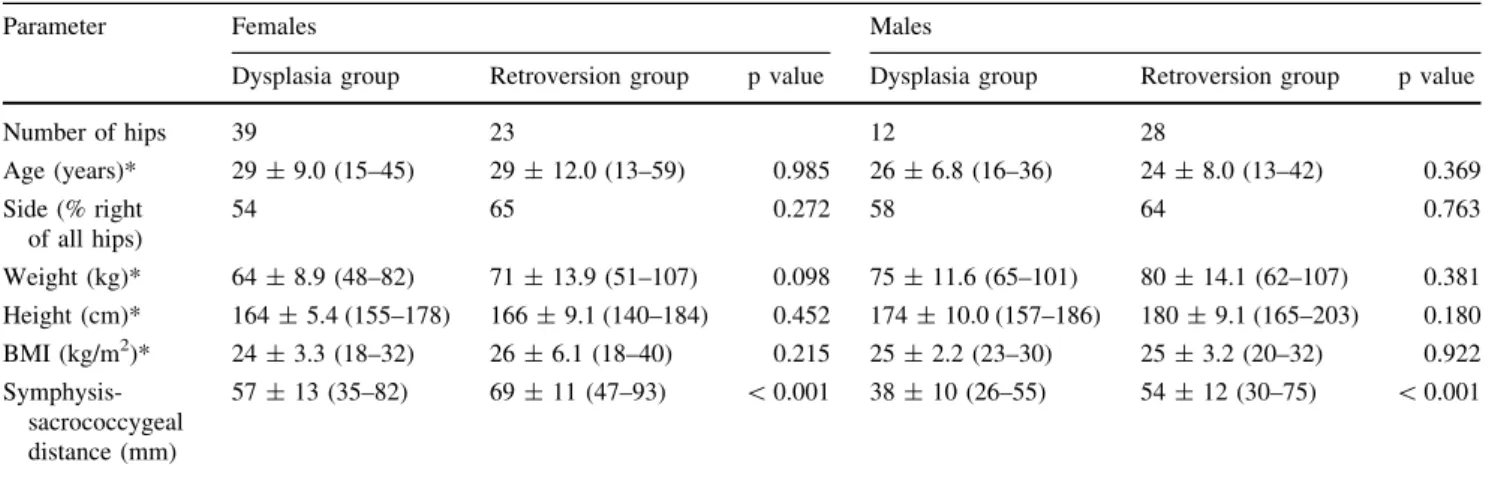
We performed a retrospective comparative study of radiographic pelvic morphology between dysplastic and retroverted hips. We reviewed radiographs from our insti-tutional database of patients who underwent periacetabular osteotomy (PAO) between March 2004 and March 2011 (n = 86) or surgical hip dislocation between March 2007 and March 2011 (n = 300). We defined the dysplasia group using the following parameters: lateral center-edge angle of less than 20° [17] and acetabular index of more than 14° [25]. We identified a total of 86 patients fitting these inclusion criteria. We excluded patients undergoing anteverting PAO (n = 17), incomplete radiographic doc-umentation (n = 10), or previous hip or pelvis surgery (n = 8). This left 51 patients in the dysplasia group. We defined the retroversion group as a lateral center-edge angle of more than 25° and the presence of three radio-graphic signs: the crossover sign [10,18], ischial spine sign [12,13], and posterior wall sign [18,23]. We identified a total of 317 patients fitting these inclusion criteria. We excluded patients in whom not all three radiographic signs were positive (n = 170) and with a previous history of hip trauma (n = 39), previous hip or pelvis surgery (n = 25), incomplete radiographic documentation (n = 14), Legg-Calve´-Perthes disease (n = 10), and protrusio acetabuli (n = 8). This left 51 patients in the retroversion group. The two groups were similar in terms of age, affected side, weight, height, and BMI (Table1). Our hospital’s institu-tional review board approved the study.
Table 1. Demographic information
Parameter Females Males
Dysplasia group Retroversion group p value Dysplasia group Retroversion group p value
Number of hips 39 23 12 28 Age (years)* 29 ± 9.0 (15–45) 29 ± 12.0 (13–59) 0.985 26 ± 6.8 (16–36) 24 ± 8.0 (13–42) 0.369 Side (% right of all hips) 54 65 0.272 58 64 0.763 Weight (kg)* 64 ± 8.9 (48–82) 71 ± 13.9 (51–107) 0.098 75 ± 11.6 (65–101) 80 ± 14.1 (62–107) 0.381 Height (cm)* 164 ± 5.4 (155–178) 166 ± 9.1 (140–184) 0.452 174 ± 10.0 (157–186) 180 ± 9.1 (165–203) 0.180 BMI (kg/m2)* 24 ± 3.3 (18–32) 26 ± 6.1 (18–40) 0.215 25 ± 2.2 (23–30) 25 ± 3.2 (20–32) 0.922 Symphysis-sacrococcygeal distance (mm) 57 ± 13 (35–82) 69 ± 11 (47–93) \ 0.001 38 ± 10 (26–55) 54 ± 12 (30–75) \ 0.001
Since a substantial number of dysplastic hips have a positive crossover sign [16], we further subdivided our DDH group into those with and without a crossover sign to evaluate how this affected our measurements. Twenty of the 39 (51%) hips in the female DDH group and 11 of the 12 (92%) hips in the male DDH group had a positive crossover sign.
A standardized radiographic technique was performed for all reviewed AP pelvis radiographs. All radiographs were performed in the supine position. A film focus dis-tance of 1.2 m was used with the beam centered between the pubic symphysis and a line connecting the anterior superior iliac spine with the pelvis in neutral rotation [12,
22,23]. The longitudinal rotation of the pelvis was verified as correct when the tip of the coccyx was in line with pubic symphysis. Images were not specifically corrected for tilt; however, we recorded the distance between the superior pubic symphysis and sacrococcygeal junction for each patient (Table1).
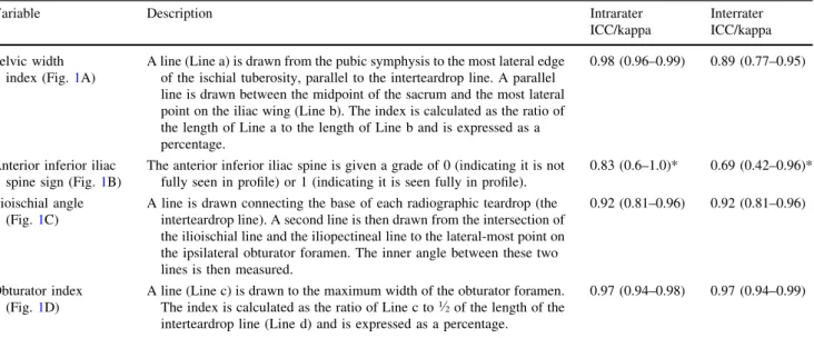
Two of us (PP, CEA) independently evaluated the following key measurement variables on each radiograph (Table2): (1) pelvic width index (Fig.1A), (2) presence of the AIIS sign (Fig.1B), (3) ilioischial angle (Fig. 1C), and (4) obturator index (Fig.1D). The two observers each performed two separate sets of measurements on deidentified preoperative plain AP pelvis radiographs. The two sets of measurements were performed a mini-mum of 7 days apart. Inter- and intraobserver reliabilities were tested for each parameter using the intraclass cor-relation coefficient (ICC) for continuous value measurements and the kappa value for ordinal measure-ments (Table2).
Table 2. Descriptions and reliability/reproducibility of the four key measurement variables
Variable Description Intrarater
ICC/kappa
Interrater ICC/kappa
Pelvic width
index (Fig.1A)
A line (Line a) is drawn from the pubic symphysis to the most lateral edge of the ischial tuberosity, parallel to the interteardrop line. A parallel line is drawn between the midpoint of the sacrum and the most lateral point on the iliac wing (Line b). The index is calculated as the ratio of the length of Line a to the length of Line b and is expressed as a percentage.
0.98 (0.96–0.99) 0.89 (0.77–0.95)
Anterior inferior iliac
spine sign (Fig.1B)
The anterior inferior iliac spine is given a grade of 0 (indicating it is not fully seen in profile) or 1 (indicating it is seen fully in profile).
0.83 (0.6–1.0)* 0.69 (0.42–0.96)*
Ilioischial angle
(Fig.1C)
A line is drawn connecting the base of each radiographic teardrop (the interteardrop line). A second line is then drawn from the intersection of the ilioischial line and the iliopectineal line to the lateral-most point on the ipsilateral obturator foramen. The inner angle between these two lines is then measured.
0.92 (0.81–0.96) 0.92 (0.81–0.96)
Obturator index
(Fig.1D)
A line (Line c) is drawn to the maximum width of the obturator foramen.
The index is calculated as the ratio of Line c to1.2of the length of the
interteardrop line (Line d) and is expressed as a percentage.
0.97 (0.94–0.98) 0.97 (0.94–0.99)
* The kappa value was calculated for this variable; the ICC was calculated for other remaining variables; values are expressed as mean, with 95% CI in parentheses; ICC = intraclass correlation coefficient.
b a
A
A
c dD
B
C
Fig. 1A–D The diagrams illustrate how to (A) calculate the pelvic
width index (a/b), (B) determine the AIIS sign, (C) determine the ilioischial angle, and (D) calculate the obturator index (c/d). For a
Due to the lack of available comparable data in the lit-erature, a pilot set of measurements was taken on a subset of our patients to perform a power analysis. Based on these, we detected a mean difference of 10% in the pelvic width index with an estimated SD of 10%. Using these numbers, a power analysis was performed and a minimum sample size of 32 hips for each group was required to provide an a of 0.01 and a b of 0.10.
Results from the complete set of measurements were collected and stratified by group and sex. Normal distri-bution was confirmed using the Kolmogorov-Smirnov test. We compared groups using a paired t-test for continuous variables and Fisher’s exact test for categorical variables. To evaluate the overall predictive performance of our key measurement variables, a receiver operating characteristic (ROC) curve was calculated for each variable. We then calculated the sensitivity and specificity of each test based on thresholds detected for the ROC curve.
Results
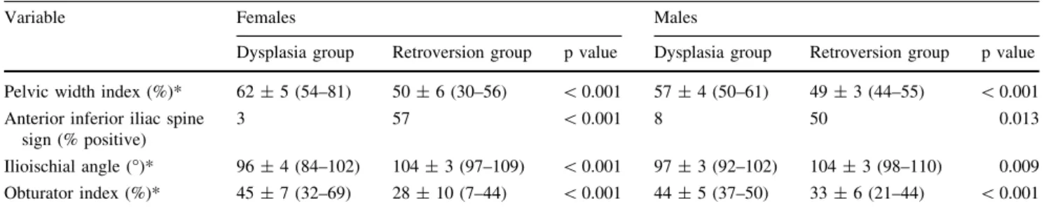
The pelvic width index was smaller (both sexes: p \ 0.001) in the retroversion group than in the dysplastic group (Table3). There was a higher (females: p \ 0.001; males: p = 0.013) prevalence of the AIIS sign in the
retroversion group than in the dysplastic group. The ili-oischial angle was higher (females: p \ 0.001; males: p = 0.009) in the retroversion group than in the dysplastic group. The obturator index was lower (both sexes: p \ 0.001) in the retroversion group than in the dysplastic group. When we subdivided our DDH hips into those with and without a crossover sign, there was no difference between the two groups for females (Table4).
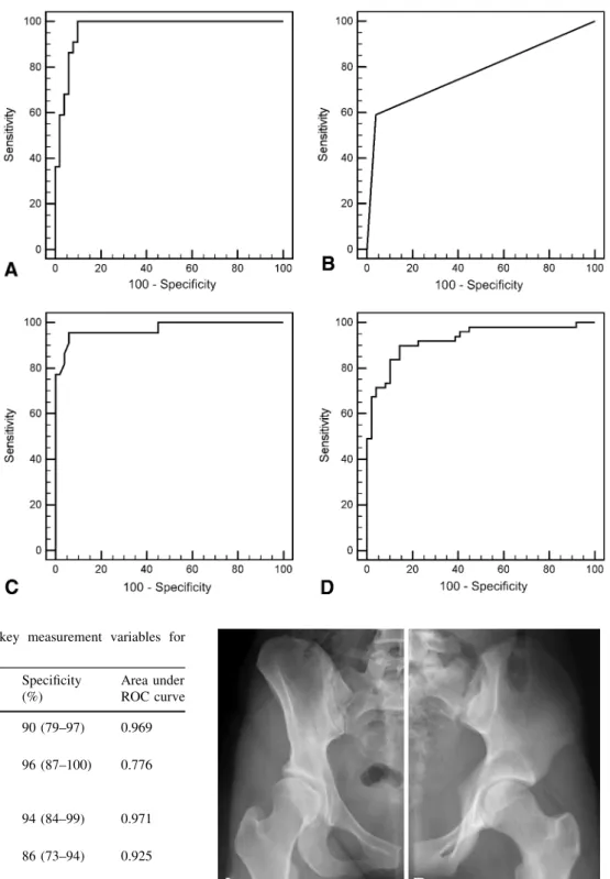
The greatest area under the ROC curve was found for the ilioischial angle with a cutoff of 100° (0.971), followed by the pelvic width index \ 56% (0.969) and the obturator index \ 40% (0.925) (Fig. 2, Table5). Compared to the other key measurement variables, the pelvic width index had the highest sensitivity (100% when pelvic width index \ 56%), and the AIIS sign had the lowest sensitivity (59%). The highest specificity was found in the AIIS sign (96%), and the lowest specificity was found in the obturator index (86%).
Discussion
DDH and acetabular retroversion represent two distinct acetabular pathomorphologies. As these disease processes have become better understood, it is clear they include not just alterations to the acetabulum but also distinct
Table 3. Results comparing the four key measurement variables for the dysplasia and retroversion groups
Variable Females Males
Dysplasia group Retroversion group p value Dysplasia group Retroversion group p value
Pelvic width index (%)* 62 ± 5 (54–81) 50 ± 6 (30–56) \ 0.001 57 ± 4 (50–61) 49 ± 3 (44–55) \ 0.001
Anterior inferior iliac spine sign (% positive)
3 57 \ 0.001 8 50 0.013
Ilioischial angle (°)* 96 ± 4 (84–102) 104 ± 3 (97–109) \ 0.001 97 ± 3 (92–102) 104 ± 3 (98–110) 0.009
Obturator index (%)* 45 ± 7 (32–69) 28 ± 10 (7–44) \ 0.001 44 ± 5 (37–50) 33 ± 6 (21–44) \ 0.001
* Values are expressed as mean ± SD, with the range in parentheses.
Table 4. Results comparing the four key measurement variables for dysplasia group with and without the COS
Variable Females Males
Dysplasia + COS (n = 19)
Dysplasia COS
(n = 20)
p value Dysplasia + COS
(n = 11)
Dysplasia COS
(n = 1)
p value
Pelvic width index (%)* 62 ± 4 (54–73) 63 ± 6 (56–81) 0.689 57 ± 4 (50–61) 52 NA
Anterior inferior iliac spine sign (% positive)
5 0 0.5 9 0 NA
Ilioischial angle (°)* 95 ± 5 (84–102) 96 ± 3 (89–100) 0.274 97 ± 3 (92–102) 98 NA
Obturator index (%)* 44 ± 8 (32–69) 46 ± 5 (35–55) 0.771 44 ± 5 (37–50) 38 NA
morphologic changes to the entire pelvis. To date, no direct comparison has been made between the morphologic pel-vic changes in these two diseases. However, there is anthropologic evidence that hip function and pelvic mor-phology are directly related [9]. We observed a similar pattern of pelvic morphology in our human subjects,
Fig. 2A–D The ROC curves
used to determine the predictive value for (A) the pelvic width index\ 56%, (B) a positive AIIS sign, (C) an ilioischial angle [ 100°, and (D) an obturator
index\40%, are shown.
Table 5. Predictive value of the key measurement variables for
detection of acetabular retroversion
Variable Sensitivity (%) Specificity (%) Area under ROC curve Pelvic width index \ 56% 100 (85–100) 90 (79–97) 0.969 Positive anterior inferior iliac spine sign 59 (36–79) 96 (87–100) 0.776 Ilioischial angle [ 100° 95 (77–100) 94 (84–99) 0.971 Obturator index \ 40% 89 (78–97) 86 (73–94) 0.925
Values are expressed as mean, with 95% CI in parentheses; ROC =
receiver operating characteristic. Fig. 3A–B (A) The right 1.2 of an AP pelvis radiograph of a
dysplastic hip is compared to (B) an AP pelvis radiograph of a left hip with acetabular retroversion. In both radiographs, the coccyx is in line with the pubic symphysis, indicating no pelvic malrotation. In addition, the vertical distance between the symphysis and the sacrococcygeal joint is similar in both patients, indicating no substantially different pelvic tilt. The right hemipelvis appears similar to an obturator oblique view while the left hemipelvis appears similar to an iliac oblique view.
Table 6. Pelvic measurement data Variable Definition Flu ¨ckiger et al. [ 6 ] Kojima et al. [ 14 ] Trousdale et al. [ 26 ] Fujii et al. [ 7 ] Current study Year 2000 2001 2002 2011 2011 Hip morphology DDH (n = 17) DDH (n = 40) DDH (n = 7) DDH (n = 50) DDH (n = 51) Retroversion (n = 51) Imaging modality Radiographs 3D CT MRI CT Radiographs Transverse pelvic inlet (cm)* Widest area of pelvis between iliac arcuate lines 15.4 (15.0–16.1) 12.3 (10.9–13.7) 12.92 (12.6–13.7) NA 14.6 (12.3–19.7) Transverse pelvic expansion (cm)* Bilateral centers of the internal surface of the acetabulum NA 11.3 (9.5–12.8) NA NA NA Pelvic interspinous distance (cm)* Line between the two ischial spines 11.8 (11.4–12.3) 11.2 (9.7–12.4) 11.97 (11.0–13.3) NA 10.7 (7.7–14.3) Transverse pelvic outlet (cm)* Bilateral internal surface of inferior ischial tuberosity 14.2 (13.6–14.5) 12.8 (10.9–14.9) NA NA 13.4 (10.7–17.7) AP inlet (cm)* Posterior-superior pubic symphysis to sacral promontory NA 12.9 (10.7–15.2) 12.54 (11.2–13.9) NA NA AP midpelvis (cm)* Posterior-inferior pubic symphysis to sacrum, passing through midpoint of bispinous line NA 11.6 (9.6–13.3) 12.24 (10.5–14.5) NA NA Sagittal diameter of the pelvic expansion (cm)* S2-S3 vertebral junction and the middle posterior pubic symphysis NA 13.4 (11.1–15.2) NA NA NA Superior iliac angle (° ) Opening angles of the ilium at the anterior superior iliac spine NA NA NA 57.0 ± 6.1 NA Inferior iliac angle (° ) Opening angles of the ilium at the anterior inferior iliac spine NA NA NA 72.0 ± 4.3 NA Ischiopubic angle (° ) Closing angle of the ischiopubic pelvis NA NA NA 30.6 ± 2.7 NA * Values are expressed as mean, with the range in parentheses; values are expressed as mean ± SD; DDH = developmental dysplasia of the hip; NA = not available; 3D = three-dimensional.
specifically associated with their hip disease. Our goal was to directly compare four key measurement variables (pelvic width index, presence of AIIS sign, ilioischial angle, obturator index) and evaluate their predictive value for these two different morphologies.
The major limitation to our study is that we only com-pared two morphologic extremes. Clearly DDH and acetabular retroversion exist on a spectrum, and mixed-type pathology is common [16]. While our key measurement variables show good predictive value and reproducibility between the two extremes, with the exception of DDH with a radiographic crossover sign, we have not explicitly evalu-ated them in the setting of mixed morphology. Second, we were unable to measure the actual rotation of the hemipelvis. CT, and specifically 3D CT, can provide precise measure-ments to determine rotational properties of the pelvis in both the coronal and sagittal planes. However, despite an inability to measure rotation directly, plain radiographs are most frequently the first, and sometimes the only, imaging studies performed on patients seeking treatment for hip disease. While using plain radiographs limits our ability to directly compare our values to previously published rotational measurements performed on CT or MRI [7, 11, 20], we believe the use of an AP pelvis radiograph is faster and more universally accessible. Finally, we did not specifically adjust for tilt when evaluating these radiographs. When we eval-uated each group (DDH and retroversion) subdivided by sex, we saw the difference in means for the symphysis-sacrococcygeal distance was 12 mm in females and 16 mm in males (Table1). This translated to a difference in pelvic tilt of 5° in females and 7° in males [24]. We do not know the extent to which this variation in tilt affected our measure-ment parameters.
Our results support the theory that the entire hemipelvis is involved in both acetabular dysplasia and retroversion. In DDH, the pelvis appears to be internally rotated around a sagittal axis. This leads to the radiographic appearance of the hemipelvis mimicking an obturator oblique view (Fig.3A). Focal acetabular dysplasia has been described in cases of neuromuscular hip disease [8], but our cohort of patients and multiple other reports [2,7, 11,20] confirm the entire hemipelvis is involved. Even in cases of hip dysplasia with a concomitant crossover sign, our mea-surements do not show any difference from those performed in hips without a crossover sign [8]. In acetab-ular retroversion, the hemipelvis appears to be externally rotated around a sagittal axis, leading to the radiographic appearance of an iliac oblique view (Fig.3B). Previously, only the inferior hemipelvis was implicated in acetabular retroversion [13].
As part of our radiographic review protocol, we also measured several standard pelvic measurements that have been reported previously (Table6). The fact that our
standard measurements correlate with the literature increases the validity of our key measurement variables. Therefore, we believe these key measurement variables allow detection of hemipelvis version on a plain AP pelvis radiograph, independent of sex.
Based on the predictive value of each key measurement variable, and independent of sex, a retroverted hip is likely to be present if the pelvic width index is less than 50%, the AIIS outline is clearly visible, the ilioischial angle is greater than 100°, and the obturator index is less than 40%. These guidelines are helpful for several reasons. First, these measurements allow one to infer acetabular mor-phology from existing pelvic mormor-phology, especially when clear radiographic visualization of the acetabulum may be difficult. It has been established that pathomorphologic pelvic changes relate to acetabular disease manifesting at an early age before ossification of the acetabulum is complete [2, 20]. Therefore, it is reasonable to assume a pediatric patient who does not yet display a fully ossified acetabular rim may have occult acetabular retroversion that can be detected by our pelvic measurements (Fig.4). It should be noted, however, we have not specifically studied these measurements in the setting of skeletally immature pelvises. Similarly, in the setting of a THA, where the normal acetabular rim may be obscured by osteophytes, these measurements may help preoperatively identify a retroverted native acetabulum (Fig.5). Finally, these measurements can assist in decision making between reorientation and rim-trimming procedures. For instance, when our four key variables indicate acetabular
Fig. 4A–B (A) A radiograph of the left hip of a child at age 13 years
shows indistinct anterior and posterior acetabular walls, an elevated pelvic width index, positive AIIS sign, elevated ilioischial angle, and decreased obturator index, all indicating likely acetabular retrover-sion. (B) By age 15 years, the acetabular walls have ossified and retroversion is plainly evident.
retroversion with associated pelvic pathomorphology, one should consider acetabular reorientation over a rim-trimming procedure [19]. In our experience, we use these measurements as an adjunct to the standard radiographic parameters to identify the major pathology to avoid inappropriate treatment, but further clinical studies could help clarify an algorithm for using these parameters.
Distinct pelvic morphology is present in both DDH and acetabular retroversion. Based on our measurements, the sagittal rotation and coronal obliquity of the entire innomi-nate bone are directly related to these two acetabular pathomorphologies. We presented indirect, sex-independent pelvimetric parameters indicating DDH and acetabular ret-roversion on an AP pelvis radiograph. Recognition of these parameters will help to understand complex morphology in hips where direct radiographic assessment of the acetabulum is difficult or impossible.
References
1. Ait-Ameur A, Wakim A, Dubousset J, Kalifa G, Adamsbaum C. The AP diameter of the pelvis: a new criterion for continence in the exstrophy complex? Pediatr Radiol. 2001;31:640–645. 2. Albin˜ana J, Morcuende JA, Delgado E, Weinstein SL. Radiologic
pelvic asymmetry in unilateral late-diagnosed developmental dysplasia of the hip. J Pediatr Orthop. 1995;15:753–762.
3. Albin˜ana-Cilveti J, Delgado-Baeza E, Miralles-Flores C. Pelvic deformity in experimental dislocation of the growing hip. Int Orthop. 1992;16:317–321.
4. Delgado-Baeza E, Albin˜ana-Cilveti J, Miralles-Flores C. Why does pelvic deformity occur in experimental dislocation of the growing hip? J Pediatr Orthop. 1992;12:376–383.
5. Dora C, Zurbach J, Hersche O, Ganz R. Pathomorphologic characteristics of posttraumatic acetabular dysplasia. J Orthop Trauma. 2000;14:483–489.
6. Flu¨ckiger G, Eggli S, Kosina J, Ganz R. [Birth after peri-acetabular osteotomy] [in German]. Orthopade. 2000;29:63–67. 7. Fujii M, Nakashima Y, Sato T, Akiyama M, Iwamoto Y. Pelvic
deformity influences acetabular version and coverage in hip dysplasia. Clin Orthop Relat Res. 2011;469:1735–1742. 8. Gugenheim JJ, Gerson LP, Sadler C, Tullos HS. Pathologic
morphology of the acetabulum in paralytic and congenital hip instability. J Pediatr Orthop. 1982;2:397–400.
9. Hogervorst T, Bouma H, de Boer SF, de Vos J. Human hip impingement morphology: an evolutionary explanation. J Bone Joint Surg Br. 2011;93:769–776.
10. Jamali AA, Mladenov K, Meyer DC, Martinez A, Beck M, Ganz R, Leunig M. Anteroposterior pelvic radiographs to assess ace-tabular retroversion: high validity of the ‘‘cross-over-sign.’’ J Orthop Res. 2007;25:758–765.
11. Jia J, Zhang L, Zhao Q, Li L, Liu X. Does medial rotational deformity of the whole pelvis universally exist in unilateral DDH? Arch Orthop Trauma Surg. 2011;131:1383–1388. 12. Kakaty DK, Fischer AF, Hosalkar HS, Siebenrock KA, Tannast
M. The ischial spine sign: does pelvic tilt and rotation matter? Clin Orthop Relat Res. 2010;468:769–774.
13. Kalberer F, Sierra RJ, Madan SS, Ganz R, Leunig M. Ischial spine projection into the pelvis: a new sign for acetabular retro-version. Clin Orthop Relat Res. 2008;466:677–683.
14. Kojima S, Kobayashi S, Saito N, Nawata M, Horiuchi H, Takaoka K. Morphological characteristics of the bony birth canal in patients with developmental dysplasia of the hip (DDH): investi-gation by three-dimensional CT. J Orthop Sci. 2001;6:217–222. 15. Kojima S, Kobayashi S, Saito N, Nawata M, Horiuchi H,
Takaoka K. Three-dimensional computed tomography evaluation of bony birth canal morphologic deformity (small pelvic cavity) after dome pelvic osteotomy for developmental dysplasia of the hip. Am J Obstet Gynecol. 2002;187:1591–1595.
16. Li PL, Ganz R. Morphologic features of congenital acetabular dysplasia: one in six is retroverted. Clin Orthop Relat Res. 2003:245–253.
17. Murphy SB, Ganz R, Mu¨ller ME. The prognosis in untreated dysplasia of the hip: a study of radiographic factors that predict the outcome. J Bone Joint Surg Am. 1995;77:985–989. 18. Reynolds D, Lucas J, Klaue K. Retroversion of the acetabulum: a
cause of hip pain. J Bone Joint Surg Br. 1999;81:281–288. 19. Siebenrock KA, Schoeniger R, Ganz R. Anterior
femoro-acetabular impingement due to femoro-acetabular retroversion: treatment with periacetabular osteotomy. J Bone Joint Surg Am. 2003;85: 278–286.
20. Suzuki S. Deformity of the pelvis in developmental dysplasia of the hip: three-dimensional evaluation by means of magnetic resonance image. J Pediatr Orthop. 1995;15:812–816.
21. Tannast M, Albers CE, Steppacher SD, Siebenrock KA. Hip pain in the young adult. In: Bentley G, ed. European Instructional Lectures. Berlin, Germany: Springer; 2011:141–154.
22. Tannast M, Mistry S, Steppacher SD, Reichenbach S, Langlotz F, Siebenrock KA, Zheng G. Radiographic analysis of femoroace-tabular impingement with Hip2Norm—reliable and validated. J Orthop Res. 2008;26:1199–1205.
23. Tannast M, Siebenrock KA, Anderson SE.
Femoroace-tabular impingement: radiographic diagnosis—what the
radi-Fig. 5 A radiograph of a 56-year old man with left hip degeneration
shows indistinct acetabular walls, an elevated pelvic width index, elevated ilioischial angle, and decreased obturator index. The patient was noted during subsequent THA to have a retroverted acetabulum.
ologist should know. AJR Am J Roentgenol. 2007;188:1540– 1552.
24. Tannast M, Zheng G, Anderegg C, Burckhardt K, Langlotz F, Ganz R, Siebenrock KA. Tilt and rotation correction of acetab-ular version on pelvic radiographs. Clin Orthop Relat Res. 2005;438:182–190.
25. To¨nnis D, Heinecke A. Acetabular and femoral anteversion: relationship with osteoarthritis of the hip. J Bone Joint Surg Am. 1999;81:1747–1770.
26. Trousdale RT, Cabanela ME, Berry DJ, Wenger DE. Magnetic resonance imaging pelvimetry before and after a periacetabular osteotomy. J Bone Joint Surg Am. 2002;84:552–556.