Anne Battisti Didier Tassaux David Bassin Philippe Jolliet
Automatic adjustment of noninvasive pressure
support with a bilevel home ventilator
in patients with acute respiratory failure:
a feasibility study
Received: 11 May 2006 Accepted: 19 January 2007 Published online: 24 February 2007 © Springer-Verlag 2007
Financial support for this study was provided by Resmed in the form of an unrestricted grant
A. Battisti · D. Tassaux · P. Jolliet (u) University Hospital, Department of Intensive Care, 1211 Geneva 14, Switzerland e-mail: [email protected] Tel.: +41-22-3729093 Fax: +41-22-3729105 A. Battisti
University Hospital, Department of Physiotherapy,
1211 Geneva 14, Switzerland D. Tassaux
University Hospital, Department of Anesthesiology,
1211 Geneva 14, Switzerland D. Bassin
ResMed Inc.,
North Ryde, NSW, Australia
Abstract Objective: To test the
feasibility of applying noninvasive ventilation (NIV) using a prototype algorithm implemented in a bilevel ventilation device designed to adjust pressure support (PS) to maintain a clinician-set alveolar ventilation in patients with acute respiratory failure after initial stabilization. Design
and setting: Prospective crossover
interventional study in an intensive care unit, university hospital.
Pa-tients: 19 patients receiving NIV
for acute hypercapnic respiratory failure (13 men, 6 women; mean age 70± 11 years). Methods: The same bilevel ventilator was used with manually adjusted PS and with the automated algorithm (autoPS), set to maintain the same alveolar ventilation as in PS. Sequence (measurements at end of each period): (a) prior to initiating NIV (baseline 1); (b) 45 min with manually set PS; (c) 60 min without NIV; (d) 45 min with autoPS;
(e) 60 min without NIV; (f) 45 min with manually set PS. Results: The magnitude of decrease in PaCO2
and increase in pH with autoPS was comparable to that of conventional PS, with the same alveolar ventila-tion and level of PS. No technical problem occurred in autoPS mode, and no NIV trial had to be discon-tinued because of patient discomfort.
Conclusions: These results suggest
that the alveolar ventilation based automatic control of PS during NIV with a bilevel device is feasible and leads to beneficial effects in patients with acute respiratory failure com-parable to those of manually set PS. Further studies should now explore the potential of this system over longer periods in patients with acute and chronic respiratory failure.
Keywords Noninvasive ventilation ·
Pressure support · Bilevel · Auto-mated modes
Introduction
Patient tolerance to noninvasive ventilation (NIV) is a key element to its success in avoiding intubations in acute respiratory failure (ARF) [1, 2]. Tolerance is highly dependent on the combination of the patient’s spontaneous breathing activity and the ventilator’s action, known as patient-ventilator interaction [3]. The latter in turn depends both on the avoidance of leaks at the mask [4, 5] and on the optimal setting of the ventilator [3, 6]. Titrating the level of pressure support (PS) is one such challenge. Unfortunately, the large number of tasks to be
performed by the clinician in the setting of ARF carries the risk of insufficient attention being given to adjusting the ventilator [7].
Automation of certain ventilator settings can prove helpful in this respect. In intubated patients the automatic titration of PS by an expert-based knowledge system has been shown to improve patient-ventilator interaction [8]. Whether the automatic setting of PS during NIV is feasible remains to be seen, but it might be an attractive option. The purpose of the present study was to test the feasibility of relying on an automated algorithm based on a closed-loop control of PS designed to guarantee a
user-set minimum minute volume on a bilevel ventilator, a type of device which, although originally designed for home ventilation, has also been shown effective in performing NIV in patients with ARF [9, 10]. Findings of this study were presented in abstract form at the 2005 European Respiratory Society annual congress (Eur Respir J 2005 26:444s).
Methods
Patients
The study included 19 patients consecutively admitted to the ICU for acute respiratory failure and in whom NIV was prescribed (13 men, 6 women; mean age 70± 11 years); all patients were hypercapnic upon inclusion (Table 1). Pa-tients were included if they had been stabilized with NIV after admission but had been treated in the ICU for 36 h or longer (mean 29± 6 h) and had received maximally four 4 NIV applications. Exclusion criteria were a very low prob-ability of short-term survival, a do not resuscitate order, the presence of pneumothorax, severe respiratory failure or hemodynamic instability with a high likelihood of im-minent intubation, impaired consciousness or absence of patient cooperation, and the presence of facial lesions pre-cluding the use of NIV. Due to the technical limitations of the bilevel device used patients requiring an FIO2 higher than 0.6 during NIV were also excluded. The protocol was accepted by the ethics committee of our institution. In-formed consent was obtained from all patients.
Table 1 Patients’ clinical characteristics (BMI body mass index, PaCO2arterial partial CO2pressure at study inclusion, Diagnosis main
cause of acute respiratory failure, COPD chronic obstructive pulmonary disease)
Patient no. Age (years) Sex BMI PaCO2(mmHg) Diagnosis
1 58 M 20 63 Empyema
2 76 F 23 65 Kyphoscoliosis
3 82 F 24 48 Pneumonia
4 85 M 28 66 Cardiogenic pulmonary edema
5 79 F 29 55 COPD
6 59 M 38 52 Pneumonia
7 78 M 22 47 Pneumonia
8 83 F 26 57 Cardiogenic pulmonary edema
9 72 M 24 45 Pneumonia 10 56 M 20 69 Pneumonia 11 64 M 26 74 Pneumonia 12 74 M 33 52 COPD 13 73 F 25 72 COPD 14 61 M 28 66 COPD 15 86 F 21 57 Restrictive disease 16 63 M 25 48 COPD 17 78 M 22 51 COPD 18 53 M 34 47 COPD 19 57 M 18 58 COPD Mean± SD 70.3± 10.9 – 25.4± 5.1 57± 9 – Methods
All patients were ventilated with a new bilevel type device (AutoVPAP, ResMed, North Ryde, Australia) allowing two modes of bilevel PS, i.e., manually set and automatic (autoPS). A detailed description of the rules on which autoPS is based is beyond the scope of this contribution, but its basic principle can be summarized as follows. AutoPS aims to guarantee a minimum clinician-set “gross alveolar ventilation” by adjusting the level of PS within manually set boundaries (minimum and maximum inspi-ratory pressures). “Gross alveolar ventilation” (termed “alveolar ventilation” below for the sake of simplicity) is defined to be the portion of minute volume minus anatomical deadspace ventilation. An estimate of the anatomical deadspace is entered by the clinician. For the purpose of this study we used the accepted approximation of 2.2 ml/kg body weight for deadspace [11]. The level of PS is continuously adjusted by comparing the recent alveolar ventilation (a continuously updated estimate obtained by low-pass filtering, related mainly to the ventilation during the two preceding respiratory cycles), with the target alveolar ventilation. This means that PS responds significantly to a change in ventilation within about three breaths. Furthermore, a variable backup rate is provided in the case of apnea. However, as this function is mainly directed at patients on long-term home NIV to deal with central apnea or an inspiratory effort too weak to trigger the ventilator occurring during sleep, this function was disabled in the present study, only the automatic titration of PS being explored. Triggering is conventional,
based on a clinician-set flow threshold. Cycling is largely conventional, with an initial absolute refractory period followed by a threshold based on a clinician-set proportion of peak inspiratory flow, the default setting being 25%.
The difference between manual PS and autoPS on the machine therefore lies in the setting of PS and determina-tion of target alveolar ventiladetermina-tion as defined above, plus the presence of a variable backup rate (which was not tested in this study). FIO2, pressurization slope, and positive end-expiratory pressure (PEEP) are similarly set by the clin-ician in both modes. In both modes the maximum level of inspiratory pressure (i.e., PS + PEEP) in this prototype ventilator is 25 cmH2O.
Design of the study
Prior to the trial, all patients had been initially stabilized with up to four runs of NIV applied by an oronasal mask (adult face mask, Vygon Schweiz, Liebefeld-Bern, Switzerland) with an ICU ventilator (Evita 4 or XL, Drägerwerk, Lübeck, Germany). The indication for NIV was as in our usual practice guidelines, based on published studies [9, 12] requiring that at least two of the following be present: worsening dyspnea over the last 10 days in cases of chronic respiratory failure; respiratory rate greater than 25/min; arterial pH less than 7.35; PaCO2 higher than 50 mmHg (6.6 kPa); PaO2below 50 mmHg (6.6 kPa), determined from arterial blood gas measurements.
All trials with the autoVPAP device were performed by the investigating chest physiotherapist (A. B.). NIV was applied with an oronasal mask (UltraMirage Full Face Mask, ResMed). For the sake of simplicity, given that only the automatic titration of PS was tested in this study, the term “autoPS” is used below for the phase of the trial during which PS was titrated automatically, as opposed to the conventional manually set mode (PS). All trials were sequentially performed in the following order: 45 min manually set PS (PS 1), 45 min autoPS mode (autoPS), and 45 min manual mode (PS 2). To avoid a carry-over effect a 60-min period without NIV was provided between each NIV period and before PS 1. Furthermore, to avoid any interference from the automatic backup rate function in autoPS mode due to apnea, the patients’ level of con-sciousness was continuously monitored during all three NIV phases. If any signs of sleepiness or apnea appeared, the patients were awakened and stimulated. The initial manual settings for PS 1 and PS 2 followed our usual prac-tice guidelines for bilevel NIV: 15 cmH2O PS, 4 cmH2O PEEP, resulting in a PS of 11 cmH2O. Thereafter PS was adjusted to obtain an expired tidal volume of 6–8 ml/kg and a respiratory rate below 30/min, with a minimal leak at the mask. In obstructive patients PEEP was titrated upwards until the number of ineffective inspiratory at-tempts either disappeared or decreased to less than 5/min. Ineffective inspiratory efforts, defined as the presence of
an inspiration initiated by the patient without any response by the ventilator, were detected by careful clinical obser-vation. In nonobstructive patients PEEP was titrated up to 6 cmH2O to maintain pulse-oximetry determined arterial O2saturation (SpO2) at 90% or higher. Cycling was set at 25% and 40% of peak inspiratory flow in nonobstructive and obstructive patients, respectively. Once the initial set-tings were made, no further modification was performed. The initial autoPS settings were made as follows: the target alveolar ventilation period was set as mean minute volume during the PS 1 period minus mean deadspace ventilation, the latter defined as estimated anatomical deadspace× mean respiratory rate during the PS 1 period. The initial PS setting was identical to that set during the PS 1, the upper and lower boundaries being 25 cmH2O (the maximum allowed by the machine) and the level of PEEP, respectively. Thereafter, PS was adjusted automatically to obtain the target alveolar ventilation. Cycling, PEEP, and pressurization slope were the same as those of PS 1.
The following measurements were taken before the start of and at the end of each NIV period (with patients still on NIV): arterial blood gases through an indwelling radial artery catheter, respiratory rate, dyspnea score (Borg visual analog scale; 0 = no dyspnea, 10 = worst pos-sible dyspnea), expired tidal volume and minute-volume (Fleisch no. 2 pneumotachograph, Fleisch, Lausanne, Switzerland; between the Y-piece and the mask), heart rate and arterial blood pressure. During NIV all ventilatory parameters (volume, pressure, flow), leaks at the mask (computed by the device using a mathematical model based on the known characteristics of the mask and the pressure and flow signals measured at the ventilator), pulse oximetry, respiratory and heart rates, and arterial blood pressure were continuously monitored. The reported level of PS for periods PS 1 and PS 2 is that set once the patient was stabilized on the machine, and for the autoPS period it is the mean of all values automatically set by the device during the 45 min trial. Of note, although the autoPS algorithm targets alveolar ventilation, the parameter that we recorded was expired minute-volume, which is reported in the results.
Statistics
Comparisons (SigmaStat 2.0–SPSS Science) between the six time-points were made by analysis of variance, with statistical significance being determined by Fisher’s pro-tected least significance test. Differences with a p-value less than 0.05 were considered statistically significant. All results are expressed as mean ± SD.
Results
The initial ventilator settings by the physiotherapist led to mean PS and PEEP levels of 15.2± 3.5 and
Table 2 Respiratory and hemodynamic parameters [HR heart rate, PS level of pressure support (inspiratory pressure minus PEEP), MAP mean systemic arterial pressure, RR respiratory rate, VE expiratory minute-volume, VTe expired tidal volume, PaCO2
arterial partial pressure of CO2, PaO2 arterial partial pressure of
O2; baseline values measured immediately prior to initiating NIV,
PEEP positive end-expiratory pressure, PS 1 and PS 2: NIV with manually set PS; autoPS NIV with automatic titration of PS
Baseline 1 PS 1 Baseline 2 autoPS Baseline 3 PS 2
RR (b/min) 19.2± 3 16.2± 5 18.8± 5 16.5± 4 18.8± 5 16± 5
HR (b/min) 89± 19 88± 19 92± 18 85± 16 90± 16 87± 15
MAP (mmHg) 75± 16 75± 14 80± 15 77± 13 77± 14 77± 14
Borg scale (points) 2.3± 1.9 1.6± 0.9 1.9± 1.6 1.7± 0.9 1.8± 1.4 1.5± 0.8
VE (l/min) n.a. 12.2± 3.1 n.a. 12.3± 2.8 n.a. 11.2± 3.3
VTe (ml) n.a. 772± 222 n.a. 773± 206 n.a. 704± 197
PS (cmH2O) n.a. 15.2± 3.4 n.a. 15.3± 3.1 n.a. 15.2± 3.4
PEEP (cmH2O) n.a. 4.9± 0.2 n.a. 4.8± 0.3 n.a. 4.8± 0.3
Leaks (l/min) n.a. 1.8± 3.5 n.a. 2.2± 2.8 n.a. 2.1± 4.4
pH 7.37± 0.04 7.39± 0.04∗ 7.37± 0.04 7.42± 0.04∗ 7.38± 0.04 7.43± 0.08
PaO2(mmHg) 77± 14 75± 14 73± 14 73± 9 71± 15 72± 8
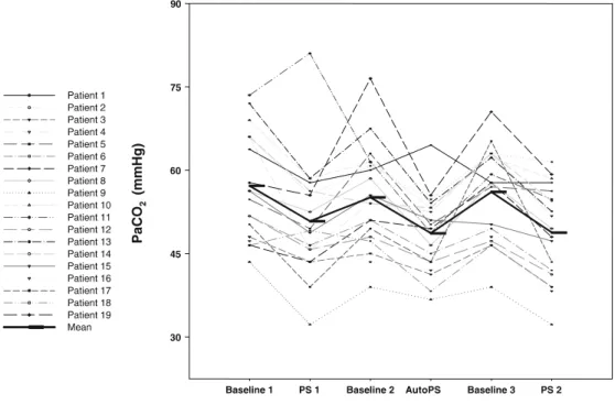
PaCO2(mmHg) 57± 9 51± 10∗ 55± 9 49± 7∗ 56± 8 49± 8∗
∗p < 0.05 vs. corresponding base line (analysis of variance)
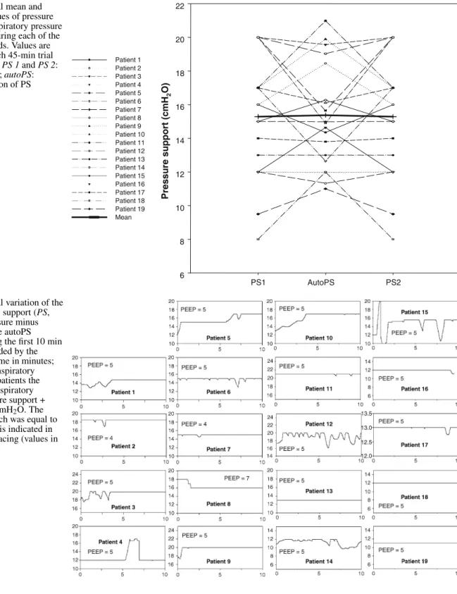
5± 0.5 cmH2O, respectively. The effects of NIV are outlined in Table 2. As shown, PaCO2and pH were com-parably improved by both manual PS and autoPS. Individ-ual changes in PaCO2 are shown in Fig. 1. PaO2 was un-changed by either mode. Respiratory rate decreased from its baseline values with both NIV modes, but the decrease did not reach statistical significance. Dyspnea was un-changed by either NIV mode. Expiratory minute-volume did not differ significantly between the two NIV modes. Overall the mean level of PS set either manually or by autoPS were comparable, although there were differences in some patients (Fig. 2). As seen in Fig. 3, the algorithm
Fig. 1 Individual and mean
variations in PaCO2. PS 1 and
PS 2: manually set PS; autoPS: automatic titration of PS. ∗p < 0.05 vs. corresponding baseline (analysis of variance)
performed little or no change in PS in some patients (e.g., nos. 2, 7, 17, 19), whereas more frequent and/or more marked modifications were made in others (e.g., nos. 12, 14, 15), during the first 10 min of the autoPS trial. Leaks at the mask were kept at a low level, and their magnitude was comparable between manual NIV and autoPS. No apneas or ineffective inspiratory efforts were documented during any of the NIV trials, with either manually set ventilation or autoPS. No change in hemodynamics occurred during NIV (Table 2). No NIV application had to be discontinued because of patient intolerance or technical problems. No patient required tracheal intubation during the study.
Fig. 2 Individual mean and
group mean values of pressure support (PS, inspiratory pressure minus PEEP) during each of the three NIV periods. Values are the mean for each 45-min trial for each patient. PS 1 and PS 2: manually set PS; autoPS: automatic titration of PS
Fig. 3 Individual variation of the
level of pressure support (PS, inspiratory pressure minus PEEP) set by the autoPS algorithm during the first 10 min of NIV, as recorded by the device; x-axis time in minutes; y-axis level of inspiratory pressure. In all patients the upper limit of inspiratory pressure (pressure support + PEEP) was 25 cmH2O. The
lower limit, which was equal to the PEEP level, is indicated in each patient’s tracing (values in cmH2O)
Discussion
The results of the present pilot study demonstrate the fea-sibility of relying on an algorithm based on volumetric closed-loop control to automatically adjust PS on a bilevel device to perform NIV in patients with acute respiratory
failure, with effects comparable to those obtained by the manual setting of PS. Before discussing the implications of our results, several limitations should be outlined. First, this was a feasibility study, aimed at examining whether the concept of automatically titrating PS during NIV was possible in patients with ARF during a 45-min period and
how this compares to a manual setting approach. The du-ration of NIV application was in accord with that of other published trials [13, 14, 15]. However, any extrapolation as to how the system would perform over longer periods remains speculative at this time. Second, our ARF patients had been stabilized on NIV, as reflected by low respiratory rate and dyspnea score. Therefore whether the same results would have been documented in more acutely decompen-sated patients is unknown. Third, the settings in all three NIV applications were performed by one of the investiga-tors (A. B.), which may have biased the results. However, safety issues mandated that an experienced therapist with in-depth knowledge of the autoPS system supervise each NIV application.
Fourth, as stated in the Methods section, the autoPS algorithm is designed not only to titrate PS but also to provide a backup rate in case of apnea. However, only PS setting was tested in the present study, given that no apnea occurred in our patients, as per our intention. Nonetheless apnea can occur both in acute NIV and chronic nocturnal home ventilation, the primary intended area of use for the device, and the performance of the algorithm in this re-spect remains to be evaluated, a path that we are currently exploring. Fifth, bilevel devices have been tailored mostly to the context of chronic long-term ventilation, and the relevance of using such a device in the acute ICU setting may seem questionable. However, the performance of bilevel machines has steadily increased over the years [16, 17, 18], and these devices have been used successfully in ARF [9, 10, 19]. Their much lower cost, relative ease of use, and ability to function without mural medical gas outlets makes them an attractive alternative to ICU ventilators, especially for NIV performed outside of that setting [20]. Finally, the number of patients was small and consisted of a heterogeneous group. However, the case-mix can be deemed representative of that usually found in studies on ARF requiring NIV [21].
At this stage the results of this pilot study show that the concept of automatically titrating PS support during NIV with a bilevel device is feasible, a finding which opens up possibilities in improving patient-ventilator interaction. In-deed, caring for an ICU patient involves considerable mul-titasking which can be a source of poor performance [22, 23]. The latter is all the more unfortunate when
apply-ing NIV since good patient ventilator synchrony, patient comfort, and minimal mask leaks are crucial in ensuring the success of the technique [1, 2]. In particular, exces-sive levels of PS can worsen dynamic hyperinflation in obstructive patients [24], in turn leading to an increase in the number of ineffective inspiratory efforts, heighten the incidence of gastric air intake leading to discomfort, vom-iting and aspiration, and increase the magnitude of leaks at the mask [25]. This being said, there is a theoretical risk that the same excessive levels of PS could be generated by the autoPS. Therefore one must ensure that the expired tidal volume does not exceed 8–9 ml/kg, and also that the inspiratory:expiratory cycling cutoff is set at a higher level (approx. 40–50% of peak inspiratory flow) than the usual 25% default value. This last point aims to minimize delayed cycling, a common occurrence in patients with chronic obstructive pulmonary disease, and one which results in incomplete lung amptying and worsening of dynamic hyperinflation [26]. At the other end of this spectrum very low levels of PS can provide insufficient respiratory muscle assistance [27]. Of note, given that the patients had acute respiratory failure, we chose to set the upper limit at the maximum inspiratory pressure that the device can generate, to avoid insufficient PS. With these settings mean expiratory minute volume was not different during the autoPS period than in either manually set periods.
The proof of concept that automation of PS can im-prove patient-ventilator synchrony and comfort has been illustrated by a study in intubated patients using an expert-based knowledge system [8]. Extending these benefits to NIV is in our view an attractive prospect, although perhaps more challenging due to the presence of mask leaks and the more unstable respiratory conditions of ARF. Nonetheless, the results of this study, using a different ventilator and al-gorithm, suggest that such an approach is possible, bearing in mind that it is a pilot study.
In conclusion, these results demonstrate the feasibility of using an algorithm to automatically adjust PS during NIV with a bilevel device (AutoVPAP) in patients with acute respiratory failure with beneficial effects comparable to those of manually set PS. Further studies should now ex-plore the potential of this approach over longer periods in patients with acute and chronic respiratory failure.
References
1. Antonelli M, Conti G et al (2001) Predictors of failure of noninvasive positive pressure ventilation in patients with acute hypoxemic respiratory failure: a multi-center study. Intensive Care Med 27:1718–1728
2. Carlucci A, Richard J, Wysocki M, Lep-age E, Brochard L (2001) Noninvasive versus conventional mechanical venti-lation. An epidemiologic survey. Am J Respir Crit Care Med 163:874–880
3. Tobin MJ, Jubran A, Laghi F (2001) Patient-ventilator interaction. Am J Respir Crit Care Med 163:1059–1063
4. Calderini E, Confalonieri M, Puccio P, Francavilla N, Stella L, Gregoretti C (1999) Patient-ventilator asynchrony during noninvasive ventilation: the role of expiratory trigger. Intensive Care Med 25:662–667
5. Prinianakis G, Delmastro M, Car-lucci A, Ceriana P, Nava S (2004) Effect of varying the pressurisation rate during noninvasive pressure support ventilation. Eur Respir J 23:314–320 6. Kondili E, Prinianakis G,
Geor-gopoulos D (2003) Patient-ventilator interaction. Br J Anaesth 91:106–119 7. Vincent JL (2005) Give your patient
a fast hug (at least) once a day. Crit Care Med 33:1225–1229
8. Dojat M, Harf A, Touchard D, Lemaire F, Brochard L (2000) Clinical evaluation of a computer-controlled pressure support mode. Am J Respir Crit Care Med 161:1161–1166 9. Plant P, Owen J, Elliott M (2000) Early
use of non-invasive ventilation for acute exacerbations of chronic obstructive pulmonary disease on general respira-tory wards: a multicentre randomised controlled trial. Lancet 355:1931–1935 10. Poponick JM, Renston JP, Bennett RP, Emerman CL (1999) Use of a ventila-tory support system (BiPAP) for acute respiratory failure in the emergency department. Chest 116:166–171 11. Cotes J (1979) Distribution of
ven-tilation and perfusion. In: Cotes J (ed) Lung function. Assessment and application in medicine. Blackwell, Oxford, pp 131–159
12. Brochard L, Mancebo J et al (1995) Noninvasive ventilation for acute exacerbations of chronic obstructive pulmonary disease. N Engl J Med 333:817–822
13. Brochard L, Isabey D et al (1990) Reversal of acute exacerbations of chronic obstructive lung disease by inspiratory assistance with a face mask. N Engl J Med 323:1523–1530
14. Hilbert G, Gruson D, Gbiki-Benissan G, Cardinaud J (1997) Sequential use of noninvasive pressure support ventila-tion for acute exacerbaventila-tions of COPD. Intensive Care Med 23:955–961 15. Nava S, Gregoretti C et al (2005)
Noninvasive ventilation to prevent respiratory failure after extubation in high-risk patients. Crit Care Med 33:2465–2470
16. Battisti A, Tassaux D, Janssens J, Michotte J, Jaber S, Jolliet P (2005) Performance characteristics of ten recent bilevel ventilators: a comparative bench study. Chest 127:1784–1792 17. Richard JC, Carlucci A et al (2002)
Bench testing of pressure support ven-tilation with three different generations of ventilators. Intensive Care Med 28:1049–1057
18. Tassaux D, Strasser S, Fonseca S, Dalmas E, Jolliet P (2002) Com-parative bench study of triggering, pressurization and cycling between the home ventilator VPAPII and three ICU ventilators. Intensive Care Med 28:1254–1261
19. Thys F, Roeseler J, Reynaert M, Liistro G, Rodenstein DO (2002) Noninvasive ventilation for acute respi-ratory failure: a prospective randomised placebo-controlled trial. Eur Respir J 20:545–255
20. British Thoracic Society Standards of Care Committee (2002) Non-invasive ventilation in acute respiratory failure. Thorax 57:192–211
21. Liesching T, Kwok H, Hill N (2003) Acute applications of noninvasive positive pressure ventilation. Chest 124:699–713
22. Reason J (1995) Understanding adverse events: human factors. Qual Health Care 4:80–89
23. Reason J (2000) Human error: models and management. BMJ 320:768–770 24. Leung P, Jubran A, Tobin M (1997)
Comparison of assisted ventilator modes on triggering, patient effort, and dyspnea. Am J Respir Crit Care Med 155:1940–1948
25. Schettino GP, Tucci MR, Sousa R, Valente Barbas CS, Passos Amato MB, Carvalho CR (2001) Mask mechanics and leak dynamics during nonin-vasive pressure support ventilation: a bench study. Intensive Care Med 27:1887–1891
26. Tassaux D, Gainnier M, Battisti A, Jolliet P (2005) Impact of expiratory trigger setting on delayed cycling and inspiratory muscle workload. Am J Respir Crit Care Med 172:1283–1289 27. Brochard L, Harf A, Lorino H,
Lemaire F (1989) Inspiratory pres-sure support prevents diaphragmatic fatigue during weaning from mechan-ical ventilation. Am Rev Respir Dis 139:513–521