HAL Id: inserm-02345466
https://www.hal.inserm.fr/inserm-02345466
Submitted on 4 Nov 2019HAL is a multi-disciplinary open access archive for the deposit and dissemination of sci-entific research documents, whether they are pub-lished or not. The documents may come from teaching and research institutions in France or abroad, or from public or private research centers.
L’archive ouverte pluridisciplinaire HAL, est destinée au dépôt et à la diffusion de documents scientifiques de niveau recherche, publiés ou non, émanant des établissements d’enseignement et de recherche français ou étrangers, des laboratoires publics ou privés.
E2F-1, Skp2 and cyclin E oncoproteins are upregulated
and directly correlated in high grade neuroendocrine
lung tumors
Caroline Salon, Galina Merdzhanova, Christian Brambilla, Elisabeth
Brambilla, Sylvie Gazzeri, Béatrice Eymin
To cite this version:
Caroline Salon, Galina Merdzhanova, Christian Brambilla, Elisabeth Brambilla, Sylvie Gazzeri, et al.. E2F-1, Skp2 and cyclin E oncoproteins are upregulated and directly correlated in high grade neuroendocrine lung tumors. Oncogene, Nature Publishing Group, 2007, 26 (48), pp.6927-36. �10.1038/sj.onc.1210499�. �inserm-02345466�
E2F-1, Skp2 and cyclin E oncoproteins are upregulated and directly correlated in high
grade neuroendocrine lung tumors.
Caroline Salon 1,2 , Galina Merdzhanova 1,2 , Christian Brambilla 1,2 , Elisabeth Brambilla 1,2 , Sylvie Gazzeri 1,2
and Beatrice Eymin 1,2,* 1
Equipe Bases Moléculaires de la Progression des Cancers du Poumon, Centre de Recherche INSERM U823, Institut Albert Bonniot, 38706 La Tronche Cedex.
2 Université Joseph Fourier, 38041 Grenoble Cedex 09, France.
Running title: Role of E2F-1, Skp2 and cyclin E proteins in lung cancer Keywords: Cyclin E, E2F-1, Skp2, lung tumors
* Corresponding author: Dr. Beatrice Eymin
Equipe Bases Moléculaires de la Progression des Cancers du Poumon Centre de Recherche INSERM U823
Institut Albert Bonniot BP170
38042 Grenoble Cedex 09, FRANCE
Tel: +33 4 76 54 94 76 / Fax: +33 4 76 54 94 13 email: Beatrice.Eymin@ujf-grenoble.fr
Abstract
The transcription factor E2F-1 plays a crucial role in the control of cellular growth. We previously reported its differential pattern of expression in human lung tumors. In this study, we have investigated the relationships linking the status of E2F-1 and a mediator of its proteasomal degradation, the Skp2 F-box protein. Using immunohistochemistry in a series of 129 lung tumors of all histological types, we demonstrate that Skp2 accumulates preferentially in high grade neuroendocrine (HGNE) lung carcinomas (86%, p<0.0001), and show that Skp2 overexpression is associated with advanced stages (p<0.0001) and nodal metastasis (p<0.0001) in NE lung tumors. Unexpectedly, we observe that Skp2 and E2F-1 expression directly correlates in NE lung tumors (p<0.0001). Moreover, using cellular models, we identify Skp2 as a new E2F-1 transcriptional target. Furthermore, we also provide evidence that Skp2 interacts physiologically with E2F-1 and stimulates its transcriptional activity towards the cyclin E promoter. Consistently, we demonstrate that cyclin E expression directly correlates with Skp2 (p<0.0001) and E2F-1 (p=0.0001) status in NE lung tumors. Overall, our data provide the first evidence of a direct and functional interconnection between the E2F-1, Skp2 and cyclin E oncoproteins in HGNE lung carcinomas.
Introduction
Bronchogenic carcinomas arise from the sequential accumulation of genetic and epigenetic abnormalities that affect various oncogene and tumor suppressor gene networks. In this respect, we have previously reported the constant alteration of p53/Rb signaling pathways in lung tumors (Brambilla et al., 1993; Brambilla et al., 1999; Gouyer et al., 1998). E2F-1, the first identified member of a family of transcription factors generically referred as E2F (DeGregori, 2002), is located at the interface of both p53/Rb pathways. E2F-1 is best-known for its role in driving cell cycle progression, a process that is tightly regulated by its interaction with Rb (Harbour & Dean, 2000). Moreover, E2F-1 can also trigger p53-dependent or -inp53-dependent apoptosis (Ginsberg, 2002). Aiming at identifying some components of the p53/Rb network that could contribute to lung carcinogenesis, we previously observed that E2F-1 is overexpressed in high grade neuroendocrine (HGNE) lung tumors, whereas it is undetectable in a majority of non small cell lung carcinoma (NSCLC) (Eymin et al., 2001a). These results indicate that a deregulated E2F-1 activity contributes to lung tumorigenesis.
The ubiquitin/proteasome pathway plays a crucial role in the selective and temporally controlled elimination of key regulatory proteins. The conjugation of a polyubiquitin chain on substrates requires the sequential activity of three distinct enzymes including an ubiquitin-protein ligase E3 involved in substrate recognition. The SKP1-CUL1-F-box-ubiquitin-protein (SCF) complexes belong to a large family of multi-subunit E3 ligases that consist of three core components: the adaptator protein SKP1, the scaffold protein cullin-1 (CUL1) and the RING-domain protein Rbx1/Roc (Zheng et al., 2002). Their association with a variable F-box-containing protein determines the substrate specificity. Initially identified as a cyclin A/CDK2-interacting protein in transformed cells (Bai et al., 1996; Zhang et al., 1995), Skp2
(S-phase kinase-associated protein 2) is the substrate-recognition subunit of the SCFskp2
E3 ligase complex. Through its capacity to target the proteasomal degradation of key regulators of the cell cycle such as the cyclin-cdk inhibitor p27Kip1
(Carrano et al., 1999; Nakayama et al., 2004), the cyclin E (Yeh et al., 2001) and the E2F-1 (Marti et al., 1999) proteins, Skp2 is required for cell cycle progression at multiple stages (Carrano et al., 1999; Deshaies, 1999; Nakayama et al., 2004; Zhang et al., 1995). Owing to this central role in the control of cell cycle regulatory proteins proteolysis, aberrant Skp2 expression is thought to exert oncogenic properties. Consistently, it has been shown that Skp2 cooperates with H-Ras in transforming primary rodent fibroblasts and in inducing tumor formation in nude mice (Gstaiger et al., 2001). Furthermore, Skp2 expression is greatly enhanced in a broad spectrum of human tumors (Nakayama & Nakayama, 2006, for review). In this setting, it is currently thought that the oncogenic phenotype resulting from high Skp2 levels is mainly related to the proteasomal degradation of various Skp2 substrates.
Based on our data demonstrating a differential pattern of E2F-1 protein expression in human lung tumors (Eymin et al., 2001a), we wondered whether some of the proteins involved in the control of E2F-1 proteolysis could be abnormally expressed in these tumors. In this context, the aim of this study was to investigate the relationships linking E2F-1 and its regulator Skp2 in lung cancer.
Results
Skp2 is overexpressed and associated with tumor progression in HGNE lung tumors.
Skp2 expression was studied by immunohistochemistry on a panel of 50 NSCLCs, 25 lung carcinoids and 54 HGNE lung carcinomas. Because Skp2 immunostaining was heterogeneous among lung tumors, differential scores were ascribed in each case according to
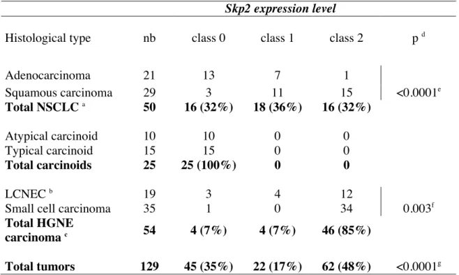
the intensity of staining and the percentage of stained cells. Tumor samples were grouped in three classes as described in the material and method section. According to this, Skp2 was undetectable (class 0) in normal lung epithelium adjacent to tumor cells on sections, as well as in normal lung tissues localized at distance from lung cancer. As compared to these normal tissues, we observed an heterogeneous pattern of Skp2 expression with tumors exhibiting either undetectable (class 0), moderate (class 1) or strong (class 2) immunostaining (Table 1, p<0.0001). In this setting, overexpression of Skp2 was observed in 48% (62/129) of all tumors tested, predominantly in high grade NE lung tumors (46/54, 85%) as compared to NSCLC (16/50, 32%) (Table 1 and Figure 1). Consistent with previous reports (Yokoi et al., 2004; Zhu et al., 2004), high levels of Skp2 were more frequent in squamous carcinomas (15/29, 51%) than in adenocarcinomas (1/21, 4%) (p<0.0001). Moreover, Skp2 was undetectable in 25/25 (100%) atypical and typical lung carcinoids, whereas it was overexpressed in the majority of high grade NE lung tumors, with SCLCs displaying higher levels (34/35, 97%) than LCNECs (12/19, 63%) (p=0.003). Therefore, these data indicate that Skp2 overexpression is associated with a high grade phenotype in NE lung tumors. To validate these IHC data, 24 of the 104 tumoral samples were analyzed for Skp2 expression by western blotting and a good concordance (88%) was observed between both techniques (data not shown).
Then, to further characterize the role of Skp2, we analyzed the relationships between its IHC status and some clinicopathological parameters (Table 2). When all histological subtypes were considered, we observed that Skp2 overexpression was associated with advanced stages III-IV (p< 0.0001) and nodal metastasis (p<0.0001). Similar correlations were observed in NE lung tumors (p<0.0001) but not in NSCLCs.
The expression of Skp2 and E2F-1 is directly correlated in NE lung tumors.
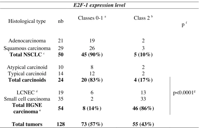
We previously reported a differential pattern of E2F-1 protein expression in a series of 60 lung tumors (Eymin et al., 2001a). In this study, we have extended these results to a larger series and included atypical and typical carcinoids (Table 3 and Figure 2). Our data confirmed our previous results with E2F-1 protein being predominantly overexpressed in HGNE lung tumors (46/54, 86%) whereas it was undetectable or faintly expressed in 90% (45/50) of NSCLC (Table 3). Importantly, high amounts of E2F-1 protein were associated with a high grade NE phenotype as compared to carcinoids (Table 3; p<0.0001 and Figure 2B).
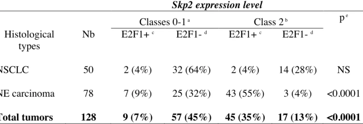
When we analyzed the status of both Skp2 and E2F-1 proteins, we found an unexpected direct correlation in all tumors tested (Table 4; p<0.0001), as well as in NE lung tumors (p<0.0001). In contrast, E2F-1 and Skp2 status was not linked in NSCLCs, but we noticed that 64% (32/50) of the cases did not express both proteins.
Skp2 is a transcriptional target of E2F-1
To further investigate the functional relationships between Skp2 and E2F-1 proteins, we undertook a series of experiments in cell lines. Firstly, we postulated that Skp2 could be a new downstream target of E2F-1. To answer, we neutralized E2F-1 using siRNA in the small cell lung carcinoma cell line H69 and analyzed Skp2 expression by immunoblotting and RT-PCR. As shown in figure 3A, the knock-down of E2F-1 was associated with a significant down-regulation of both Skp2 protein (left panel) and mRNA levels (right panel). Furthermore, using a stable E2F-1 tetracycline-inducible clone (H358/Tet-On) that we previously established in the human lung adenocarcinoma cell line H358 (Salon et al., 2006), we observed an increase in Skp2 mRNA (Figure 3B, right panel) and protein (Figure 3B, left panel) expression upon doxycyclin induction. Altogether, these data indicate that Skp2 expression is positively regulated by E2F-1. Then, we investigated whether E2F-1 was able to
control Skp2 promoter activity. A549 cells were transfected with a plasmid encoding the luciferase protein under the control of the human Skp2 promoter, together with increasing amounts of an E2F-1 expression vector. Because of their low transfection efficiency, H69 cells could not be used here. As reflected by the increase in luciferase activity, E2F-1 was able to activate in a dose dependent manner the Skp2 promoter in these cells (Figure 3C, left panel). Similar results were obtained in the H1299 cell line (Figure 3C, right panel). Therefore, these data demonstrate that Skp2 is a transcriptional target of E2F-1. Interestingly, we noticed that the Skp2 promoter response to E2F-1 increasing levels was stronger in H1299 cells deriving from a large cell carcinoma with neuroendocrine features than in A549 adenocarcinoma cells. These data were consistent with our findings of a direct correlation between E2F-1 and Skp2 in NE lung tumors.
Skp2 interacts with and stimulates the transcriptional activity of E2F-1
It was previously demonstrated that Skp2 interacts with and acts as a transcriptional cofactor of c-Myc (Kim et al., 2003; Von der Lehr et al., 2003). These data prompted us to investigate whether Skp2 could also regulate E2F-1 transcriptional activity. Firstly, we tested the possibility that E2F-1 and Skp2 could physiologically interact in our cellular models. As illustrated, E2F-1 was clearly recovered from Skp2 immunoprecipitates obtained from H69 (Figure 4A) or H810 (data not shown) neuroendocrine lung carcinoma cell lines. More importantly, an E2F-1/Skp2 complex was also detected in cellular extracts obtained from three small cell lung carcinomas with high levels of both proteins (Figure 4B). Altogether, these data demonstrate that Skp2 interacts with E2F-1 in HGNE human lung cell lines as well as in primary tumors.
Then, we asked whether Skp2 could regulate E2F-1 transcriptional activity. To answer, A549 cells were transfected with plasmids encoding the luciferase under the control
of the cyclin E promoter, a well-known E2F-1 target gene (Soucek et al., 1997), E2F-1 and increasing amounts of Skp2. As reflected by the increase in luciferase levels, the cyclin E promoter was activated upon E2F-1 expression (Figure 4C). Importantly, the addition of Skp2 stimulated in a dose-dependent manner the transcriptional activity of E2F-1 in this setting (Figure 4C). In contrast, Skp2 alone was unable to promote cyclin E activation, nor to stimulate the activity of a control pGLuc vector (Figure 4C). Taken together, these results indicate that Skp2 stimulates the transcriptional activity of E2F-1. Similar results were obtained in H1299 cells as well as in another cellular model, the human osteosarcoma cell line SAOS2 (Figure 3D).
Relationships between Skp2, E2F-1 and cyclin E status in human lung tumors.
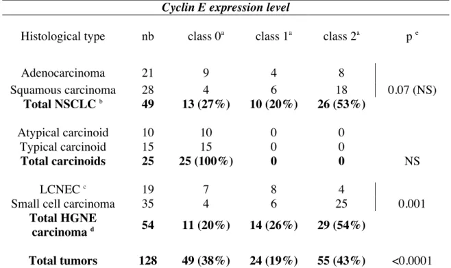
The above results led us to examine whether Skp2, E2F-1 and cyclin E expression was linked in human lung tumors. First, we analyzed the status of the cyclin E protein in our series of lung tumors (Table 5 and Figure 5). As compared to normal lung tissues in which immunostaining was not detectable (Figure 5A, a), we observed an heterogeneous pattern of cyclin E expression with tumors exhibiting either undetectable (class 0), moderate (class 1) or strong (class 2) immunostaining (Table 5, p<0.0001). Interestingly, we noticed that cyclin E was undetectable in 100% of carcinoids whereas high levels were observed in 21% (4/19) of LCNEC and 71% of SCLC (25/35) (Table 5, p=0.001). In addition, a strong cyclin E immunostaining was associated with advanced stages III-IV (p< 0.0001) and nodal metastasis (p<0.0001) in these tumors (data not shown). Thus, as for Skp2 and E2F-1, accumulation of cyclin E appears to be a hallmark of a high grade phenotype in NE lung tumors.
Then, we studied the relationships linking Skp2, E2F-1 and cyclin E proteins in our series of lung tumors. As shown in table 6, we found a direct correlation between cyclin E and Skp2 (p<0.0001) or E2F-1 (p=0.0001) expression in NE lung tumors. In contrast, no
significant correlation was found in NSCLC. Furthermore, the expression profile of the 3 proteins was associated (either undetectable or overexpressed) in 56% (71/126) of all tumors tested, more frequently in NE tumors (66%) as compared to NSCLC (40%) (Supplementary Table 1, p=0.006).
Discussion
The Skp2 protein, one of the F-box protein of the E3-ubiquitin ligase SCF complexes, targets a variety of cell cycle regulators for proteasomal degradation (Nakayama & Nakayama, 2006, for review). In this respect, Skp2 acts as a growth promoter which deregulation is thought to exert oncogenic functions. The Skp2 gene is located at 5p13, a region that is commonly amplified in lung cancer. Consequently, Skp2 amplification and overexpression have been reported in NSCLC cell lines and primary tumors (Takanami, 2005; Yokoi et al., 2004; Zhu et al., 2004). In contrast, Skp2 status has been poorly investigated in NE lung tumors (Yokoi et al., 2002). In agreement with previous studies, we report that Skp2 is overexpressed in 32% of NSCLC, especially in squamous carcinomas. More importantly, we demonstrate for the first time that HGNE lung tumors display a more frequent Skp2 upregulation (46/54; 86%) than NSCLC (p <0.0001), and that Skp2 protein expression is undetectable in low grade NE typical and atypical carcinoids. Elevated expression of Skp2 was previously linked to positive lymph node metastasis, advanced stages and poor or moderate differentiation in NSCLC (Yokoi et al., 2004). In addition, a high level of Skp2 protein was also reported as an independent poor prognostic marker (Osoegawa et al., 2004; Zhu et al., 2004). In this study, we did not find any correlation between Skp2 overexpression and either of these clinicopathological parameters in NSCLC. In contrast, we observe that high levels of Skp2 expression are associated with advanced stages (III-IV) and nodal
metastasis in NE lung tumors. Overall, our data support the notion that overexpressed Skp2 acts as an oncogenic protein in HGNE lung tumors.
It is currently thought that the oncogenic properties of Skp2 are related to its ability to induce the ubiquitination and proteasomal degradation of its cell cycle targets (Nakayama & Nakayama, 2006, for review). Therefore, basedon our previous demonstration of an altered pattern of E2F-1 expression in human lung cancer (Eymin et al., 2001a), we had hypothesized that the status of E2F-1 and Skp2 proteins could be inversely related in these tumors. In this study, we did not observe any significant correlation between both proteins in NSCLC. In contrast and unexpectedly, Skp2 and E2F-1 status was directly correlated in NE lung tumors, with a predominant upregulation of both proteins in HGNE lung tumors (78% of the cases). In addition, a direct correlation between Skp2 and cyclin E (p<0.0001, this study) or p27KIP1
(p=0.006; data not shown), another target of Skp2-mediated proteolysis (Carrano et al., 1999; Nakayama et al., 2004), was also observed in these carcinomas. Taken together, these results clearly demonstrate that overexpression of Skp2 does not always correlate with the downregulation of its main targets in tumors. What might be the potential explanations for such observation? One plausible hypothesis is that the activity of SCFSkp2 complexes is altered
in HGNE lung tumors, maybe due to mutations or loss of expression of either SCF members. This could explain the concomittant expression of Skp2, E2F-1 and cyclin E proteins in these tumors. It remains to determine whether alterations of other SCF components exist in human lung tumors. In addition, our results suggest that Skp2 overexpression might have proteasomal-independent functions.Another but not exclusive explanation is the existence of a positive loop linking E2F-1 and Skp2 protein expression. In agreement with such hypothesis, we provide evidence that Skp2 is a transcriptional target of E2F-1. Interestingly, in a gene expression profiling analysis, Vernell and co-workers had observed that Skp2 expression is upregulated in response to E2F-1 (Vernell et al., 2003). Furthermore, during the
course of our study, it was demonstrated that E2F-1 directly interacts with the human Skp2 promoter and positively regulates its activity in various cellular models (Zhang & Wang, 2006). Therefore, our demonstration of a direct correlation between E2F-1 and Skp2 in HGNE lung tumors provides a physiological relevance to the observations made in cell lines.
Finally, how could Skp2 and E2F-1 overexpression contribute to the development of HGNE lung tumors ? We demonstrate that endogenous Skp2 and E2F-1 proteins interact in HGNE lung carcinoma cell lines and identify for the first time a Skp2/E2F-1 complex in small cell lung carcinomas. Moreover, using various cellular models, we show that Skp2 stimulates the transcriptional activity of E2F-1 towards the cyclin E promoter and identify a direct relationship linking cyclin E with Skp2 (p<0.0001) and E2F-1 (p=0.0001) proteins in NE lung tumors. Interestingly, 93% (27/29) of the tumors overexpressing the 3 proteins were HGNE lung tumors, indicating that their concomittant upregulation is highly predictive of a high grade NE phenotype. Therefore, besides its ability to induce cyclin E proteasomal degradation (Nakayama et al., 2000; Yeh et al., 2001), Skp2 could also induce cyclin E expression, notably through E2F-1 activation. Such differential Skp2-mediated cyclin E control could depend on the cellular context (normal versus tumoral tissues) and/or on the co-activated signals (E2F-1 upregulation).
To conclude, our data provide the first evidence of a direct correlation linking the E2F-1, Skp2 and cyclin E oncoproteins in high grade neuroendocrine lung tumors and suggest that their cooperation might contribute to the tumorigenesis of these highly aggressive lung carcinomas.
Materials and Methods
Tissue samples
One hundred and twenty nine human lung tumors were included in this study. Tissue samples were taken at surgical resection of lung tumors in 103 cases or at mediastinoscopy of node metastases in 5 cases of Large Cell Neuroendocrine Carcinomas (LCNECs) and 21 cases of Small Cell Lung Carcinomas (SCLCs). Tumor tissues and normal lung parenchyma were immediately frozen and stored at -80°C until use. For histological classification, tumor samples were fixed in formalin and the diagnoses were made on paraffin-embedded material using the current World Health Organization International Histological Classification of Lung Tumors criteria (Travis et al., 1999). They consisted of 29 squamous cell carcinomas, 21 adenocarcinomas, 19 LCNECs and 35 SCLCs, 15 typical carcinoids and 10 atypical carcinoids . Sixty three patients were suffering from nodal metastasis at the time of diagnosis.
Immunohistochemistry (IHC)
Skp2 and cyclin E immunohistochemical analyses were carried-out on formalin-fixed and paraffin-embedded tissue sections. Four-micrometer sections were dehydrated in xylene and rehydrated in graded ethanol solutions. Endogenous peroxydase activity was blocked by immersion in H2O2 solution. For Skp2, the antigen immunoreactivity was enhanced by microwaving the sections for 3 x 5 minutes in EDTA buffer, pH 8. Then, a classical three step immunohistochemical method was applied. The primary antibody (clone 2C8D9) was used at a 1:100 dilution. Cyclin E immunodetection was performed using the Ventana Discovery Autostainer (Ventana Medical International Inc). Cut sections were boiled for 60 minutes in citrate buffer pH 6 for heat-induced epitope retrieval. The primary antibody (clone 13A3) was applied at a 1:10 dilution for one hour. E2F-1 protein expression was detected on frozen
sections using the KH95 monoclonal antibody diluted at 1:100, as previously described (Eymin et al., 2001a). Slides incubation with normal rabbit or mouse IgG at the same concentration as the primary antibodies served as negative controls.
Immunohistochemical staining evaluation
Skp2, E2F-1 and cyclin E immunostainings were evaluated by two independent observers (CS and EB) in distinct areas of the slide sections for correlation and confirmation of tissues analysis and scored by taking into account the tumor heterogeneity. A final score (0-300) was established by multiplying the percentage of labeled cells (0-100%) with the intensity of staining (1+, 2+, 3+). According to the final scores, tumoral samples were divided in three classes as follow : class 0, no staining, class 1, score < 60 for Skp2 or < 40 for cyclin E and E2F-1, class 2, score ≥ 60 for Skp2 or ≥ 40 for cyclin E and E2F-1. Samples with a final score ≥ 60 were considered as tumors overexpressing Skp2. Cyclin E and E2F-1 overexpressing cases displayed a final score ≥ 40. Stromal cells and background normal lung were negative for all the three tested proteins. Correlations between the expression of various factors and clinicopathological parameters were based on the chi-square (X2
) test, with a p value ≤ 0.05 considered significative.
Cell lines , plasmids, siRNA and transfection
The A549 human lung adenocarcinoma, H1299 human large cell lung carcinomawith neuroendocrine features and SAOS2 human osteosarcoma cell lines were cultured as previously described (Eymin et al., 2001b). The H69 small cell lung carcinoma cell line was cultured in 5% CO2 at 37°C in RPMI-1640 medium supplemented with 10% (v/v)
heat-inactivated FCS, 2.5 mg/ml glucose, 10 mM HEPES and 1 mM sodium pyruvate. The H358/Tet-On/E2F-1 inducible clones were obtained as previously described (Salon et al.,
2006). Plasmids used were pcDNA3.1, pCMV-E2F-1, pGL2-cyclin E encoding the luciferase protein under the control of the cyclin E promoter, pcDNA3-Myc-Skp2 and Skp2-luc encoding the luciferase under the control of the human Skp2 promoter region spanning from – 272 to +244 residues. Luciferase assays were performed as previously described (Eymin et al., 2001b). The sequences specifically targeting human e2f-1 RNA were as follow: 5’-GUCACGCUAUGAGACCUCATT-3’ and 5’-ACAAGGCCCGAUCGAUGUUTT-3’. The scrambled siRNA oligonucleotides were as follow: 5’-AAAGGUGACGCUGACGAAGTT-3’; 5’-CAAGAAAGGCCAGUCCAAGTT-3’. Cells were transiently transfected either with Fugene 6 (Roche Diagnostic) or JetSITM
(Polyplus transfection).
Immunoblotting and immunoprecipitation experiments
Immunoblotting and immunoprecipitation experiments were performed as previously described (Eymin et al., 2001a). The antibodies were anti-actin (Sigma), anti-E2F-1 (C-20, Santa Cruz; KH-95, Pharmingen), anti-cyclin E (13A3, Novocastra) and anti-Skp2 antibodies (Skp2-2B12 and 2C8D9, Zymed; H-435, Santa-Cruz).
RT/PCR analysis of Skp2 mRNA level
PCR was carried out for 30 cycles using the following conditions : 94°C for 30 sec ; 57°C for 30 sec and 72°C for 30 sec. The primers used were : Skp2 forward (sense) 5’-TCAACTACCTCCAACACCTATCAC-3’ ; Skp2 reverse (antisense) 5’-GACAACTGGGCTTTTGCAGT-3’. Amplification of a fragment of the cDNA of G3PDH (Invitrogen) was performed in the same PCR reaction as an internal control.
Aknowledgements
We thank Kristian Helin for providing us with the plasmid encoding E2F-1, Didier Trouche for the plasmid pGL2-cyclin E, Lars-Gunnar Larsson for the pcDNA3-Myc-Skp2 and Günter Schneider (Technical University of Munich, Munich, Germany) for the plasmid Skp2-luc. We thank Patricia Betton, Pascal Perron, Sylvie Veyrenc and Aurelie Micoud for technical assistance. This work was supported by grants from the Region Rhône Alpes (Thématique Prioritaire Cancer and Canceropole 2003: Oncocell, Epimed and INACancer) and by the Ligue Nationale contre le Cancer (Equipe Labellisée). C.S. was supported by a “poste accueil” INSERM.
References
Bai C, Sen P, Hofmann K, Ma L, Goebl M, Harper JW, et al. (1996). SKP1 connects cell cycle regulators to the ubiquitin proteolysis machinery through a novel motif, the F-box. Cell 86: 263-74.
Brambilla E, Gazzeri S, Moro D, Caron de Fromentel C, Gouyer V, Jacrot M, et al. (1993). Immunohistochemical study of p53 in human lung carcinomas. Am J Pathol 143: 199-210.
Brambilla E, Moro D, Gazzeri S, Brambilla C. (1999). Alterations of expression of Rb, p16(INK4A) and cyclin D1 in non-small cell lung carcinoma and their clinical significance. J Pathol 188: 351-60.
Carrano AC, Eytan E, Hershko A, Pagano M. (1999). SKP2 is required for ubiquitin-mediated degradation of the CDK inhibitor p27. Nat Cell Biol 1: 193-9.
DeGregori J. (2002). The genetics of the E2F family of transcription factors: shared functions and unique roles. Biochim Biophys Acta 1602: 131-50.
Deshaies RJ. (1999). SCF and Cullin/Ring H2-based ubiquitin ligases. Annu Rev Cell Dev
Biol 15: 435-67.
Eymin B, Gazzeri S, Brambilla C, Brambilla E. (2001a). Distinct pattern of E2F1 expression in human lung tumours: E2F1 is upregulated in small cell lung carcinoma. Oncogene
20: 1678-87.
Eymin B, Karayan L, Seite P, Brambilla C, Brambilla E, Larsen CJ, et al. (2001b). Human ARF binds E2F1 and inhibits its transcriptional activity. Oncogene 20: 1033-41. Ginsberg D. (2002). E2F1 pathways to apoptosis. FEBS Lett 529: 122-5.
Gouyer V, Gazzeri S, Bolon I, Drevet C, Brambilla C, Brambilla E. (1998). Mechanism of retinoblastoma gene inactivation in the spectrum of neuroendocrine lung tumors. Am J
Respir Cell Mol Biol 17: 1-9.
Gstaiger M, Jordan R, Lim M, Catzavelos C, Mestan J, Slingerland J, et al. (2001). Skp2 is oncogenic and overexpressed in human cancers. Proc Natl Acad Sci U S A 98: 5043-8. Harbour JW , Dean DC. (2000). The Rb/E2F pathway: expanding roles and emerging
paradigms. Genes Dev 14: 2393-409.
Kim SY, Herbst A, Tworkowski KA, Salghetti SE , Tansey WP. (2003). Skp2 regulates Myc protein stability and activity. Mol Cell 11: 1177-88.
Marti A, Wirbelauer C, Scheffner M , Krek W. (1999). Interaction between ubiquitin-protein ligase SCFSKP2 and E2F-1 underlies the regulation of E2F-1 degradation. Nat Cell
Biol 1: 14-9.
Nakayama K, Nagahama H, Minamishima YA, Matsumoto M, Nakamichi I, Kitagawa K, et
al. (2000). Targeted disruption of Skp2 results in accumulation of cyclin E and
Nakayama K, Nagahama H, Minamishima YA, Miyake S, Ishida N, Hatakeyama S, et al. (2004). Skp2-mediated degradation of p27 regulates progression into mitosis. Dev Cell
6: 661-72.
Nakayama KI , Nakayama K. (2006). Ubiquitin ligases: cell-cycle control and cancer. Nat Rev
Cancer 6: 369-81.
Osoegawa A, Yoshino I, Tanaka S, Sugio K, Kameyama T, Yamaguchi M, et al. (2004). Regulation of p27 by S-phase kinase-associated protein 2 is associated with aggressiveness in non-small-cell lung cancer. J Clin Oncol 22: 4165-73.
Salon C, Eymin B, Micheau O, Chaperot L, Plumas J, Brambilla C, et al. (2006). E2F1 induces apoptosis and sensitizes human lung adenocarcinoma cells to death-receptor-mediated apoptosis through specific downregulation of c-FLIP(short). Cell Death
Differ 13: 260-72.
Soucek T, Pusch O, Hengstschlager-Ottnad E, Adams PD, Hengstschlager M. (1997). Deregulated expression of E2F-1 induces cyclin A- and E-associated kinase activities independently from cell cycle position. Oncogene 14: 2251-7.
Takanami I. (2005). The prognostic value of overexpression of Skp2 mRNA in non-small cell lung cancer. Oncol Rep 13: 727-31.
Travis WD, Colby TV, Corrin B, Shimosato Y, Brambilla E. (1999). WHO international Histological Classification of Tumours: Histological Typing of Lung and Pleural Tumours. Ed Springer Third Edition.
Vernell R, Helin K , Muller H. (2003). Identification of target genes of the p16INK4A-pRB-E2F pathway. J Biol Chem 278: 46124-37.
Von der Lehr N, Johansson S, Wu S, Bahram F, Castell A, Cetinkaya C, et al. (2003). The F-box protein Skp2 participates in c-Myc proteosomal degradation and acts as a cofactor for c-Myc-regulated transcription. Mol Cell 11: 1189-200.
Yeh KH, Kondo T, Zheng J, Tsvetkov LM, Blair J, Zhang H. (2001). The F-box protein SKP2 binds to the phosphorylated threonine 380 in cyclin E and regulates ubiquitin-dependent degradation of cyclin E. Biochem Biophys Res Commun 281: 884-90. Yokoi S, Yasui K, Mori M, Iizasa T, Fujisawa T, Inazawa J. (2004). Amplification and
overexpression of SKP2 are associated with metastasis of non-small-cell lung cancers to lymph nodes. Am J Pathol 165: 175-80.
Yokoi S, Yasui K, Saito-Ohara F, Koshikawa K, Iizasa T, Fujisawa T, et al. (2002). A novel target gene, SKP2, within the 5p13 amplicon that is frequently detected in small cell lung cancers. Am J Pathol 161: 207-16.
Zhang H, Kobayashi R, Galaktionov K, Beach D. (1995). p19Skp1 and p45Skp2 are essential elements of the cyclin A-CDK2 S phase kinase. Cell 82: 915-25.
Zhang L, Wang C. (2006). F-box protein Skp2: a novel transcriptional target of E2F.
Oncogene 25: 2615-27.
Zheng N, Schulman BA, Song L, Miller JJ, Jeffrey PD, Wang P, et al. (2002). Structure of the Cul1-Rbx1-Skp1-F boxSkp2 SCF ubiquitin ligase complex. Nature 416: 703-9.
Zhu CQ, Blackhall FH, Pintilie M, Iyengar P, Liu N, Ho J, et al. (2004). Skp2 gene copy number aberrations are common in non-small cell lung carcinoma, and its overexpression in tumors with ras mutation is a poor prognostic marker. Clin Cancer
Titles and legends to figures
Figure 1. (A) Immunostaining of normal lung parenchyma and lung cancer tissue on paraffin
embedded sections using an anti-Skp2 monoclonal antibody (2C8D9, Zymed). (a) Absence of immunostaining in the columnar epithelium lining a normal bronchial structure (less than 1% of positive cells). (b) Negative immunostaining (class 0) in a typical carcinoid. (c,d) Strong positive nuclear immunostaining (class 2) on (c) most or (d) 50% of tumor cells in small cell lung carcinomas. (e) Weak nuclear immunostaining (class 1) in a squamous cell carcinoma. (f) Negative immunostaining (class 0) in an adenocarcinoma. (B) Mean scores ± standard deviation of Skp2 protein expression according to the histological sub-types.
Figure 2. (A) Immunostaining of normal lung parenchyma and lung cancer tissue on paraffin
embedded sections using an anti-E2F-1 monoclonal antibody (KH95, Pharmingen). (a) Absence of immunostaining in hyperplastic bronchiolar cells. (b) Weak positive immunostaining (class 1) in an atypical carcinoid consisting in few scattered positive cells (arrow). (c) Strong positive nuclear staining (class 2) detected on at least 50% of tumor cells in a small cell lung carcinoma. (d) Negative immunostaining (class 0) in an adenocarcinoma.
(B) Mean scores ± standard deviation of E2F-1 protein expression according to the
histological sub-types.
Figure 3. Skp2 is a transcriptional target of E2F-1. (A) The small cell lung carcinoma cell
line H69 was transfected with either mismatch or E2F-1 siRNA as indicated. Skp2 protein and mRNA levels were detected by immunoblotting (left panel) or RT-PCR (right panel), respectively. (B) H358 stable E2F-1-inducible clones were incubated in the presence (+) or absence (-) of 1µg/ml doxycyclin for 72 hours as indicated. E2F-1 and Skp2 protein
expression were detected by immunoblotting (left panel). Skp2 mRNA level was analyzed by RT-PCR (right panel). (C) A549 lung adenocarcinoma and H1299 large cell lung carcinoma cell lines were co-transfected for 48 hours with 1µg of a Skp2-luc plasmid encoding the luciferase under the control of the Skp2 promoter and increasing amounts of a pCMV-E2F-1 vector. The luciferase activity obtained in cells transfected with Skp2-luc alone was normalized to 1 and a relative luciferase activity was then calculated for each condition.
Figure 4. Skp2 interacts with E2F-1 and stimulates its transcriptional function towards the
cyclin E promoter. (A, B) Endogenous Skp2 protein was immunoprecipitated from (A) H69 or (B) small cell lung carcinoma extracts (T1, T2, T3) with an anti-Skp2 antibody.. Immunoblotting was carried-out with an anti-E2F-1 antibody. An irrelevant IgG was used as a negative control for immunoprecipitation. (C, D) Luciferase experiments were performed in A549, H1299 and SAOS2 cell lines co-transfected for 48 hours with 1µg pGL2-cyclin E encoding the luciferase under the control of the cyclin E promoter or 1 µg pGL2 as a control, in the presence or absence of 50ng pCMV-E2F-1 and increasing amounts of pcDNA3-Myc-p45SKP2 (100 and 250ng), as indicated. The luciferase activity obtained in cells transfected
with pGL2-cyclin E or pGL2 was normalized to 1 and a relative luciferase activity was then calculated for each condition.
Figure 5. (A) Immunostaining of lung cancer tissues on paraffin embedded sections using a
monoclonal anti-cyclin E antibody. (a) Absence of staining in epithelial cells lining a bronchiolar structure and in pneumonocytes. (b) Negative immunostaining (class 0) in a typical carcinoid. (c) Strong positive nuclear staining (class 2) on a majority of tumor cells in a small cell lung carcinoma. (d) Strong positive nuclear staining (class 2) detected on most
tumor cells in a squamous cell carcinoma. (B) Mean scores ± standard deviation of cyclin E protein expression according to the histological sub-types.
Table 1 : Immunohistochemical analysis of Skp2 expression in lung cancer according to
histological type.
Immunostaining scores were calculated by multiplying the number of labeled cells (0 to 100%) by the level of intensity (1 to 3). According to this, tumor samples were grouped in three classes (see materials and methods section): class 0 (undetectable staining), class 1 (moderate staining) and class 2 (high staining considered as Skp2 overexpression). nb= number of cases in each histological type. a Non small cell lung carcinoma. b Large cell neuroendocrine carcinoma. c Neuroendocrine tumors. d χ2 test. e p represents the comparison
of Skp2 expression level between adenocarcinoma and squamous carcinoma. f
p represents the comparison of Skp2 protein level between LCNEC and SCLC. g
p represents the comparison between each class of Skp2 expression level in all tumors tested.
Skp2 expression level
Histological type nb class 0 class 1 class 2 p d
Adenocarcinoma 21 13 7 1 Squamous carcinoma 29 3 11 15 <0.0001e Total NSCLC a 50 16 (32%) 18 (36%) 16 (32%) Atypical carcinoid 10 10 0 0 Typical carcinoid 15 15 0 0 Total carcinoids 25 25 (100%) 0 0 LCNEC b 19 3 4 12
Small cell carcinoma 35 1 0 34 0.003f
Total HGNE
carcinoma c 54 4 (7%) 4 (7%) 46 (85%)
Table 2 : High levels of Skp2 correlate with advanced stages and nodal metastasis in NE lung carcinomas.
a
Undetectable or low level of Skp2 staining. b
Strong Skp2 staining considered as Skp2-overexpressing cases.
c
χ2 test. d p represents the comparison of Skp2 classes of expression between low stages and high stages in all tumors, NSCLC, or NE carcinoma. e p represents the comparison of Skp2 protein expression between tumors with (N1-N3) or without (N0) nodal metastasis in all
tumors, NSCLC, or NE carcinoma. nb: number of cases. NS: non significant.
Total tumors NSCLC NE carcinoma
nb Classes 0-1a Class 2b pc Classes 0-1a Class 2b pc Classes 0-1a Class 2b pc Stage I-II 61 45 16 19 9 26 7 Stage III-IV 68 22 46 <0.0001d 15 7 NSd 7 39 <0.0001d N0 56 43 13 19 8 24 5 N1-N3 73 24 49 <0.0001e 15 8 NSe 9 41 <0.0001e
Table 3 : Immunohistochemical analysis of E2F-1 expression in lung cancer according to
histological type.
Tumors were grouped in two classes according to their score of E2F-1 staining. a Tumors with
undetectable or low level of E2F-1 staining. b
Tumors with high level of E2F-1 staining, considered as overexpressing cases. nb= number of cases in each histological type. c Non small cell lung carcinoma. d Large cell neuroendocrine carcinoma. e Neuroendocrine tumors. f
χ2 test. g p represents the comparison of E2F-1 expression level between total carcinoids and
total HGNE carcinoma.
E2F-1 expression level Histological type nb Classes 0-1
a Class 2 b p f Adenocarcinoma 21 19 2 Squamous carcinoma 29 26 3 Total NSCLC c 50 45 (90%) 5 (10%) Atypical carcinoid 10 8 2 Typical carcinoid 14 12 2 Total carcinoids 24 20 (83%) 4 (17%) LCNEC d 19 6 13 p<0.0001g
Small cell carcinoma 35 2 33
Total HGNE
carcinoma e 54 8 (14%) 46 (86%)
Table 4. Skp2 and E2F1 expression is directly correlated in NE lung carcinoma.
Skp2 expression level
Classes 0-1 a Class 2 b p e Histological
types
Nb E2F1+ c E2F1- d E2F1+ c E2F1- d
NSCLC 50 2 (4%) 32 (64%) 2 (4%) 14 (28%) NS NE carcinoma 78 7 (9%) 25 (32%) 43 (55%) 3 (4%) <0.0001
Total tumors 128 9 (7%) 57 (45%) 45 (35%) 17 (13%) <0.0001
a
Undetectable or low level of Skp2 staining. b
High levels of Skp2 staining and considered as Skp2-overexpressing cases. c E2F1+: tumors overexpressing E2F1. d E2F1-: cases without E2F1 overexpression. e
Table 5. Immunohistochemical analysis of cyclin E expression in lung cancer according to
histological type.
Tumors were grouped in three classes according to their score of cyclin E staining. a
Class 0 (undetectable staining), 1 (moderate staining) and 2 (high staining considered as overexpressing cases). nb= number of cases in each histological type. b Non small cell lung carcinoma. c Large cell neuroendocrine carcinoma. d Neuroendocrine tumors. e
χ2 test. Cyclin E expression level
Histological type nb class 0a class 1a class 2a p e
Adenocarcinoma 21 9 4 8 Squamous carcinoma 28 4 6 18 0.07 (NS) Total NSCLC b 49 13 (27%) 10 (20%) 26 (53%) Atypical carcinoid 10 10 0 0 Typical carcinoid 15 15 0 0 Total carcinoids 25 25 (100%) 0 0 NS LCNEC c 19 7 8 4
Small cell carcinoma 35 4 6 25 0.001
Total HGNE
carcinoma d 54 11 (20%) 14 (26%) 29 (54%)
Table 6. Direct correlation between cyclin E and Skp2 or E2F1 immunostaining in NE lung
carcinoma.
Skp2 expression level Histological
types nb Classes 0-1 a Class 2 b p g
Cyclin E+ c Cyclin E- d Cyclin E+ c Cyclin E- d
NSCLC 49 15 18 11 5 NS
NE carcinoma 79 1 32 28 18 <0.0001
E2F-1 expression level Histological
types
nb
Tumors w/o E2F-1 overexpression e
Tumors with E2F-1 overexpression f
p g Cyclin E+ c Cyclin E- d Cyclin E+ c Cyclin E- d
NSCLC 49 22 23 4 0 NS
NE carcinoma 78 2 26 27 23 0.0001
a
Tumors with an undetectable or moderate Skp2 immunostaining. b
Skp2-overexpressing tumors. c Cyclin E +: tumors overexpressing cyclin E using immunohistochemistry. d Cyclin E -: cases without cyclin E overexpression. e
Tumors with undetectable or low level of E2F-1 immunostaining. f
Tumors overexpressing E2F-1. nb: number of tumoral samples. g
χ2 test. NS: non significant.
Supplementary Table 1. Association of Skp2, E2F-1 and cyclin E immunostaining in lung tumors. Skp2/E2F-1/Cyclin E status Histological types nb Skp2/E2F-1/Cyclin E + or Skp2/E2F-1/Cyclin E - a Dissociation of at least one protein b
p c NSCLC 49 20 (40%) 29 (60%) 0.006 NE carcinoma 77 51 (66%) 26 (33%) Total tumors 126 71 (56%) 55 (43%) a
Tumors displaying either undetectable (-) or upregulation (+) of Skp2, E2F-1 and cyclin E proteins. b
Tumors in which the status of at least one protein is dissociated from that of the others. c χ2 test. nb: number of tumoral samples. NS: non significant.