HAL Id: hal-02323542
https://hal.archives-ouvertes.fr/hal-02323542
Submitted on 6 Oct 2020HAL is a multi-disciplinary open access archive for the deposit and dissemination of sci-entific research documents, whether they are pub-lished or not. The documents may come from teaching and research institutions in France or abroad, or from public or private research centers.
L’archive ouverte pluridisciplinaire HAL, est destinée au dépôt et à la diffusion de documents scientifiques de niveau recherche, publiés ou non, émanant des établissements d’enseignement et de recherche français ou étrangers, des laboratoires publics ou privés.
Polyelectrolyte Multilayers: A Versatile Tool for
Preparing Antimicrobial Coatings.
Lydie Séon, Philippe Lavalle, Pierre Schaaf, Fouzia Boulmedais
To cite this version:
Lydie Séon, Philippe Lavalle, Pierre Schaaf, Fouzia Boulmedais. Polyelectrolyte Multilayers: A Ver-satile Tool for Preparing Antimicrobial Coatings.. Langmuir, American Chemical Society, 2015, 31 (47), pp.12856-12872. �10.1021/acs.langmuir.5b02768�. �hal-02323542�
Polyelectrolyte Multilayers: A Versatile Tool for Preparing
Antimicrobial Coatings
Lydie Séon,
†,‡,§Philippe Lavalle,
‡,§Pierre Schaaf,
†,‡,§,∥,⊥,#and Fouzia Boulmedais
*
,†,⊥,∇†Centre National de la Recherche Scientifique, Institut Charles Sadron, UPR 22, 23 rue du Loess, 67034 Strasbourg, France
‡Institut National de la Santé et de la Recherche Médicale, Biomaterials and Bioengineering, UMR 1121, 11 rue Humann, 67085
Strasbourg, France
§Université de Strasbourg, Faculté de Chirurgie Dentaire, 2 rue Sainte-Elisabeth, 67000 Strasbourg, France
∥Université de Strasbourg, Ecole Européenne de Chimie, Polymères et Matériaux, 25 rue Becquerel, 67087 Strasbourg, France
⊥International Center for Frontier Research in Chemistry, 8 allée Gaspard Monge, 67083 Strasbourg, France
#Institut Universitaire de France, 103 boulevard Saint-Michel, 75005 Paris, France
∇University of Strasbourg Institute of Advanced Study, 5 allée du Général Rouvillois, 67083 Strasbourg, France
ABSTRACT: The prevention of pathogen colonization of
medical implants represents a major medical and financial
issue. The development of antimicrobial coatings aimed at protecting against such infections has thus become a major field of scientific and technological research. Three main strategies are developed to design such coatings: (i) the prevention of microorganisms adhesion and the killing of microorganisms (ii) by contact and (iii) by the release of active compounds in the vicinity of the implant. Polyelectrolyte multilayer (PEM) technology alone covers the entire wide-spread spectrum of functionalization possibilities. PEMs are obtained through the alternating deposition of polyanions and polycations on a substrate, and the great advantages of PEMs
are that (i) they can be applied to almost any type of substrate whatever its shape and composition; (ii) various chemical, physicochemical, and mechanical properties of the coatings can be obtained; and (iii) active compounds can be embedded and
released in a controlled manner. In this article we will give an overview of thefield of PEMs applied to the design of antimicrobial
coatings, illustrating the large versatility of the PEM technology.
■
INTRODUCTIONPolyelectrolyte multilayers (PEMs) were introduced in 1991 by
Decher1 by extending to polyelectrolytes a technique initially
developed by Iller2for colloidal particles. PEMs are obtained by
the step-by-step deposition of polyanions and polycations
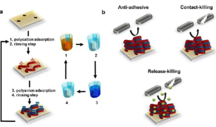
interacting electrostatically with each other (Figure 1a).
This step-by-step deposition process of macromolecules was later extended to other types of interactions such as hydrogen bonding and even covalent bonding between macromolecules. PEMs can be constructed by dipping the substrate alternately in
the different polyelectrolyte solutions, by spin-coating, or by
spraying them.3 Different physicochemical and mechanical
properties can be obtained depending on the buildup conditions (pH, ionic strength) and the polyelectrolytes used.
The growth of PEMfilms can be either linear or exponential.
For linearly growingfilms, the thickness increases linearly with
the number of deposition steps, and for the others, the
thickness increases exponentially, at least during the first
deposition steps. Whereas the thickness of the first films
remains in the nanometer range per bilayer, the thickness of
exponentially growingfilms can reach several micrometers after
10−15 bilayer deposition steps.4 Linearly growing films are
encountered for polyelectrolytes which interact strongly at each
deposition step.5 The resulting multilayer is thus a stratified
architecture with no diffusion of the polyelectrolytes
perpendicular to the film.6
Poly(styrenesulfonate)/poly-(allylamine) (PSS/PAH) constitutes a prominent example of
this type offilm. Exponentially growing PEMs are obtained for
polyanions and polycations that interact weakly,5 leading to
much more hydrated and softer films than linearly growing
ones.7This growth process is observed when at least one of the
polyelectrolytes constituting thefilm diffuses in and out of the
whole structure during each “bilayer” deposition step.8
Hyaluronic acid/poly(L-lysine) (HA/PLL) is the most
investigated exponentially growing multilayer. Thesefilms are
of interest because they can be used as reservoirs for embedded
proteins or enzymes.9A recent review of Caruso discusses how
to engineer PEM films with appropriate physicochemical
Received: July 27, 2015
Revised: October 27, 2015
Published: October 29, 2015
Invited Feature Article
pubs.acs.org/Langmuir
copying and redistribution of the article or any adaptations for non-commercial purposes.
Downloaded via BIBCNRS INC on January 16, 2020 at 11:09:25 (UTC).
properties by making judicious choices of the assembly
technology.10 The great advantages of the method are that
(i) it can be applied to almost any type of substrate whatever its shape and composition; (ii) various chemical, physicochemical, and mechanical properties of the coatings can be obtained; and (iii) active compounds can be embedded and released in a control manner. They thus represent a method of choice for developing antimicrobial coatings.
The prevention of pathogen colonization of medical implants
constitutes a major medical andfinancial issue since nosocomial
infections represent one of the most serious complications after surgery or critical care. The development of antimicrobial coatings aimed at protecting against such infections has thus
become a majorfield of scientific and technological research. A
wide range of strategies have been developed to design new
antimicrobial coatings.11 Various approaches based on the
immobilization of antifouling polymers or bactericidal
sub-stances using self-assembled monolayers,12 covalent
attach-ment, or graft polymerization13 have been explored and
extensively reviewed. Chemical surface treatments are focused on preventing adhesion and killing or inhibiting the growth of pathogens before they settle on a surface. These strategies make
use not only of the chemical,14 mechanical,15 and
morpho-logical properties of the coatings16but also of different active
compounds such as antibiotics and other antibacterial
compounds incorporated into the coatings.17 Numerous
surfaces and commercially used objects have already been
coated with different antimicrobial agents showing all potential
applications (Table 1). The PEM technology allows us to cover
the entire widespread spectrum of functionalization possibil-ities. Three main kinds of PEM coatings have been developed to limit microbial colonization on material surfaces: adhesion-resistant, contact killing, and antimicrobial agent leaching
coatings (Figure 1b). In this feature article, we will try to give
an overview, often based on our own contribution, to thefield
of antimicrobial coatings related to PEMs. We will not cover the whole body of literature but rather will try to illustrate the
different possibilities opened by PEM technology to address the
different strategies mentioned above.
■
STRATEGIES TO PREVENT BACTERIAL ADHESIONBecause thefirst step in bacterial infection on implants is the
bacterial adhesion on the substrate, thefirst strategy to create
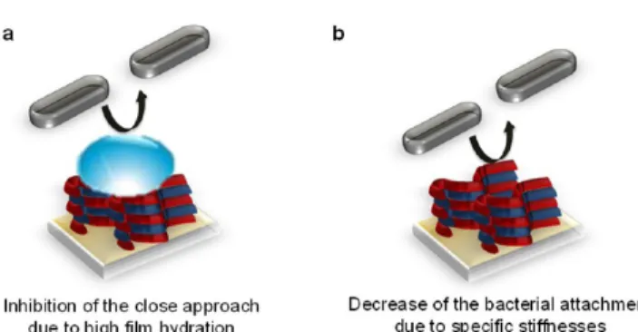
antimicrobial coatings that comes to mind is the design of substrates that prevent bacterial adhesion. Adhesion-resistant
PEMs can be obtained in two different ways: (1) by inhibiting
the close approach or the contact of the bacteria with the surfaces through the use of hydrophilic polymer-based PEM
films18,19,45,46
(Figure 2a) or (2) by using films with specific
stiffnesses so that the bacteria do not adhere once entering into
contact with thefilm (Figure 2b).47,48
To develop bacterial adhesion-resistant surfaces, one of the strategies consists of the use of hydrophilic polymers that can inhibit contact with or the close approach of bacteria to the
surface due to the strong affinity of the polymers for water
molecules. The simplest way to build hydrophilic PEMs is to Figure 1.(a) Schematic representation of a polyelectrolyte multilayer
(PEM)film buildup by successive adsorption steps of polycations and polyanions followed by rinsing steps using the dipping method. (b) Three main strategies were followed to design antimicrobial PEM: antiadhesivefilms inhibiting the close approach of pathogens, contact-killing films by exposing antimicrobial agents on the surface, and release-killingfilms delivering antimicrobial agents in the supernatant, with the last two strategies leading to the death of pathogens.
Table 1. Examples of the Different Surfaces That Have Been
Coated with PEM Films and Their Applicationsa
coated surfaces
antimicrobial
agent application references Polymers
poly(dimethylsiloxane) antiadhesive PEM
microfluidic devices 18 poly(ethylene
terephthalate)
polycation water purification 19
antiseptic textile 20
poly(L-lactic acid) AgNP tissue engineering 21
poly(propylene) polycation packaging 22 polystyrenefilms polycation laboratory plates 23 polyurethane polycation medical devices
(catheters)
24
styrene-butadiene-styrene block copolymer
polycation wound dressing 25
Metals
copper AgNP marine coatings 26
stainless steel polycation marine coatings 27 AgNP orthopedic implants 28 AMP medicine application 29 titanium/titanium antibiotic orthopedic implants 30
alloy AgNP 31
Fiber Mats
cellulosefibers layered silicate/ polycation
healthcare product 32
polycation 33
nylon and silkfibers AgNP clothing product 34 polyacrylonitrilefibers polycation filtration, wound
dressing, vascular graft
35
Medical Devices
Biobrane AgNP biological dressing 36
catheter tube AMP fluid or gas
administration
37 cortical bone polycation bone graft 38
cotton gauzes AMP wound dressing 39
gelatin sponge antibiotic adsorption of blood during invasive surgeries
40
intraocular lenses antibiotic intraocular lense implantation 41,42 microfiltration membranes antiadhesive PEM drinking water treatment 43 suture materials antibiotic medical device
coating
42,44 aAgNP, silver nanoparticles; AMP, antimicrobial peptides.
use polyelectrolytes known for their high hydrophilicity.
Heparin (HEP), a strong polyacid (with a pKa ranging from
0.5 to 1.5), and hyaluronic acid (HA), a weak polyacid (with a
pKa of 2.9), are two ideal candidates known for this. As a
polycation, chitosan (CHI), which is a weak polybase (with a
pKaof 6.5), was also often chosen because it is the only known
cationic polysaccharide that also possesses an intrinsic
antibacterial property.49 Because CHI is a weak polycation,
the pH condition used to build the PEM plays a critical role in
the antiadhesive properties of CHI/HEPfilms.19By varying the
assembly pH from 2.9 to 6, the degree of ionization of CHI can be adjusted. At high pH values, CHI chains, with a low charge density, adopt loopy structures and tend to adsorb as thick
layers. At low pH values, CHI chains adopt flatter structures,
and the adsorbed layer is thinner. The surface composition of
PEMfilms can thus be tuned by the assembly pH. The most
hydrophilic PEM film, prepared at low pH, rich in HEP,
entailed a better antiadhesive property whereas the most
CHI-rich surfaces prepared at pH ∼6 had the most efficient
antimicrobial (killing-effect) property against E. coli.19 Our
group investigated the effect of the ionic strength on the
antiadhesive properties of CHI/HA films.50 Compared to a
bare glass substrate, CHI/HAfilms assembled at 0.15 M NaCl,
pH 5, led to a decrease of about 80% in E. coli adhesion whereas only a 40% decrease was found when the multilayer was
assembled at 10−2 M NaCl. The higher adhesion of bacteria
under this last condition could be explained by the high rigidity
of these thinfilms obtained at 10−2M NaCl.
Another way to create highly hydrophilic PEMs is to use
polyelectrolytes chemically modified by grafting hydrophilic
chains or moieties. These modified polyelectrolytes are then
incorporated into multilayers or can be deposited on top of a multilayer. In this way the hydrophilicity of the coating can be changed by varying the nature of the grafted chains and the grafting ratio. Adsorbed or grafted onto a surface, poly(ethylene glycol) (PEG) molecules act as a highly hydrated polymer layer
reducing protein adsorption and bacterial adhesion.51In 2004,
our group was thefirst to report the insertion of PEG into PEM
in order to enhance its antiadhesive properties toward
bacteria.45A PEG-functionalized poly(L-glutamic acid),
PGA-g-PEG (Figure 3a), was synthesized and combined with poly(L
-lysine) (PLL) to build PEM films. Composed of 45 ethylene
glycol monomers, PEG chains were grafted at a ratio of 16% on
PGA. (PLL/PGA-g-PEG)n multilayers were deposited on a
PEM precursor film and were tested with respect to bacterial
adhesion for 30 min. In comparison with the uncoated glass substrate, the adhesion of E. coli was reduced by 72% on PEM films composed of one PLL/PGA-g-PEG bilayer and by 92% on
films terminated by three PLL/PGA-g-PEG bilayers (Figure
3b).
In order to minimize the nonspecific adhesion of a yeast,
Saccharomyces cerevisiae, on poly(dimethylsiloxane)
(PDMS)-based microfluidic devices, a pegylated poly(acrylic acid)
(PAA-g-PEG) was adsorbed as the terminal layer on a PAA/ poly(allylamine hydrochlroride) (PAH) or a PAA/poly-(diallyldimethylammonium chloride) (PDADMAC) PEM
film.18
The PEG chains were constituted of 110 monomer units, and the grafting ratio was 20%. Compared to a bare PDMS substrate, a reduction of yeast cell adhesion of at least 2
orders of magnitude was found for the most efficient coating,
i.e., a PAA-g-PEG-terminated PAA/PDADMACfilm. Yet, it has
to be noted that hydrated PAA/PAH and PAA/PDADMAC films already possess some antiadhesive properties when Figure 2.Scheme of the two strategies that have been used to build
adhesion-resistant PEMs: (a) highly hydrated films prevent the adhesion of pathogens and (b) a range of specific stiffnesses of the films allow decreases in the attachment of pathogens.
Figure 3.(a) Chemical structure of PGA-g-PEG, where k≈ 45, j ≈ 330, and m ≈ 60. (b) Bacterial adhesion per mm2on bare silica, mPLL-g-PEG =
PLL-g-PEG monolayer, mPGA-g-PEG = (PLL/PGA-g-PEG)1, and multilayerfilms ∼PLL 3 = Pre-(PLL/PGA)2,∼PLL-g-PEG 3 = Pre-PLL-(PGA/
PLL-g-PEG)3,∼PGA 3 = Pre-(PLL/PGA)3,∼PGA-g-PEG 1 = Pre-(PLL/PGA-g-PEG)1, and∼PGA-g-PEG 3 = Pre(PLL/PGA-g-PEG)3with Pre =
deposited on PDMS substrates.18 This is attributed to their highly hydrated structure.
As for eukaryotic cells, the elastic modulus of a substrate also seems to play a role in bacterial adhesion, but the results are still controversial. Polyelectrolyte multilayers make it easier to
modulate the elastic modulus of a film without significantly
changing its chemical surface properties. The first study
reporting the influence of the elastic moduli on bacterial
adhesion was performed by Lichter et al. in 2008 using PEMs.47
The effective stiffness of PAH/PAA films was varied over
several orders of magnitude (elastic modulus ranging from 1 to 100 MPa) by tuning the assembly pH. The bacterial attachment of S. epidermidis positive bacteria) and E. coli
(Gram-negative bacteria) increased with the stiffness of the PAH/PAA
films independently of other surface characteristics such as surface roughness, surface interaction energy, or surface charge density. The elastic modulus of PEM can also be varied by
cross-linking the films. This is most effective by using
exponentially growing multilayers whose Young modulus can be varied over a wide range simply by changing the
cross-linking degree. In this way Saha et al.48 used PLL and HA
modified by photoreactive vinylbenzyl groups (HAVB) to
prepare photo-cross-linkable PEMfilms with different
mechan-ical properties (an elastic modulus ranging from 30 to 150 kPa)
under UV light illumination (Figure 5a). PLL/HAVB films
were placed in contact for 1 h with positive and Gram-negative bacterial strains. While the growth of Gram-positive Lactococcus lactis was independent of the rigidity of PLL/HAVB
PEM films, the growth of Gram-negative E. coli was slowed
down on stiffer films compared to on softer ones. When
incubated onto photopatterned PLL/HAVB films having a
rigidity micropattern, a larger amount of E. coli was observed on the softer background and on the border between softer and
stiffer regions. After image analysis inFigure 5b, we notice a
greater number of adhered bacterial colonies on the non-cross-linked region (red patches) than on the photo-cross-non-cross-linked
regions (white patches) (Figure 5c). Atfirst sight, these results
seem to contradict the study described previously by Lichter et
al.47 This probably comes from the difference in the elastic
modulus range studied. Indeed, the elastic moduli varied from 1 to 100 MPa for Lichter et al. and from 30 to 150 kPa for Saha
et al.48 In addition, the chemistry of PEM films is quite
different: synthetic polyelectrolytes were used in the first study,
and a polypeptide and a natural biopolymer were used in the second one. The chemical composition of the surface is an Figure 4. (a) Chemical structure of PAA-(EO)3-PC with a 25%
grafting ratio. (b) Adhesion of C. albicans onto a silicone sheet functionalized with one or two bilayers of (PAH/PAA), (PAH/PAA-(EO)3-PC), and (PAH/PAA-(EO)3) deposited onto a (PSS/PAH)2
precursorfilm compared to the (PAH/PSS)2PAHfilm (A) at rest and
(B) under a stretching degree of 1.5. Reproduced from ref52 with permission fromThe Royal Society of Chemistry.
Figure 5.(a) Preparation of photo-cross-linked (PLL/HAVB) LbLfilms. (b) Microscopy epifluorescence image showing E. coli M6155 after 4.5 h of growth on photopatternedfilms; the lighter 20 × 20 μm2 features correspond to stiffer photo-cross-linked regions while the dark background
corresponds to a non-cross-linkedfilm. The scale bar represents 40 μm. (c) Image analysis of the same area to discriminate the bacterial colonies adhered to the photo-cross-linked regions (white patches) and the non-cross-linked background (red patches). Reprinted with permission from ref 48. Copyright 2013 American Chemical Society.
important aspect of the bacterial response. To conclude, the film stiffness seems to play an important role in the adhesion of
bacterial cells. However, the influence of the stiffness on
bacterial adhesion remains an open question, and PEMs could represent an ideal tool for further investigating this issue.
Adhesion-resistant PEMs have the advantage of preventing
thefirst step in biofilm formation, i.e., pathogen adhesion, but
pathogens in solution could stay alive and can further colonize another surface area.
■
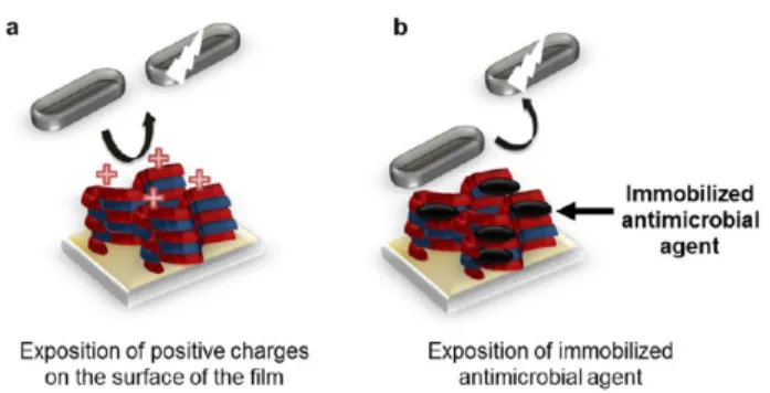
CONTACT-KILLING PEMSSurrounded by a capsule which is mostly constituted of acidic polysaccharides, most bacterial cell membranes are negatively charged. Hence most antimicrobial polymers are positively charged to promote the interaction with the membrane. Due to cationic charges available on their surface, they inhibit bacterial proliferation by disrupting their membranes, leading to death.
Contact-killing PEMfilms terminated by polycations were thus
developed (Figure 6a).22,27,32,33,53−58 Other contact-killing
films incorporate other antimicrobial agents such as carbon
nanoparticles59,60 and enzymes61 (Figure 6b). The design of
thesefilms is thus based on the ease of the PEM technology to
control the surface charges and to incorporate almost any kind of molecule or particle in a multilayer.
The easiest way to create a multilayer with a positively
charged surface is to terminate the film construction with a
polycation. The advantage of using multilayers instead of a simple cationic layer to cover the substrate is (i) to render the coating substrate-independent and universal, i.e., less sensitive to small composition variations, and (ii) to have a reservoir of polycations of a controlled amount. As already mentioned, CHI
is the only known polycationic polysaccharide (Figure 7a).
Associated with other natural polymers such as pectin,22
carrageenans,55 HA,56 or alginate,32 CHI-based PEM films
inhibited bacterial and fungal growth when a sufficient number
of layers is reached. In particular, PEMs terminated by a CHI layer were reported to possess antibacterial properties which are explained by a larger number of amino groups available on
the surface compared to films with a polyanion terminating
layer.55The antibacterial properties of CHI/alginate32or CHI/
pectin57 films deposited on nanofibrous mats were slightly
improved by binding a layered silicate, organic rectorite, with one of the polyelectrolytes during assembly. Although not
antimicrobial, clay with a large specific surface area, such as
organic rectorite, could immobilize the bacteria with the help of its excellent adsorption capacity. The antibacterial properties of CHI and rectorite nanocomposites result from the synergy between the adsorption and immobilization of the bacteria on the surface of the clay and the stronger interaction between amino groups and bacteria due to the accumulation of CHI on the surface of the clay.
Most often, there is almost an exact compensation between positive and negative charges of the polyelectrolytes and of ions
constituting the film. After buildup, when using weak
polyelectrolytes, the positive/negative charge ratio coming
from polyelectrolytes can be tuned in the film simply by
changing the pH of the supernatant solution. Along this line,
the assembly and postassembly pH values of PEMfilms such as
PAH/PSS and PAH/PAA were modified in order to expose
Figure 6.Scheme of the different strategies to obtain contact-killing PEMs: (a) highly positively charged surface and (b) immobilized antimicrobial agents, on the surface and inside thefilm, kill pathogens by contact.
Figure 7. (a) Chemical structure of chitosan. (b) Schematic representation of the construction, cross-linking, degradation, and antibacterial properties of the (HEP/CHI)10-(PVP/PAA)10multilayerfilm. (c) SEM images of water-borne assays of S. aureus adhesion on a silicon wafer (a1−
a3), the (HEP/CHI)10multilayerfilm (b1−b3), and the (HEP/CHI)10-(PVP/PAA)10multilayerfilms cross-linked at 110 °C for 16 h (c1−c3) or at
enough mobile cationic charges and to obtain antimicrobial activity against S. epidermidis and E. coli. At high pH, near or
above their pKa, PAH chains were incorporated with many
uncharged amine groups into PEMfilms, revealing no
contact-killing ability. By lowering the pH, these uncharged amines
were protonated, creating sufficient positive charges to induce
cell death.53By simple pH variation, PAH protonation can be
varied, leading to a conformational change which allows
exposing sufficient positive charges to obtain an antibacterial
property.
Phenolic compounds are known to cause bacterial cell death through the disintegration of the cell membrane. They interact with the surface of the cell through van der Waals interactions of the phenyl ring and the hydrophobic tails of the lipids. Pinto et al. reported the use of poly(4-vinylphenol) (PVPh) assembled with PAH or PDADMAC at pH ranging from
10.5 to 12.5.54Both systems led to a maximum of S. epidermidis
growth inhibition of about 60−70% at pH 10.5. Only a small
antibacterial activity (maximum of 35% inhibition) was found
against E. coli. The increase in antimicrobial efficacy at the
lower assembly pH was likely due to the protonation of the phenol moiety that increased the mode of action of the multilayer.
Quaternary ammonium compounds (QAC) present the advantage of being permanently charged, independently of the solution pH, and are known to possess antimicrobial
proper-ties.62 Thus, to improve the antibacterial activity of CHI,
quaternized derivatives of CHI (QCHI) were synthesized and
incorporated into PEMfilms.56These polyelectrolytes were not
used as substrate coating but to build up PEM capsules, another
majorfield of research in PEM. The degree of substitution of
QCHI was demonstrated to be a crucial parameter in inhibiting bacterial growth. The higher degree of substitution led to the lowest survival of E. coli bacteria. Contact-killing QCHI/HA microcapsules, based on QCHI with the highest degree of
substitution, show the most efficient effect on killing E. coli.56
QCHI-terminated capsules appeared to be more effective at
killing bacteria than HA-terminated ones. Bacteria appeared to be stuck on microcapsules, probably due to attractive electrostatic interactions between the positively charged capsules and the negatively charged bacteria.
Other polymeric quaternary ammonium salts have also been
incorporated into PEM films.27,58 The association of
N,N-dodecyl,methyl-poly(ethylenimine) (DMLPEI) and PAA when built under optimal conditions leads to the growth inhibition of both Gram-positive and Gram-negative bacteria as well as of an
influenza A/WSN (H1N1) virus.58Following Rubner and
co-workers’ study,53Wong et al. demonstrated the importance of
the pH assembly by making use of the fact that PAA is a weak
polyacid.58 The bactericidal activity of DMLPEI/PAA films
against S. aureus increases as the pH of the PAA solution used for assembly is lowered. This is consistent with the current view that mobile positive charges are necessary for bactericidal activity. PAA is weakly negatively charged at low pH and adopts a loop conformation in solution. The adsorbed PAA layer is thus relatively thick. In this case, the following adsorbed polycation (DMLPEI) layer is thick and loopy, with many of its positive charges available to interact with bacterial cell membranes. At high pH due to deprotonation, there is an increase in the number of negative charges of PAA which
adopts a flat random-coil conformation. The adsorbed PAA
layer isflat, and the following polycation layer that is adsorbed
interacts tightly. Thus, practically no polycations in brush
conformations are generated on the surface. By varying the length of the polycation alkyl chains, they proved the importance of the hydrophobicity of the polycation for its antibacterial activity. The proposed mechanism of action is based on a direct disruption of the bacterial membrane by
hydrophobic polycationic chains upon contact with thefilm.
Polyvinylamines (PVAm) (highly cationically charged
polymers) associated with PAA33were deposited on cellulosic
fibers, reducing bacterial growth by 99.9% after 1 h of contact with at least one layer. Using a quarternized poly(4-vinyl-pyridine)/carboxymethyl-cellulose (QPVP/CMC) system, Amim et al. showed the importance of the hydrophobicity of the substrate on the antibacterial activity of the resulting
PEM.63This is due to the chain conformation of thefirst QPVP
layer. In the case of a hydrophilic surface (Si/SiO2 wafer),
QPVP chains are thicker (more expanded) due to better
hydration, while for a hydrophobic surface (polystyrenefilms)
the chains seem to be less hydrated and tend to be in a more compact conformation. The biocidal activities of
QPVP-terminated PEM films built on hydrophobic surfaces were
generally weaker than those observed for the ones deposited on hydrophilic ones. Less exposure of pyridinium groups to the
aqueous dispersion is obtained in thefirst case.
To improve their stability under external parameter changes
(such as ionic strength or pH changes), PEM films can be
either cross-linked after their buildup or treated in a
step-by-step manner during each layer deposition.64Along these lines,
the first reported PEM films that are both antibacterial and
antifungal were obtained using carboxymethyl, CHI, and pectin
cross-linked by glutaraldehyde.22 Coated on a polypropylene
film, these multilayers were used for tomato packaging and remained almost intact with no apparent rotting infection for 13 days. Yang et al. used click chemistry to assemble, via the layer-by-layer technique, a QAC-containing polymer and a PEG
copolymer.27 The two polymers exhibit antibacterial and
antiadherent properties. The final coating exhibited good
resistance to bacterial adhesion (97% of reduction) and a high efficiency against marine Pseudomonas sp. Moreover, the antibacterial activity of the coating was maintained after
exposition tofiltered natural seawater at 30 °C for 30 days.
Because PEM buildup is a step-by-step process, multistrata films can be designed, with each stratum having a different property. Therefore, in order to further enhance the properties
of antibacterial coatings, in particular during the first 24 h
decisive period, a strategy was developed by Wang et al. where
an adhesion-resistant PEM film, a thermally cross-linked
poly(vinylpyrrolidone) (PVP)/PAA film, was built on top of
a contact-killing CHI/HEP film (Figure 7b,c).65 Incubated in
phosphate buffer at pH 7.4 and at 37 °C, the continuous
removal of the top PVP/PAAfilm inhibited S. aureus adhesion.
After the degradation of the outmost surface, the underlying
CHI/HEPfilm was exposed to the external environment and
showed contact-killing properties.
As already mentioned, nanoparticles can be embedded into PEM during the buildup process. This property was used by
Van Tassel’s group. Single-walled carbon nanotubes (SWNTs)
proved to exhibit antimicrobial activity by direct contact,
causing severe bacterial membrane damage.66
This group dispersed SWNTs in an aqueous solution by
using different amphiphile PEG-functionalized phospholipid
(PL-PEG) and incorporated them into PLL/PGA films.59
Compared to pure PLL/PGAfilms, the antibacterial activity of
epidermidis at up to 90% inhibition after 24 h of incubation. They also demonstrated the importance of tube bundling
effects.60Films containing isolated SWNTs inactivated 90% of
E. coli after 24 h, whereasfilms containing bundled nanotubes
reached this level in only 1 h (Figure 8e). This suggests a
fast-acting mechanism possibly related to enhanced SWNT content
and/or bacterial contact due to the engulfing of bacteria in
SWNT bundles (Figure 8a−d).
PEMs also have the interesting property to allow the loading
and the immobilization of enzymes. Sukhishvili’s group made
use of this property to develop an anti-infective surface by
incorporating a biofilm-dispersing enzyme within PEM-coated
hydrogels.61This can thus be considered to be an active,
self-defensive multilayer, a concept that will be further developed
later. A biofilm-degrading glycoside hydrolase dispersin B,
which cleaves polysaccharides contained in the biofilm matrix, was immobilized in PAH/poly(methacrylic acid) (PMAA)
cross-linked PEM films. DispersinB-containing PEMs showed
98% inhibition in biofilm growth in 12 h and good
biocompatibility toward osteoblasts. Recently, negatively charged gold nanoparticles and positively charged lysozyme
were alternately deposited on negatively charged cellulose mats
to obtain an antimicrobial effect with better results against S.
aureus than against E. coli. The weak antimicrobial activity of lysozyme against E. coli (Gram-negative bacteria) is due to the protection of the lipopolysaccharide layer surrounding their
outermost membrane.67
Contact-killing PEM films have a constant efficiency with
time, but their action is restricted to the vicinity of the functionalized surface.
■
RELEASE-KILLING PEMSWhereas contact-killing PEMfilms have an action restricted to
their surface, release-killing PEM films contain antimicrobial
agents that leach out in their environment.Figure 9shows the
two main strategies that have been used to develop
release-killing PEMfilms: (i) by direct diffusion of antimicrobial agents
outside the PEMfilms28,30,34,36,44,68−75and (ii) by degradation
of the PEMfilms and thus the liberation of the antimicrobial
agents.31,76−81 An important parameter to take into account
when designing release-killing PEMs is the time scale over which the PEM has to be active. In the case of an implantation, Figure 8. Scanning electron microscopy images of (a) (PLL/SWNT-PL-PEGbundled/PGA)4, (b) (PLL/SWNT-PL-PEGisolated/PGA)4, (c)
sample (a) following 24 h of E. coli incubation, and (d) sample (b) following 24 h of E. coli incubation. Red arrows identify some of the SWNTs present. E. coli are clearly visible as intact, black objects in (c) and (d). Bacteria appear to be engulfed by the bundled (c) but not isolated (d) SWNT-PL-PEG. (PL-PEG, poly(ethylene glycol)-functionalized phospholipid, PL-PEG). (e) Inactivation of E. coli at 1 and 24 h on various substrates, as determined by LIVE/DEAD assay. Reproduced from ref60with the permission of The Royal Society of Chemistry.
a 6 h postimplantation period has been identified (decisive period), during which it is of paramount importance to prevent bacterial adhesion and proliferation to ensure the long-term
success of the implant.82,83 This shows that it is usually
interesting to target short-time antimicrobial active coatings
lasting over 6−12 h up to several days.
The most common antimicrobial agents embedded in PEM
films are silver and silver ions (Ag+). The bactericidal properties
of silver have been known for centuries. Metallic silver exhibits
antibacterial properties when it is dissociated in silver ions.84
Silver ions act against a broad spectrum of bacterial and fungal strains through binding to the microbial cell membrane and
diffusion into the cell. Then Ag+ions aggregate with proteins or
enzymes and inactivate membrane-related enzymes and prevent DNA replication.
Silver nanoparticles (AgNPs) can be directly incorporated
into PEMfilms. For example, PMAA-coated AgNPs could be
assembled with PDADMAC in a step-by-step manner onfibers
to obtain 80% S. aureus growth inhibition for 20 bilayers
deposited on silk fibers.34 To enhance the PEM mechanical
properties for implant device functionalization, Kotov’s group
developed a hybrid PEM film composed of montmorillonite
clay nanosheets, PDADMAC, and uncoated AgNPs.68 To
incorporate AgNPs in PEMfilms, a second strategy based on
two steps was used: (i) the buildup offilms containing Ag+ions
and (ii) the reduction of Ag+ions to silver metal. Two methods
can be used to obtain Ag+-ion-containing PEM films: loading
Ag+in preassembled PEMfilms or direct incorporation during
PEM buildup. Preassembled PEMfilms36,69,70containing free
carboxylic acid or sulfonate groups were immersed in an Ag+
ion solution to trap them in the films by exchange with acid
protons. The amount of trapped ions could be modulated by tuning the concentration of free carboxylic acid groups available
in the PEMfilms depending on the pH assembly.70Silver ions
within thefilms were then reduced in a second step, in situ, to
AgNPs by a chemical reducing agent such as NaBH436or UV
radiation.34,69 After the formation of AgNPs, this procedure
regenerated the carboxylic groups of the polyanions within the
PEMs, allowing the additional incorporation of Ag+ ions by
another immersion of PEM in a Ag+ion solution.36The use of
nonphysiological solutions, such as reducing agents, is not suitable for building PEMs directly on biological tissues, including wound beds. To address this limitation, Abbott and co-workers proposed the prefabrication of PEMs loaded with AgNPs on elastomeric stamps and the mechanical transfer of AgNP-loaded PEMs onto the surfaces of biological tissues and
biological wound dressings (Biobrane).36 The mechanical
transfer onto soft materials was greatly facilitated by the
incorporation of polymeric microspheres (1 to 2 μm in
diameter) into PEMs. The antibacterial activity of Ag-coated Biobrane was tested against S. aureus in vitro and in vivo. After 72 h, wounds treated with Ag-coated Biobrane showed
significantly (P < 0.001) fewer colony-forming units than
wounds treated with unmodified Biobrane (more than a 4 log
10 difference).36To avoid the addition of any reducing agent
for silver ion reduction, the latent reactivity of catechol
functional groups within the PEM was exploited.30A titanium
alloy surface coated with a PEM film composed of CHI/
dopamine-modified alginate was simply immersed in an AgNO3
solution to produce and to immobilize AgNPs on the surface.30
The self-polymerization of dopamine groups was reported to be able to spontaneously reduce noble metal ions to metal nanoparticles, requiring no aid of reducing agents or surfactants and no energy-consuming steps.
In order to even further improve the antibacterial properties
of the coatings, Rubner’s group designed coatings presenting
both a contact-killing ability and containing AgNPs to allow for
the continuous release of Ag+ ions. They made use of several
potentialities of the step-by-step buildup method to achieve this
goal. A multistrata PAA/PAH and PAH/SiO2nanoparticlefilm
was built and capped with a quaternary ammonium silane. The
quaternary ammonium groups conferred to the film strong
contact-killing properties. Ag+ions were then trapped inside the
PAA/PAHfilm by simple immersion in a Ag+ion solution. By
the reduction of Ag+, AgNPs were formed and concentrated at
the interface between the PAA/PAH film and the SiO2
nanoparticle layer (Figure 10). This coating showed a killing
efficiency higher than 99.9% against both strains of bacteria (E.
coli and S. epidermidis) over 4 days.85This constitutes a nice
example of the great possibilities of the layer-by-layer buildup method in designing sophisticated multifunctional coatings.
Silver ions can also be directly incorporated in PEMfilms by
the alternate deposition of silver ions/polyelectrolyte
com-plexes and oppositely charged polyelectrolytes.28,71,72As for the
previous strategy, silver ions within thefilms are then reduced
in situ to AgNPs by chemical reducing agents, heating, or UV irradiation. The advantage of this strategy is greater control of
the amount of metal ions incorporated into thefilm, which in
turn allows control over AgNP size by changing the Figure 9.Scheme of the different strategies used to obtain
release-killing PEMs: (a) diffusion of the antimicrobial agent through the film and (b) release of the antimicrobial agent by degradation of thefilm.
Figure 10. Antimicrobial coating presenting both contact-killing properties and Ag+-release-killing properties. The PEM reservoir is
constituted of a PAA/PAH multilayer, and the cap layer is constituted of a PAH/SiO2nanoparticlefilm where the SiO2nanoparticles were
further functionalized with a quaternary ammonium silane which confers the contact-killing property. The presence of a thin AgNP layer underneath allows the release of Ag+ions, inducing a release-killing
property in thefilm. Reprinted with permission from ref85. Copyright 2006 American Chemical Society.
concentration of metal ions present during the PEM buildup.72
Using SiO2 NPs in suspension with a bisamine silver nitrate
solution ([Ag(NH3)2]NO3) and a PAH solution, AgNP
synthesis occurred in situ during LbL assembly, where the
amino groups of PAH acted as reducing agents.72PAH/SiO2
films were previously reported to be antibacterial coatings, probably due to a combination of their cationic properties and
their nanotexturized rough surfaces.86 To increase the
long-term antibacterial performance, Yin and co-workers deposited
silanes on a PEI-Ag+/PAA film, creating a superhydrophobic
surface that (i) enhanced the stability of the coating, (ii) prevented bacterial adhesion, and (iii) prolonged the release of
silver.73
Our group introduced an original approach to include silver
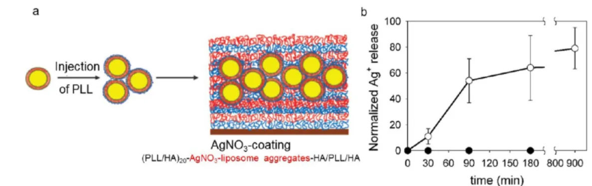
in a PEM by embedding liposomesfilled with AgNO3(Figure
11a).74No silver ions leaked out of the AgNO3-PEM film at
room temperature whereas leaking was observed when the
temperature was raised to 37°C (Figure 11b). After 30 min of
incubation at 37°C, 11% of the silver ions encapsulated in the
coating were released. This percentage increased to 100% after 15 h. A 4 log reduction in the number of viable E. coli was
obtained after 2 h of contact at 37 °C. Thus, by raising the
temperature to above the transition temperature of the vesicles
(∼34 °C), the release of silver ions effectively killed E. coli
populations.
Compounds forming oxygen reactive species known to kill
bacteria were also incorporated into PEM films.
Polyoxome-talates, nanoclusters of transition metals embedded in a
CHI-based PEM film, have been used to kill bacteria by the
oxidation of their cell membrane.87Titanium dioxide (TiO2),
known to form biocidal radicals upon UV irradiation, was also used in combination with silver nanoparticles. Yuan et al. built
silver-loaded CHI-TiO2/HEP films to obtain a coating with
both contact-active properties due to titanium and release-active properties due to silver nanoparticles. In the dark or at low UV-light irradiation, equivalent to the ambient
environ-ment in daylight, this coating’s antibacterial property is stronger
than that of CHI-TiO2/HEP.88 After 7 days of immersion in
PBS, the silver-loaded PEM film maintained its antibacterial
activity under low UV-light irradiation. Corbitt et al. built photoactive conjugated polyelectrolyte capsules that can entrap
and kill bacteria under exposure to white light (Figure 12).89
Illumination of the capsules leads to the production of an oxygen singlet at the polymer/bacteria interface, and this oxygen singlet or some subsequently reactive oxygen intermediates interact with the bacteria, resulting in bacterial
killing.Figure 12b-c shows that the live-to-dead ratio decreases
from 7 to 0.33 when hollow polyelectrolyte capsules (CPE)
with trapped P. aeruginosa are irradiated for 15 min with a
fiber-optic lamp. Gabriel et al. reported a photoactive surface coating
consisting of cross-linked hyaluronic acid and poly(L-lysine)
modified with a photoactive molecule triggered upon
irradiation with near-infrared (NIR) light.90 The strength of
such a coating lies in the fact that it can be triggered from outside once capsules are inside the body since NIR light can penetrate tissue. This is not the case for UV light that is used to trigger titanium oxide, for example.
Beside metal ions, the multilayer technology allows also to incorporate antimicrobial organic small molecules into PEMs which can be further released. Antiseptics and antibiotics
belong to this category. The chemical structure of different
antiseptics and antibiotics that have been embedded into PEMs
are represented inFigure 13.
Applied externally to tissues or skin, antiseptics prevent bacteria, fungi, yeasts, and other microorganisms from colonizing a wound. Antiseptics only weaken and slow down the growth of microorganisms, which prevent bacteria from Figure 11.(a) Liposome complexation with PLL resulted in the formation of AgNO3−liposome aggregates. They were deposited on a (PLL/HA)20
film followed by an additional coating with HA/PLL/HA layers. (b) Release of silver ions from the AgNO3coating versus time (●) at room
temperature and (○) at 37°C. Reprinted with permission from ref74. Copyright 2008 American Chemical Society.
Figure 12.(a) Chemical structures of the two charged poly(phenylene ethynylene)-type conjugated polyelectrolytes. Composite confocal microscope images of the hollow CPE capsules with trapped P. aeruginosa (b) before and (c) after 15 min of irradiation with the fiber-optic lamp. Live and dead bacteria are labeled in red and green, respectively. Approximate live-to-dead ratios are (b) 7.0 and (c) 0.33. Moderate killing is observed before irradiation by contact with conjugated CPE. Reprinted with permission from ref89. Copyright 2009 American Chemical Society.
causing further infection. Antibiotics are used to kill bacteria by inhibiting the cell wall, nucleic acid, and protein syntheses. They are used to deal with systemic infections. Unlike most antiseptics, bacteria may become resistant to antibiotics after prolonged use.
The insertion of antimicrobial organic molecules into PEMs
has been widely studied, and different strategies have been
developed. Positively or negatively charged molecules can bind
to polyelectrolyte chains. Afirst strategy to load PEM consisted
of a simple immersion of preassembledfilms into an antibiotic
or an antiseptic solution.20A second strategy consisted of the
direct deposition of molecules either by association with one of the polyelectrolytes or as one of the multilayer
compo-nents.31,44,75−78 The loading and release of antibiotics was
shown to be tunable by varying the number of deposited layers, the assembly pH, the incubation time, and also by using heat
treatment after film buildup.91 A variety of PEM films have
been used to control drug release by diffusion through the
films,44,75
by hydrolytic degradation of one of the
polyelec-trolytes,31,76,77by pH changes,78or by external triggers such as
the electric potential,79laser light,80,81and ultrasound.80,81
Among the antiseptics used in clinical practice, chlorhexidine (Figure 13) has antimicrobial activity through its positive charge at physiological pH. It destabilizes bacterial cell walls and alters bacterial osmotic equilibrium, leading to the precipitation of the cytoplasmic contents and triggering microbial cell death. Adsorbed alternately with a polyanion, positively charged chlorhexidine was successfully incorporated
into PEM films further stamped on wound dressings.75
Stamped chlorhexidine/PAA PEM films on Biobrane wound
dressings led to a localized nontoxic release of chlorhexidine, allowing the decrease in S. aureus colonization and promoting
normal wound healing in mice.75Methylene blue (Figure 13), a
cationic dye known for its antimicrobial activity, has been inserted into PEMs as a model bioactive compound, but only
Martel’s group used its antimicrobial properties.20 Using the
ability of methylene blue to form stable inclusion complexes
with β-cyclodextrin (β-CD), methylene blue-loaded CHI/
poly(β-CD) PEM films were built and showed sustained
antibacterial activity against S. epidermidis over 72 h of contact. Due to its hydrophobic nature, it is difficult to incorporate
triclosan (Figure 13) directly into LbL-assembled films.
Triclosan has a broad antibacterial spectrum and is also an
antifungal agent. It is found in numerous consumer products such as soaps or detergents. It is also used for healthcare in surgical scrubs and personnel hand washes. By using dendritic block copolymer micelles, composed of a hydrophobic poly(propylene oxide) core and a positively charged poly-(amidoamine) corona, triclosan could be released over a period of several weeks and its activity could be maintained against S.
aureus growth over this period.92 Recently, triclosan was
encapsulated into cetyltrimethylammonium bromide (CTAB)
surfactant micelles that were then inserted by diffusion into a
preformed PEI/PAA PEM film.93 The triclosan-loaded PEI/
PAAfilms could inhibit the growth of both Gram-negative and
Gram-positive bacteria by the sustained release of triclosan molecules for over 20 days. On the basis of an exponentially
growing PEM film, this coating has the property of healing
scratches and restoring its transparency in the presence of water.
Hammond and co-workers developed PEMfilms based on a
hydrolytically degradable poly(β-amino ester) and different
polyanionic polysaccharides that efficiently released gentamicin
(Figure 13) against S. aureus proliferation in vitro and in vivo.31
Later, vancomycin (Figure 13), a glycopeptide antibiotic, was
incorporated into PEMs containing hydrolytically degradable
cationic poly(β-amino esters) and anionic polysaccharides.
Both the dipping and the spraying methods were used. Whereas films obtained by dipping were exponentially growing, sprayed films grew linearly with the number of deposition steps. The amount of drug incorporated was also very dependent on the
buildup method: sprayed films have a 3−8 times higher drug
density than the dipped ones. The release time from sprayed films was greatly accelerated compared to the release from
dippedfilms. By combining the dipping and spraying assembly
with dextran sulfate and alginate, a composite architecture could be designed that has a rapid initial drug release followed by a linear release above the antibiotic minimum inhibitory
concentration.76This study provides a new example of the high
versatility of the PEM technology to finely tune the coating
properties. To prolong the release period of antibiotics
embedded in hydrolytically degraded PEM films, one can
even add into the architecture inorganic particles such as a
laponite clay interlayer that physically block antibiotic diffusion.
Using this strategy with two drugs embedded in the same PEM Figure 13. Chemical structure, with their respective net charge at physiological pH, of different antiseptics (methylene blue, triclosan, and chlorhexidine) and antibiotics (ciprofloxacin, minocycline, gentamicin, and vancomycin) that have been embedded in PEMs.
film, Min et al. obtained a rapid release of gentamicin for the
first few days before reaching a sustained release for weeks.77
Since tissue injuries, inflammation, and infections commonly
lead to a reduced extracellular pH, a pH-triggered release of
antibiotics at the implant−tissue interface is very pertinent.
Depending on the system, the increase or decrease in pH
induces the diffusion of the antibiotics by permeability
changes94 or by a decrease in the binding affinity.78 Hollow
capsules composed of two weak polyelectrolytes PAH and PMAA prepared by LbL assembly showed excellent loading
capacity for ciprofloxacin (Figure 13). The pH-induced
permeability of microcapsules allowed the loading of
ciprofloxacin at low pH and its subsequent release at neutral
pH. The release of ciprofloxacin from microcapsules showed
significant antibacterial activity against bacterial pathogen E. coli
for 7 h.94While most of the coatings can release antibiotics for
a period of some hours to a few days, Zhong and co-workers
reached a sustained release over 35 days of minocycline (Figure
13), an antibiotic and anti-inflammatory drug.78On the basis of
binding-mediated interactions between calcium and minocy-cline, the antibiotic was incorporated and released from a
dextran/gelatin PEM. As the binding affinity decreases with the
pH, minocycline was released at pH 6.0 in 13 days with a high initial burst release and at physiological pH in 35 days with a weak burst release.
Recently, several studies have emerged that used an external
trigger for the release of antibiotics. Gentamicin (Figure 13)
was incorporated into a CHI/Prussian blue PEMfilm deposited
on an electrode to obtain electrically triggered release by the application of a small (<1.0 V) electric potential. When oxidized, the Prussian blue nanoparticles shift from negatively
charged to neutral, inducing the dissolution of the film.
Different drug release kinetics, i.e., burst, on/off, or pulsatile
releases, can be achieved depending on the applied electric
potential profiles. The in vitro efficacy of the released drug was
confirmed against S. aureus bacteria.79Combining antimicrobial
properties of antibiotics and AgNPs, Raishur and co-workers triggered the release of the antimicrobial agents by laser light or
ultrasound due to the rupture of the PEMfilm.80,81
The last family of antimicrobial agents that have been
included into release-killing PEMs are AMPs95−99 and our
group was a pioneer in this domain.95 AMPs, polypeptides
secreted by numerous living organisms against pathogens, gained increased attention due to their broad spectrum of antimicrobial activity and their low cytotoxicity. They predominantly act by disrupting the membrane integrity of pathogen agents and thus are unlikely to initiate the
development of pathogen resistance. Different strategies were
applied to incorporate AMPs in PEM films: either by
electrostatic adsorption when they are cationic,39,95,96,98,100,101
by association with one of the polyelectrolytes when they are
poorly water-soluble,97 or by grafting them on one of the
polyelectrolyte components.29,99Constituted of 40 amino acids
and positively charged, defensin was thefirst AMP embedded
in a PEMfilm.95The antibacterial activity was obtained only for
PLL-terminated PEM films. The adhesion of bacteria on the
PEM surface was needed for the peptide to be sufficiently in
contact with the pathogens, thus exhibiting a contact-killing mechanism. Another positively charged AMP, Ponericin G1, was successfully incorporated into a hydrolytically degradable
PEM, based on poly(β-amino ester), to obtain a release killing
mechanism.96The degradation of thefilm and thus the release
profile depended on the polyanion itself.
Our group introduced antifungal AMP-loaded PEM films
and tested them in vivo.100Embedded in PLL/PGA PEMfilms,
a small polycationic AMP, chromofungin, was able to interact with the surface of the membrane of the fungi, reducing the growth of C. albicans by 64% and fully inhibiting the growth of Neurospora crassa. Tested in vivo on rats with an oral candidosis, the chromofungin-loaded PEMs postponed the
fungal infection.100Using the same PEM system, Karlsson et al.
incorporated cationic amphiphilic oligomers of β-substituted
amino acids (β-peptides) designed to mimic AMPs that
exhibited toxicity toward C. albicans.β-peptides were released
over a period of 17 days without physical PEMfilm erosion.101
Similarly, a cationicβ-peptide-based antifungal agent against C.
albicans was loaded inside PGA/PLL PEM postfabrication by
the incubation of β-peptides. Then these agents were release
Figure 14.(a) Procedure for the preparation of functional antibacterial films. (b) Effect of pH on the retention of peptide L5 from EDA- and AADH-stabilized (PMAA)10hydrogels. The fraction released was determined as the ratio of ellipsometric thickness of peptide loading after
pH-triggered release into 0.01 M phosphate buffer containing 0.2 M NaCl thickness to the thickness of antibiotics loaded into (PMAA)10films at pH 7.5.
All error bars represent the average standard deviation obtained from three separate experiments. Reprinted with permission from ref98. Copyright 2010 American Chemical Society.
over ∼4 months when incubated in physiological media.37 Using amphiphilic polysaccharides, poorly water soluble AMP
(gramicidin A) was incorporated into PEM films.97 The
antibacterial activity of the functionalized PEM films resulted
from a double mechanism: contact-killing and release of the
peptide into the solution surrounding thefilm. To functionalize
stainless steel, widely used in medical devices, Detrembleur and
co-workers developed antimicrobial PEM films based on a
water-soluble poly(methacrylamide) bearing oxidized 3,4-dihydroxyphenylalanine groups (Pox(mDOPA)), PAH, and
an AMP (nisin).29Cross-linking of the PEMfilms and covalent
attachment of nisin were obtained thanks to DOPA moieties at room temperature and without the use of any toxic reagent. Tested against Bacillus subtilis, the coating showed sustainable antibacterial activity even after immersion for one night in tap water or after several mechanical cleanings with a wet sponge.
■
TOWARD A SELF-DEFENSIVE COATINGSelf-defensive coatings can be defined as coatings that become
active only in the presence of pathogens. Along this line, Pavlukhina et al. reported the release of antimicrobial agents using pH variations associated with growth of bacteria as an internal releasing trigger. The self-defensive property of the coating is related to a local change in the environment of the coating due to the presence of the pathogens themselves. This
was the first system developed on the basis of this idea.
Obtained from a preassembled PEM film, Pavlukhina et al.
developed an ultrathin PMAA hydrogel, obtained by the layer-by-layer method, able to incorporate and release polycationic
charged antimicrobial agents, antibiotics, or AMPs.98After the
buildup of PVP/PMAA hydrogen-bonded PEM films, PMAA
was cross-linked with the use of a cross-linker, ethylenediamine
(EDA), or adipic acid dihydrazide (AADH) before the removal
of PVP (Figure 14a).
The ultrathin PMAA hydrogel obtained can thus retain its cationic antimicrobial property by an electrostatic mechanism. pH variations related to the growth of S. epidermidis were used as an internal trigger to release an antibiotic, gentamicin, and an AMP, peptide L5. In response to this pH variation, the
EDA-cross-linked hydrogel swelled while releasing its load (Figure
14b). The antibacterial activity, mainly obtained on the surface
of the hydrogel, was demonstrated after 4 h of incubation with
Gram-positive bacterium S. epidermidis.98 Sukhishvili’s group
further developed the concept of a self-defensive coating using
the acidification of the immediate environment of pathogenic
bacteria. The layer-by-layer assembly of tannic acid (negatively charged natural molecule) and one cationic antibiotic allow the immobilization of a large amount of antibiotics in a stable manner at physiological pH and its release due to the pH
decrease.102A multilayer nanocomposite,
PAA/montmorillon-ite nanoplatelets, was impregnated by an antibiotic, gentamicin, to further release the antibiotic. PAA-bound gentamicin is
released due to the acidification of the environment, whereas
gentamicin adsorbed to montmorillonite remained within the coating. Combining both release-killing and contact-killing properties, this coating is also subjected to a swelling that
hindered bacterial adhesion.103Finally, they developed
poly(2-alkylacrylic acid) hydrogels which become hydrophobic and
bactericidal in response to bacterially induced acidification of
the medium.104
Recently our group pushed even further the concept of self-defensive PEM but based on enzymatically degradable PEM films. The self-defensive property was due to the release of AMP triggered only by the hyaluronic acid degradation induced by the pathogens themselves, thanks to hyaluronidase secretion Figure 15.(a) Schematic representation of chitosan/hyaluronic acid (CHI/HA) multilayers functionalized by an antimicrobial peptide cateslytin (CTL) and its activity toward bacteria and yeasts based on the enzymatic degradation of thefilm and the normalized growth of (b) S. aureus and (c) C. albicans incubated for 1 to 24 h in contact with PEI-(HA/CHI)15−n-(HA-CTL/CHI)nwithn = 0 to 15 and (HA-CTL/CHI)30multilayerfilms.
(Figure 15a).99 Cateslytin (CTL), an antibacterial and antifungal peptide, was directly grafted onto HA (HA-CTL) which was then assembled with chitosan. After 24 h of
incubation, HA-CTL/CHIfilms fully inhibit the development
of S. aureus and C. albicans (Figure 15b,c). Furthermore, the
coating preventsfibroblast adhesion without inducing
cytotox-icity. This highlights a medically relevant application for
prevent infections on catheters or tracheal tubes wherefibrous
tissue encapsulation is undesirable.
■
SUMMARY AND PERSPECTIVES IN PEM-BASED ANTIMICROBIAL SURFACESThe first antimicrobial PEM films were reported in the early
2000s, which was only 10 years after the introduction of PEMs
by Decher.1Since then, the different teams involved in this field
aimed at improving the coating properties in order to render
them usable for specific application. With rare exceptions,
systematic evaluations of the biocompatibility of the antimicro-bial PEM coatings were performed in vitro and even sometimes
in vivo.31,75,100Interestingly and even if complementary studies
are needed, it seems that the stiffness of the coating has
antagonistic effects on bacteria and cells, i.e., stiffer films
promote cell adhesion while they limit bacterial proliferation.48
Each strategy to design an antimicrobial PEM film, i.e.,
adhesion-resistant, contact-killing, and release-killing, was extensively studied. In particular, new polymers were
synthesized to bring about better contact-killing properties,56
and new antimicrobial agents such as antimicrobial peptides
were incorporated into release-killing PEMfilms. Each of the
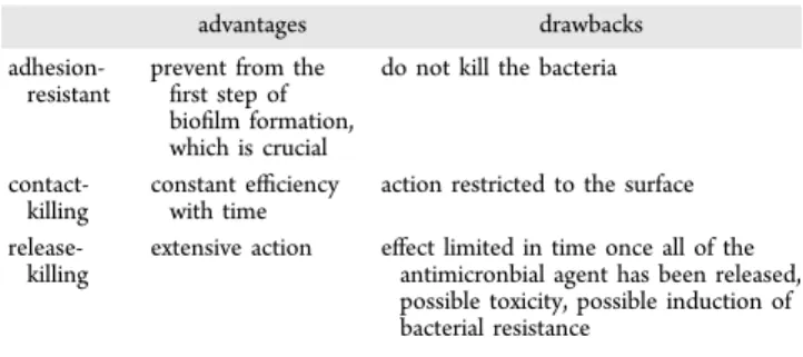
strategies possesses advantages but also drawbacks which can
be summarized inTable 2.
To further increase the antimicrobial abilities of the coatings, two strategies out of the three main ones were combined,
allowing a limitation of the drawbacks. PEM films exhibiting
both adhesion-resistant and contact-killing27,65or both
contact-killing and release-contact-killing properties85,88 were developed.
Release-killing coatings based on the embedding of two
different antimicrobial agents were also prepared.77,88 Some
studies were also performed to increase the durability of the
coatings. The first aspect was to improve the mechanical
properties. This was done by chemically cross-linking the
films29
or by introducing PS microspheres.36The second aspect
was to obtain long-term release periods, especially by slowing
down the diffusion of antimicrobial agents outside the film. For
example, a coating with a clay interlayer barrier could slowly
release antibiotics for weeks.77
An important evolution in PEM coatings was the design of triggered release systems. An internal trigger such as
temper-ature74or pH78,98or an external trigger such as white light,89
laser light,80 near-infrared light,90 or ultrasound81 was
successfully used. However, the problem of the internal trigger is that the release can be immediate after the pH or the temperature change, and thus the antimicrobial agent can be exhausted before the bacteria are even present. Concerning the
external trigger, besides the need for specific equipment, it is
not possible to know when the bacteria are present and thus when it is necessary to activate the trigger. One further step was to obtain a smart release that is triggered only by the presence
of the pathogen. This wasfirst introduced by Sukhishvili’s team
using pH changes induced through bacteria proliferation.98Our
group introduced in 2013 the concept of self-defensive PEM films where the film releases antimicrobial compounds by the
enzymatic degradation of the PEM film, which is due to the
presence of the bacteria themselves.99 In contrary to the pH
change induced by bacteria,98this concept can be extended by
using PEMfilms degradable by only specific enzymes secreted
by pathogens. On the contrary to other release-killing strategies, this smart release strategy allows us to maintain the antibacterial property of the coating as long as pathogens are missing.
All of these results suggest that future antimicrobial PEM films will consist of coatings simultaneously presenting several functionalities and also of smart coatings that will become
active only in the presence of specific pathogens.
■
AUTHOR INFORMATIONCorresponding Author
*E-mail:[email protected]. Tel: +33 (0) 3
88 41 41 60. Fax: +33 (0) 3 88 41 40 00. Author Contributions
The manuscript was written through the contributions of all
authors. All authors have given approval to thefinal version of
the manuscript. Notes
The authors declare no competingfinancial interest.
Biographies
Lydie Séon studied polymer science at the European Engineering School of Chemistry, Polymers and Materials Science in Strasbourg and obtained her M.Sc. in 2011. She received her Ph.D. in physical chemistry from the University of Strasbourg in 2014. Her research interest was the development of new polyelectrolyte-based coatings exhibiting antimicrobial properties. She is now head of R&D projects at VonRoll Meyzieu (Lyon area, France).
Table 2. Main Advantages and Drawbacks of the PEM Films Built Following an Adhesion-Resistant, Contact-Killing, or Release-Killing Strategy
advantages drawbacks
adhesion-resistant
prevent from the first step of biofilm formation, which is crucial
do not kill the bacteria
contact-killing
constant efficiency with time
action restricted to the surface
release-killing
extensive action effect limited in time once all of the antimicronbial agent has been released, possible toxicity, possible induction of bacterial resistance
Philippe Lavalle received his Ph.D. degree in biophysics in 1998 from the University of Strasbourg. After a 2 year postdoctoral position at the Biozentrum in Basel, Switzerland, he was recruited as a researcher at INSERM in 2000 in the biomaterials unit in Strasbourg. He is deputy director of the new INSERM unit“Biomaterials and Bioengineering”. The focus of Lavalle’s research is the design of mechanical stimuli-responsive materials, smart surface coatings preventing nosocomial infection, immunomodulatory coatings to control monocyte differ-entiation, and personalized biomaterials.
Pierre Schaaf earned an engineering degree from ESPCI (Paris) in 1982 and a Ph.D. in physical chemistry from the University of Strasbourg in 1986. He was appointed full professor in 1991 at the Chemistry Engineering School of Strasbourg (ECPM). Since January 2013, he has been the director of the new INSERM unit“Biomaterials and Bioengineering”. His research interests include polyelectrolyte multilayers and stimuli-responsive and bioactivefilms.
Fouzia Boulmedais received her Ph.D. in physical chemistry from University Louis Pasteur in Strasbourg (France) in 2003. Her postdoctoral research at ETH-Z in Zürich (Switzerland) and MPI in
Gölm (Germany) focused on the electrochemical response of multilayers of polyelectrolytes. She joined the Institut Charles Sadron in Strasbourg (France), where she took a position as CNRS researcher in 2006. Since January 2012, she has been the deputy director of Institut Charles Sadron. Her current research involves polyelectrolyte films: their applications in biomaterials and tissue engineering and their buildup by an electrical stimulus.
■
ACKNOWLEDGMENTSL.S. thanks the Ministère de l’Enseignement Supérieur et de la
Recherche for financial support. We acknowledge the MICA
Carnot Institute (SELF-DECAMP project) and the Faculty of
Dentistry of the University of Strasbourg for financial
contributions.
■
ABBREVIATIONSAADH, adipic acid dihydrazide; AgNPs, silver nanoparticles;
AMP, antimicrobial peptide; β-CD, β-cyclodextrin; Biobrane,
biological wound dressing; C. albicans, Candida albicans; CHI, chitosan; CTL, cateslytin; CTAB, cetyltrimethylammonium bromide; DMLPEI, N,N-dodecyl,methyl-poly(ethylenimine); E. coli, Escherichia coli; EDA, ethylenediamine; HA, hyaluronic
acid; HA-CTL-C, cateslytin-grafted HA; HA-VB, HA modified
by photoreactive vinylbenzyl groups; HEP, heparin; PAA, poly(acrylic acid); PAH, poly(allylamine hydrochloride); PC,
p h o s p h o r y l c h o l i n e ; P D A D M A C , p o l y
-(diallyldimethylammonium chloride); PDMS, (dimethylsiloxane); PEG, poly(ethylene glycol); PEM,
poly-electrolyte multilayer; PGA, poly(L-glutamic acid); PLL, poly(L
-lysine); PL-PEG, poly(ethylene glycol)-functionalized phos-pholipid; PMAA, poly(methacrylic acid); Pox(mDOPA), poly(methacrylamide) bearing oxidized 3,4-dihydroxyphenyla-lanine groups; PSS, poly(styrenesulfonate); PVP, poly-(vinylpyrrolidone); PVPh, poly(4-vinylphenol); QAC, quater-nary ammonium compounds; QCHI, quaternized derivatives of CHI; QPVP, quarternized poly(4-vinylpyridine); S. epidermidis, Staphylococcus epidermidis; S. aureus, Staphylococcus aureus;
SWNTs, single-walled carbon nanotubes; TiO2, titanium
dioxide
■
REFERENCES(1) Decher, G.; Hong, J. D.; Schmitt, J. Buildup of ultrathin multilayer films by a self-assembly process: Iii. Consecutively alternating adsorption of anionic and cationic polyelectrolytes on charged surfaces.Thin Solid Films 1992, 210-211, 831−835.
(2) Iler, R. Multilayers of c+olloidal particles. J. Colloid Interface Sci. 1966, 21, 569.
(3) Schaaf, P.; Voegel, J.-C.; Jierry, L.; Boulmedais, F. Spray-assisted polyelectrolyte multilayer buildup: From step-by-step to single-step polyelectrolyte film constructions.Adv. Mater. 2012, 24, 1001−1016. (4) Lavalle, P.; Gergely, C.; Cuisinier, F. J. G.; Decher, G.; Schaaf, P.; Voegel, J. C.; Picart, C. Comparison of the structure of polyelectrolyte multilayer films exhibiting a linear and an exponential growth regime: An in situ atomic force microscopy study. Macromolecules 2002, 35, 4458−4465.
(5) Laugel, N.; Betscha, C.; Winterhalter, M.; Voegel, J.-C.; Schaaf, P.; Ball, V. Relationship between the growth regime of polyelectrolyte multilayers and the polyanion/polycation complexation enthalpy. J. Phys. Chem. B 2006, 110, 19443−19449.
(6) Decher, G. Fuzzy nanoassemblies: Toward layered polymeric multicomposites. Science 1997, 277, 1232−1237.
(7) Richert, L.; Engler, A. J.; Discher, D. E.; Picart, C. Elasticity of native and cross-linked polyelectrolyte multilayer films. Biomacromo-lecules 2004, 5, 1908−1916.