Epidemiol. Infect. (1994). 112. 533-542 5 3 3 Copyright © 1994 Cambridge University Press
Comparison in the immunological properties of Borrelia
burgdorferi isolates from Ixodes ricinus derived from three endemic
areas in Switzerland
C. M. HU1, S. LEUBA-GARCIA1, M. D. KRAMER2, A. AESCHLIMANN1 AND L. GERN1*
1
Institut de Zoologie, Neuchdtel University, CH-2000 Neuchdtel, Switzerland
2
Institut fiir Immunologie, Ruprecht-Karls-Universitdt, Heidelberg, Germany (Accepted 17 January 1994)
SUMMARY
Borrelia burgdorferi isolates were obtained from Ixodes ricinus from three sites in Switzerland. They were examined by SDS-PAGE and immunoblotting. The phenotypes. in respect of three outer surface proteins (Osp), differed between the sites of collection. In site 1, most isolates had an OspA of 31 kDa and an OspB of 34 kDa: in site 2, isolates presenting an OspA of 33 kDa dominated and in site 3, the isolates with an OspA of 32 kDa and an OspB of 35 kDa were most frequent. This distribution differed significantly. About half of the isolates from sites 1 and 3 reacted with anti-OspA monoclonal antibody H5332 compared to 29% from site 2. Site 1 isolates reacted significantly more frequently (81 %) with another anti-OspA monoclonal antibody LA-31 than isolates from site 3 (P < 0-0001). These findings have implications for the epidemiology of Lyme borreliosis, for the further development of serodiagnostic reagents and for the development of a vaccine.
INTRODUCTION
In humans, Lyme borreliosis is a disease caused by infection with Borrelia burgdorferi [1]. B. burgdorferi is transmitted by infected ticks belonging primarily to the Ixodes ricinus complex [2]. In Europe, B. burgdorferi can be isolated from infected ticks, animals and patients. The outer membrane of B. burgdorferi contains at least three outer surface proteins (Osp) A (31-33 kDa), B (34-36 kDa) and C (20-24 kDa) [3-6]. These lipoproteins are embedded in the fluid outer membrane of B. burgdorferi and are encoded by linear plasmids [6, 7]. Although their exact functions have not yet been defined, the outer surface proteins of B. burgdorferi are generally thought to have an important role in the host-parasite interactions during the course of infection. The European isolates are more heterogeneous with respect to their antigenie profiles than the American isolates [8-12].
* Author for correspondence: Institut de Zoologie. Chantemerle 22, CH-2000 Neuehatel. Switzerland.
534 C. M. H r AND OTHERS
Table 1. Characterization of B. burgdorferi isolates (n = 92)
Osp Mws (kDa) MoAbs PoAb
t A ( ( A ^
anti-Strains A B C H5332 LA-2 LA-4 LA-31 LA-25 LA 27 LA-7 p39 22 kDa Site 1 XE83 XE84 XE85 XE190 XE192 XE193 XE308 XE317 XE323 XE15 XE12 XE14 XE196 XE303 XE304 XE305 XE378 XE20 XE25 XE26 XE8 XE19 XE550 XE21 XE24 XE23 Site 2 XE2 XE4 XE58 XE60 XE173 XE352 XE355 XE363 XE3 XE9 XE10 XE22 XE16 XE1 XE5 XE6 XE27 Site 3 XE413 XE443 XE450 XE454 XE456 XE460 33 33 33 31 33 31 33 32 32 31 31 31 31 32 32 31 33 32 31 33 32 32 31 32 31 32 33 33 33 33 33 32 33 32 33 32 32 33 32 32 33 33 32 33-5 31 32 32 31 32 a a a 34 a 34 34 34 35 34 34 34 34 35 a 34 a a 34 a a 35 34 a 34 a a a a a a 34 a 35 a a a a a a a a 35 a a 35 35 34 35 22 22 22 23 22 a a 22 a a 22 a a 23 22 22 22 23 23 22 23 a a 22 23 22 22 22 a a 22 23 22 a a 22 23 a 22 22 22 a 23 22 23 23 21 22 22
Geographical diversity of B. burgdorferi
535 Table 1 {cont.) Strains XE461 XE462 XE472 XE474 XE477 XE478 XE485 XE490 XE496 XE506 XE.5O7 XE517 XE519 XE558 XE601 XE603 XE606 XE607 XE608 XE623 XE624 XE629 XE630 XE632 XE418 XE426 XE429 XE435 XE438 XE463 XE467 XE471 XE470 XE493 XE508 XE537 XE605 XE200 XE207 XE201 XE202 XE203 XE204 Osp A 32 33 32 31 32 32 32 32 32 a 32 32 32 32 32 32 32 32 32 32 32 32 32 32 32 33 32 32 33 31 32 32 32 31 32 32 32 32 32 32 31 32 32 Mus A B 35 a 35 a 35 35 35 a 35 a 35 35 35 a a a 35 a a 35 35 35 35 35 35 a 35 35 a 34 35 35 35 34 35 35 35 a a a a 35 .36 (kDa) (' 22 23 23 23 22 22 22 22 23 22 22 22 22 22 21 22 22 22 22 22 22 a 22 23 a 22 a 22 a 21 21 22 a 21 22 22 22 22 23 a 22 a a MoAbs PoAb anti-22 kDa+ . positive reaction: —. negative reaction: a. absent; n, not tested.
In this study. B. burgdorferi strains from B. burgdorferi ticks collected in three different endemic areas were screened for evidence of phenotypic differences using immunoehemical methods.
536 C. M. H u AND OTHERS MATERIAL AND METHODS
Collection of ticks and isolation of B. burgdorferi
/. ricinus ticks (adults and nymphs) were collected by nagging lower vegetation using a white cotton flannel flag (1 m2) which was dragged behind collectors in three endemic areas: site 1, Bois de l'Hopital forest close to Neuchatel; site 2. Staatswald forest and site 3, Karoline forest around Aarberg. Ticks were collected during April-June and August-November 1987 to 1992 in sites 1 and 2. and in 1989 and 1992 in site 3. For isolation of B. burgdorferi, the midgut of the tick was incubated for 10 days at 34 °C in individual culture tubes containing 4 ml BSK II medium [13] supplemented with rifampicin (50/tg/ml) and phosphomycin (50/^g/ml).
SDS-PAGE and immunoblot analysis
Each isolate was inoculated in 25 ml BSK II medium and after 10 days, the cultures were centrifuged and washed twice with PBS+ 5 mil MgCl2. Whole-cell
lysates (equivalent to 107 cells/lane) were separated by SDS-PAGE using a 12-5% polyacrylamide gel. The gels were stained with Coomassie brilliant blue R 250 [14].
The separated proteins were transferred onto nitrocellulose paper using a transit cell (2117-250 Nova Blot Electrophoretic Transfer Kit, LKB AM Bromma. Sweden) [14]. The monoclonal antibodies (MoAbs): H5332. LA-2, LA-4. LA-31 (anti-OspA) [8, 15], LA-25, LA-27 (anti-OspB) [15], LA-7 (anti-20 kDa protein) [15], and polyclonal antibodies (PoAbs): anti-22 kDa/NE4 [14] and anti-B31. produced by immunizing a New Zealand white rabbit with strain B31 [9]. were used for immuno-blotting. Bound antibodies were visualized by using peroxidase labelled anti-rabbit IgG or anti-mouse IgG antibodies (1:1000, Nordic Immuno-logical Laboratories, The Netherlands).
Statistical analyses
The Fischer's exact test was used to compare the distribution of the different characterized B. burgdorferi isolates derived from different areas. The difference was considered as significant if/3 value was < 0-017 [16].
RESULTS
Characterization o/B. burgdorferi isolates by immunological methods Twenty-six B. burgdorferi isolates were obtained from ticks of the Bois de l'Hopital forest (site 1), 17 isolates from the Staatswald forest (site 2) and 49 isolates from the Karoline forest (site 3). OspA, OspB and OspC were expressed by 91 (99%), 50(54%) and 70(76%) of the 92 isolates (Table 1). Four different phenotypes could be distinguished on the basis of the expression pattern of the Osps, namely, A (11% of isolates), AB (16%), AC (41%) and ABC (32%).
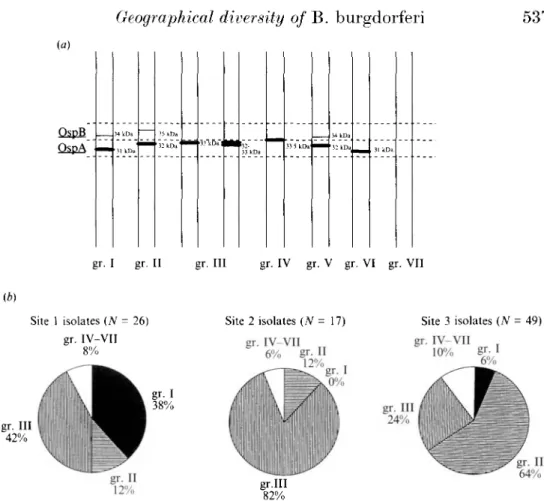
Peter and colleagues [11] distinguished four typing groups of B. burgdorferi (I. II, III and IV) according to the molecular weight of OspA and OspB (Fig. la). Most of our isolates belong to groups I, II and III and only one. from site 3. is in group IV. Seven isolates did not fit into this classification and comprised three additional groups: V. VI and VII (Fig. la).
Geographical diversity o/B. burgdorferi
537 (a) OspB OspA 34 kDa 31 kDa ' 35 k nd 32 k D a 'mm3j'kDa"|Ml! 32-33 kDa 33 5 kDa 34 kDa 32 kDa 31 kDa gr. I gr. II gr. Ill gr. IV gr. V gr. VI gr. VII (b) Site 1 isolates (N = 26) gr. IV-VII 8% Site 2 isolates (N = 17) gr. IV-VII 6% gr- II 12% Site 3 isolates (N = 49) gr. IV-VII 10% gr. Ill 42% gr. Ill 24% gr.III 82%Fig. la and b. Typing groups of B. burgdorferi isolates and their geographical distribution, (a) Schematic description of the typing groups according to the molecular weights of OspA and OspB. The first four groups (gr. I-IV) were suggested by Peter and colleagues [11] and the groups V -VII were proposed by us. Group VII comprised one isolate (XE506) which did not express OspA and OspB. (b) Relative distribution of the different typing groups in the three sites of isolation: groups I and III prevailed in site 1. group III or II prevailed in site 2 and site 3, respectively.
Immunologieal characterization of these isolates revealed a heterogeneous reactivity with the different antibodies used (Table 1).
The different phenotvpes obtained from April to November did not show any differences in their seasonal distribution (data not shown).
Distribution of the characterized isolates in the three studied sites
The isolates from the three different geographic sites of isolation were compared for: (1) the apparent molecular weights (Mw) of OspA. OspB and OspC; (2) their classification into the respective typing groups; (3) differences in the Osp phenotvpes; (4) their reactivity with mono- and polyclonal antibodies specific for B. burgdorferi antigens.
The apparent molecular weights of OspA, OspB and OspC
538 C M . Hu AND OTHERS
Table 2. The presence of the major proteins in isolates derived from three dijferent sites
OspA OspB OspC'
Site 1 (N = 26) Site 2 (N= 17) Site 3 (.V=49) Statistical results P I * P2* P3*
31 kDa 32 kDa 33 kDa 34 kDa 35 kDa 36 kDa 21 kDa 22 kDa 23 kDa
10 9 7 12 3 0 0 12 6 (38%) (35%) (27%) (46%) (12%) (0%) (0%) (46%) (23%) 0 7 10 1 2 0 0 8 3 (0%) (41%) (59%) (6%) (12%) (0%) (0%) (47%) (18%) 6 38 3 3 29 1 5 28 8 (12%) (78%) (6%) (6%) (59%) (2%) (10%) (57%) (16%) 00072 0-7521 00565 00062 10000 10000 0 0158 0 0004 00170 0 00008 0 00007 10000 01889 00081 000002 10000 00014 10000 10000 10000 0-7223 01567 0-4669 05398 0-3165 0-5753 10000 * P I : was as compared between site 1 isolates to site 2 isolates.
P2: was between the site 1 isolates to the site 3 isolates. P 3 : was between the site 2 isolates to site 3 isolates. Bold type: significant differences between both.
however, they varied in molecular weight with the 31, 32 and 33 kDa protein predominating in isolates from sites 1, 3 and 2 respectively (Table 2). OspB was more prevalent among isolates from sites 1 and 3 than site 2; the 34 kDa protein predominated in site 1 and the 35 kDa protein in site 3. OspC was detected in 69. 65 and 8 3 % of isolates from sites 1, 2 and 3 respectively with the 22 kDa protein being the most prevalent in all three sites.
The distribution of typing groups in different areas
Between the sites of isolation, the distribution of the main typing groups differed significantly (Fig. Ib). In site 1, group I and III were most frequent (38 and 42%) and group I was more prevalent than in site 2 (0%, P = 0-0072) and in site 3 (3%, P = 0-0008). Group III was the main group in site 2 (87%). Its presence differed significantly from site 1 (42%, P = 0-0058) and site 3 (24%. P = 0-00004). Group II was more frequent in site 3 (60%) than in site 1 (12%. P = 0-00007) and site 2 (12%, P = 0-0014). About 8% of isolates from each site belonged to groups IV, V, VI or VII.
The distribution of the dijferent Osp phenotypes
Differences in the distribution of the Osp phenotypes according to the expression pattern of the different Osps were observed between the three areas studied (Fig. 2). Phenotype AC (OspA + OspC) was the most frequent in sites 1 and 2. whereas phenotype ABC (OspA + OspB + OspC) predominated in site 3 (54%) and was less frequent in site 2 (12%) than in site 3 (P = 0-0038). Phenotype A (OspA only) was more prevalent in site 2 (29%) than in the other sites.
The immuno-reactivity with dijferent MoAbs and PoAbs
Geographical diversity of H. burgdorferi 539
Site 1 isolates (N = 26) Site 2 isolates (N = 17) Site 3 isolates (N - 49)
53% 54%
Fig. 2. Relative distribution of the Osp phenotypes (in percentage) in B. burgdorferi isolates derived from the three sites of isolation. A, expression of OspA only; AB, expression of OspA and OspB; AC, expression of OspA and OspC: ABC, expression of OspA. OspB and OspC.
Table 3. The reaction of B. burgdorferi isolates derived from three different sites with specific mono- and polyclonal antibodies
MoAbs PoAb Site 1 (X = 26) Site 2 (.V= 1") Site 3 (.Y=49) Statistif-al results P I * P2* P3* H5332 13 (50%) 5 (29%) 29 (59%) O2194 0-4733 00490 LA-2 10 (30%) 3 (18%) 18 (37%) 01874 1-0000 0-2273 LA-4 3 (12%) 0 (0%) 9 (18%) 0-2658 0-5256 0-0977 LA-31 21 (81 %) 7 (41%) 14 (29%) 0-0200 000002 0-3566 LA-25 5 (19%) 2 (13%) 6 (12%) 0-6897 0-4984 10000 LA-27 11 (42%) 1 (6%) 5 (10%) 0 0149 00023 10000 LA-7 4 (15%) 0 (0%) 3 (6%) 01498 0-2270 0-5692 anti-22 kDa 11 (42%) 8 (50%) 37 (76%) 10000 00059 00386 * PI: was as compared between the site 1 isolates to site 2 isolates.
P2: was between the site 1 isolates to site 3 isolates.
P 3 : was between the site 2 isolates to site 3 isolates. Bold type: significant differences between both. MoAbs: monoclonal antibodies.
PoAbs: polyclonal antibodies.
from site 1 reacted more frequently (81 %) with MoAb LA-31 (anti-OspA of B31) than isolates from site 3 (29%; P = 000002) (Table 3). Reactions of the isolates with MoAb LA-27 (anti-OspB) were more frequent in site 1 (42 %) as compared to site 2 (6%) and site 3 (10%); the differences were statistically significant (P = 0-0149 and P = 0-0023). In contrast, 76% of site 3 isolates reacted with anti-22 kDa/NE4 (PoAb) and this was significantly different from isolates from site 1 (42%. P = 00059).
To reveal the number B31-like strains in each area, we also compared our isolates with the B. burgdorferi strain B31, a prototype strain isolated from I. dammini. Five of the site 1 isolates had the same Osp phenotype as B31. Five additional site 1 isolates and three site 3 isolates, all of which expressed the OspC
540 C M . HIT AND OTHERS
protein, were similar to the B31 strain in the protein llw of their OspA (31 kDa) and OspB (34 kDa). However, only two of the site 1 isolates displayed the same reactivity as B31 with the MoAbs described by Barbour and colleagues [8J. Kramer and colleagues [15] and Wallich and colleagues [12].
DISCUSSION
Our study confirms the antigenic heterogeneity of the European B. burgdorferi isolates [8, 9. 11, 12]. In this study we compared isolates from ticks from three endemic areas and showed significant differences in the distribution of the different phenotypes among the isolates of these sites.
According to the molecular weight of OspA and OspB (Fig. 2). each endemic-area presented a main typing group which differed significantly from the two other sites: group I prevailed in site 1, group III in site 2 and group II in site 3. A recent study demonstrated that the molecular weights of OspA and OspB and the representative phenotypes of borrelia isolates from patients with disseminated Lyme borreliosis were different from those obtained from patients with the cutaneous form of Lyme borreliosis [20]. It was reported that most skin isolates presented proteins of 32 kDa (OspA) and 35 kDa (OspB) (group II in our study), whereas the isolates from the disseminated Lyme borreliosis patients expressed an OspA of 32-5 kDa and an OspB of 33-34 kDa (groups III and V in our study). In view of our results, it may be suggested that the clinical manifestations of Lyme borreliosis may differ in different geographical areas. This hypothesis remains to be confirmed.
Immunoreactivity of the isolates varied between the different sites of isolation. Site 1 isolates reacted most with MoAbs LA-31 and LA-27. Site 3 isolates reacted most frequently with the MoAb H5332 and PoAb anti-22 kDa/XE4. In addition, the frequency of isolates reacting with MoAb LA-2, a MoAb which recognizes a protective epitope against B. burgdorferi infection [21], was different in each site. On the other hand, the comparison of our isolates with the strain B31 showed that only two isolates presented exactly the same reactivity with the MoAbs described by Barbour and colleagues [8], Kramer and colleagues [15] and Wallich and colleagues [12]. In view of this, it is suggested that European isolates could elicit specific antibody responses during infection, which differ from one site to another site and which is different from that induced by the strain B31. Therefore, a correct selection of the antigen or antigens seems to be necessary for the serodiagnosis of Lyme disease.
The different protein profile and immunoreactivity of the isolates from different geographical locations may account for seroconversion in people who do not develop Lyme borreliosis, as described in Aarberg (site 3, in this study) where 26 % of an asymptomatic population was seropositive by ELISA and Western blotting [22,23]. Among isolates from the Aarberg area (site 3), 83% expressed the 21-23 kDa proteins and 76% reacted with PoAb anti-22 kDa/XE4 and this was significantly different from the isolates from site 1. Several studies showed that these proteins are important immunogens which elicit the antibody response early after the tick bite [5, 17-19]. It remains to be elucidated whether these proteins could be responsible for the presence of asymptomatic seropositive people in
Geographical diversity o/B. burgdorferi 541
Aarberg by eliciting a protective antibody response. Another explanation could be that some of the strains present in this area were less pathogenic or non-pathogenic and that could depend on their antigenic profiles.
Heterogeneity observed among B. burgdorferi isolates from different sites which are fairly close to each other may have implications for human health if some strains are capable of producing early or late manifestations of Lyme borreliosis, on serodiagnosis and also on the production of protective vaccine since OspA and a 22 kDa protein are actually vaccine candidates. In fact, several studies showed that these antigens elicit protective antibody responses in animal models [21.24-26].
The reasons leading to such a geographic diversity remain unknown but could be due to differences in the reservoir hosts in these three areas. However, our results suggest that studies on the local distribution of B. burgdorferi strains in each endemic area should represent an important step toward understanding the epidemiology of Lyme borreliosis, toward improving serological testing and toward developing an efficient vaccine for populations exposed to bites by infected ticks.
ACKNOWLEDGEMENTS
This work is part of the PhD thesis of one of the authors (Hu C. M.) and it was supported by the Swiss National Science Foundation. We thank Jacqueline Moret for assistance in statistical analysis, Alan Barbour (University of Texas, San Antonio. USA) for providing monoclonal antibodies, Olivier Rais for technical assistance and Larry Kendall for advice on the manuscript.
REFERENCES
1. Steere AC. Bartenhagen NH, Craft JE. et al. Clinical manifestations of Lyme disease. Zentralblatt fur Bakteriologie. Mikrobiol Hyg 1986; A263: 201-5.
2. Burgdorfer W. Discovery of the Lyme disease spirochete and its relation to tick vectors. Yale .I Biol Med 1984: 57: 515-2(1
3. Barbour AG. Tessier SL. Todd WJ. Lyme disease spirochetes and ixodid tick spirochetes share a common surface antigenic determinant defined by a monoclonal antibody. Infect Immun 1983: 41: 795-804.
4. Barbour AG. Tessier SL. Hayes SF. Variation in a major surface protein of Lyme disease spirochete. Infect Immun 1984: 45: 94-100.
5. Wilske B. Preac-Mursie V. von Busch K. Immunochemical and immunological analysis of European Borrelia burgdorferi strains. Zentralblatt Bakteriol Hyg 1986; 263: 92-102. 6. Fuchs R. Jauris S. Lottspeich F. Preac-Mursic V. Wilske B. Soutschek E. Molecular
analysis and expression of a Borrelia burgdorferi gene encoding a 22 kDa protein (pC) in Escherirhia coli. Molec Microbiol 1992; 6: 503-9.
7. Barbour AG. Garon CF. Linear plasmids of the bacterium Borrelia burgdorferi have covalently closed ends. Science 1987; 237: 409-11.
8. Barbour AG. Heiland RA. Tessin HR. Heterogeneity of major protein in Lyme disease borrelia: a molecular analysis of American and European isolates. J Infect Dis 1985; 152: 478-84.
9. Wilske B. Preac-Mursic V, Schierz G. Kiihbeck R, Barbour AG. Kramer MD. Antigenic variability of Borrelia burgdorferi. Annals NY Acad Sci 1988; 593: 126-43.
10. Rosa PA. Hogan DM. Colony formation by Borrelia burgdorferi in solid medium clonal: analysis of Osp locus variants. First International Conference on Tick-Borne Pathogens at the Host-Vector Interface: An Agenda for Research. Saint Paul. Minnesota. USA, 1992; 95-103.
542 C. M. HU AND OTHERS
11. Peter O, Bretz AG. Polymorphism of outer surface proteins of Borrelia burgdorferi as a tool for classification. Zentralblatt Bakteriol 1992; 277: 28-33.
12. Wallich R, Moter SE, Kramer MD, et al. Untersuchungen zur genotypischen und phanotypischen Heterogenitat von Borrelia burgdorferi, dem Erreger der Lyme-Borreliose Infection. In: Hassler D, Kramer M, Maiwald M. Marget W, Zoller L. eds. Forschritte der Infektiologie. Miinchen: MMV Verlag 1992: 176-91.
13. Barbour AG. Isolation and cultivation of Lyme disease spirochete. Yale J Biol Med 1984: 57: 521-5.
14. Hu CM, Gern L, Aeschlimann A. Changes in the protein profile and antigenicity of different Borrelia burgdorferi strains after reintroduction to Ixodes ricinus. Parasite Immunol 1992:
14: 415-27.
15. Kramer MD, Schaible UE, Wallich R, Moter SE, Petzoldt D. Simon MM. Characterization of Borrelia burgdorferi associated antigens bv monoclonal antibodies. Immunobiol 1990:
181: 357-66.
16. Kirk, R. Experimental design. Procedure for behavioral sciences. Belmont. California: Brooks, 1968.
17. Wilske B, Preac-Mursic V, Schierz G, Liegl G, Gueye W. Detection of IgM and IgG antibodies to Borrelia burgdorferi using different strains as antigen. Zentralblatt Bakteriol. Mikrobiol Hyg 1989; Suppl. 18: 299-309.
18. Ma B, Christen B, Leung D, Vigo-Pelfrey C. Serodiagnosis of Lyme Borreliosis by Western immunoblot: reactivity of various significant antibodies against Borrelia burgdorferi. J Clin Microbiol 1992; 30: 370-6.
19. Gern L, Schaible UE. Simon MM. Mode of inoculation of the Lyme disease agent Borrelia burgdorferi influences infection and immune responses in inbred strains of mice. J Infect Dis
1993; 167: 971-5.
20. van Dam AP, Kuiper H, Vos K, et al. Different genospecies of Borrelia burgdorferi are associated with clinical manifestations of Lyme borreliosis. Clin Infect Dis 1993: 17: 708-17.
21. Schaible UE, Kramer MD, Eichmann K. Modolell M. Museteanu C. Simon MM. Monoclonal antibodies specific for the outer surface protein A (OspA) of Borrelia burgdorferi prevent Lyme borreliosis in severe combined immunodeficiency (scid) mice. Proc Nat Acad Sci USA 1990:87:3768-72.
22. Gern L, Frossard E. Walter A, Aeschlimann A. Presence of antibodies against Borrelia burgdorferi in a population of the Swiss Plateau. Zentralblatt Bakteriol, Mikrobiol Hyg
1989; Suppl. 18: 321-8.
23. Gern L, Garcia S, Frossard E. Characterization and follow-up the IgG antibody response against Borrelia burgdorferi using Western blot in a seropositive (ELLSA) population from an endemic area. Bull Soc Neuchateloise Sciences Naturelles 1993; 116: 5-14.
24. Fikrig R, Barthold SW, Kantor FS, Flavell RA. Protection of mice against the Lyme disease agent by immunizing with recombinant OspA. Science 1990; 250: 553-6.
25. Fikrig R, Barthold SW, Kantor FS, Flavell RA. Long term protection of mice from Lyme disease by vaccination with OspA. Infect Immun 1992; 60: 773-7.
26. Simon MM, Schaible UE, Kramer MD, et al. Recombinant outer surface protein A from Borrelia burgdorferi induces antibodies protective against spirochetal infection in mice. J Infect Dis 1991: 164: 123-32.