Angiotensin ll-receptor subtypes in human atria and
evidence for alterations in patients with cardiac
dysfunction
H. Rogg*, M. de Gasparo*, E. Graedelf, P. Stulzt, F. Burkartf*, M. Eberhardt
and P. Erne§
"Research Department, Pharmaceuticals Division, Ciba-Geigy Limited, CH-4002 Basel, \Cardio-Thoracic Surgery Unit, University Hospital, CH-4031 Basel, \Division of Cardiology and Department of Research, University
Hospital, CH-4031 Basel, and the ^Department of Cardiology, Cantonal Hospital, CH-6000 Lucerne 16, Switzerland
Angiotensin II (All) has been implicated as an important factor in the pathophysiology of heart diseases. Following the recent identification of two subtypes of the AH receptor in cardiac tissue of animals, we investigated the possible occurrence of these, or similar, subtypes in human atrial tissue. In right-atrial tissue from patients undergoing heart surgery, we determined the All-receptor profile in receptor binding studies, using [l25I]-angiotensin as radioligand and All as well as two compounds selective for the receptor subtypes to identify and quantify All-receptor subpopula-tions. In 35 patients (23 requiring coronary bypasses, 10 valvular surgery and two combined coronary and valvular surgery), the left-ventricular ejection fraction was deter-mined in the preoperative phase, and right- and left-atrial pressure during surgery. In membranes of human right atria, All receptors are present in high density (median: Bmax=294 fmol. mg ~ ' protein, range: 111-2073) and two different subtypes can be distinguished. Type-1 receptors (AT,) accounted for 33 ± 10% of the population whereas
type-2 receptors (ATj) made up 67 ± 10% of the popu-lation. There was no correlation between any of the measured cardiac functions and total All-receptor density or receptor affinity. However, the percentage of AT, recep-tors was higher in the atria of patients with normal right-atrial pressure; left-ventricular ejection fraction was positively and right-atrial pressure inversely correlated with the percentage of AT, receptors (r=0-740 and -0-901, respectively; P<0-001, for both). Moreover, the percentage of AT2 receptors was directly correlated with the levels of left-atrial pressure (r=0-853; P<0001). It is concluded that the ratio of AT, to AT2 receptors correlates well with right-atrial pressure and left-ventricular function. This is a first indication of a possible involvement of All-receptor subtypes in the pathophysiology of cardiac dysfunctions. (Eur Heart J 1996; 17: 1112-1120)
Key Words: Human, angiotensin II, receptor, subtype,
AT,, AT2, cardiac, atrium, CGP 42 112 A, Losartan.
Introduction
Angiotensin II (All) is the principal effector peptide of the renin-angiotensin system, which is the major regulat-ory mechanism for electrolyte balance and blood pres-sure. There is also increasing evidence of the existence of an intrinsic renin-angiotensin system in the heart: find-ings made mainly in animals show the presence in myo-cardial tissue of mRNAs coding for angjotensinogen and renin'1"41, and of renin, angiotensin-converting enzyme, and All, as well as its receptor15"131.
Manuscript submitted 6 September 1995, and accepted 13 November 1995.
• Since this paper was submitted this author has died.
Correspondence. Dr H. Rogg, Ciba-Geigy Ltd., K-125.14.19,
CH-4002 Basel, Switzerland.
The physiological and pathophysiological roles of this All-generating pathway and the circulating All in the heart have not yet been fully elucidated. However, they may include inotropic and chronotropic activities, effects on coronary vascular tone, arrhythmogenesis, and impairment of relaxation, as well as a permissive or regulatory role in modulating cardiac growth and development'14"181. There is indirect evidence tending to implicate the renin-angiotensin system in the initiation of proto-oncogene expression and cell growth in smooth muscle and myocardial cells'19"251, and inhibition has been shown to prevent left ventricular hypertrophy in rats with pressure overload'26"281 and to promote the regression of chronic pressure-overload hypertrophy in man'29"301.
Recently, two distinct subtypes of All receptors have been discovered'31"341. They can be identified
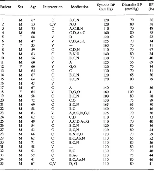
Table 1 Patient characteristics Patient 1 2 3 4 5 6 7 8 9 10 11 12 13 14 15 16 17 18 19 20 21 22 23 24 25 26 27 28 29 30 31 32 33 34 35 Sex M M M M F M F M M M M F M M M M M F M M M M M M M M M M M M M M M M M Age 67 53 73 60 68 47 33 59 63 56 60 60 65 67 64 43 67 65 58 72 60 58 63 62 49 54 53 66 62 71 58 53 59 41 67 Intervention C C,V C C V V V C
c
c
V Vc
c
c
Vc
Vc
c
c
c
Vc
Vc
c
c
c
c
Vc
c
c
c,v
Medication B,C,N N,O A,C,B,N C,D,Ao,O D C,D,Ao,G — C,D,N B,N,0 B,C,N A G,0 C B,C,N B,C,N — A D,G,O B,C,N C,O B.C.N B,C A,B,C,N,G,T CD A,C,D,Ao,G B,C,N B,C,N B,N,C,O B,C,Ao,N B,C,N — B,C B,Ao B,C,Ao,N D, 0 Systolic BP (mmHg) 120 120 110 160 120 125 105 110 140 130 125 120 130 120 170 140 160 100 130 165 150 125 110 110 120 130 120 110 110 120 130 110 110 110 Diastolic BP (mmHg) 70 80 70 80 60 70 70 70 80 70 56 70 70 65 90 — 80 100 80 75 65 90 70 70 70 80 80 70 65 80 80 70 75 80 80 EF (%) 66 58 49 68 62 34 31 67 64 40 69 34 51 50 79 — 36 41 58 59 50 46 56 33 40 56 64 59 52 36 35 48 44 46 41C=coronary bypass surgery; V = replacement of valve(s).
A = ACE inhibitor; Ao = anticoagulant; B=/J-adrenergic antagonist; C = calcium antagonist; D=diuretic; G=glycoside; N = nitrate; T=other cardiotonics; O=others.
BP=blood pressure. EF=ejection fraction.
unequivocally with the aid of the novel agents Losartan and CGP 42 112 A, which have an inverse affinity for the subtypes and selectivity factors of more than lOOO13'1. Losartan is a specific non-peptidic AH antagonist hav-ing a high affinity for the AT, subtype, to which several functions, such as vasoconstriction, can be attributed; the function of the AT2 subtype, which shows the greatest affinity for CGP 42 112 A remains unclear131"351. Both subtypes have been demonstrated in rabbit myocardial tissue1361 and rat myocardium'371.
The potential functional significance of the two cardiac All-receptor subtypes is unknown. Up to now, no attempt had been made to differentiate All-receptor subtypes in human myocardial tissue. This paper reports, for the first time, the existence and the proper-ties of two subtypes in human right atria. If All-receptor subtypes play a role in cardiac function, their abundance or properties may differ according to the degree of dysfunction. Studies in rats have indicated changes in All-receptor subtypes with cardiac hypertrophy using
different models'38'39'. To test this hypothesis, we inves-tigated the All-receptor subtypes in right-atrial tissue from patients with varying degrees of cardiac dysfunc-tion. Our results demonstrate that the relative propor-tions of the All-receptor subtypes are closely associated with altered diastolic and systolic properties of the heart.
Methods
Patients and study design
Atrial-tissue specimens (0-6 ± 0-2 g; mean ± SD) were excised from the right-atrial appendage during cannu-lation in 35 patients (31 males, four females, aged 60 ± 9 years, weighing 81 ±12 kg and 173 ± 8 cm in height) undergoing open-heart surgery. The left-ventricular ejection fraction was derived from the levocardiogram recorded during preoperative left-heart catheterization in all but one patient (51 ± 13%, n = 34).
Prior to surgery, all but three patients had been receiving medical therapy (Table 1): two patients had been on monotherapy and the remaining 30 on com-bined medication with two or more drugs of the follow-ing categories (calcium-channel blockers in 22; nitrates in 17, beta-blockers in 18; antiarrhythmic agents in eight; glycosides in six; diuretics in eight; converting-enzyme inhibitors in five; antiplatelet drugs in 10; oral anticoagulants in seven). The antiplatelet agents were withdrawn 10 days before the operation and the anticoagulants on the day before surgery.
On the night before surgery, the patients received flunitrazepam (1 mg) orally and 1 h before induction of anaesthesia morphine (01 mg. kg"1) with hyoscine hydrobromide (01 mg . 25 kg" ') intramuscularly. Anaesthesia was induced with either sodium thiopental (2-3 mg. k g " ' ) or etomidate (0-2 mg . k g " ' ) plus fen-tanyl citrate (3-5 ug. kg"1). To maintain anaesthesia, enfluorane (0-3-10 volume % in an air/oxygen mixture) and supplementary doses of fentanyl were used. Pan-curonium bromide (01 mg . k g " ' ) was given as muscle relaxant. Ventilation was controlled to maintain the partial pressure of carbon dioxide between 4-5 and 5 0 kPa and the inspiratory flow of oxygen was set at 0 6 . Right- (7 ± 4 mmHg) and left-atrial pressures (8 ± 5 mmHg) were recorded, and the values closest to the time of excision of right-atrial tissue (maximum 20 min) were taken for analysis. In 23 patients, coronary-bypass surgery was performed, in 10 patients replacement of one or two valves (aortic valves in six; four for aortic stenosis, three for aortic regurgitation: mitral valves in four; three for mitral regurgitation and one for mitral stenosis) and two patients received bypass grafts and aortic valve replacements (both for aortic stenosis, with additional regurgitation in one). The clini-cal and biochemiclini-cal evaluations were performed separ-ately, and the investigators were blinded for putative dependent variables. The codes were not broken until the two sets of data were ready to be collated.
Membrane preparation
Intraoperatively excised right-atrial tissue was immedi-ately rinsed in ice-cold cardioplegic solution, frozen in liquid nitrogen and stored at — 80 °C. Atrial membranes were prepared using a slight modification of a published procedure'361. In brief, the tissue was thawed on ice and a homogenate (20% w/v) was prepared in 0-25 M sucrose, 25 mM Tris pH 7-5, with a Polytron (2 x 30 s) at setting 6. The homogenate was sedimented at 10 000 g for 20 min, the pellet discarded and the supernatant centrifuged at 100 000 g for 30 min. The pellet was resuspended in 5 ml of 0 6 M K G and 30 mM histidine at pH 7 0 (per g of tissue) and resedimented at 100 000 g for 30 min. The pellets obtained from the final centri-fugations were washed three times and resuspended in 25 mM Tris pH 7 5 and 10 mM MgCl2 using a tight-fitting Teflon-pestle homogenizer. AH steps above were carried out at 0-4 °C. The membrane preparations
were frozen in liquid nitrogen and held in aliquots at - 80 °C until used. There was no apparent loss of All-binding activity with time. Protein was assayed by the method of Bradford1401 using bovine serum albumin from Sigma as standard.
Binding assay
Equilibrium-binding studies were performed at 25 °C for 50 min in 01 ml Tris pH 7-5 containing MgCl2 (10 mM), bovine serum albumin (0-2%), membranes (4-9 ± 2-8 ug protein; mean ± SD), [I25I]-AII (2200 Ci/mmol pur-chased from Anawa, Wangen, Switzerland; Concen-trations as stated in the figures), and the peptidase inhibitors antipain, phosphoramidon, leupeptin, pepsta-tin, bestapepsta-tin, amastapepsta-tin, each at 1 ug . m l " ' (all obtained from Novabiochem, Laufelfingen, Switzerland), and bacitracin at lOOug.ml"1. Control experiments showed that under these conditions equilibrium was reached and the binding was linearly related to the amount of protein used. Bound and free ligand were separated by addition of 4 ml of ice-cold Tris pH 7-5 and subsequent rapid filtration through Whatman GF/F filters presoaked in 0 1 % bovine serum albumin. This was followed by three additional washes with 4 ml of the same buffer, and radioactivity trapped on the filter was then measured in a gamma-counter (Pharmacia LKB, Uppsala, Sweden) at 80% efficiency. Non-specific binding was determined in the presence of 10 uM un-labelled All (human sequence; obtained from Bachem, Bubendorf, Switzerland). Typically, values of total bound All were about 2000 cpm and non-specific binding amounted to about 500 cpm.
The proportions of the receptor subtypes were determined in inhibition experiments performed in trip-licate, using selective concentrations of CGP 42 112 A (0-3 uM) and Losartan (3 uM), respectively (Losartan and CGP 42 112 A were synthesized by Ciba-Geigy). The integrity of the radioligand before and after incu-bation was determined by reversed-phase thin-layer chromatography, as described elsewhere'361. These con-trols showed that under the incubation conditions used as much as 84 ± 1% (n = 35) of the radioligand remained intact.
Data analysis
The binding data were analysed using the LIGAND curve-fitting program'4'1. The data were obtained from experiments each performed in duplicate, and the geo-metric means are given when dose-response curves were averaged. The means of the percentages of AT! and AT2 receptors calculated from the two determinations using selective concentrations of CGP 42 112 A and Losartan, respectively, were averaged. All other data were ana-lysed with Student's t-test and linear regression. Results are expressed as mean ± SD, unless otherwise stated. Differences at /*<0-05 were accepted as statistically significant.
12 a.
i
8 -4 - /' /
1
^^
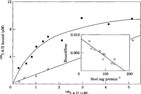
0.010 0.005 0 I ^__^_ " • ^ v O o\. o "bs. 1 \ 100 finol mg protein" 1 _• .— A 200 1 12SI-AII(nM)Figure 1 Saturation binding of |I25I|-AII to human right-atrial membranes.
Specific ( • ) and non-specific binding (A) as a function of increasing concen-trations of | IJ-AII is shown. A Scatchard analysis of the data is given in the inset. The values given are representative of three experiments. The experiment is an example showing the occurrence in human atrial membranes of ATI receptors having a high affinity and density.
Results
Identification of All-receptor subtypes in
human atrial membranes
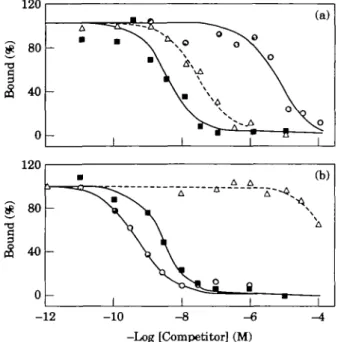
Saturation binding of [125I]-AJI to human right-atrial membranes is depicted in Fig. 1. The data demonstrate the presence of specific sites with high affinity for AIL In the 35 specimens investigated, the median receptor density was 294 fmol. mg~' protein (range 111-2073). Half-saturation of the receptors is reached at 1-3 nM (range 0-6-2-0). To characterize the All receptors in human atria, we performed separate competition-binding experiments (n = 35 for All; n = 2 for each subtype-selective competitor) (Fig. 2). AH displaces the radioligand monophasically. The Hill coefficient is near unity. These data indicate the presence of one homo-geneous class of high-affinity receptors. The highly selective ligands, CGP 42 112 A and Losartan, possess-ing an inverse affinity for AH-receptors subtypes, how-ever, displace the radioligand in a biphasic manner, unmasking the presence of two distinct binding sites. These were further characterized in competition-binding experiments performed in the presence of CGP 42 112 A and Losartan in concentrations sufficient to block their high-affinity sites (Fig. 3). These experiments show that
All possesses the same affinity for the two receptor
subtypes. However, Losartan has a 1000 times higher affinity for the AT, than for the AT2 receptor and, conversely, CGP 42 112 A has a high affinity for the AT2 receptor and only a very weak affinity for the AT, receptor. This demonstrates the pharmacological differ-ence between these two entities. Furthermore, it was
100
o m
-Log [Competitor] (M)
Figure 2 Competition curves with AH ( • ) , CGP 42 112
A ( • ) and Losartan (A) in human right-atrial membranes. The experiments were carried out in the presence of 0-5 nM |12SIJ-A1I. The biphasic displacement curves of the
subtype-specific antagonists demonstrate the presence of AT, and AT2 receptors.
found that they are chemically distinguishable. They are, for instance, affected differently by the disulphide-reducing reagent dithiothreitol: the AT, receptor is inhibited, whereas the binding of the hormone to the AT2 receptor is stimulated (Fig. 4).
AH receptors and correlation with cardiac
parameters
Figure 5 illustrates the lack of correlation between the density of total All receptors and the ejection fraction.
120 * 80 (S 40 0 - i i 1 H (a) > \ o 1 2250 1ZU 80 40 0 • \ \ \ \ 1 1 A A L
a
i (b) \ \\ 1 -12 -10 -Log [Competitor] (M)Figure 3 Inhibition of [I25I1-AII binding, by A l l ( • ) ,
CGP 42 112 A ( • ) and Losartan (A) to AT, and AT2
receptors in human right-atrial membranes, (a) The AT, receptor was unmasked by blocking AT2 receptors with
CGP 42 112 A (0-3 uM). (b) The AT2 receptor was
unmasked by blockade of AT, receptors using Losartan (3 uM). The experiments were carried out in the presence of 0-5 nM [I25I]-AII, and the data given are
represen-tative of two experiments each. The two sets of exper-iments show the pharmacological profile of the AT, receptor (binding affinity orden AII>Losartan>CGP 42 112 A) and the AT2 receptor (CGP 42 112
A > A D > Losartan).
200
-1 -10 Dithiothreitol (mM)
100
Figure 4 Effect of various concentrations of
dithiothrei-tol on the binding of 0-5 nM |125I1-AII, to subtypes of A l l
receptors of human atrial tissue. The experiments were carried out in the presence of 0-3 uM CGP 42 112 A (O) and 3 uM Losartan ( • ) , respectively, to investigate the effect of DTT on subtypes 1 and 2 separately (see Figs 2 and 3). The data given are representative of two experiments each.
n
30 40 50 60 70
Ejection fraction (%)
Figure 5 Lack of relation of between density of total All
receptors and ejection fraction.
Moreover, there was no demonstrable association between alterations in left- or right-atrial pressure and changes in the density of total All receptors, or their affinity for the hormone (data not shown).
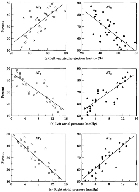
Both AH-receptor subtypes were detected in the atria of all 35 patients. Although the mean distribution comprised 33 ± 10% AT, receptors and 67 ± 10% of the AT2 type, there was a fairly wide range of interindi-vidual variation: some patients had almost identical proportions of the two subtypes, but in extreme cases the ratio of AT^AT, was up to 7:1. The analysis of the data revealed a striking association between the propor-tions of the receptor subtypes and the various cardiac parameters measured (Fig. 6). It was found that the proportion of AT, receptors increases significantly with the left-ventricular ejection fraction (Fig. 6(a)). There was a highly significant, direct correlation between the height of the left-atrial pressure and the proportion of AT2 receptors (Fig. 6(b)). Furthermore, the height of right-atrial pressure and the percentage of AT2 receptors were positively correlated (Fig. 6(c)). In summary, these findings indicate that the proportion of AT, receptors in right-atrial tissue decreases, and concomitantly the pro-portion of AT2 receptors increases with the degree of impairment of the cardiac functions measured.
Analysis of relationships between the absolute density of the receptors was hampered by the vari-ation in the total receptor number, mainly caused by two extreme values with densities exceeding 1000 fmol. mg~ ' of protein (see Fig. 5). Hence, with the exception of two individuals the patients had a receptor density with a variation expected in biological samples. We carefully investigated all characteristics including medication of two 'outlayers' and found no explanation for this phenomenon. Re-analysis of the data after exclusion of these two values revealed significant corre-lations between the absolute density of AT, receptors (%AT, x BmaJ and alterations in the various cardiac functions: the height of the ejection fraction correlates positively (r=0-44, P<002, n = 32) and the heights of both left- and right-atrial pressures showed an inverse correlation (r= -0-45, P<0-02, n = 31) and (r= -0-40,
40 60 80 20 40 (a) Left ventricular ejection fraction (%)
80
12 16 0 4 (b) Left atria! pressure (mmHg)
8 12
s,
4 8 12 16
(c) Right atrial pressure (mmHg)
Figure 6 Relationship between tbe proportion of receptor subtypes and cardiac
par-ameters. The data show significant correlations between tbe proportions of AH receptor subtypes and ejection fraction (a), left-atrial pressure (b) as well as right-atrial pressure (c). In tbe panels depicting tbe fraction of AT2 receptors, the circles indicate tbe values
from patients who underwent coronary-bypass surgery and tbe triangles those from patients who underwent valvular surgery. The data indicate a decrease in the proportion of AT, receptors with increasing degree of dysfunction of all cardiac parameters measured (and a reciprocal situation for tbe AT2 receptors) irrespective of tbe underlying disease
state. For (a), r=O740 and /»<0-001; (b), r= - 0-853 and /><0-001; (c) r= - 0-901 and
P<0-03, n = 33). This indicates that a decrease in AT,
receptor numbers in the right atrium is associated both with increased right-atrial pressure and with impaired left systolic and diastolic function. The AT2 receptor density showed an opposite tendency [ejection fraction:
r= -0-20 (ns); left-atrial pressure: r=0-34, .P=0-06 (ns), right-atrial pressure: r=0-40, P<003]. This was ex-pected, since the data on total receptor density and the parameters of cardiac function showed no correlation. However, no significant correlation between any
receptor parameters and the patients' ages or body weights was demonstrable (data not shown).
In conclusion, these data indicate that the rela-tive proportions of AT, and AT2 receptors in human atrial tissue show a good correlation with right-atrial pressure and left-ventricular functions in these patients.
Discussion
In right-atrial tissues from 35 patients undergoing car-diac surgery, we have demonstrated the presence of a dense population of high-affinity All receptors, compris-ing both AT, and AT2-receptor subtypes, and found the proportions as well as the absolute density of these two subtypes to be related to the height of right- and left-atrial pressures and the left-ventricular ejection frac-tions. Our findings contrast with the results of previous studies in human cardiac tissue1'0'42', in which very low densities of All receptors (Bmax<7 fmol , m g " ' protein) were detected in normal subjects and patients with idiopathic or dilated cardiomyopathy: the values quoted for right-atrial tissue (the highest density recorded) were more than 40 times less than those determined in our patients. This discrepancy may have been due to metho-logical differences, such as dissimilarities in the purity of the membranes, or possibly to denaturation of the receptors, since the cardiac tissue had been kept for several hours in ice-cold cardioplegic solution before being frozen'101 whereas in the present study fresh, snap-frozen tissue was used. In addition, we investigated the receptors in atrial tissue, which in rabbit hearts has been shown to possess an All receptor density about 10 times higher than that of ventricular tissue151. In very preliminary experiments this difference has also been found in human hearts in our laboratory (H. Rogg, unpublished observations). However, the reported absence of any clear-cut difference in the density of total All receptors in normal and failing hearts agrees with our findings showing a lack of correlation between this parameter and the various cardiac functions.
This first study of human cardiac All receptors was carried out prior to the initial report of the existence of All-receptor subtypes'3'1 and the discovery of their coexistence in rabbit ventricular tissue1361, and the tissues investigated were cardiac explants. In our study, we examined specimens excised during heart surgery. The possibility that the anaesthetics or the cardiac drugs administered prior to surgery may have influenced the results cannot be excluded. Although the tissue originated from patients on multiple drugs, specimens exposed to single drugs were too few in number to be analysed separately. At least no obvious effect of angiotensin-converting enzyme inhibitors (n = 5 of 35 patients) on the proportions of All receptor subtypes was detectable (data not shown).
The analysis of the 35 specimens revealed a direct relation between the proportion of All receptor subtypes and right- and left-atrial pressures, as well as left-ventricular ejection fraction. Since these three
parameters were interrelated in the patients studied, the influence of a single abnormal parameter cannot be determined separately.
The main outcome of our study is the demon-stration of the occurrence of the two All-receptor sub-types in human atrial tissue and their close relations to various cardiac functions. The properties of the two atrial subtypes are indistinguishable from those in other tissues such as uterus, adrenal glands and vascular smooth-muscle cells'3'1, as well as in rabbit ventricles'361. The order of affinity of the AT, subtype (AII>Losartan£>CGP 42 112) clearly differentiates it from the AT2 subtype (CGP 42 112 A>AIl£>Losartan). Furthermore, the two subtypes have different suscepti-bilities to the disulphide-reducing reagent dithiothreitol in human atria and other tissues'31"34'361: the AT! recep-tors is inhibited, whereas the binding of the hormone to the AT2 receptor is stimulated by DTT. This furnishes additional evidence for the distinct nature of the two subtypes of All receptors in human atrial tissue.
Atrial tissue was chosen for these investigations of human cardiac All-receptor subtypes because it can be obtained fresh and because earlier reports indicated that atria may be the cardiac tissue with the highest receptor density, both in animals and in man'5'101. The demonstrable correlations between different proportions of the two All-receptor subtypes and right-atrial pres-sure, and especially left-ventricular systolic and diastolic function, afford the first indication of the possible involvement of these entities in a pathophysiological process. The individuals investigated have the character-istics of a large proportion of cardiac patients suitable for heart surgery. The receptor profile of patients with either valvular disease or coronary artery disease did not deviate from the correlations (Fig. 6). However, none of them had other heart disease such as dilated cardiomy-opathy. Accordingly, our finding cannot be extrapolated to such conditions. Although at this stage it is obviously impossible to tell whether these changes are a cause or a consequence of cardiac dysfunction, it is tempting to speculate that the cardiac AT, receptor, which is known to be coupled to a G protein'431, may be down-regulated with the degree of cardiac dysfunction, in a similar fashion to the alterations in beta-adrenergic receptors in these conditions'441. Thus, the effects of AT, receptor, known to be mediated via phosphoinositol break-down'341 leading to an elevation of intracellular free calcium'35', might conceivably result in sluggish calcium transients and hence reduced contractile force in patients with altered left-ventricular systolic function as a consequence of receptor down-regulation.
An increase in angiotensin-converting enzyme activity and mRNA expression has been reported in rats with pressure-overload left-ventricular hypertrophy1451. This indicates that in some forms of cardiac dysfunction the local renin-angiotensin system may be activated and linked to cardiac hypertrophy. Moreover, All is thought to play a permissive or regulatory role in modulating cardiac growth and development'14"18'. The reported alteration in the proportion of the All-receptor subtypes
associated with cardiac dysfunction in this study may have both theoretical and practical implications. The possibility cannot be excluded that these alterations may play an important role in the progression of structural changes in the myocardium, either linked to ischaemic heart disease or to chronic pressure overload. Unfortu-nately, the effector pathway(s) of the AT2 receptors, and their function is still controversial. Therefore, it would be premature to speculate on a permissive or restrictive involvement of AT2 receptors in the regulation of growth. However, the high density of this subtype found in the fetus may play a functional role in development146-471.
The association between both the proportion and the density of the AT, receptor subtype and cardiac dysfunction reported in this study may be of direct therapeutic relevance. The non-peptidic AT,-specific receptor antagonist Losartan and other compounds of the same class are in preclinical or clinical development. Recent initial reports of the effects of Losartan in man describe a marked elevation of circulating AII[481. In a recent study comparing the effects of Losartan and an angiotensin-converting enzyme inhibitor in rats, it was demonstrated that the latter compound causes no appre-ciable change in circulating All1491. It is consequently not admissible to draw inferences regarding the effects of AT,-selective inhibition from the cardiac effects of angiotensin-converting enzyme inhibition. Given the presence of both the AT, and AT2 receptor subtypes on human cardiac tissue, blockade of the AT, subtype by treatment with such drugs exposes the AT2 subtype to elevated All levels, with unpredictable, either desirable or undesirable, long-term cardiac effects.
We are indebted to Dr W. Kremers for his expert assistance in the statistical analysis and to Mr A. H. Kirkwood for his invalu-able help in the preparation of this manuscript. The contributions of our chemists Dr B. Kamber and Dr F. Ostermayer to this work are gratefully acknowledged, and we thank Mr R. Reut and Mr A. Schmid for their excellent technical assistance. P. Erne and M. Eberhard were supported by the Swiss National Foundation Grant No. 32/029 975.90, the Swiss Foundation of Cardiology, and the K. Maier Stiftung.
References
[1] Jin M, Wilhelm MJ, Lang RE, Unger T, Lindpaintner K, Ganten D. Endogenous tissue renin-angiotensin systems. Am J Med 1988; 84 (Suppl 3A): 28-36.
[2] Dzau VJ, Ellison KE, Brody T, Ingelfinger J, Pratt RE. A comparative study of the distributions of renin and angio-tensinogen messenger ribonucleic acids in rat and mouse tissues. Endocrinology 1987; 120: 2334-8.
[3] Ohkubo H, Nakayama K, Tanaka T, Nakanishi S. Tissue distribution of rat angiotensinogen mRNA and structural analysis of its heterogeneity. J Biol Chem 1986; 261: 319-23. [4] Kanapuli SP, Kumar A. Molecular cloning of human angi-otensinogen cDNA and evidence for the presence of its mRNA in rat heart. Circ Res 1987; 60: 786-90.
[5] Baker KM, Campanile CP, Trachte GJ, Peach MJ. Identifi-cation and characterization of the rabbit angiotensin II myocardial receptor. Cir Res 1984; 54: 286-93.
[6] Rogers TB, Gaa ST, Allen IS. Identification and characteriz-ation of functional angiotensin II receptors on cultured heart myocytes. J Pharmacol Exp Therap 1986; 236: 438-444.
[7] Wright GB, Alexander RW, Ekstein LS, Gimbrone MA. Characterization of the rabbit ventricular myocardial receptor for angiotensin II. Mol Pharmacol 1983; 24: 213-21. [8] Saito K, Gutkind JS, Saavedra JM. Angiotensin II binding
sites in the conduction system of rat hearts. Am J Physiol 1987; 253: H1618-22.
[9] Mukherjee A, Kulkarni PV, Haghani Z, Sutko JL. Identifica-tion and characterizaIdentifica-tion of angiotensin II receptors in cardiac sarcolemma. Biochem Biophys Res Comm 1982; 105: 575-81. [10] Urata H, Healy B, Stewart RW, Bumpus FM, Husain A. Angiotensin II receptors in normal and failing human hearts. J Clin Endocrin Metabol 1989; 69: 54-66.
[11] Allen AM, Yamada H, Mendelsohn FAO. In vitro autoradio-graphic localization of binding to angiotensin receptors in the rat heart. Internat J Cardiol 1990; 28: 25-33.
[12] Peach MJ. Actions of angiotensin on elements of the vascular wall and myocardium. In: Harding JW, Wright JW, Speth RC, Barnes CD, eds. Angiotensin and Blood Pressure Regulation. New York: Academic Press, 1988; 35-59. [13] Dostal DE, Baker KM. Evidence for a role of an intracardiac
renin-angiotensin system in normal and failing hearts. Trends Cardiovasc Med 1993; 3: 67-74.
[14] Knape JTA, van Zwieten PA Positive chronotrophic activity of angiotensin II in the pithed normotensive rat is primarily due to activation of cardiac ^,-adrenoceptors. Naunyn-Schmiedeberg's Arch Pharmacol 1988; 338: 185-90. [15] Neyses L, Vetter H. Impaired relaxation of the hypertrophied
myocardium is potentiated by angiotensin II. J Hypertens 1989; 7 (Suppl 6): S104-5.
[16] Dzau VJ. Cardiac renin-angiotensin system. Molecular and functional aspects. Am J Med 1988; 84 (Suppl 3A): 22-7. [17] Lindpaintner K, Jin M, Wilhelm MJ el al. Intracardiac
generation of angiotensin and its physiologic role. Circulation 1988; 77 (Suppl I): 118-23.
[18] Aceto JF, Baker KM. [Sar'Jangiotensin II receptor-mediated stimulation of protein synthesis in chick heart cells. Am J Physiol 1990; 258: H806-13.
[19] Naftilan AJ, Pratt RE, Eldridge CS, Lin HL, Dzau VJ. Angiotensin II induces c-fos expression in smooth muscle via transcriptional control. Hypertension 1989; 13: 706-11. [20] Naftilan AJ, Pratt RE, Dzau VJ. Induction of platelet-derived
growth factor A-chain and c-myc gene expressions by angio-tensin II in cultured rat vascular smooth muscle cells. J Clin Invest 1989; 83: 1419-24.
[21] Geisterfer AAT, Peach MJ, Owens GK. Angiotensin II induces hypertrophy, not hyperplasia, of cultured rat aortic smooth muscle cells. Circ Res 1988, 62: 749-56.
[22] Robertson AL, Khairallah PA. Angiotensin II: rapid local-ization in nuclei of smooth and cardiac muscle. Science (Washington DC) 1971; 172: 1138-9.
[23] Khairallah PA, Kanabus J. Angiotensin and myocardial protein synthesis. Perspect Cardiovasc Res 1983; 8: 337-47. [24] Hori M, Iwai K, Iwakara K, Sato H, Kitabatake A.
Angio-tensin II stimulates protein synthesis in neonatal rat cardio-myocytes through enhanced N a+/ H+ exchange. Circulation
1989; 80 (Suppl II): 11-450 (Abstr.).
[25] Katoh Y, Komuro I, Shibasaki Y, Yamaguchi H, Yazaki Y. Angiotensin II induces hypertrophy and oncogene expression in cultured rat heart myocytes. Circulation 1989; 80 (Suppl II): 11-450 (Abstr.).
[26] Kromer EP, Riegger GAJ. Effects of long-term angiotensin converting enzyme inhibition on myocardial hypertrophy in experimental aortic stenosis in the rat. Am J Cardiol 1988; 62: 161-3.
[27] Sen S. Regression of cardiac hypertrophy: experimental animal model. Am J Med 1983; 75 (Suppl 3A): 87-93. [28] Pfeffer JM, Pfeffer MA. Angiotensin converting enzyme
inhi-bition and ventricular remodeling in heart failure. Am J Med 1988; 84 (Suppl 3A): 37-44.
[29] Nakashima Y, Fouad FM, Tarazi RC. Regression of left ventricular hypertrophy from systemic hypertension by enalapril. Am J Cardiol 1984; 53: 1044-9.
[30] Devereux RB, Pickering TG, Cody RJ, Laragh JH. Relation of renin-angiotensin system activity to left ventricular hyper-trophy and function in experimental and human hypertension. J Clin Hypertens 1987; 3: 87-103.
[31] Whitebread S, Mele M, Kamber B, de Gasparo M. Pre-liminary biochemical characterization of two angiotensin II receptor subtypes. Biochem Biophys Res Commun 1989; 163: 284-91.
[32] Chiu AT, Herblin WF, McCall DE et al. Identification of angiotensin II receptor subtypes. Biochem Biophys Res Commun 1989; 165: 196-203.
[33] Chang RSL, Lotti VJ. Two distinct angiotensin II receptor binding sites in rat adrenal revealed by new selective non-peptide ligands. Mol Pharmacol 1990; 37: 347-51.
[34] Bumpus FM, Can KJ, Chiu AT et al. Nomenclature for angiotensin-receptors. Hypertension 1991; 17: 720-1. [35] Catt K, Abbott A. Molecular cloning of angiotensin II
receptors may presage further receptor subtypes. TIPS 1991; 12:279-81.
[36] Rogg H, Schmid A, deGasparo M. Identification and charac-terization of angiotensin II receptor subtypes in rabbit ven-tricular myocardium. Biochem Biophys Res Commun 1990; 173: 416-22.
[37] Suzuki J, Matsubara H, Urakami M, Inada M. Rat angio-tensin II (type 1A) receptor mRNA regulation and subtype expression in myocardial growth and hypertrophy. Circ Res
1993; 73: 439-47.
[38] Lopez JJ, Lorell BH, Ingelfinger JR et al. Distribution and function of cardiac angiotensin II AT, and AT2 receptor subtypes in hypertrophied rat hearts. Am J Physiol 1994; 267: H844-52.
[39] Poole TD, Holder MS, Gipson D. Cardiac angiotensin II receptor populations during aortocaval fistulae, All and fl adrenergic receptor blockade. Biochem Biophys Res Commun
1994; 203: 1865-74.
[40] Bradford MM. A rapid and sensitive method for the quanti-tation of microgram quantities of protein utilizing the principle of protein-dye binding. Anal Biochem 1976; 72: 248-54.
[41] Munson P, Rodbard D. LIGAND: A versatile computerized approach for characterization of ligand-binding systems. Anal Biochem 1980; 107: 220-39.
[42] Nozawa Y, Haruno A, Oda N et al. Angiotensin II receptor subtypes in bovine and human ventricular myocardium. J Pharmacol Exp Therap 1994; 270: 566-71.
[43] Sechi LA, Griffin CA, Grady EF, Kalinyak JE, Schambelan M. Characterization of angiotensin II receptor subtypes in rat heart. Circ Res 1992; 71: 1482-9.
[44] Bristow MR, Ginsburg R, Minobe W et al. Decreased cat-echolamine sensitivity and beta-adrenergic-receptor density in failing human hearts. N Engl J Med 1982; 307: 205-11. [45] Schunkert H, Dzau VJ, Tang SS, Hirsch AT, Apstein CS,
Lorell BH. Increased rat cardiac angiotensin converting en-zyme activity and mRNA expression in pressure overload left ventricular hypertrophy. J Clin Invest 1990; 86: 1913-20. [46] Feuillan P, Millan M, Aguilera G. Angiotensin II receptor
subtypes in the rat fetus. FASEB J 1991; 5: A872 (abstr). [47] Grady EF, Sechi LA, Griffin CA, Schambelan M, Kalinvak
JE. Expression of the type 2 angiotensin receptor in the developing rat fetus. FASEB J 1991; 5: A869 (abstr). [48] Christen Y, Waeber B, Nussberger J et al. Oral administration
of Dup 753 a specific angiotensin II receptor antagonist, to normal male volunteers. Inhibition of pressor response to exogeneous angiotensin I and II. Circulation 1991; 83:
1333-42.
[49] Bunkenburg B, Schnell C, Baum H-P, Cumin F, Wood JM. Prolonged angiotensin II antagonism in spontaneously hyper-tensive rats: Hemodynamic and biochemical consequences. Hypertension 1991; 18: 278-88.