Schistosoma mansoni TOR is a tetraspanning orphan receptor
on the parasite surface
C. LOCHMATTER*, J. A. SCHIFFERLI and P. J. MARTIN
Department of Biomedicine, Immunonephrology, University Hospital Basel, Hebelstrasse 20, CH-4031 Basel, Switzerland (Received 19 September 2008; 15 December 2008 and 18 January 2009; accepted 18 January 2009; first published online 13 March 2009)
S U M M A R Y
A trispanning orphan receptor (TOR) has been described in Schistosoma haematobium and S. mansoni. Here we report the complete molecular organization of the S. mansoni TOR gene, also known as SmCRIT (complement C2 receptor inhibitor trispanning). The SmTOR gene consists of 4 exons and 3 introns as shown by cloning the single exons from S. mansoni genomic DNA and the corresponding cDNA from the larval stage (cercaria) and the adult worm. The SmTOR ORF consists of 1260 bp and is longer than previously reported, with a fourth trans-membrane domain (proposed new name : Tetraspanning Orphan Receptor) and with, however, an unchanged C2-binding domain on the extracellular domain 1 (ed1). This domain differs in S. japonicum. A protein at the approximate expected molecular weight (55 kDa) was detected in adult worm extracts with polyclonal and monoclonal antibodies, and was found to be expressed on the tegumental surface of cercariae.
Key words : CRIT, TOR, Schistosoma, complement regulation.
I N T R O D U C T I O N
Schistosomiasis is a parasitic infection also known as bilharzia named after Theodor Bilharz who first described it (Ross et al. 2002). There are 5 species of schistosoma that are known to infect humans by contact with its larval stage, the cercariae : Schisto-soma mekongi, S. intercalatum, S. mansoni, S. japo-nicum and S. haematobium, the latter 3 being the main schistosome species that affect humans (Hamburger et al. 1998). Adult worm pairs reside in host veins and produce eggs which penetrate the tissue. They end up excreted with feces where they complete the schistosoma life cycle by hatching and re-infecting fresh-water snails as intermediate hosts (Ross et al. 2002). Between the mid-1990s and 2003 the esti-mated number of people at risk from schistosomiasis increased from 702 million to 779 million and the estimated number of individuals infected increased from 193 million to 207 million (Steinmann et al. 2006). The treatment of choice is the chemothera-peutic agent praziquantel. However, drug treatment alone might not be sufficient and a vaccine-linked chemotherapy to control schisotomiasis is recom-mended (Bergquist et al. 2005). Naturally acquired immunity or vaccination in animals strongly dimishes the pathology associated with schistosome in-fection. There are efforts to develop a vaccine against schistosomiasis. Radio-attenuated cercariae induced
a high level of protection in animal models (Minard et al. 1978 ; Stek et al. 1981) and naturally resistant population groups exist (Correa-Oliveira et al. 1989, 2000), which suggests that development of an effective vaccine is likely to be possible (McManus and Loukas, 2008). There is a list of recombinant proteins that correlate with resistance in human studies and/or have shown efficacy in animal models (McManus and Loukas, 2008). Among these pro-mising vaccine candidates are the tetraspanins, SmTSP-1 and SmTSP-2, that are both recognized by IgG1 and IgG3 from putatively resistant indi-viduals, SmTSP-2 providing high level of protection in the mouse vaccination model in addition (Tran et al. 2006). Another candidate, Fatty Acid-Binding Protein (FABP)-Sm14 is at the stage of planned clinical trials after scale-up and industrial production processes have been put in place (Tendler and Simpson, 2008).
The host complement system participates in the first line of immune defences against the invading parasite. Of the 3 pathways of complement acti-vation, the alternative pathway was shown to attack schistosomula (Ruppel et al. 1984 ; Pearce et al. 1990) and S. mansoni adult worms (Linder and Huldt, 1983 ; Rasmussen and Kemp, 1987). Schistosomes are sensitive to killing by complement, but lose their sensitivity with loss of the glycocalyx and maturation. Partial tryptic digestion of adult worm tegumental proteins rendered them sensitive to complement attack as shown for S. mansoni by Fishelson’s group (Marikovsky et al. 1990). The
* Corresponding author. Tel :+41 61 2653891. Fax: +41 61 2652350. E-mail : c.lochmatter@unibas.ch
mannose-binding lectin pathway has also been shown to be activated (Klabunde et al. 2000), but there has been no consensus regarding the activation of the classical pathway. Some researchers find IgG (different subtypes) and IgM deposits at the parasite surface and some do not (Kemp et al. 1976, 1978, 1980). Identification of host IgG1, IgG3, IgM, C3 degradation products (Braschi and Wilson, 2006) and C4 (Braschi et al. 2006 a), but no known proteins with homology to Fc receptors by proteomic analysis of the adult worm tegument, provide a strong argu-ment for activation of the classical pathway (Braschi et al. 2006 a). In addition, there have been reports of complement regulators being present on schisto-somes, including C1q binding proteins, surface C3 receptor, host acquired DAF and SCIP-1/para-myosin (Skelly, 2004), which might regulate the ter-minal membrane attack complex insertion, although this remains a matter of debate (Skelly and Wilson, 2006).
A specific surface receptor for C2, the Schistosoma trispanning orphan receptor (TOR) was described by Inal some time ago (Inal, 1999). In S. haema-tobium and S. mansoni TOR was found to be a 32 kDa trans-membrane protein located at the tegumental surface of adult worms and was also expressed in the larval stage (cercaria). Inal showed that a short amino acid sequence of the ShTOR extracellular domain 1 (ed1) binds C2, resulting in competitive inhibition of the binding of C2 to C4b. In addition it inhibits the cleavage of C2 by C1s. Further experiments in-dicated that this sequence was a strong inhibitor of classical pathway activation. This sequence, corre-sponding to the C-terminal 11 amino acids of ed1 (termed H17 peptide), has homologies with a specific sequence of the beta chain of C4, which may explain the competitive binding between H17 and C4 for C2 (Inal and Schifferli, 2001, 2002). Hui et al. (2005) defined more precisely the binding site of H17 to be the vWFA domain of C2. Therefore, TOR was re-named CRIT for ‘ complement C2 receptor inhibitor trispanning ’ (Inal and Sim, 2000 ; Inal et al. 2005 b). Recently, H17 was shown to interfere also with the formation of the alternative pathway C3 convertase by binding to FB (Hui et al. 2005, 2006). Evidently, TOR might be a central element for schistosomes to escape innate immunity.
Here we report the exon/intron structure of the SmTOR gene. Four exons were amplified and sequenced from S. mansoni genomic DNA based on database analysis of the S. mansoni genome and comparing it with S. japonicum TOR (SjTOR) cDNA. The full-length construct was amplified from the adult worm and cercarial cDNA preparations. Further evidence for expression of the TOR protein was gained by Western blotting with S. mansoni proteins and probing these with antibodies directed against the extracellular domain. Based on these re-sults we propose a new structure of SmTOR/CRIT.
M A T E R I A L S A N D M E T H O D S
Alignment of S. japonicum cDNA with S. mansoni genome : derivation of SmTOR exon/intron gene organization
SjTOR cDNA (PubMed Accession number AY-814912 ; http://www.ncbi.nlm.nih.gov/) was aligned with S. mansoni GeneDB database entry Smp_ 093840 (http://www.genedb.org/genedb/smansoni/) designated as a putative trispanning orphan receptor gene. The resulting overlapping sequences served as a basis to define SmTOR exon/intron boundaries.
PCR amplification and sequencing of SmTOR fragments from S. mansoni genomic DNA
S. mansoni genomic DNA was prepared from cercariae-infected water. Cercariae in suspension were washed by pelleting for 5 min at 1800 g and resuspending in 1rPBS and an additional centri-fugation step as before. The pellet was resuspended in 1rTE (Fluka 86377)/100 mM NaCl and snap frozen in liquid nitrogen. After thawing, 20 % SDS (Fluka 05030) and Proteinase K (Fluka 82456) were added to a final concentration of 1 % and 1mg/ml respectively. The mixture was incubated at 60 xC overnight. One volume of TE saturated phenol/ chloroform was added to the sample and mixed by inversion for 15 min. After spinning at full speed in a microfuge, the aqueous supernatant was transferred to a new tube and the extraction step repeated twice, but using chloroform only in the last cycle. DNA in the aqueous phase was then precipitated by adding 1/10 (v/v) NaOAc, pH 5.5, overlaid with 2.5 volumes of ethanol and incubating atx20 xC overnight. DNA was pelleted at full speed, air-dried and resuspended in 1rTE. Primers (Microsynth) flanking the puta-tive SmTOR exons were designed according to the exon/intron map (Table 1). PCR was performed with Taq PCR core kit (Quiagen) using 0.75 mMspecific primers and 30 ng S. mansoni genomic DNA as template. The PCR programme was 95 xC for 5 min, then 35 cycles of 95 xC for 45 s, 59 xC for 1 min, 72 xC for 1 min followed by a final extension step of 72 xC for 15 min. Reaction products were separated by 1 % agarose gel electrophoresis, excised bands were purified (QIAquick1PCR purification kit) and cloned into a TOPO vector (Invitrogen) for se-quencing. Blanks for each PCR reaction using water only were negative (not shown). All PCR products were sequenced in both directions, using plasmid preparations from 3 different clones respectively.
RNA isolation from S. mansoni adult worms or cercariae and full length cDNA preparation
S. mansoni RNA was isolated from adult worm preparations of S. mansoni (Liberian strain, kindly donated by Dr J. Chollet, STI, Basel) isolated from
NMRI mice or cercariae. Worm pairs were briefly rinsed with 1rPBS and preserved in RNAlater1 reagent (Ambion). Worm tissue or cercariae were homogenized by mechanical disruption with Mol-ecular Grinding ResinTM(G-Biosciences) resuspend-ed in lysis buffer RLT (AllPrepTM DNA/RNA/ Protein extraction kit, Quiagen). After removal of resin and cell debris by centrifugation at 10 800 g, for 5 min at 4 xC, the homogenate was applied on a Quiagen AllPrep column and RNA extracted ac-cording to the manufacturer’s protocol.
RT-PCR and cDNA alignment SmTOR/SjTOR Single strand cDNA synthesis was performed using random hexamers, oligo dT primers or gene specific primer SmTOR_exon4_rev (Table 1). Fifty ng of RNA was reverse transcribed per reaction with SuperScriptTM III First-Strand Synthesis System for RT-PCR (Invitrogen). Using the transcribed single stranded cDNAs as a template, the SmTOR transcribed sequence was amplified with gene specific primer pair SmTOR_ex1_x15_fwd and SmTOR_exon4_rev (Table 1) using the same cycling conditions as described above. Reaction mixtures were run on a 1 % agarose gel and the single bands purified, cloned and analysed as described above. SmTOR ORF on sequenced cDNA was aligned with SjTOR ORF on cDNA entry (AY814912) using EMBOSS pairwise alignment algorithm (http:// www.ebi.ac.uk/emboss/align/).
Real-time quantitative PCR analysis (qPCR)
Total RNA was isolated from S. mansoni cercaria, schistosomula, adult worm pairs, eggs and miracidia as described above. Eggs and miracidia were isolated as reported previously (Dalton et al. 1997). Schisto-somula were generated by in vitro transformation
(Ramalhopinto et al. 1974). cDNA was generated as described above and 1ml per reaction was used, performing qPCR on an ABI 7900 (Applied Bio-systems) instrument using SYBR1green as a fluor-escence dye (Power SYBR1Green PCR Master Mix, Applied Biosystems). Primers targeting a 219 bp region of the constitutively expressed SmTPI (triose phosphate isomerase) (Hooker and Brindley, 1996) and primers used for SmTOR (SmTOR_ex1_x15_ fwd and SmTOR_ex2_+33_rev) are listed in Table 1. Amplification of contaminating genomic DNA was avoided by using sets of primers located in different exons. Results were evaluated using the 2xDDCTmethod (Livak and Schmittgen, 2001).
Protein sequence alignment of S. japonicum and S. mansoni TOR and secondary structure prediction of SmTOR
Molecular weight analysis was performed using the EMBOSS Pepstats program at the European Bioinformatics Institute web site (http://www.ebi. ac.uk/emboss/pepinfo). Proteins were aligned using EMBOSS pairwise alignment algorithm (http:// www.ebi.ac.uk/emboss/align/). TopPred was used for transmembrane prediction analysis (Claros and Vonheijne, 1994) using upper cutoff hydrophobicity values. Secondary structure prediction analysis was performed using PSIPREDView (Jones, 1999 ; McGuffin et al. 2000). The signal sequence predic-tion was performed using SignalP 3.0 server (http:// cbs.dtu.dk/services/SignalP).
Adult worm tegument membrane preparations and Western blot analysis
Tegument surface membranes of S. mansoni adult worms were prepared as done previously, applying Table 1. Primer list used for amplification of single exons of SmTOR and amplifications from cDNA
(Expected fragment lengths are indicated for primer pairs used to amplify single exons. Numbers used in the primer names indicate its 5k annealing position within the exon or the flanking intron, preceded by a positive or negative sign respectively, if not annealing at the ends of the exons of interest due to issues of melting temperature when designing.)
Primer Sequence 5kp3k Expected product (bp) SmTOR_ex1_x15_fwd GTCTCGTTAACTGTCGTTGTTGAATAATTG 246 bp SmTOR_ex1_+12_rev TCTTGTCCTCTGATGGGTCTGTATTTCCAT 246 bp SmTOR_ex2_x12_fwd TTCTACCCTAGGTTTTTATGTTTTCTCGAC 538 bp SmTOR_ex2_rev TTTTGTGTGAATCATCAAGCGTAGATCTGA 538 bp SmTOR_ex3_fwd ACGGGGCCTATTTACATCAAATCTACA 325 bp SmTOR_ex3_rev CTCATACTTTGGTAGATCGTTAGCTGG 325 bp SmTOR_exon4_7_fwd TATTGAAAATTCCGGCAAATGCCTACGCTC 248 bp SmTOR_exon4_rev TTAGCAAGAAGAGTGAGCATTCGATGGTGC 248 bp SmTPI_fwd GTTGGGGGGAACTGGAAAATGAA 219 bp SmTPI_rev TTCTCCGGTGAATGCACCCTTTG 219 bp SmTOR_ex1_x15_fwd GTCTCGTTAACTGTCGTTGTTGAATAATTG 291 bp
the freeze-thaw method followed by vortex pulses in order to strip the parasites (Roberts et al. 1983 ; Brouwers et al. 1999). Samples were run on a stan-dard 12 % SDS-PAGE acrylamide/bisacrylamide (30 %/0.8 %, Bio-Rad) gel and proteins then trans-ferred to a nitrocellulose membrane (162-0115, Bio-Rad). After blotting, the membrane was stained with Ponceau red. Subsequently, the membrane was blocked in 1rPBS/0.05% Tween20 (Sigma)/5% milk (170-6404 Bio-Rad) for 1 h at RT. Incubations with primary and secondary antibodies described below were done in 1rPBS/Tween 0.05%/1% blotting milk (Bio-Rad) with 3 washing steps of 5 min shaking at RT before developing the blot. Two polyclonal antibodies generated respectively against SmTOR peptide ed1 NH2 -MSPSLVSYTQKNERGSHEV-KIKHFSP-COOH (Inal and Schifferli, 2002) and against ShCRIT peptide ed2 NH2 -SSTSDIRL-VIHTKTGPIYIKST-COOH 1 : 1000 were used in PBS/T 1 : 1000 (Inal et al. 2005 b), followed by goat anti-rabbit IgG-HRP coupled (Bio-Rad, #170-6515). The blot was developed using the ECLTM Western blotting detection system (Amersham Biosciences). Alternatively, a human monoclonal antibody against SmTOR peptide ed1 isolated from a human monoclonal antibody library (HuCAL) was used at a dilution of 1 : 1000 (2.52 mg/ml stock) in PBS/T (AbyD04644.1, AbD Serotec, Martinsried). Mouse anti-Histidine-tag :HRP (MCA1396P, Sero-tec) 1 : 3000 in PBS/T was used as secondary anti-body and the blot revealed as described above. The antibody was blocked by pre-incubation of AbyD0644.1 with a 100-fold molar excess of ed1 peptide in PBS/T for 60 min at RT, and subsequent steps were performed as described above.
Electron microscopy
Cercariae were fixed in 3 % paraformaldehyde and 0.5 % glutaraldehyde in 10 mMPBS (pH 7.4). After washing with PBS, they were treated with 0.5 % OsO4 for 30 min followed by dehydration in a series of graded ethanol solutions and embedding in LR White resin at 60 xC. Ultrathin sections (60 nm) were cut on an UltracutE Leica ulramicrotome and col-lected on copper 200 mesh grids. Grids were blocked in PBS/2 % BSA for 2r5 min. Polyclonal anti-ed1 or anti-ed2 antibodies described before were diluted in blocking buffer (1 : 50) and pre-immune serum was used as control. After incubation with primary anti-bodies for 2 h, grids were washed twice for 5 min with blocking buffer and incubated for 1 h with goat anti-rabbit IgG (EM-GAHL10, British BioCell Laboratories) diluted 1 : 20 in blocking buffer. Final washings were performed followed by staining with 6 % uranyl acetate, for 1 h, and then Millonigs lead acetate for 2 min. The sections were then dried, ex-amined and photographed using a Philips Morgani transmission electron microscope.
Cryosections and immunolocalization
Thin sections (9mm) of OCT-embedded frozen S. mansoni cercariae were cut on a cryostat (Microm HM 560) and fixed in ice-cold methanol for 10 min. After blocking in 1rPBS/3% BSA sections were stained with monoclonal antibodies against ed1 (AbyD04644.1, described above) 1 : 50 in 1rPBS/ 1 % BSA or anti-GFP antibody (AbyD04652, AbD Serotec, Martinsried, 1.14 mg/ml stock) diluted to the according concentration for 2.5 h at RT. A fluorescein-labelled goat anti-human IgG F(abk)2 specific secondary antibody (# 109-095-006, Jackson Immuno Research Laboratories) was used at a dilution of 1 : 100 in PBS/BSA for 30 min at RT. Slides were mounted with Vectashield fluorescence mounting medium (Vector Laboratories) and ex-amined using LSM 510 META confocal laser scan-ning microscopy system (Carl Zeiss, Feldbach, Switzerland) with a Zeiss Plan Neofluar 63x/1.25 numeric aperture oil (1/0.17) objective.
R E S U L T S
S. mansoni TOR gene comprises four exons
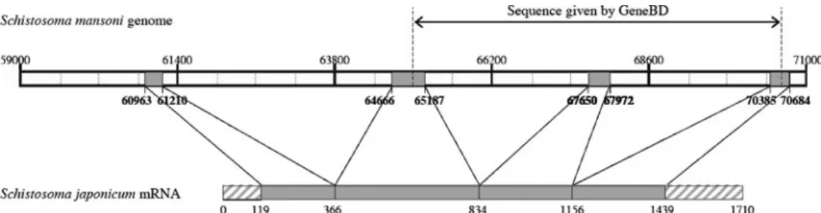
Thanks to S. mansoni genome sequence data pub-lished on the GeneDB website, we performed an alignment with S. japonicum mRNA sequence available in the NCBI database in order to define exon/intron boundaries of the SmTOR gene. The analysis suggested that the SmTOR gene comprised 4 exons, instead of the 3 suggested in GeneDB. Figure 1 represents the exon localizations along the Sm genome, showing that the gene contains 3 introns in the range of 2500 to 3500 bp, resulting in a 9721 bp length gene. The hatched boxes in Sj mRNA rep-resent the 5k and 3k untranslated regions. The re-sulting transcript and protein would thus be longer than observed before (Inal, 1999). Thanks to this alignment, we were able to locate the ATG start codon of the SmTOR gene Smp_093840 at position 60992 in scaffold Smp_scaff000547.
Cloning and sequencing of SmTOR exons
We cloned the single exons from genomic DNA extracted from S. mansoni cercariae using primers flanking the exons (Table 1). Separation of the PCR products by agarose gel electrophoresis showed bands at the expected size (Fig. 2A). The product for the amplification of exon 2 was expected to be 538 bp long but appears to be about 50 bp smaller. After gel extraction, cloning and sequencing of the single bands, the PCR product of exon 2 was found to be 481 bp long. The difference of 50 bp is because part of this region in the S. mansoni GeneDB is unknown and designated as multiple NNN. The length and sequencing results of the other 3 PCR products did match the information deposited at the S. mansoni
GeneDB database. All the fragments were cloned at least 3 times and sequenced in both directions. Sequencing results of the single exons are in com-plete concordance with the cDNA sequence shown below in an alignment with SjTOR cDNA (Fig. 3). This delineation of sequencing information rep-resents the merged information of single exon se-quencing results. How the gained information is related to the GeneDB entry is listed in the next section.
Expression of full-length SmTOR
PCR amplification of SmTOR ORF from cDNA preparations from S. mansoni adult worms and cer-cariae generated with different primers (as described above) yielded the same result for all the reactions. Size-separation of reaction products amplified with smTOR_ex1_x15_fwd and smTOR_exon4_rev showed a single band at the expected size (Fig. 2B).
The gel-purified and cloned fragments all held the 1260 bp sequence coding for full length SmTOR. The results were identical using different clones and performing sequencing reactions. The sequencing result for SmTOR cDNA is shown in alignment with SjTOR cDNA (AY814912) starting at the trans-lation initiation codon (Fig. 3). When aligning the results from the single exons described in the pre-vious section with the sequenced SmTOR cDNA the correlation was 100 %. Splicing sites are marked with black arrows on the SmTOR cDNA (Fig. 3).
Sequencing data for SmTOR cDNA perfectly match the corresponding sequences in the S. mansoni GeneDB database (positions 60 991 to 70 636, scaffold Smp_scaff000547) on S. mansoni genome (starting at ATG on exon 1) for exons 1, 3 and 4 (single mutations differing between the clones and occuring at a frequency of 2 per 1260 bp were not taken into account). The gap in the S. mansoni GeneDB database lying within SmTOR exon 2
Fig. 2. Electrophoretic separation of PCR amplification products from Schistosoma mansoni genomic DNA or adult worm cDNA. (A) Lanes 1–4 correspond to PCR amplification of exon 1–4 respectively, primers used Table 1.
(B) SmTOR ORF amplified from adult worm cDNA, primers : smTOR_ex1_x15_fwd, smTOR_exon4_rev (Table 1). Fig. 1. Alignment of Sm (Schistosoma mansoni) genome (Smp_093840) and Sj (Schistosoma japonicum) mRNA of CRIT. Representation of the gene structure of SmCRIT exons and introns. The grey boxes represent the exons, the hatched boxes represent the sequences not found in Sm genome. Numbers indicate positions on scaffold Smp_scaff000547.
(position 368–400 on SmTOR cDNA, 64 816 to 64 908, Smp_scaff000547 on S. mansoni GeneDB entry Smp_093840) could be filled in (Fig. 3).
Furthermore, we found 13 single nucleotide differ-ences or gaps (indicated with stars in Fig. 3) within exon 2 in the range of position 348 to 400 when
Fig. 3. SmTOR and SjTOR cDNA alignment ; identical nucleotides are linked with a bar. Filled triangles indicate the positions of splice site on SmTOR cDNA (positions 214/215, 684/685 and 1009/1010, numbering with respect to the SmTOR start codon). Stars indicate mutations or gaps comparing sequencing data and GeneDB entry data ; ATG of smTOR 0.86 kb ORF (AF051138) is indicated by a triangle. Bases 368–400 were newly sequenced on Schistosoma mansoni cDNA and from genomic DNA as described.
comparing S. mansoni GeneDB sequence and SmTOR cDNA. The newly sequenced SmTOR cDNA compared with the shorter SmTOR mRNA database entry (AF051138) of Inal (1999) was nearly identical (18 single bases differed ; the ATG is marked with a opened arrow (Fig. 3)). The former 5k UTR region published at the same time for S. hae-matobium TOR (64 base pairs upstream of the former ATG) matches the sequence we now find to be part of exon 2 of the longer SmTOR version, with only 5 bases being altered (not shown).
Developmental expression profiling of SmTOR as compared to SmTPI showed that the receptor mRNA was expressed at all the stages examined (Fig. 4). The receptor was expressed at a higher level in cercariae as compared to schistosomula, adult worm, egg and miracidia.
The cDNA sequences aligned for S. mansoni and S. japonicum TOR showed 46 % homology at the nucleotide level.
SmTOR translates into a protein similar in length to SjTOR and possesses four transmembrane domains SmTOR cDNA contains a 1260 bp open reading frame which should generate a protein of 419 amino acid length with a theoretical mass of 46.7 kDa. Its amino acid sequence is shown in alignment with SjTOR, a 414 amino acid protein based on pro-teomics data (Liu et al. 2006). Protein alignment of a possible SmTOR transcript and SjTOR shows 76.7 % homology on the protein level (Fig. 5).
Based on results from transmembrane prediction analysis we suggest SmTOR to have 4 transmem-brane segments and a long C-terminal tail in hom-ology with SjTOR (Fig. 6). In the absence of an N-terminal signal peptide, the transmembrane do-main 1 (aa 31–53, Fig. 6) is most likely to function as a
signal anchor sequence for ER targeting and mem-brane insertion, as predicted with the SignalP soft-ware. The 4 transmembrane segments have an alpha helical conformation predicted with high confidence, and the amphipatic alpha helix in the intracellular domain 2 is likely to be partially inserted into the plasma membrane. The domain organization is the same for SmTOR and SjTOR. The overall trans-membrane architecture remains the same as com-pared to the truncated protein version (UniProtKB/ TrEMBL entry Q9U597) (Inal et al. 2005 a, 2006).
The 11 amino acids in the C-terminal part of ed1 that have been described to bind to C2 and interfere with its cleavage are boxed (Fig. 6). This sequence is different in SmTOR and SjTOR.
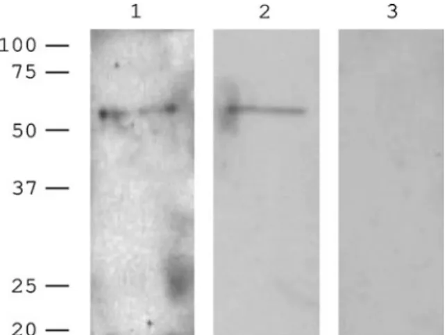
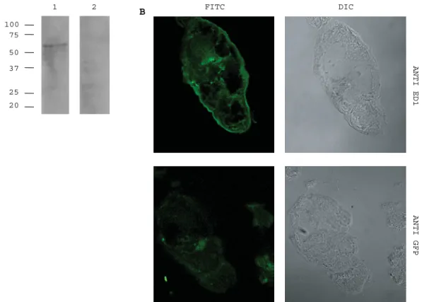
Performing Western blot analysis of adult worm tegument preparations we detected a protein of about 55 kDa using antibodies generated against peptide sequences of ed1 and ed2 (Fig. 7). The same result was obtained using a monoclonal antibody generated against ed1 (Fig. 8A). Staining of cryosections of cercariae with the monoclonal antibody generated against ed1 showed surface labelling, as compared to the negative control (Fig. 8B). The presence of SmTOR on the tegument surface was confirmed by electron microscopy. In sections stained with the polyclonal antibodies against SmTOR ed1 and ed2 respectively, immunogold labelling was detected at the tegument surface (Fig. 9).
D I S C U S S I O N
The data presented here clarify the exon/intron gene structure of SmTOR and provide evidence that S. mansoni expresses the corresponding protein.
We found that SmTOR gene structure contains 4 exons. A transcript containing the 1.26 kb SmTOR ORF showing 46 % identity with the 1.25 kb SjTOR ORF was found in adult worm and cercariae cDNA preparations. The sequencing data generated not only complete the gaps present in the S. mansoni GeneDB for SmTOR, but also correct some likely sequencing errors present in the database entry (stars on SmTOR cDNA Fig. 3). The sequencing data are likely to be correct. SmTOR ORF presented here aligns with the previously described SmTOR ORF on mRNA entry AF0511378 from position 418 on (alignment not shown), and translates into a functional protein aligning with SjTOR. SmTOR is expressed at various developmental stages of S. mansoni. The highest expression was in cercariae, where we were able to detect it at the surface by confocal microscopy of cryosections and by electron microscopy. The higher expression in cercariae as compared to adult worms has previously been de-scribed for the truncated receptor version (Inal, 1999). This observation is of interest since cercariae are the first to come into contact with the human skin, an encounter that might determine the fate of the
Fig. 4. Quantitative RT-PCR analysis of SmTOR mRNA in different Schistosoma mansoni stages : (1) eggs, (2) miracidiae, (3) cercariae, (4) schistosomulae, (5) adult worm. Expression of SmTOR quantitated relative to SmTPI as the control gene with bars representing 2xDDCTvalues.
Fig. 5. SjTOR and SmTOR protein sequence alignment. Identical amino acids are linked with a bar (76.7 % identity), similar amino acids are connected via ‘ . . ’ (84 % similarity). Exon/Intron boundaries on cDNA level for SmTOR are indicated with filled triangles. SmTOR protein (AF051138) starting with former extracellular domain 1 is marked by a triangle. Transmembrane domains 1–4 are shaded in grey as well as partially inserted helix 5. Domain designation : ed1, new extracellular domain 1 ; ed2, extracellular domain 2 ; id1, id2 intracellular domains 1 and 2.
Fig. 6. SmTOR protein model according to data generated with TopPred and PSIPREDView program. Numbers indicated refer to the first and last amino acid of the corresponding intra- or extracellular domain. The peptide sequence shown to bind C2 is boxed. Alphahelical transmembrane domains 1–4 are at positions 32–52, 165–185, 199–219 and 236–256 respectively. Transmembrane domain one acting as signal anchor is shaded in grey.
infection. SmTOR might be essential at that specific time-point. Many complement proteins are pro-duced by epidermal cells (Timar et al. 2007) and several authors have shown that complement pro-teins diffuse from the vascular compartment to the dermis and epidermis (Kool et al. 2007 ; Hansell et al. 2008). The complete complement cascade is active in the skin as indicated by the role of complement in many human blistering skin diseases (Lessey et al. 2008). SmTOR might at that time help the parasite to escape complement attack.
We had previously not been able to replicate the amplification of the short version ORF of the TOR receptor homologue from Schistosoma genomic DNA or from vertebrate genomic DNA of various sources (Inal et al. 2005 b). This is now under-standable since the gene has 3 large introns not described previously. In addition, we did not find the sequence or fragments of it when performing BLASTn searches on different vertebrate genomes. DeMarco et al. (2007) recently analysed the contro-versy about the origin of schistosome albumin, and demonstrated it to be of hamster origin. In his dis-cussion, he suggested that CRIT might be an ex-ample of reverse contamination from the schistosome into the vertebrate samples. Our present data are in agreement with DeMarco’s comments. We must now consider our previous observations as being due to contamination and non-specific antibody staining (Inal et al. 2005 a, b ; Moll et al. 2006).
Previously, for both ShTOR and SmTOR, tran-scripts of 1.2 kb and 1.35 kb cloned from adult worm cDNA libraries had been reported to contain an open reading frame of 0.86 kb, as proteins of 31.7 and 31.2 kDa were detected in adult worm preparations (Inal, 1999). Alignment of the region published as 5k UTR of ShTOR (Inal, 1999) with SmTOR exon 2 sequenced resulted in a nearly perfect match (align-ment not shown). SjTOR cDNA and protein (Liu et al. 2006) are of the same length as we now found in
S. mansoni. We therefore suggest SmTOR to be longer than originally described with the former ATG lying within the second exon of the gene. The translation initiation site we suggest for SmTOR does not lie in a classical Kozak sequence (Kozak, 1984), but possible variation around the translation initiation codon has been reported for invertebrates (Nakagawa, 2008).
TOR protein had been shown on the surface of the adult worm by immunohistochemistry (Inal, 1999). We detected a band at approximately 55 kDa in adult worm tegument membrane preparation using 2 different polyclonal antibodies generated against ed1 and ed2 respectively. The SmTOR amino acid sequence contains a potential N-linked glycosylation site at position 138. The protein detected might be the glycosylated form, as its theoretical molecular weight was calculated as 46.7 kDa. SmTOR in alignment with SjTOR protein shows 76 % identity and 84 % similarity. TOR was found by Liu in S. japonicum in cercariae, schistosomula and adult worm (Liu et al. 2006), whereas no proteomic analysis of the schistosome tegument of adult worms identified peptides belonging to SmTOR (van Balkom et al. 2005 ; Braschi et al. 2006 b ; Braschi and Wilson, 2006). This might be due to the low abun-dance of the protein in the tegument of the adult worm (Skelly and Wilson, 2006).
Based on the secondary structure analysis de-scribed above we generated a hypothetical protein model for SmTOR. SmTOR (or CRIT) was pre-viously thought to span the membrane 3 times (hence the ‘ T ’ for rispanning). The longer protein we found is very likely to be tetraspanning, as suggested also for SjTOR, which allows us to suggest a change in the name of the molecule, but without a change in the abbreviations used. The amphiphatic helix inserted partially in the membrane is present in SjTOR and SmTOR, although there is high variability of amino acid sequence at this site between the two species.
SmTOR had been renamed to CRIT (comp-lement C2 receptor inhibitor trispanning) according to its putative function to bind C2. The former N-terminal extracellular domain ed1 of ShTOR as isolated peptide had been shown to bind C2 and with its C-terminal 11 amino acids designated as H17 peptide being the active binding site (Inal and Schifferli, 2002). Incubation with this peptide has been shown to interfere with the binding of C2 to C4b and with C2 cleavage by complement C1s, thus blocking complement activation by the classical pathway. The new tetraspanning SmTOR protein still contains the same amino acid sequence in the extracellular domain 1 known to bind C2. Whether this sequence binds C2 in vivo on schistosomes remains to be explored, and it is interesting to note several differences in it between S. mansoni and S. japonicum. It will be interesting to test whether these differences alter C2 binding to TOR/CRIT.
Fig. 7. Western blot of adult worm tegument
preparation. Polyclonal anti-ed1, anti-ed2 antibody and pre-immune serum (lanes 1, 2 and 3 respectively).
1 2 100 75 50 37 25 20 B A FITC DIC ANTI ED1 ANTI GFP
Fig. 8. SmTOR detection with a monoclonal anti-ed1 antibody AbyD04644.1. (A) Adult worm tegument preparation. Western blot probed with monoclonal anti-ed1 (lane 1) or anti-ed1 pre-incubated with 100-fold excess of ed1 peptide as control (lane 2). (B) Immunofluorescent labelling of cryosections through cercariae. Anti-ed1 labelled section top left panel and anti-GFP labelled control section bottom left panel.
Fig. 9. Localization of SmTOR on tegument of cercariae using electron microscopy. Immunogold labelling is indicated by arrows. Sections stained with (A) polyclonal anti-ed1 antibody, (B) polyclonal anti-ed2 antibody and (C) pre-immune serum. Secondary antibody only was negative (not shown).
We thank Dr J. Chollet, Y. Hagenmu¨ ller and C. List (Swiss Tropical Institute) for providing the Schistosoma material. Further thanks go to Vesna Olivieri and Ursula Sauder (Microscopy Center, Pharmacenter, University of Basel) for performing the electron microscopy, Brigitte Schneider and Beatrice Dolder (Infectious Diseases and Neurosurgery, University Hospital Basel) for helping with the cryosections and to the group of Professor A. Merlo (Neurosurgery, University Hospital Basel) for providing technical resources and support. Sequence data used to generate the genomic map were produced by the Wellcome Trust Sanger Institute and are available from www. genedb.org. This work was supported by the Swiss National Foundation (320000-116839).
R E F E R E N C E S
Bergquist, N. R., Leonardo, L. R. and Mitchell, G. F. (2005). Vaccine-linked chemotherapy : can
schistosomiasis control benefit from an integrated approach ? Trends in Parasitology21, 112–117. Braschi, S., Borges, W. C. and Wilson, R. A. (2006 a).
Proteomic analysis of the schistosome tegument and its surface membranes. Memorias Do Instituto Oswaldo Cruz101, 205–212.
Braschi, S., Curwen, R. S., Ashton, P. D., Verjovski-Almeida, S. and Wilson, A. (2006 b). The tegument surface membranes of the human blood parasite Schistosoma mansoni : a proteomic analysis after differential extraction. Proteomics6, 1471–1482.
Braschi, S. and Wilson, R. A. (2006). Proteins exposed at the adult schistosome surface revealed by biotinylation. Molecular & Cellular Proteomics5, 347–356. doi : 10.1074/mcp.M500287-MCP200
Brouwers, J., Skelly, P. J., Van Golde, L. M. G. and Tielens, A. G. M. (1999). Studies on phospholipid turnover argue against sloughing of tegumental membranes in adult Schistosoma mansoni. Parasitology 119, 287–294.
Claros, M. G. and Vonheijne, G. (1994). TopPred-II – an improved software for membrane-protein structure predictions. Computer Applications in the Biosciences10, 685–686.
Correa-Oliveira, R., Caldas, I. R. and Gazzinelli, G. (2000). Natural versus drug-induced resistance in Schistosoma mansoni infection. Parasitology Today 16, 397–399.
Correa-Oliveira, R., Pearce, E. J., Oliveira, G. C., Golgher, D. B., Katz, N., Bahia, L. G., Carvalho, O. S., Gazzinelli, G. and Sher, A. (1989). The human immune-response to defined immunogens of Schistosoma mansoni – elevated antibody levels to Paramyosin in stool-negative individuals from 2 endemic areas in Brazil. Transactions of the Royal Society of Tropical Medicine and Hygiene83, 798–804. Dalton, J. P., Day, S. R., Drew, A. C. and Brindley, P. J.
(1997). A method for the isolation of schistosome eggs and miracidia free of contaminating host tissues. Parasitology115, 29–32.
DeMarco, R., Mathieson, W., Dillon, G. P. and Wilson, R. A. (2007). Schistosome albumin is of host, not parasite, origin. International Journal for Parasitology37, 1201–1208. doi : 10.1016/j.ijpara. 2007.03.004
Hamburger, J., Xu, Y. X., Ramzy, R. M., Jourdane, J. and Ruppel, A. (1998). Development and laboratory evaluation of a polymerase chain reaction for monitoring Schistosoma mansoni infestation of water. American Journal of Tropical Medicine and Hygiene59, 468–473. Hansell, E., Braschi, S., Medzihradszky, K. F.,
Sajid, M., Debnath, M., Ingram, J., Lim, K. C. and McKerrow, J. H. (2008). Proteomic analysis of skin invasion by blood fluke larvae. PLoS Neglected Tropical Diseases2, e262.
Hooker, C. W. and Brindley, P. J. (1996). Cloning and characterisation of strain-specific transcripts encoding triosephosphate isomerase, a candidate vaccine antigen from Schistosoma japonicum. Molecular and Biochemical Parasitology82, 265–269.
Hui, K. M., Magnadottir, B., Schifferli, J. A. and Inal, J. M. (2006). CRIT peptide interacts with factor B and interferes with alternative pathway activation. Biochemical and Biophysical Research Communications 344, 308–314. doi: 10.1016/j.bbrc.2006.03.101
Hui, K. M., Orriss, G. L., Schirmer, T., Magnadottir, B., Schifferli, J. A. and Inal, J. M. (2005). Expression of functional recombinant von Willebrand factor-A domain from human complement C2 : a potential binding site for C4 and CRIT. The Biochemical Journal389, 863–868. doi : 10.1042/bj20050183 Inal, J. M. (1999). Schistosoma TOR (trispanning orphan
receptor), a novel, antigenic surface receptor of the blood-dwelling, Schistosoma parasite. Biochimica et Biophysica Acta-Gene Structure and Expression1445, 283–298.
Inal, J. M. (2005). Complement C2 receptor inhibitor trispanning : from man to schistosome. Springer Seminars in Immunopathology27, 320–331. doi : 10.1007/s00281-005-0009-9
Inal, J. M., Hui, K. M., Miot, S., Lange, S., Ramirez, M. I., Schneider, B., Krueger, G. and Schifferli, J. A. (2005 b). Complement C2 receptor inhibitor trispanning : A novel human complement inhibitory receptor. Journal of Immunology174, 356–366. Inal, J., Miot, S. and Schifferli, J. A. (2005 a). The
complement inhibitor, CRIT, undergoes clathrin-dependent endocytosis. Experimental Cell Research 310, 54–65. doi : 10.1016/j.yexcr.2005.07.003 Inal, J. M. and Schifferli, J. A. (2001). C4 beta
chain peptide interferes with the formation of the classical pathway C2 convertase. Molecular Immunology 38, 97.
Inal, J. M. and Schifferli, J. A. (2002). Complement C2 receptor inhibitor trispanning and the beta-chain of C4 share a binding site for complement C2. Journal of Immunology168, 5213–5221.
Inal, J. M. and Sim, R. B. (2000). A Schistosoma protein, Sh-TOR, is a novel inhibitor of complement which binds human C2. FEBS Letters470, 131–134. Jones, D. T. (1999). Protein secondary structure
prediction based on position-specific scoring matrices. Journal of Molecular Biology292, 195–202.
Kemp, W. M., Brown, P. R., Merritt, S. C. and Miller, R. E. (1980). Tegument-associated antigen modulation by adult male Schistosoma mansoni. Journal of
Immunology124, 806–811.
Kemp, W. M., Damian, R. T. and Greene, N. D. (1976). Immunocytochemical localisation of IgG on
adult Schistosoma mansoni tegumental surfaces. Journal of Parasitology62, 830–832.
Kemp, W. M., Merritt, S. C. and Rosier, J. G. (1978). Schistosoma mansoni : Identification of immunoglobulins associated with tegument of adult parasites from mice. Experimental Parasitology45, 81–87.
Klabunde, J., Berger, J., Jensenius, J. C., Klinkert, M. Q., Zelck, U. E., Kremsner, P. G. and Kun, J. F. J. (2000). Schistosoma mansoni : Adhesion of mannan-binding lectin to surface glycoproteins of cercariae and adult worms. Experimental Parasitology 95, 231–239.
Kool, J., Reubsaet, L., Wesseldijk, F., Maravilha, R. T., Pinkse, M. W., D’Santos, C. S., Van Hilten, J. J., Zijlstra, F. J. and Heck, A. J. (2007). Suction blister fluid as potential body fluid for biomarker proteins. Proteomics7, 3638–3650.
Kozak, M. (1984). Compilation and analysis of sequences upstream from the translation start site in eukaryotic mRNAs. Nucleic Acids Research12, 857–872. Lessey, E., Li, N., Diaz, L. and Liu, Z. (2008).
Complement and cutaneous autoimmune blistering diseases. Immunologic Research41, 223–232.
Linder, E. and Huldt, G. (1983). Antibody-independent binding and activation of complement by Schistosoma mansoni adult worms. Parasite Immunology5, 183–194. Liu, F., Lu, J., Hu, W., Wang, S. Y., Cui, S. J., Chi, M.,
Yan, Q., Wang, X. R., Song, H. D., Xu, X. N., Wang, J. J., Zhang, X. L., Zhang, X., Wang, Z. Q., Xue, C. L., Brindley, P. J., McManus, D. P., Yang, P. Y., Feng, Z., Chen, Z. and Han, Z. G. (2006). New perspectives on host-parasite interplay by comparative transcriptomic and proteomic analyses of Schistosoma japonicum. Plos Pathogens2, 268–281. doi : 10.1371/ journal.ppat.0020029
Livak, K. J. and Schmittgen, T. D. (2001). Analysis of relative gene expression data using real-time quantitative PCR and the 2(-Delta Delta C(T)) method. Methods25, 402–408.
Marikovsky, M., Parizade, M., Arnon, R. and Fishelson, Z. (1990). Complement regulation on the surface of cultured schistosomula and adult worms of Schistosoma mansoni. European Journal of Immunology 20, 221–227.
McGuffin, L. J., Bryson, K. and Jones, D. T. (2000). The PSIPRED protein structure prediction server. Bioinformatics16, 404–405.
McManus, D. P. and Loukas, A. (2008). Current status of vaccines for schistosomiasis. Clinical Microbiology Reviews21, 225–242.
Minard, P., Dean, D. A., Jacobson, R. H., Vannier, W. E. and Murrell, K. D. (1978). Immunization of mice with Co-60 irradiated Schistosoma mansoni cercariae. American Journal of Tropical Medicine and Hygiene27, 76–86.
Moll, S., Lange, S., Mihatsch, M., Dragic, Z., Schifferli, J. and Inal, J. (2006). CRIT is expressed on podocytes in normal human kidney and upregulated in membranous nephropathy. Kidney International69, 1961–1968.
Nakagawa, S. (2008). Diversity of preferred nucleotide sequences around the translation
initiation codon in eukaryote genomes. Nucleic Acids Research36, 861–871.
Pearce, E. J., Hall, B. F. and Sher, A. (1990). Host-specific evasion of the alternative complement pathway by schistosomes correlates with the presence of a phospholipase C-sensitive surface molecule resembling human decay accelerating factor. Journal of Immunology144, 2751–2756.
Ramalhopinto, F. J., Gazzinelli, G., Howells, R. E., Motasantos, T. A., Figueiredo, E. A. and
Pellegrino, J. (1974). Schistosoma mansoni – defined system for stepwise transformation of cercaria to schistosomule in vitro. Experimental Parasitology36, 360–372.
Rasmussen, K. R. and Kemp, W. M. (1987). Schistosoma mansoni : interactions of adult parasites with the complement system. Parasite Immunology9, 235–248.
Roberts, S. M., MacGregor, A. N., Vojvodic, M., Wells, E., Crabtree, J. E. and Wilson, R. A. (1983). Tegument surface membranes of adult Schistosoma mansoni : development of a method for their isolation. Molecular and Biochemical Parasitology9, 105–127. Ross, A. G. P., Bartley, P. B., Sleigh, A. C., Olds, G. R.,
Li, Y. S., Williams, G. M. and McManus, D. P. (2002). Current concepts – schistosomiasis. New England Journal of Medicine346, 1212–1220. Ruppel, A., McLaren, D. J., Diesfeld, H. J. and
Rother, U. (1984). Schistosoma mansoni : escape from complement-mediated parasiticidal mechanisms following percutaneous primary infection. European Journal of Immunology14, 702–708.
Skelly, P. J. (2004). Immunoparasitology series : intravascular schistosomes and complement. Trends in Parasitology20, 370–374. doi : 10.1016/j.pt.2004.05.007 Skelly, P. J. and Wilson, R. A. (2006). Making sense
of the schistosome surface. Advances in Parasitology 63, 185–284. doi : 10.1016/s0065-308x(06)63003-0 Steinmann, P., Keiser, J., Bos, R., Tanner, M. and
Utzinger, J. (2006). Schistosomiasis and water resources development : systematic review, meta-analysis, and estimates of people at risk. Lancet Infectious Diseases6, 411–425.
Stek, M., Minard, P., Dean, D. A. and Hall, J. E. (1981). Immunization of baboons with Schistosoma mansoni cercariae attenuated by gamma irradiation. Science212, 1518–1520.
Tendler, M. and Simpson, A. J. (2008). The
biotechnology-value chain : development of Sm14 as a schistosomiasis vaccine. Acta Tropica108, 263–266. Timar, K. K., Dallos, A., Kiss, M., Husz, S., Bos, J. D.
and Asghar, S. S. (2007). Expression of terminal complement components by human keratinocytes. Molecular Immunology44, 2578–2586.
Tran, M. H., Pearson, M. S., Jeffrey, M. B., Smyth, D. J., Jones, M. K., Duke, M., Don, T. A.,
McManus, D. P., Correa-Oliveira, R. and Loukas, A. (2006). Tetraspanins on the surface of Schistosoma mansoni are protective antigens against schistosomiasis. Nature Medicine12, 835–840.
Van Balkom, B. W. M., Van Gestel, R. A., Brouwers, J., Krijgsveld, J., Tielens, A. G. M., Heck, A. J. R. and Van Hellemond, J. J. (2005). Mass spectrometric analysis of the Schistosoma mansoni tegumental
sub-proteome. Journal of Proteome Research4, 958–966. doi : 10.1021/pr050036w