HAL Id: hal-02952514
https://hal.archives-ouvertes.fr/hal-02952514
Submitted on 29 Sep 2020
HAL is a multi-disciplinary open access
archive for the deposit and dissemination of
sci-entific research documents, whether they are
pub-lished or not. The documents may come from
teaching and research institutions in France or
abroad, or from public or private research centers.
L’archive ouverte pluridisciplinaire HAL, est
destinée au dépôt et à la diffusion de documents
scientifiques de niveau recherche, publiés ou non,
émanant des établissements d’enseignement et de
recherche français ou étrangers, des laboratoires
publics ou privés.
Connexin 43 inhibiting astrocytic gap junctional
intercellular communication
Amos Fumagalli, Joyce Heuninck, Anne Pizzoccaro, Enora Moutin, Joyce
Koenen, Martial Séveno, Thierry Durroux, Marie-Pierre Junier, Géraldine
Schlecht-Louf, Françoise Bachelerie, et al.
To cite this version:
Amos Fumagalli, Joyce Heuninck, Anne Pizzoccaro, Enora Moutin, Joyce Koenen, et al.. The
atyp-ical chemokine receptor 3 interacts with Connexin 43 inhibiting astrocytic gap junctional
intercel-lular communication. Nature Communications, Nature Publishing Group, 2020, 11 (1), pp.4855.
�10.1038/s41467-020-18634-y�. �hal-02952514�
The atypical chemokine receptor 3 interacts with
Connexin 43 inhibiting astrocytic gap junctional
intercellular communication
Amos Fumagalli
1
, Joyce Heuninck
1
, Anne Pizzoccaro
1
, Enora Moutin
1
, Joyce Koenen
2,3
, Martial Séveno
4
,
Thierry Durroux
1
, Marie-Pierre Junier
5
, Géraldine Schlecht-Louf
2
, Francoise Bachelerie
2
, Dagmar Schütz
6
,
Ralf Stumm
6
, Martine J. Smit
3
, Nathalie C. Guérineau
1
, Séverine Chaumont-Dubel
1
& Philippe Marin
1
✉
The atypical chemokine receptor 3 (ACKR3) plays a pivotal role in directing the migration of
various cellular populations and its over-expression in tumors promotes cell proliferation and
invasiveness. The intracellular signaling pathways transducing ACKR3-dependent effects
remain poorly characterized, an issue we addressed by identifying the interactome of ACKR3.
Here, we report that recombinant ACKR3 expressed in HEK293T cells recruits the gap
junction protein Connexin 43 (Cx43). Cx43 and ACKR3 are co-expressed in mouse brain
astrocytes and human glioblastoma cells and form a complex in embryonic mouse brain.
Functional in vitro studies show enhanced ACKR3 interaction with Cx43 upon ACKR3 agonist
stimulation. Furthermore, ACKR3 activation promotes
β-arrestin2- and dynamin-dependent
Cx43 internalization to inhibit gap junctional intercellular communication in primary
astro-cytes. These results demonstrate a functional link between ACKR3 and gap junctions that
might be of pathophysiological relevance.
https://doi.org/10.1038/s41467-020-18634-y
OPEN
1Institut de Génomique Fonctionnelle, Université de Montpellier, CNRS, INSERM, Montpellier, France.2Université Paris-Saclay, Inserm, Inflammation,
Microbiome and Immunosurveillance, 92140 Clamart, France.3Amsterdam Institute for Molecules Medicines and Systems, Division of Medicinal Chemistry,
Faculty of Sciences, VU University Amsterdam, 1081 HV Amsterdam, The Netherlands.4Biocampus Montpellier, Université de Montpellier, CNRS, INSERM,
Montpellier, France.5CNRS UMR8246, Inserm U1130, Neuroscience Paris Seine-IBPS, Sorbonne Universités, Paris, France.6Institute of Pharmacology and
Toxicology, Jena University Hospital, 07747 Jena, Germany. ✉email:philippe.marin@igf.cnrs.fr
123456789
T
he atypical chemokine receptor 3 (ACKR3, previously
called CXCR7) is a seven-trans-membrane (7-TM)
recep-tor belonging to the CXC chemokine receprecep-tor family that
binds to CXCL12 and CXCL11. ACKR3 is expressed in various
tissues such as the heart
1, kidney
2, and brain
3. In the last, ACKR3
is found in neurons, astrocytes, and vascular cells
3and plays a
pivotal role in interneuron migration
4,5and leukocyte entry into
the brain parenchyma
6. ACKR3 is over-expressed in numerous
cancer types
7, including glioma, where both its expression and
activation have been positively correlated with increased
pro-liferative state and invasiveness
8.
Despite this accumulating knowledge, the intracellular
path-ways underlying ACKR3-dependent effects remain poorly
char-acterized. Several reports suggest that ACKR3 operates as a
molecular chemokine sink
4,9,10lowering the extracellular
che-mokine concentration without activating any intracellular
path-way. Several lines of evidence indicate that ACKR3 does not
couple to and activate G proteins but only engages
β-arrestin-dependent pathways
11–13. A single study suggests that some
ACKR3 effects are mediated by G proteins in glial cells
14.
Over the last two decades, it has become evident that 7-TM
receptors interact with large networks of proteins that
finely
control their targeting to specific subcellular compartments, their
trafficking in and out of the plasma membrane and the nature of
receptor-operated signal transduction
15. In line with these
find-ings, studies have shown that ACKR3 exerts its biological effects
by interacting with diverse membrane receptors
16. For example,
ACKR3 might indirectly influence Gα
iactivation via its
interac-tion with the other CXCL12 receptor CXCR4
13. ACKR3 also
associates with the epidermal growth factor receptor
17to promote
proliferation of tumor cells in an agonist-independent manner.
More recently, ACKR3 was found to interact with the G
protein-coupled receptor kinase 2 that is involved in ACKR3
endocy-tosis
9. These results suggest a strong influence of
ACKR3-interacting proteins in ACKR3 pathophysiological functions.
They provided the impetus for the extensive characterization of
the ACKR3 interactome, thanks to a proteomic strategy
com-bining affinity purification of receptor-interacting proteins and
their identification by high-resolution mass spectrometry.
This interactomic screen identified the gap junction protein
Connexin 43 (Cx43) as an ACKR3-interacting protein. In the
brain, Cx43 is mainly expressed in astrocytes where Cx43
chan-nels are involved in several important physiological processes
including glucose diffusion
18and propagation of Ca
2+waves
19.
Several studies have demonstrated that Cx43-mediated gap
junctional intercellular communication (GJIC) is
finely controlled
by astrocytic receptors, especially 7-TM receptors
20. Furthermore,
both ACKR3 and Cx43 have been involved in glioma progression.
While ACKR3 was found to be upregulated in glioma, promoting
tumor cell proliferation, angiogenesis and resistance to
che-motherapy
8, suppression of Cx43-dependent communication in
glioma cells has been correlated with enhanced proliferation and
invasiveness
21,22. On the other hand, Cx43 was found to promote
glioma cell migration and resistance to apoptosis, suggesting
opposing roles of Cx43 in glioma progression
21,22. However, the
functional link between ACKR3 and GJIC remains unexplored.
In light of these
findings, we explored whether ACKR3 controls
Cx43-mediated GJIC in primary astrocytes by combining dye
diffusion experiments with electrophysiological recordings of
junctional currents. We show that agonist stimulation of
ACKR3 substantially reduces GJIC in astrocytes through a
mechanism involving Gα
i/oproteins and
β-arrestin2-dependent
Cx43 internalization. These
findings provide evidence of a
phy-sical and functional interaction between ACKR3 and Cx43 that
might underlie their influence on the pathogenesis of glioma.
They shed light on the key role played by ACKR3-interacting
proteins in determining the functional outcome of this
chemo-kine receptor upregulated and mobilized in a broad range of
pathologic conditions such as cancers.
Results
ACKR3 interacts with the gap junction protein Cx43. Due to
the lack of an ACKR3 antibody providing receptor
immunopre-cipitation yields compatible with mass spectrometry analysis, we
expressed hemagglutinin (HA)-tagged ACKR3 in human
embryonic kidney (HEK293T) cells. ACKR3-interacting proteins
were immunoprecipitated using an anti-HA monoclonal antibody
immobilized onto agarose beads. Control immunoprecipitations
were performed using cells transfected with an empty plasmid.
Only proteins identified in all three biological replicates in at least
one group were considered for further analysis. Label-free
quantification (LFQ) of the relative protein abundances in
immunoprecipitates obtained from cells expressing ACKR3 and
control cells showed that 151 proteins were significantly more
abundant in immunoprecipitates from ACKR3-expressing cells,
than in immunoprecipitates from control cells (Supplementary
Data 1). As expected, ACKR3 (bait protein) was the most
enri-ched one (Fig.
1
a). The ACKR3 interactome also included
Cla-thrin (CLTC) and accessory proteins of the Rab5 and Rab3
complexes involved in ACKR3 internalization
23. In addition, we
identified several E3 ubiquitin-protein ligases (HUWE1,
HECTD1, and AMFR), which might be responsible for basal
ubiquitination of the receptor
12. In line with previous
findings
indicating that ACKR3 promotes ERK1/2 phosphorylation via
activation of MAP2K2
24, MAP2K2 was retrieved in our
inter-actomic screen. Consistent with a previous study showing a
constitutive interaction between ACKR3 and G proteins
13, we
also identified Gα
i3(GNAI3) as a putative ACKR3 partner.
The ACKR3 interactome also included the Gap Junction
Alpha-1 protein (GJAAlpha-1, also called Connexin 43, Cx43) as well as
additional proteins previously shown to be physically or
function-ally linked to Cx43 and to regulate Cx43-mediated GJIC. These
included (i) Dynactin 1 (DCTN1)
25, which is known to affect the
cellular localization of Cx43; (ii) Ubiquilin-4 (UBQLN4)
26, which
interacts with and promotes the degradation of Cx43; (iii)
Cytochrome
P450
oxidoreductase
(POR)
27,
whose
down-regulation triggers transcriptional repression and inhibition of
Cx43; (iv) Solute carrier family 1 member 5 (SLC1A5)
28, which
interacts with Cx43 to stimulate cytotrophoblast fusion; (v) the
beta-subunit of the electron-transfer protein (ETFB)
29, which
interacts with Cx43 to regulate mitochondrial respiration and
reactive oxygen species signaling; and vi) the tubulin beta-3 chain
(TUBB3, Fig.
1
a), suggesting that Cx43 might regulate microtubule
stability in contacting cells
30.
Given the recruitment of Cx43-associated proteins by ACKR3
(Supplementary Data 1) and the co-expression of ACKR3 and
Cx43 in GFAP-positive astrocytes of the subventricular zone and
surrounding blood vessels in several brain regions such as the
striatum and the hippocampus (Fig.
1
b), we next sought to
validate ACKR3-Cx43 interaction in the brain. As ACKR3
exhibits a peak of expression in the brain at the embryonic
stage
3, we immunoprecipitated HA-ACKR3 from embryonic
brains
of
HA-ACKR3
knock-in
mice
9.
Cx43
co-immunoprecipitation with the receptor (Fig.
1
c) indicates that
both proteins form a complex in vivo.
We next assessed ACKR3-Cx43 interaction in living cells using
bioluminescence resonance energy transfer (BRET) (Fig.
1
d). We
monitored the BRET signal variation as a function of the
Cx43-YFP expression under conditions of constant ACKR3-NLuc or
CXCR4-NLuc expression. BRET variation was significantly better
fitted with a one-site saturation curve than a line through origin
(P < 0.0001) only for ACKR3 (Fig.
1
d), arguing for a specific
association of Cx43 with ACKR3 in living cells. Consistent with
these
findings, negligible Cx43 amounts co-immunoprecipitated
with HA-CXCR4 in HEK293T cells, compared with HA-ACKR3,
further supporting the specificity of ACKR3-Cx43 interaction
(Fig.
1
e). Double immunostaining of Cx43 and either ACKR3 or
CXCR4 in two patient-derived glioblastoma-initiating cell lines
with endogenous expression of the three proteins further
indicated that a higher fraction of Cx43 is co-localized with
ACKR3, in comparison with CXCR4 (Fig.
1
f and Supplementary
Fig. 1).
Collectively, the aforementioned results suggest that Cx43
already interacts with ACKR3 in the absence of an agonist. BRET
experiments in HEK293T cells also showed that treating cells with
1 2 3 Cx43 Merge 1 2 3 ACKR3 ETFB SLC1A5 GNAI3 UBQLN4 POR TUBB3 DCTN1 CLTC FDR = 1% S0 = 0.1 –5 0 5 10 0 2 4 6 8
Log2(LFQACKR3/LFQmock)
–Log 10 (P ) Cx43-YFP ACKR3/CXCR4-NLuc YFP/Nluc BRET (%) Cx43 overlap (%)
a
c
d
b
100 50 0 R633 TG1 Mouse brain HEK293T cellse
HEK293T cells Mouse brain
****
****
GJA1 ACKR3 0 0.01 0.02 0.03 0.04 0 50 100 ACKR3CXCR4 IP 55 43 34 Input Input Co-IP Cx43 55 43 34 Cx43 55 43 34 55 43 34 HA ACKR3 HA ACKR3 KI WTGlioblastoma cells (R633 and TG1) CXCR4 Cx43 Overlapping volume Overlapping volume Cx43 ACKR3 R633 TG1
f
Time [min] BRET (%) 95 0 5 10 15 20 25 30 100 110 105* *
* *
*
* *
** *
**
*
* **
g
h
CXCL12 CXCL11 Vehicle***
**
200 150 100 50 0 Cx43 IP (%) ACKR3 CXCL11 CXCL12 + + + + + – – – – HEK293T cells HEK293T cells ACKR3 CXCL11 43 43 Input Cx43 Co-IP Input IP CXCL12 HA ACKR3 – + – – – – + – – + + + – – – + – – – – + + + + Input 55 43 34 Cx43 ACKR3 CXCR4 – + – – + – – – + – – + Co-IP IP 100 50 0 kDa kDa kDa 43 34 Input HEK293T cells HA-ACKR3/ CXCR4 A CKR3 CXCR4 **** ACKR3 CXCR4 GFAPCXCL12 (10 nM) or CXCL11 (100 nM) time-dependently
enhanced the interaction of ACKR3 with Cx43 (Fig.
1
g).
Co-immunoprecipitation of Cx43 with ACKR3 was also
incremen-tally increased upon ACKR3 stimulation by CXCL12 or CXCL11
for 30 min (Fig.
1
h), suggesting that the Cx43-ACKR3 interaction
is a dynamic process depending on receptor activation.
ACKR3
stimulation
inhibits
Cx43-mediated
GJIC
in
astrocytes. Given the co-expression of ACKR3 and Cx43 in
astrocytes (Fig.
1
b), we next investigated the effect of ACKR3
activation on GJIC in primary cultures of astrocytes. We
first
assessed GJIC by scrape loading followed by measuring the
dif-fusion of the
fluorescent dye Lucifer yellow (LY) from the scrape
through the astrocytic syncytium
31. As previously described
32and in line with a high Cx43 expression
33, LY showed a large
diffusion in untreated astrocytes, arguing for a robust GJIC
(Fig.
2
). Further supporting astrocyte coupling through gap
junctions, treating cells with the gap junction inhibitor
carbe-noxolone (CBX, 50
μM) strongly inhibited LY diffusion (53 ±
6.5% inhibition vs. vehicle-treated cells, p < 0.0001, n
= 3). Cell
exposure to CXCL12 (10 nM) for 30 min also inhibited LY
dif-fusion (47 ± 6.5% inhibition vs. vehicle-treated cells, p < 0.0001,
n
= 3). A similar level of inhibition (37 ± 6.5% inhibition vs.
vehicle-treated cells, p < 0.0001, n
= 3) was reached following
exposure of astrocytes to CXCL11 (100 nM) for 30 min. In
con-trast, 5-min treatments with either CXCL12 or CXCL11 did not
affect LY diffusion (Supplementary Fig. 2). CXCL12 is known to
bind to CXCR4 in addition to ACKR3, while CXCL11 is also an
agonist of CXCR3, another chemokine receptor subtype.
Pre-treatment of cells with the CXCR4 antagonist AMD3100 (1
μM,
30 min), which alone did not change LY diffusion, did not
pre-vent CXCL12-induced inhibition of GJIC. This suggests that the
CXCL12 effect is mediated by ACKR3 and not CXCR4 (Fig.
2
).
Likewise, blocking CXCR3 by its antagonist NBI-74330 (1
μM, 30
min), which alone did not affect GJIC, did not prevent the ability
of CXCL11 to inhibit GJIC. This result indicates that the CXCL11
effect is mediated by ACKR3.
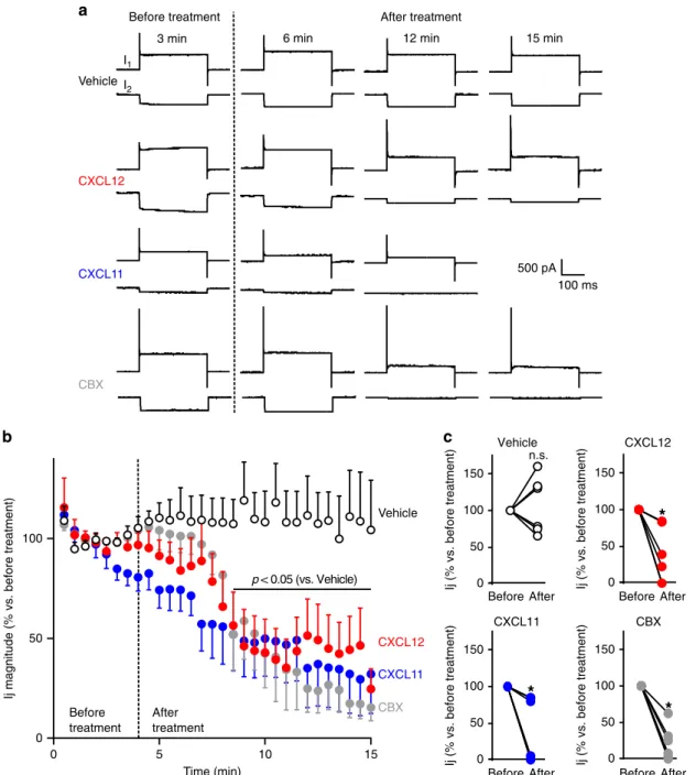
We next performed double patch-clamp recordings of paired
astrocytes in secondary culture to get more direct evidence that
ACKR3 stimulation inhibits the electrical coupling between
astrocytes.
The junctional current (Ij) was continuously monitored in cell
pairs voltage-clamped at
−50 mV. Ij was recorded for a 4-min
control period before challenging cells with either the chemokines
or CBX (Fig.
3
a, b). While Ij magnitude remained stable during
the 15-min recording of vehicle-treated astrocytes (+4.6 ± 20.8%,
p
= 0.9997, n = 6), it strongly decreased as soon as 4.5 min after
the onset of bath-application of CXCL12 or CXCL11, dropping
down to 56.5 ± 16.7% (p
= 0.0194, n = 6) and 52.0 ± 18.3% (p =
0.0093, n
= 6), respectively, and remained inhibited as long as 10
min after the onset of treatments (Fig.
3
b, c). As expected for a
gap junction-mediated electrical coupling, CBX (100
μM) also
drastically reduced Ij magnitude (51.9 ± 17.8%, p
= 0.0091, n =
6).
Altogether,
these
findings
indicate
that
agonist
ACKR3 stimulation inhibits GJIC in primary mouse astrocytes.
ACKR3 inhibits GJIC through G proteins in astrocytes. We
next examined the possibility that G proteins could be involved in
ACKR3-mediated GJIC inhibition, in light of our interactomic
screen, which revealed constitutive association of ACKR3 with
the Gα
i3protein (Fig.
1
a and Supplementary Data 1), and of
previous
findings suggesting that ACKR3 might activate G
i/oproteins in primary rodent astrocytes
14. Overnight pre-treatment
of primary cultures of astrocytes with pertussis toxin (PTX, 100
ng/mL) abolished CXCL12- and CXCL11-induced inhibition of
GJIC, whereas it did not reverse CBX-induced GJIC inhibition
(44.6 ± 6.5% inhibition vs. vehicle, p < 0.0001, n
= 3, Fig.
2
).
Interestingly, PTX significantly increased basal GJIC
commu-nication in astrocytes (130 ± 6.5% increase vs. vehicle, p
= 0.007,
n
= 3, Fig.
2
). Further confirming functional coupling between
ACKR3 and Gα
iproteins in astrocytes, treating cells with
CXCL12 (10 nM) or CXCL11 (100 nM) for 5 min inhibited
for-skolin (10
μM)-induced cAMP production (40.8 ± 6.5, p < 0.0001
and 20.7 ± 6.5% inhibition, p
= 0.0254 in cells exposed to
CXCL12 and CXCL11, respectively, n
= 3). Pre-treatment of cells
with the CXCR4 antagonist AMD3100 (1
μM) for 30 min did not
prevent CXCL12-induced inhibition of cAMP production (44.9 ±
6.5% vs. control, p < 0.0001 n
= 3), indicating involvement of
ACKR3 (and not CXCR4) in the CXCL12 effect (Supplementary
Fig. 3a). As expected, and reminiscent of GJIC measurements,
pre-treating astrocytes with PTX prevented CXCL12- and
CXCL11-induced inhibition of cAMP production
(Supplemen-tary Fig. 3a).
These results contrast with previous
findings indicating that
ACKR3 is not able to activate G proteins in other cellular
contexts
11,12,34, such as in HEK293 cells
13. Corroborating the
results of our interactomic screen performed in HEK293T cells
(Fig.
1
a), saturation BRET analysis confirmed that ACKR3 does
interact with Gα
i3proteins (Supplementary Fig. 3b). However,
CXCL12 (10 nM) did not affect forskolin-induced cAMP
production in HEK293T cells expressing human ACKR3
(Supplementary Fig. 3c), while it did inhibit cAMP production
Fig. 1 ACKR3 physically interacts with ACKR3. a Volcano plot representing proteins identified by nanoLC-MS/MS in immunoprecipitations from HEK293T cells transfected with HA-ACKR3 or empty plasmid. Proteins known to physically or functionally interact with Cx43 (GJA1) and ACKR3 are represented in orange and green, respectively.b Representative images of GFP (Ackr3), Cx43 and GFAP immunostaining in brain from 8-week-old Ackr3 -EGFP BAC mice. Scale bars= 500 μm for mosaics and 100 μm for others. c Representative WB of HA-immunoprecipitations from WT or HA-ACKR3 knock-in (KI) embryonic mouse brains.d Normalized BRET values measured in HEK293T cells co-expressing the indicated proteins. ACKR3-Cx43 BRET signal variations werefitted with the one-site total binding equation and background constraint to constant value of 0 using Prism (Extra-sum-of Square, F-test,P < 0.001 vs. line through the origin, F(2,114)). e Representative WB of immunoprecipitations performed from HEK293T cells expressing HA-ACKR3 or HA-CXCR4. The histogram shows the immunoreactive signals (normalized to the value in HA-HA-ACKR3-expressing cells) of Cx43 co-immunoprecipitated with HA-receptors (Two-tailed unpairedt test, F(2,2)). f 3D reconstruction of ACKR3, CXCR4, and Cx43 immunostainings in TG1 and R633 cells. The overlapping volume represents the volume where each receptor is co-expressed with Cx43 (see Supplementary Fig. 1 for original immunoreactive signals). The histogram illustrates the percentage of Cx43 immunostaining in the overlapping volume (Two-way ANOVA, Bonferroni post-hoc, F(1,17)). Scale bar= 3 μm. g Kinetics representing normalized BRET values measured in HEK293T cells co-expressing Cx43-YFP and ACKR3-NLuc challenged with either vehicle, 10 nM CXCL12 or 100 nM CXCL11. (Two-way ANOVA, Bonferroni post-hoc, F(22,99)).h HEK293T cells expressing HA-ACKR3 challenged with vehicle, CXCL12 (10 nM) or CXCL11 (100 nM) for 30 min. Representative WB of HA-immunoprecipitations are illustrated. The histogram shows the immunoreactive signals (normalized to values from cells challenged with vehicle) of Cx43 co-immunoprecipitated with HA-ACKR3 (One-way ANOVA, Bonferroni post-hoc, F(2,12)). All data are represented as means ± SEM. See the Statistics and Reproducibility section for the number of repetitions, exactP values and symbol (*) legend. Source data and uncropped Western blots are provided as a Source Data file.
in cells expressing CXCR4 (43.9 ± 10.5% inhibition, p
= 0.0018,
n
= 3). Likewise, activation of mouse recombinant CXCR4 by
CXCL12, but not mouse ACKR3, caused a decrease in the BRET
signal between venus-Gγ
2and Gα
i3-RLuc in HEK293T cells
(Supplementary Fig. 3d), suggesting that the ability of ACKR3 to
activate Gα
i/oproteins in mouse astrocyte cultures does not reflect
different coupling properties of mouse and human ACKR3.
ACKR3 stimulation induces Cx43 internalization. Consistent
with already published results
9,35, ACKR3 was constitutively
internalized in HEK293T cells in much higher amounts than
CXCR4 (Supplementary Fig. 4a). Agonist stimulation of ACKR3
further increased the rate of receptor internalization
(Supple-mentary Fig. 4b). As Cx43 activity is often regulated by the
alteration of its trafficking
36, we next investigated if
ACKR3-induced inhibition of GJIC in primary cultured astrocytes is
mediated by Cx43 internalization. Cx43 immunostaining of
vehicle-treated astrocyte cultures showed that Cx43 is primarily
localized at the interface of cell–cell contacts, where Cx43 is
organized as clusters (gap junctional plaques). This results in a
typical
“pavement-like” staining, but a little proportion of Cx43
was also detected intracellularly (Fig.
4
a). In contrast, Cx43 was
mainly detected in intracellular compartments in cultures
exposed to CXCL12 (10 nM) or CXCL11 (100 nM). Furthermore,
treatment of astrocytes with the dynamin inhibitor dynasore (80
μM) prevented CXCL12 and CXCL11 ability to promote Cx43
internalization (Fig.
4
a). Dynasore also prevented the inhibition
of GJIC induced by CXCL12 and CXCL11 treatments (Fig.
4
b). In
contrast, CBX was still able to decrease GJIC in dynasore-treated
cells (57.0 ± 8.8% inhibition, p < 0.0001, n
= 3, Fig.
4
b).
Collec-tively, these results suggest that ACKR3 stimulation promotes
Cx43 internalization and inhibits GJIC through a
dynamin-dependent mechanism in astrocytes.
To get more direct evidence that ACKR3 stimulation promotes
Cx43 internalization, we performed time-lapse recordings of
Cx43-GFP in living HEK293T cells. Treatment of cells
co-expressing RedCherry-ACKR3 and Cx43-GFP with CXCL12 (10
nM) or CXCL11 (100 nM) removed Cx43 plaques localized at
cell-cell contacts, whereas plaques were not affected in cells that
do not express ACKR3 (Fig.
5
and Supplementary Fig. 5). Further
supporting
the
internalization
of
Cx43
plaques
upon
ACKR3 stimulation, the formation of Cx43-containing buds
was transiently observed prior to the removal of plaques (Fig.
5
).
Furthermore, removal of Cx43 plaques often started from their
central part (Supplementary Fig. 5), consistent with previous
findings indicating that Cx43 internalization primarily occurs
from the center of the plaques
37.
We next explored whether ACKR3-induced Cx43
internaliza-tion modifies the subcellular localizainternaliza-tion of Cx30 in astrocytes,
another connexin isoform abundantly expressed in this cell
type
38. Contrasting with what was observed for Cx43, Cx30 was
mainly detected intracellularly in untreated astrocytes and
exposure to CXCL12 or CXCL11 did not affect its intracellular
localization (Supplementary Fig. 6, left panel). Treatment of
astrocytes with dynasore (80
μM) induced a redistribution of Cx30
to the cell surface in cells exposed or not to CXCL11 and CXCL12
LY diffusion (%) PTX Vehicle PTX 150 50 0 100 Vehicle Control Control CXCL12 CXCL12 AMD3100 AMD3100+CXCL12 CXCL11 CXCL11 NBI-74330+CXCL11 NBI-74330 CBX CBX **** **** **** *** **** **** ** *** $$$ $$ n.s. n.s. CXCL12 – + – + – – – – – + – – AMD3100 – – + + – – – – – – – – CXCL11 – – – – + – + – – – + – NBI-74330 – – – – – + + – – – – – CBX – – – – – – – + – – – +
Fig. 2 ACKR3 stimulation inhibits intercellular dye coupling in astrocytes. Representative photomicrographs of scrape loading performed in confluent primary astrocyte cultures, taken ten minutes after the scrape in the presence of Lucifer yellow (1 mg/mL). All treatments (CXCL12 (10 nM), AMD3100 (1μM), CXCL11 (100 nM), NBI-74330 (1 μM)) were applied for 30 min except for CBX (50μM) and PTX (100 ng/mL) that were applied overnight. Scale bar= 200 μm. LY diffusion was calculated by measuring the distance from the scrape where LYfluorescence intensity is 50% of the maximalfluorescence. Values were normalized to LY diffusion in vehicle-treated astrocytes. Results represent the means ± SEM. One-way ANOVA was used for comparison within the vehicle-treated group F(11,24), whereas the two-way ANOVA was used for the comparison across vehicle and PTX groups F(3,16). For both Bonferroni post-hoc was used. See the Statistics and Reproducibility section for the number of repetitions, exactP values and symbol (*,$) legend. Source data are provided as a Source Datafile.
(Supplementary Fig. 6, right panel). Collectively, these results
suggest that Cx30 is
“constitutively” internalized in astrocytes
through a mechanism independent of ACKR3 stimulation and that
ACKR3-induced internalization of Cx43 does not influence
Cx30 subcellular localization.
ACKR3-mediated GJIC inhibition depends on
β-arrestin2.
ACKR3 is known to signal through
β-arrestin2, which has also
been involved in its internalization
12,35. Corroborating these
observations, BRET experiments showed a constitutive
interac-tion between ACKR3 and
β-arrestin2 in HEK293T cells
(Sup-plementary Fig. 4c) that increased in a concentration-dependent
manner upon CXCL12 and CXCL11 challenge (Supplementary
Fig. 4d). In contrast, no constitutive association of CXCR4 with
β-arrestin2 was observed (Supplementary Fig. 4c). Both
constitu-tive and agonist-induced interaction of ACKR3 with
β-arrestin2
was confirmed by co-immunoprecipitation experiments in
b
c
VehicleCBX CXCL12
Ij (% vs. before treatment) Ij (% vs. before treatment) Ij (% vs. before treatment) Ij (% vs. before treatment)
0 50 100 0 5 10 15 Before treatment After treatment Time (min) p < 0.05 (vs. Vehicle) Vehicle CXCL12 CXCL11 CBX
Ij magnitude (% vs. before treatment)
CXCL11
*
0 50 150 100 n.s. 0 50 150 100*
0 50 150 100Before After Before After Before After Before After
*
0 50 150 100 Before treatment After treatment
3 min 6 min 12 min 15 min
CBX Vehicle 500 pA I1 I2 100 ms CXCL12 CXCL11
a
Fig. 3 ACKR3 stimulation impairs gap junctional electrical coupling between astrocytes. a Representative membrane currents recorded in cell pairs of secondary astrocyte cultures (whole-cell configuration of the patch-clamp technique, holding potential −50 mV). Each chart recording illustrates the current recorded in the stimulated celI (I1, upper trace, in response to depolarizing voltage steps, 40 mV amplitude, 300 ms duration, every 30 s) and the junctional current recorded in the non-stimulated cell (I2, bottom trace). I1 and I2 were continuously monitored before and after the treatment of cells with either vehicle, CXCL12 (10 nM), CXCL11 (100 nM), or CBX (100μM). b Time-course of the effect of CXCL12, CXCL11, and CBX on Ij magnitude. Perfusion of the recorded cell pairs with the different compounds started at the time indicated by the dotted line. Data are expressed as a percentage of Ij (normalized to the average ratio recorded during thefirst 4 min before any treatment). Values are means ± SEM (Two-way ANOVA, Bonferroni post-hoc, F(87,573)).c Ij before and after treatment for each cell pair recorded in the different conditions are plotted. Data are expressed as a percentage of Ij (normalized to the average ratio recorded during thefirst 4 min before any treatment, Two-tailed Wilcoxon test). See the Statistics and Reproducibility section for the number of repetitions, exactP values and symbol (*) legend. Source data are provided as a Source Data file.
HEK293T cells co-expressing HA-tagged ACKR3 and
FLAG-tagged
β-arrestin2 (Supplementary Fig. 4e). Therefore, agonist
stimulation of ACKR3 induces concomitant recruitment of
β-arrestin2 and Cx43 (Supplementary Fig. 4e). However, no BRET
signal was detected in HEK293T cells expressing YFP-tagged
Cx43 and RLuc-tagged
β-arrestin2 in the presence of ACKR3
with or without CXCL12 stimulation, thereby ruling out a direct
interaction between Cx43 and
β-arrestin2 (Supplementary
Fig. 4f).
We next examined the impact of CXCL12 and CXCL11 on
Cx43 subcellular localization in astrocytes from
β-arrestin2 knock
out (βarr2
−/−) mice. Contrasting to what was observed in
WT WT Dynasore β-arr2–/– β-arr2–/– β-arr2–/– WT WT Dynasore β-arr2–/– Vehicle CXCL12 CXCL11 WT WT Dynasore 100 150 50 0Cx43 gap junctional plaque size (%)
**
**
Vehicle CXCL12 100 150 50 0 L Y dif fusion (%) * ** $$ $$ $ $$$ CBX CXCL11 *** **** **** WT WT Dynasoreb
a
CXCL11 CXCL12 Vehicle CXCL11 CXCL12 Vehicle CXCL11 CXCL12 Vehicle Vehicle CXCL12 CXCL11 CBX Vehicle CXCL12 CXCL11 CBX Vehicle CXCL12 CXCL11 CBXastrocytes from wild type mice, exposing primary astrocyte
cultures from
βarr2
−/−mice to CXCL12 (10 nM) or CXCL11
(100 nM) did not promote Cx43 internalization (Fig.
4
a).
Consistently, neither CXCL12 nor CXCL11 inhibited GJIC in
astrocytes from these mice (Fig.
4
b). In addition, the extent of LY
diffusion through vehicle-treated astrocytes was similar to that
measured in astrocytes from WT mice and CBX was still able to
inhibit astrocytic GJIC in
βarr2
−/−mice (67.8 ± 8.2% inhibition,
p < 0.0001, n
= 3, Fig.
4
b).
Discussion
ACKR3 is involved in key biological processes such as
migra-tion
10, proliferation
39, and differentiation
40,41of different cell
populations, but the cellular pathways transducing these effects
Fig. 4 ACKR3 stimulation regulates Cx43 cellular localization and inhibits GJIC in astrocytes. a Confocal images of Cx43 immunostaining of confluent primary astrocyte cultures from WT orβ-arr2−/−mice representative of three independent experiments performed on different sets of cultured cells exposed for 30 min to either vehicle or CXCL12 (10 nM) or CXCL11 (100 nM) in the absence or presence of Dynasore (Dyn, 80μM). Scale bar = 50 μm. The histograms show the size of Cx43 gap junctional plaques (clusters of channels present at the cell-cell interface). Data were normalized to the one measured in vehicle-treated cultures. They are the means ± SEM of values (Two-way ANOVA, Bonferroni post-hoc F(4,18)).b Representative photomicrographs of scrape loading in confluent primary astrocyte cultures obtained from embryonic WT and β-arr2−/−mice taken 10 min after the scrape in the presence of LY. CXCL12 (10 nM), CXCL11 (100 nM), and Dynasore (Dyn, 80μM) were applied for 30 min. CBX (50 μM) was applied overnight. Scale bar= 200 μm. LY diffusion was calculated by measuring the distance from the scrape where LY fluorescence intensity is 50% of the maximalfluorescence. Values were normalized to LY diffusion in vehicle-treated astrocytes. Results represent the means ± SEM of values (Two-way ANOVA, Bonferroni post-hoc, F(6,24)). See the Statistics and Reproducibility section for the number of repetitions, exactP values and symbol (*/$)
legend. Source data are provided as a Source Datafile.
Cx43
ACKR3
0 min
0 min
5 min
10 min
15 min
20 min
27 min
26 min
28 min
29 min
30 min
25 min
24 min
23 min
22 min
21 min
*
*
*
*
Fig. 5 ACKR3 activation promotes Cx43 internalization. Time-lapse images of gap junction plaques in HEK293T cells co-expressing Cx43-GFP and RedCherry-ACKR3 and exposed to CXCL12 (10 nM) for 30 min. The two upper images show GFP (Cx43, left panel) and RedCherry (ACKR3, right panel) fluorescence signals, respectively, before the onset of chemokine application. Images below show Cx43-GFP fluorescence at the times indicated. A zoomed image of a Cx43-GFP plaque is illustrated at 0, 20 and 30 min in the left bottom corner of the image. The arrows show the removal of the plaque and the arrowheads Cx43-GFP vesicle budding. The decrease in Cx43-GFPfluorescence was not due to photobleaching, as CXCL12 exposure did not induce the removal of a gap junction plaque located at the interface of cells that do not express ACKR3 (*). Scale bars= 10 and 1 μm for time-lapse and magnified pictures, respectively. See the Statistics and Reproducibility section for the number of repetitions.
remained poorly understood. To address this issue, we
char-acterized the ACKR3 interactome in HEK293T cells and
identi-fied Cx43 as an ACKR3-interacting protein. Intriguingly, ACKR3
was previously found to interact with the gap junction beta-2
protein GJB2 (or Cx26), but not Cx43, in a large interactomic
screen exploring the interactome of 1125 GFP-tagged proteins
expressed in HeLa cells
42. These distinct results might reflect the
different cell backgrounds (HEK293T vs. HeLa) used in both
studies and suggest that ACKR3 physically associates with gap
junctions composed of distinct connexin families in a cell
type-dependent manner. The ability of ACKR3 and Cx43 to form a
complex in vivo was further validated by co-IP in mouse brain,
where both proteins are co-expressed in GFAP-positive astrocytes
of the subventricular zone and surrounding blood vessels. ACKR3
is also present in neurons
3that do not express Cx43
43, indicating
that the interaction between ACKR3 and Cx43 only occurs in
restricted brain cell populations. Whether ACKR3 modulates
GJIC in other cell types expressing different connexin isoforms
such as neurons remains to be explored. Moreover, using
com-plementary approaches, we showed that Cx43 interacts with
ACKR3 but not with the functionally related CXCR4 in both
HEK293T cells and glioblastoma-initiating cell lines.
There is convergent evidence indicating that ACKR3 and Cx43
exert an opposite influence upon various physiological and
pathological processes such as neuronal migration
4,5,44,45,
leu-kocyte entry
6,46into the brain and formation or progression of
certain cancer types
8,47. For instance, a large body of evidence
indicates an increased expression of ACKR3, which favors tumor
cell proliferation and invasion as well as angiogenesis
8, in various
cancers such as glioma
48. In contrast, studies support the notion
that connexins, including Cx43, are tumor suppressors even
though recent evidence suggest that Cx43 may also promote
tumor cell migration and metastasis through both
GJIC-dependent and inGJIC-dependent pathways
47. Likewise, ACKR3
expression is increased during experimental autoimmune
ence-phalomyelitis (EAE), a preclinical model of multiple sclerosis
(MS), which is ameliorated by treatment with an ACKR3
antagonist
6, whereas astrocytic Cx43 expression is decreased in
MS and other inflammatory demyelinating brain disorders.
Fur-thermore, Cx43 loss is mainly found in actively demyelinating
and chronically active lesions and is associated with rapid disease
progression
49. Corroborating this opposite influence upon
com-mon pathophysiological processes, we show that agonist
stimu-lation of ACKR3 inhibits GJIC in primary astrocyte cultures using
fluorescent dye diffusion and whole-cell patch-clamp recordings.
These observations provide evidence of a direct functional link
between ACKR3 and gap junctions that might contribute to the
role of ACKR3 in glioma progression. Reminiscent of the specific
association of Cx43 with ACKR3 but not with CXCR4, only
ACKR3 stimulation reduced GJIC in astrocytes. The role of the
physical ACKR3-Cx43 interaction in receptor-operated inhibition
of GJIC could not be fully established in the absence of the
identification of binding motifs within the sequences of both
partners.
In an effort to identify the signaling pathways involved in the
ACKR3-mediated inhibition of GJIC, we showed that inhibiting
Gα
i/oprotein activation by PTX treatment abolished the ACKR3
inhibitory effect in primary cultured astrocytes. Consistent with
previous studies indicating that Cx43 is negatively regulated by
the activation of numerous GPCRs
20, the increase in basal GJIC
elicited by PTX might reflect a blockade of G
i/o-coupled receptors
endogenously expressed in astrocytes. In untreated conditions,
these GPCRs would inhibit Cx43-mediated GJIC as a result of
their activation by gliotransmitters released by astrocytes or
constitutive activity. The inhibitory effect of PTX upon
ACKR3-mediated inhibition of GJIC was more unexpected given the
current classification of ACKR3 as an atypical chemokine
receptor unable to activate G proteins. However, it corroborates a
previous report indicating that the receptor can signal through
Gα
i/oproteins in rodent astrocytes and human glioma cells
14. The
ability of ACKR3 to activate G proteins has so far only been
demonstrated in glial cells, but not in other cell types like
HEK293 cells
11–13,34. Consistent with these
findings, we showed
that ACKR3 stimulation inhibits cAMP production through a
Gα
i/o-dependent mechanism in primary astrocytes, but not in
HEK293T cells. Furthermore, this different capability to activate
G
i/oproteins was not due to different coupling properties between
human and mouse ACKR3, as stimulation of mouse ACKR3
expressed in HEK293T cells did not activate G proteins. Though
one cannot entirely rule out that ACKR3 stimulation promotes
the release of GPCR agonists by glial cells, such as adenosine and
glutamate, which would in turn activate their respective
endo-genously expressed receptors to inhibit cAMP production, such
an indirect activation of other Gα
i/o-coupled receptors is unlikely,
due to the short chemokine challenging protocol used.
Accord-ingly, the present
findings together with previously published
results suggest that astrocytes constitute a unique cellular
envir-onment where ACKR3 can activate G proteins. This unique
capability of astrocytic ACKR3 to activate Gα
i/oproteins might
rely on the specific expression of not yet identified receptor
partners or adaptor proteins.
Beyond G protein-dependent mechanisms, Cx43 activity is
often regulated by the alteration of its trafficking
36. Several
pro-tein kinases including some members of the MAP kinase
path-ways and Cx43 partners such as clathrin, myosin, actin, and
drebrin, have been involved in Cx43 endocytosis
50–53. Here, we
provide convergent evidence indicating that GJIC inhibition
eli-cited by ACKR3 stimulation in astrocytes is a consequence of
internalization of Cx43 involved in gap junctions: (i) treatment of
astrocyte cultures with either CXCL12 or CXCL11 triggered
concomitant Cx43 internalization and GJIC inhibition; (ii)
inhi-bition of Cx43 internalization by the dynamin inhibitor Dynasore
prevented the ability of ACKR3 stimulation to inhibit GJIC; and
(iii) GJIC inhibition and concomitant Cx43 internalization
eli-cited by ACKR3 activation were abolished in astrocytes from
β-arr2
−/−mice, consistent with the ability of the receptor to
interact with
β-arrestin2
9. Previous studies have demonstrated
that recruitment of
β-arrestin2 by ACKR3 depends on its
C-terminal domain
34and likely involves the phosphorylation of Ser/
Thr clusters by protein kinase(s) such as GRK2 and GRK5
9.
Co-IP experiments indicated that ACKR3 forms a complex with both
Cx43 and
β-arrestin2 and that their recruitment is enhanced
upon agonist receptor stimulation, suggesting that ACKR3 might
concomitantly recruit Cx43 and
β-arrestin2 via different binding
motifs.
The prevailing view on GPCR endocytosis is that the
phos-phorylation of ligand-activated GPCRs by GRKs and other
kinases induces recruitment of
β-arrestins, which then promotes
clathrin-dependent receptor endocytosis. In agreement with this
model, both basal and agonist-induced ACKR3 internalization
were initially reported to depend on
β-arrestins
34,35. These
findings favor the possibility that Cx43 is directly co-internalized
with activated ACKR3 within a complex comprising ACKR3,
Cx43, and
β-arrestin2. Nevertheless, the notion that ACKR3
requires
β-arrestins to internalize and sequester chemokines has
been challenged by recent reports indicating that
β-arrestins are
dispensable for ACKR3 endocytosis and ACKR3-mediated
scavenging of CXCL12 in mammalian cell cultures
54,
zebra-fish
23, and mouse brain
9. Accordingly, Cx43 internalization in
CXCL12/CXCL11-exposed astrocytes might occur independently
of ACKR3 internalization through an indirect,
β-arrestin2-dependent, signaling mechanism.
Protein–protein interactions play a key role in numerous
bio-logical processes, and their dysfunctions have been involved in
the pathogenesis of a range of human diseases, including cancer.
Abnormal protein–protein interactions contribute to all phases of
oncogenesis, including tumor cell proliferation and survival,
inflammation, invasion, and metastasis. Identifying and
char-acterizing the function of protein–protein interactions involving
molecules upregulated and mobilized in a diversity of cancers,
such as ACKR3, is thus crucial to understand mechanisms
underlying cancer progression and develop new therapeutic
strategies
55. In this context, the current study provides evidence
for a physical and functional link between the CXCL11/CXCL12/
ACKR3 axis and Cx43, one of the members of the gap junction
protein family that can act as tumor suppressor or promote
tumor progression and metastasis, depending on the cancer type
and cancer stage
47. This interaction might not only contribute to
the development of various cancers such as glioma, but also play a
key role in physiological processes under the control of ACKR3 or
depending on proper function of gap junction channels in
astrocytes, such as interneuron migration
4,5, blood brain barrier
permeability
6, and synaptic transmission and plasticity
56. Finally,
the present study provides insight into the persistent conundrum
of the role of G proteins in ACKR3-mediated effects by
con-firming the previously described engagement of G
protein-dependent signaling in astroglial cells
14, thereby suggesting a cell
type-dependent involvement of G proteins in ACKR3-operated
signaling and function.
Materials and methods
Materials. HA-ACKR3 was obtained introducing a single HA-tag at the N-terminal by PCR. The construct was then cloned in the pCDNA3.1 vector using the BamHI and XbaI restriction enzymes. 3XHA-CXCR4-pCDNA3.1 construct was obtained from cDNA.org (Bloomsburg University). The pcDNA 3.1 plasmids encoding ACKR3-NLuc and CXCR4-NLuc were provided by Dr. M.J. Lohse (Max-Delbrück-Zentrum für Molekulare Medizin (MDC), Berlin, Germany). The Cx43-YFP-pCDNA3.1, Cx43-GFP-pEGFPN1 and RedCherry-ACKR3-pCDNA3.1 plas-mids were obtained from the human ORFeome collection (IGMM, Montpellier, France). The FLAG-SNAP-ACKR3 and FLAG-SNAP-CXCR4 constructs (in pcDNA3.1) were provided by Cisbio Bioassays (Codolet, France). The ACKR3-YFP and the CXCR4-YFP constructs were described in13and57, respectively. pcDNA3.1
plasmids encoding Venus-Tagged G protein subunit gamma 2 (Venus-γ2), FLAG-tagged G protein subunit beta 2 (FLAG-β2), β-arrestin2-RLuc and Gαi3-RLuc were
provided by Dr. D. Maurel (IGF, Montpellier, France). Human β-arrestin2-FLAG-pcDNA3.1 plasmid was provided by Dr. C. Mendre (IGF, Montpellier, France). Mouse ACKR3 and CXCR4 constructs were described in refs.58,59, respectively.
EGFP-ACKR3 BAC mice were from GENSAT and characterized in ref.2,4.
They were maintained heterozygous on Hsd:ICR (CD-1®) background. The HA-ACKR3 mouse models were designed and generated by GenOway (Lyon, France) and already characterized in9. C57BL/6Jβ-arr2−/−mice were generated from
heterozygousβ-arr2+/−mice generated in Prof J. Lefkowitz’s laboratory (Duke
University, USA). Mice were housed under standardized conditions with a 12‐h light/dark cycle, stable temperature (22 ± 1 °C), controlled humidity (55 ± 10%), and free access to food and water. Experiments on animals conformed to European ethics standards (86/609-EEC) and to decrees of the French National Ethics Committee (N°87/848) for the care and use of laboratory animals. Protocols were approved by the University of Montpellier ethics committee for animal use (CEEA LR 34, #7251).
Recombinant mouse CXCL11 (Ref 572-MC), Human/Feline/Rhesus Macaque CXCL12 (Ref 350-NS) and NBI-74330 (Ref 4528/10) were purchased from R&D System, AMD3100 from Tocris (Ref 3299), Lucifer Yellow CH dilithium salt (LY, Ref L0259), Dynasore hydrate (Ref D7693), carbenoxolone disodium salt (CBX, Ref C4790) and pertussis toxin (Ref 516560) from Merck.
Primary antibodies. For the complete list of primary antibodies and related information, see Supplementary Table 1.
Cell cultures and transfection. HEK293T cells, purchased from ATCC (Anassas, VI, ATCC, CRL-1573), were grown in Dulbecco’s Modified Eagle’s Medium (DMEM, Thermo Fisher Scientific, Ref 419960) supplemented with 10% heat-inactivated fetal bovine serum (Thermo Fisher Scientific, Ref 10099-133) and maintained in humidified atmosphere containing 5% CO2at 37 °C. Cells were
passed twice a week and used between passages 10 and 20. Absence of mycoplasma was assessed every month using the MycoAlert Mycoplasma Detection Kit (Lonza,
Ref LT07-118). HEK293T cells were transfected in suspension using poly-ethylenimine (PEI, Polyscience, Ref 24765). Cells were seeded one day prior transfection and used two days after transfection for all experiments except for BRET. For BRET cells were transfected in suspension 24 h before experiment. Mock cells were transfected with empty plasmids.
Primary and secondary cultures of astrocytes were prepared from wild type and β-arr2−/−mice embryos (embryonic day E16.5). Forebrains were dissected and
cells were dissociated mechanically before seeding in culture medium containing a 1:1 mixture of DMEM and F-12 nutrient (Merck, Ref D057) supplemented with glucose (30 mM), glutamine (2 mM), NaHCO3(13 mM), HEPES buffer (5 mM, pH
7.4), penicillin-streptomycin (100 unit/mL-0.1 mg/mL, Thermo Fisher Ref 15140-122) and 10% heat-inactivated Nu-Serum (Corning, Ref 355500). The medium was changed one week after seeding and then twice a week. Five weeks after seeding, cultures were starved from serum in DMEM supplemented with penicillin-streptomycin (100 unit/mL–0.1 mg/mL) overnight before experiment. For secondary culture preparation, cells were washed with PBS and incubated for 15 min with trypsin-EDTA 0.05% (Thermo Fisher Scientific, Ref 25300-054) at 37 °C. Cells were then centrifuged at 200 × g for 5 min, suspended in complete DMEM/F-12 medium, plated on polyornithine-coated glass coverslips and processed for double patch-clamp recordings the following day. Primary cultures were used for all experiments but double patch-clamp recordings that were performed on secondary cultures.
Patient-derived cell lines TG1 and R633, isolated from neurosurgical biopsy samples of human glioblastoma and characterized for their stem-like and tumor-initiating properties, were cultured in Dulbecco’s modified Eagle’s:F-12 medium (1:1) containing the N2, G5 (containing FGF and EGF) and B27 supplements (all from Invitrogen, France)60,61. Samples were obtained with informed consent of
patients to use them in research. The institutional review board of the Sainte-Anne Hospital Center - University Paris Descartes (Comité de protection des personnes Ile de France III) approved the study protocol (Protocol number DC-2008-323). Co-immunoprecipitation. HEK293T cells were lysed in ice-cold lysis buffer con-taining 1% n-Dodecyl-β-D-Maltopyranoside (DDM, Antrace, Ref D310), 20 mM Tris-HCl (pH 7.5), 100 mM NaCl, 2.5 mM CaCl2, phosphatase inhibitors (NaF, 10
mM; Na+-vanadate, 2 mM; Na+-pyrophosphate, 1 mM; andβ-glycerophosphate, 50 mM) and the cOmplete Protease Inhibitor Cocktail (Merck, Ref 11836145001). Samples were maintained under gentle agitation for 1 h at 4 °C and centrifuged for 15 min at 15,000 × g to eliminate insoluble material. Soluble proteins were quan-tified by bicinchoninic acid assay (BCA, Merck, Refs B29643 and C2284) and equal protein amounts (5 or 1.5 mg for IP followed by MS/MS or WB, respectively) were incubated with agarose-conjugated anti-HA antibody (Merck, Ref A2095) over-night at 4 °C. Samples were then washed twice with an ice-cold solution of 0.5 M of NaCl and phosphatase inhibitors and three times with 150 mM NaCl and phos-phatase inhibitors. Immunoprecipitated proteins were then eluted in Laemmli sample buffer.
Co-immunoprecipitation from embryonic brains (E17) was performed as described in9. Briefly, brains were isolated and snap frozen in liquid nitrogen. Two
brains per conditions were pooled and sonicated in lysis buffer containing (150 mM NaCl, 5 mM EDTA, 50 mM Tris-HCl, pH 7.4 and DDM 1.5%) plus the cOmplete Protease Inhibitor Cocktail. Samples were maintained for 1 h at 4 °C under gentle agitation and centrifuged for 30 min at 21,000 × g. Soluble proteins were quantified by BCA and equal protein amount (5 mg) per each condition was incubated with the agarose-conjugated anti-HA magnetic beads (Thermo Fisher Scientific, Ref 88836) overnight at 4 °C under constant rotation.
Immunoprecipitated proteins were then eluted in Laemmli sample buffer. Protein identification by mass spectrometry. Immunoprecipitated proteins were separated by SDS-PAGE and stained with Protein Staining Solution (Euromedex, Ref 10-0911). Gel lanes were cut into seven gel pieces and destained with 50 mM TriEthylAmmonium BiCarbonate (TEABC, Merck, Ref T7408) followed by three washes in 100% acetonitrile. After reduction (with 10 mM dithiothreitol in 50 mM TEABC at 60 °C for 30 min) and alkylation (with 55 mM iodoacetamide TEABC at room temperature for 60 min), proteins were digested in-gel using trypsin (500 ng/ band, Gold, Promega, Ref V5280)62. Digest products were dehydrated in a vacuum
centrifuge and reduced to 3μL. The generated peptides were analyzed online by nano-flow liquid chromatography coupled to tandem-mass spectrometry (nanoLC-MS/MS) using an Orbitrap Elite mass spectrometer (Thermo Fisher Scientific, Waltham USA) coupled to an Ultimate 3000 HPLC (Thermo Fisher Scientific). Desalting and pre-concentration of samples were performed on-line on a Pepmap® pre-column (0.3 mm × 10 mm, Dionex). A gradient consisting of 0–40% B for 60 min and 80% B for 15 min (A= 0.1% formic acid, 2% acetonitrile in water; B = 0.1% formic acid in acetonitrile) at 300 nL/min was used to elute peptides from the capillary reverse-phase column (0.075 mm × 150 mm, Acclaim Pepmap 100®C18, Thermo Fisher Scientific). Eluted peptides were electrosprayed online at a voltage of 1.8 kV into the Orbitrap Elite mass spectrometer. A cycle of one full-scan mass spectrum (MS1, 400–2,000 m/z) at a resolution of 120,000 (at 400 m/z), followed by 20 data-dependent tandem-mass (MS2) spectra was repeated continuously throughout the nanoLC separation. All MS2 spectra were recorded using nor-malized collision energy (33%, activation Q 0.25 and activation time 10 ms) with an isolation window of 2 m/z. Data were acquired using the Xcalibur software (v 2.2).
For all full scan measurements with the Orbitrap detector a lock-mass ion from ambient air (m/z 445.120024) was used as an internal calibrant63. Mass spectra
were processed using the MaxQuant software package (v 1.5.5.1) and MS2 using the Andromeda search engine“[http://coxdocs.org/doku.php?id=maxquant:
andromeda:start]” against the UniProtKB Reference proteome UP000005640
database for Homo sapiens (release 2017_10) and the contaminant database in MaxQuant. The following parameters were used: enzyme specificity set as Trypsin/ P with a maximum of two missed cleavages, Oxidation (M) and Phosphorylation (STY) set as variable modifications and carbamidomethyl (C) as fixed modification, and a mass tolerance of 0.5 Da for fragment ions. The maximum false peptide and protein discovery rate was specified as 0.01. Seven amino acids were required as minimum peptide length. When precursor peptides were present in MS1 spectra but not selected for fragmentation and identification by MS2 in given runs, peptide identification were based on accurate mass and retention times across LC-MS runs using the matching between runs tool in MaxQuant. Only proteins identified in all three biological replicates in at least one group were considered for further analysis. Relative protein quantification in IP from ACKR3-expressing cells and Mock cells was performed using the label-free quantification (LFQ) algorithm “[https:// maxquant.net/maxquant/]”. For statistical analysis, missing values were defined
using the imputation tool of the Perseus software (v. 1.5.6.072)“[https://maxquant. net/perseus/]”.
Western blotting. Proteins were separated by SDS-PAGE onto 10% poly-acrylamide gels and transferred electrophoretically to nitrocellulose membranes (Bio-Rad, Ref 1704271). Membranes were incubated in blocking buffer (Tris-HCl, 50 mM, pH 7.5; NaCl, 200 mM; Tween-20, 0.1% and skimmed dried milk, 5%) for 1 h at room temperature and overnight with primary antibodies in incubating buffer (Tris-HCl, 50 mM, pH 7.5; NaCl, 200 mM; Tween-20, 0.1% and Bovine Serum Albumin (Merck, Ref A2153), 5%) at 4 °C. Then membranes were immu-noblotted with either anti-mouse (Merck, Ref GENA931V) or anti-rat (Jackson ImmunoResearch, Ref 112-035-003) or anti-rabbit (Merck, Ref GENA934V) horseradish peroxidase (HRP)-conjugated secondary antibodies (1/5,000) in blocking buffer for 1 h at room temperature. Immunoreactivity was detected with an enhanced chemiluminescence method (Western lightning®Plus-ECL, Perkin Elmer, Ref NEL103E001EA) on a ChemiDoc™ Touch Imaging System (Bio-Rad). Quantification was performed using the Image Lab software (Bio-Rad). Uncropped blots corresponding to the blots illustrated on thefigures are provided in the Source Datafile.
Immunohistochemistry. Eight-week-old mice were anesthetized with pento-barbital (100 mg/kg i.p., Ceva SA) and perfused transcardiacally withfixative solution containing 4% w/v paraformaldehyde, 0.1 M sodium phosphate buffer (pH 7.5), NaF (10 mM), and Na+-vanadate (2 mM). Brains were post-fixed for 48 h in the same solution. For cryoprotection, tissue was transferred to PBS buffer con-taining 10 then 20 andfinally 30% sucrose. They were then embedded in the embedding medium for cryotomy (Optimal Cutting Temperature, OCT, VWR Chemicals, Ref 361603E) and rapidly frozen with SnapFrost®(Excilone) and stored at−80 °C. 50 μm-thick coronal cryosections were cut using a Leica CM3050 Cryostat and kept freefloating in PBS at 4 °C. Slices were incubated in PBS solution containing 10% heat-inactivated goat serum (Vector Laboratories, Ref S-100) and 0.3% Triton X-100 for 20 min. They were then incubated overnight at 4 °C in PBS containing 3% heat-inactivated goat serum, 0.1% Triton X-100, anti-Cx43 (Rabbit), anti-GFP (Chicken) and anti-GFAP (Mouse) antibodies. After extensive PBS washings, slices were incubated for 2 h with the Alexa Fluor®488-conjugated anti-chicken antibody (1/1,000, Thermo Fisher Scientific, Ref A-11039), the Alexa Fluor®594-conjugated anti-rabbit antibody (1/1,000, Thermo Fisher Scientific Thermo Fisher Scientific, Ref A-11037) and the Alexa Fluor®680-conjugated
anti-mouse antibody (1/1,000, Thermo Fisher Scientific, A-21057) in PBS containing 3% heat-inactivated goat serum, 0.1% Triton X-100 and Hoechst 33342 (2μM, Thermo-Scientific, Ref 62249). After three washes in PBS, slices were mounted on superfrost ultra plus slides (Thermo-Scientific, Ref 10417002) using fluorescent mounting medium (Dako, Ref S3023). Pictures were taken with a Leica SP8-UV confocal microscope. Quantification of the Cx43 gap junctional plaque size was performed using the Fiji software. Each image was converted into binary and threshold was automatically defined. Plaque areas were then calculated by means of the Particle Analysis Fiji algorithm. The quantified areas were then divided by the number of cells counted in thefield.
Immunocytochemistry. Astrocytes cultivated on coverslips werefixed with a 4% solution of PFA in PBS for 10 min. Glioblastoma cell lines cultured as free-floating cell spheres were mechanically dissociated, transferred onto SuperFrost glass slides (Dutscher) andfixed with 4% PFA in PBS. Excess of PFA was quenched by washing cells three times in a 0.1 M solution of glycine in PBS. Cells were per-meabilized with a PBS solution containing 5% heat-inactivated goat serum and 0.1% Triton X-100 for 20 min. When needed, antigen retrieval was performed by heating up cells to 80 °C for 20 min in a citrate buffer solution (pH 6) containing Tween 0.05% before any staining. After cooling down to room temperature, cells were washed three times and incubated with primary antibodies (anti-Cx43 (rabbit or mouse), anti-ACKR3 (mouse) and anti-CXCR4 (rabbit)) in PBS containing 2.5%
heat-inactivated goat serum and 0.05% Triton X-100. After three washes in PBS, cells were incubated for 2 h at room temperature in PBS containing 2.5% goat serum, 0.05% Triton X-100, the appropriate secondary antibody (Alexa Fluor® 488-conjugated anti-mouse antibody (1/1,000, Thermo-Scientific, Ref A-11029), Alexa Fluor®594-conjugated anti-rabbit antibody (1/1,000)) and Hoechst 33342 (2μM). Cells were then washed three times with PBS and coverslips were mounted on superfrost ultra plus usingfluorescent mounting medium. Pictures were taken at different confocal plans with a Leica SP8-UV confocal microscope. For the inter-nalization assay the different confocal plans were then merged using the Z project algorithm of Fiji with the maximal intensity for projection. Plaque size was quantified using the counting particle plug-in of Fiji. For co-localization studies, 3D reconstructions from imaged confocal plans (between 20 and 50 plans for each cells) were performed using the Imaris software (Bitplane). Overlapping volume wasfirstly defined by identifying the center of mass of each object belonging to group A (Cx43) or B (ACKR3/CXCR4). Then, two objects, defined by setting an automatic threshold64, were considered as co-localized if the center of one of them
falls into the area of the other65. The intensity of Cx43 signals within the
over-lapping volume was quantified. For comparison this intensity was divided by the total intensity of each group before plotting.
Time-lapse imaging of Cx43-GFP in living cells. Transfected HEK293T cells were seeded (50,000 cells/well) in polyornitine-coated 35 mm-Nunc™ Glass Bottom Dishes (Thermo-Scientific, Ref 150680). Eighteen hours before the experiments, the medium was replaced by DMEM without serum. Cells were then washed with PBS and incubated in medium containing 130 mM NaCl, 2.8 mM KCl, 1 mM CaCl2, 2
mM MgCl2and 10 mM HEPES (pH 7.2) before challenge. Cells were next treated
with either CXCL12 (10 nM) or CXCL11 (100 nM) and imaged on a Leica SP8-UV inverted confocal microscope using a 488-nm argon/krypton laser line and a 561-nm DPSS laser line with a 63x oil (1.4 numerical aperture) objective. Cells were maintained at 37 °C, 95% O2/ 5% CO2for the duration of the experiment during
which the focus, contrast, or brightness settings remained constant. Optical scans were collected continuously at a scan speed of 32 s for 30 min after the treatment onset. For analysis, images were organized sequentially in a movie sequence using the LAS X software (v. 3.5, Leica).
BRET. Transfected HEK293T cells were seeded (50,000 cells/well) in white and black polyornithine-coated-96-well plates (Greiner, Ref 655083 and 655079). After 24 h, cells were washed twice with PBS. Coelenterazine (Nanolight Technology, Ref 50909-86-9) was added at afinal concentration of 5 μM in the white plate before incubation at 37 °C for 5–10 min. Reading was immediately performed after the addition of different ligands or PBS, using a Mithras LB 940 plate reader (Berthold Biotechnologies) that allows the sequential integration of light signals detected with twofilter settings (Rluc/NLuc filter, 485 ± 20 nm; and YFP filter, 530 ± 25 nm). Data were collected using the MicroWin2000 software (Berthold Biotechnologies). The YFP signal was measured from the black plate using an INFINITE F500 plate reader (TECAN) by setting the excitationfilter at 485 ± 20 nm and the emission filter at 520 ± 10 nm.
DERET. DERET internalization assay was performed as described in66. Briefly,
transfected HEK293T cells were seeded in white polyornithine-coated 96-well plates (25,000 cells/well). Forty-eight h after seeding, culture medium was sub-stituted with 75 µl Tag-lite®labelling medium (Cisbio, Ref LABMED) containing 100 nM SNAP-Lumi4-Tb (Cisbio, Ref SSNPTBX). Cells were then incubated for 1.5 h at 16 °C. After washing the excess of SNAP-Lumi4-Tb, internalization experiments were carried out by incubating cells in the absence or presence of incremental concentrations of CXCL12 in Tag-lite®labeling medium containing fluorescein (25 µM, Merck, Ref 46955) at 37 °C for 1.5 h. Lumi4-Tb was excited at 340 nm and signals emitted at 620 and 520 nm were collected using the INFINITE F500 plate reader (TECAN). Ratio (620/520) was obtained by dividing the donor signal by the TR-FRET signal and multiplying this value by 10,000. Data were expressed as % of maximal internalization after subtraction of the internalization at the onset of treatment.
Scrape loading. Five-week-old astrocytes were serum-starved overnight in the presence or absence of PTX (100 ng/mL) or CBX (50μM). Cultures were exposed to the indicated treatment in medium containing 130 mM NaCl, 2.8 mM KCl, 1 mM CaCl2, 2 mM MgCl2and 10 mM HEPES (pH 7.2), for 30 min or 5 min at room
temperature. Cells were then exposed to the same medium without CaCl2for 1 min
and the cell monolayer was scraped using a razor blade in the presence of Lucifer yellow (1 mg/mL) dissolved in the calcium-free medium. LY was then let diffusing into cells for 1 min. Cells were then washed 5 times with the calcium-containing medium. After 10 min, images were taken using an invertedfluorescence micro-scope (Zeiss Axiovert 40CFL) equipped with a CCD camera (Axiocam ICCL1, Zeiss), using the Axiovision 4.8 software (Zeiss). For quantification of LY diffusion from the scrape, images were analyzed with the Fiji software. Fluorescence was plotted against the distance from the cut. The averagefluorescence of the 50 most distant pixels from the cut was considered as background and subtracted from all values. Fluorescence was then normalized to the maximalfluorescence value and one exponential decay regression wasfitted using Prism (v. 8.0, GraphPad Software