HAL Id: hal-02906984
https://hal.sorbonne-universite.fr/hal-02906984
Submitted on 26 Jul 2020
HAL is a multi-disciplinary open access
archive for the deposit and dissemination of
sci-entific research documents, whether they are
pub-lished or not. The documents may come from
teaching and research institutions in France or
abroad, or from public or private research centers.
L’archive ouverte pluridisciplinaire HAL, est
destinée au dépôt et à la diffusion de documents
scientifiques de niveau recherche, publiés ou non,
émanant des établissements d’enseignement et de
recherche français ou étrangers, des laboratoires
publics ou privés.
Distribution of free-living and particle-attached aerobic
anoxygenic phototrophic bacteria in marine
environments
Raphaël Lami, Z Âuperová, J. Ras, P. Lebaron, M Koblíïek
To cite this version:
Raphaël Lami, Z Âuperová, J. Ras, P. Lebaron, M Koblíïek. Distribution of free-living and
particle-attached aerobic anoxygenic phototrophic bacteria in marine environments. Aquatic Microbial
Ecol-ogy, Inter Research, 2009, 55, pp.31-38. �10.3354/ame01282�. �hal-02906984�
INTRODUCTION
Recent studies have revealed that a large fraction of marine prokaryotes are not strictly heterotrophic or au-totrophic, but adopt a variety of mixotrophic strategies (Béjà et al. 2000, Kolber et al. 2000, Zubkov & Tarran 2005). One group of these organisms, aerobic anoxy-genic phototrophic (AAP) bacteria, performs
photo-heterotrophic metabolism. They require organic sub-strates for their metabolism and growth and they are able to supplement a fraction of their energy demands using light (Yurkov & Beatty 1998). AAP bacteria con-tain bacterial photosynthetic reaction centers with bac-teriochlorophyll a (Bchl a) as the main light-harvesting
pigment. The abundance of AAP bacteria varies greatly among different marine environments (Kolber
© Inter-Research 2009 · www.int-res.com *Email: lami@udel.edu
Distribution of free-living and particle-attached
aerobic anoxygenic phototrophic bacteria in
marine environments
Raphaël Lami
1, 2, 7,*, Zuzana >uperová
3, 4, Josephine Ras
5, 6, Philippe Lebaron
1, 2,
Michal Koblí=ek
3, 41UPMC Univ Paris 06, UMR 7621, LOBB, Observatoire Océanologique, 66651 Banyuls-sur-Mer, France 2CNRS, UMR 7621, LOBB, Observatoire Océanologique, 66651 Banyuls-sur-Mer, France
3Institute of Physical Biology, University of South Bohemia, Zámek 136, 373 33 Nové Hrady, Czech Republic 4Institute of Microbiology CAS, Opatovick) ml)n, 379 81 Trˇeboˇn, Czech Republic
5UPMC Univ Paris 06, UMR 7093, Lab. d’Océanographie de Villefranche-sur-Mer, 06230 Villefranche-sur-Mer, France 6CNRS, UMR 7093, LOV, 06230 Villefranche-sur-Mer, France
7Present address: College of Marine and Earth Studies, University of Delaware, Lewes, Delaware 19958, USA
ABSTRACT: Aerobic anoxygenic phototrophic (AAP) bacteria are bacteriochlorophyll a-containing
prokaryotes which can use both light and organic compounds as energy sources. This functional group is ubiquitous in the euphotic zone of the oceans. Nevertheless, life strategies, distribution pat-terns and physiology of AAP bacteria remain largely unknown. We combined infrared fluorometry, microscopic counts and HPLC pigment analysis to characterize free-living and particle-attached AAP bacterial populations. Using a size-fractionation approach, we found that the size distribution of AAP bacteria and the fraction of particle-attached cells varied greatly among different marine environ-ments. In the open sea environments (Atlantic Ocean, offshore Mediterranean Sea), the main portion of AAP bacterial fluorescence was in the < 0.8 µm fraction, which indicates that the majority of AAP bacteria in these regions were free-living cells < 0.8 µm. In these environments, only a few particle-attached AAP bacteria were found. In coastal Mediterranean waters, the fraction of larger cells increased together with a few particle-attached cells, but > 50% of AAP bacteria were free living. In a coastal lagoon and in the deep chlorophyll a maximum at an offshore Mediterranean station,
parti-cle-attached AAP bacteria formed up to half of the AAP bacterial community. The results presented here suggest that AAP bacteria can take on either free-living or particle-attached lifestyles depend-ing on environmental conditions.
KEY WORDS: AAP bacteria · Photoheterotrophy · Free-living bacteria · Particle-attached bacteria
Aquat Microb Ecol 55: 31–38, 2009
et al. 2001, Cottrell et al. 2006, Ma$ín et al. 2006, Lami et al. 2007), but on average these bacteria account for about 2 to 4% of the total prokaryotes (Jiao et al. 2007). Little is known about the distribution and physiology of marine AAP bacteria. Originally it was suggested that they would be abundant in oligotrophic environ-ments, where the capacity to use light might represent a significant ecological advantage (Kolber et al. 2001). Later, AAP bacteria were also found in the highly eutrophic Baltic Sea (Koblí=ek et al. 2005), as well as in many freshwater environments (Ma$ín et al. 2008), which suggests that different AAP bacterial species can inhabit various environments and that their distri-bution is not determined solely by trophic status. Other factors such as light, temperature and nitrate availabil-ity were identified as potentially important factors con-trolling the abundance of AAP bacteria (Kolber et al. 2001, Ma$ín et al. 2006, Waidner & Kirchman 2007).
In the pioneering study by Kolber et al. (2000), the authors suggested that the majority of the AAP bacter-ial population in the tropical Pacific is composed of free-living cells < 0.8 µm. In contrast, Allgaier et al. (2003) concluded that dinoflagellates could represent an ‘important ecological niche’ for AAP bacteria and speculated that these prokaryotes are directly attached to dinoflagellate cells. In addition, large fractions of particle-attached AAP bacteria were found in the Delaware estuary (Waidner & Kirchman 2007). Fur-thermore, analyses of the metagenomic data sets from the Global Ocean Sampling expedition indicated a sig-nificant fraction of particle-attached AAP bacteria in the open ocean (Yutin et al. 2007). These conflicting results raise the question of whether these microorgan-isms in the natural environment are mostly free-living or particle-attached, and how this changes under dif-ferent conditions. To address these questions, we used size fractionation to examine free-living and particle-attached AAP bacteria in several contrasting marine environments.
MATERIALS AND METHODS
Sample collection. Three Mediterranean sites (SOLA, POLA and MOLA) in the Gulf of Lion were re-peatedly sampled in spring and early summer 2007 (Table 1). The observatory Stn SOLA is in the Bay of Banyuls-sur-Mer, Stn POLA is on the continental shelf (7.4 km offshore), and the offshore Stn MOLA is on the continental slope (37 km from the coast). Nine stations in the North Atlantic Ocean were sampled in April 2008 during the OC443 cruise aboard the RV ‘Oceanus’. The Sargasso Sea Stn 13.5 was sampled repeatedly during diel studies conducted on April 9 and 10, 2008; the other 8 stations were sampled only once. In the Mediterranean Sea and North Atlantic Ocean, samples were collected with General Oceanics 12 l Niskin bot-tles mounted on a rosette equipped with a SeaBird SBE911+ (SBE19 at the bay station) conductivity-tem-perature-depth (CTD) instrument. The coastal lagoon Lapalme was sampled on April 13 and May 1, 2007. Here, the samples were collected using acid-washed Nalgene PC bottles rinsed 3 times with sampled water.
Fluorometry and particle size distributions.Bchl a
fluorescence was measured with an infrared (IR) kinetic fluorometer (Photon Systems Instruments) as described previously (Koblí=ek et al. 2007). To sepa-rate chlorophyll a (chl a) and Bchl a contribution in the
IR fluorescence signal (> 850 nm), the oxygenic phyto-plankton was selectively blocked by adding 10– 5 M
(final concentration) of 3-(3, 4-dichlorophenyl)-1,1-dimethylurea (Diuron; PESTANAL, Fluka), which specifically binds photosystem II, but does not affect the bacterial reaction centers. Then, only the variable part of the kinetic IR signal originating from bacterial reaction centers was determined (Koblí=ek et al. 2005). This approach avoids potential interferences from other fluorescing compounds (not exhibiting variable fluorescence) or from electrical drifts within the detec-tion system (occuring at different time scale).
32
Location Position Regime Depth Sampling date Temp. Salinity Chl a Bchl a
(m) (°C) (PSU) (ng l–1) (ng l–1)
Sargasso Sea
Stn 13.5 35° 5’ N, 65° 0’ W Open ocean 4875 9–10 Apr 08 21.2 nd 50 ~1.5a Mediterranean Sea
Stn MOLA 42° 27’ N, 3° 32’ E Offshore 600 10 Apr 07 14.4 38.1 590 0.9
23 May 07 18.0 37.6 223 1.8
Stn POLA 42° 28’ N, 3° 15’ E Shelf 90 21 May 07 18.6 37.8 nd ~4a
Stn SOLA 42° 29’ N 3° 08’ E Coastal bay 27 9 May 07 16.5 36.6 318 3.9 Lapalme lagoon 42° 58’ N 3° 00’ E Coastal lagoon 1.2 13 Apr 07 11.0 28.0 800 22 aEstimated from the infrared fluorometry data
Table 1. Main sampling stations. Samples were collected in surface waters (3 m) at all stations, and every 20 m between 0 and 100 m at Stn MOLA. nd: no data
Particle size distributions were measured with the Multisizer 3 Coulter Counter (Beckmen Coulter). Only particles with equivalent spherical diameters of 6 µm and greater were considered and quantified using the total cumulative volume.
AAP bacteria enumeration.AAP bacteria were enu-merated using previously described methods (Ma$ín et al. 2006). Briefly, bacterial cells were fixed using 2% paraformaldehyde (final concentration) and stored at 4°C. Later, the samples were collected onto 0.2 µm polycarbonate filters, dried, and stained with DAPI using a 3:1 mixture of Citifluor AF1 and Vectashield containing 1 µg ml–1DAPI. First, the total DAPI-stained
bacteria were recorded in the blue part of the spectrum (100 to 200 ms exposure). Then, IR emission (> 850 nm) images were captured, showing both AAP bacteria and phytoplankton (from 15 to 35 s exposure). Finally, red chl a autofluorescence was recorded to identify
chla-containing organisms (0.5 to 1 s exposure). The
acquired images were saved and semi-manually ana-lyzed with the aid of AnalySiS software (Soft Imaging Systems) to distinguish between heterotrophic bacte-ria, picocyanobacteria and AAP bacteria for each sam-ple. To obtain net AAP bacteria counts, the contribu-tion of chl a-containing organisms to the IR image was
subtracted (Schwalbach & Fuhrman 2005). For each individual sample, 8 to 12 fields of view were recorded and analyzed (total count of 400 to 600 DAPI cells sample–1).
Pigment concentrations. Pigment concentrations were determined using HPLC, following previously published protocols (Ras et al. 2008). Between 1 and 5 l seawater (depending on the trophic conditions) were filtered under low vacuum (< 500 mbar) onto 2 stacked 25 mm GF/F filters which were frozen immediately at –80°C until analysis. The samples were extracted in 3 ml methanol (100%) for a minimum of 1 h, and soni-cated. The clarified extracts were injected onto an Agi-lent Technologies 1100 series HPLC system equipped with a refrigerated autosampler and a column thermo-stat, according to a modified version of the method described by Ras et al. (2008). The sample extracts (100% methanol), premixed (1:1) with a buffer solution (28 mM tetrabutylammonium acetate), were injected onto a narrow reversed-phase C8 Zorbax Eclipse XDB column (3 × 150 mm; 3.5 µm particle size) which was heated to 60°C. Separation was achieved within 28 min with a gradient between a solution (A) of 28 mM tetra-butylammonium acetate: methanol (30:70; vol:vol) and a solution (B) of 100% methanol according to the fol-lowing program: 0 min: 90% A, 10% B; 25 min: 5% A, 95% B; 28 min: 5% A, 95% B. The chromatogram was recorded by the on-line diode array detector. Caro-tenoids were recorded at 450 nm, while chl a and its
derivatives were detected at 667 nm and Bchl a at
770 nm. The retention times and diode array absorp-tion spectra of each peak were used for identificaabsorp-tion purposes. Pigment concentrations were calculated from the peak areas with an internal standard correc-tion (Vitamin E acetate, Sigma) and external calibra-tion standards which were provided by DHI Water and Environment and Sigma. This method has proven to be satisfactory in terms of resolution, sensitivity, accuracy and precision, with the detection of about 25 separate phytoplankton pigments (Ras et al. 2008), and a lower limit of detection (3 times signal:noise ratio) for chl a
and Bchl a of 0.1 ng l–1. The injection precision was
0.4%.
RESULTS
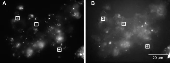
The objective of the present work was to examine free-living and particle-attached AAP bacteria in 5 con-trasting marine environments: a Mediterranean coastal lagoon, Mediterranean coastal and offshore waters, North Atlantic Ocean (including the Gulf Stream) and the Sargasso Sea. AAP bacterial cells were frequently seen attached to particles in samples collected from coastal waters by IR microscopy: the individual AAP bacterial cells were typically attached together with other bacteria to irregular particles of organic matter as shown in Fig. 1. Monospecific aggregates of AAP bac-terial cells or phytoplankton cells selectively colonized by AAP bacterial cells were extremely rare (data not shown). Unfortunately, direct microscopic observations are labor-intensive and proper quantification of parti-cle-attached cells is extremely complicated. For this reason Bchl a fluorescence was used in this study as an
easy and rapid way to detect AAP bacteria (Kolber et al. 2000, 2001, Koblí=ek et al. 2005, 2007).
Evaluation of the filtration methods
Size fractionations were conducted to determine the percentage of total Bchl a fluorescence associated with
particles. For each sample, < 0.8 µm and < 2 µm Bchl a
fluorescence signals were recorded with the original non-filtered water sample and the collected fractions, and the percentage of Bchl a signal in individual
frac-tions was then calculated. Gravity filtration was used to minimize potential particle dislodging during the filtration processes (Ghiglione et al. 2007, Waidner & Kirchman 2007).
In preliminary experiments we found that as ex-pected a 0.2 µm filtration always removed all the Bchl
a signal. In a preliminary evaluation of our methods
conducted at Stn POLA, we found that the majority (67%) of the fluorescence signal was in the 0.4 to
Aquat Microb Ecol 55: 31–38, 2009
0.8 µm fraction, with no signal below 0.4 µm (Fig. 2). Only small part of the total signal was found in the larger size classes (13% in 0.8 to 2.0 µm fraction, 7% in 2 to 5 µm and 12% in > 5 µm fraction), which indicates that the majority of the AAP bacterial population was formed by small individual cells sized 0.4 to 0.8 µm (Fig. 2).
The disadvantage of this size-fractionation protocol was lengthy filtration leading to slow deterioration of the samples. To avoid potential artifacts, the procedure was simplified to a 2-step protocol separating only 3 size classes: small cells passing through 0.8 µm mem-brane filters, larger cells between 0.8 and 2 µm, and mostly particle-attached cells which do not pass through 2 µm membrane filters. Using this protocol, the standard error of triplicate determinations on the same sample was 2.8%. As the experimental error was
small, in later experiments the samples were analyzed only once to speed up the fractionation protocol.
Collection of water samples onto GF/F filters is a very common approach in marine microbiology. For this reason, we decided to test (in addition to nucleo-pore filtration) the ability of GF/F filters to retain the Bchl a fluorescence signal. The water samples
col-lected at the coastal Stn SOLA in March and April 2007 were filtered through GF/F filters without vacuum. The filters retained 70% (in March) or 85% (April) of the Bchl a signal, which equals 15 to 30% of the signal
passing through the filter. At Stn POLA, GF/F retained only 42% of the Bchl a signal (Fig. 2). The more dense
MN GF-5 fiber glass filters (Macherey-Nagel) retained a larger fraction of the signal, but even these filters missed up to 30% of the Bchl a signal in the
oligotrophic regions (data not shown).
Mediterranean waters
The size distribution of the Bchl a fluorescence signal
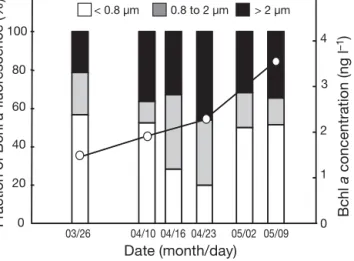
was examined through March to May 2007 at the coastal bay Stn SOLA (Fig. 3). During this period, Bchl a concentrations increased from 1.5 ng l–1 in
March to 3.5 ng l–1in May. The < 0.8 µm fraction (trac-ing free-liv(trac-ing AAP bacteria) was the largest one (50 to 55%, except April 16 to 23, during which it was 25 to 30%). The > 2 µm fraction (tracing particle-attached AAP bacteria) ranged from 20 to 40%, while the 0.8 to 2 µm fraction varied between 10 and 40% (Fig. 3).
The spatial changes in the AAP bacterial size distrib-ution were examined at 3 time-series stations in off-shore waters of Banyuls-sur-Mer in April and May 2007 (Table 1). At these studied Mediterranean sites, the AAP bacterial abundance in surface waters
(deter-34
Fig. 1. (A) Particle-attached bacteria stained with DAPI and (B) infrared emission (> 850 nm) of the sample. The squares indicate 3 aerobic anoxygenic phototrophic bacteria attached to a particle
Size fraction (µm) 0 20 40 60 80 >5 GF/F 0.4-0.8 <0.4 0.8-2 2-5 Fraction of Bchl a fluor escence (%)
Fig. 2. Size fractionation of Bchl a fluorescence signal
conducted at Stn POLA at 3 m depth on April 16, 2007. GF/F fraction indicates the percentage of the signal trapped by
mined with IR epifluorescence microscopy) ranged from 4.4 × 10³to 65 × 103cells ml–1. Bchl a
concentra-tion measured by HPLC varied from 0.9 to 4.3 ng l–1,
and the Bchl a:chl a ratio was between 0.6 and 1%. At
all these sites, the fraction of fluorescence in the < 0.8 µm size fraction dominated the signal; however, its proportion varied. The highest fraction of the < 0.8 µm signal was observed at the offshore Stn MOLA (up to 74% of the total signal in April), whereas at the shelf Stn POLA and the coastal site SOLA this fraction was smaller (55 to 65% of the total signal respectively) (Fig. 4). The 0.8 to 2 µm fraction was pre-sent in all samples and was 10 to 40% of the total sig-nal depending on the station and sample. The opposite pattern was found for the large fraction (> 2 µm), which mostly traces particle-attached AAP bacteria. Whereas at the offshore and shelf stations, this > 2 µm fraction was about 10% of the total signal, it accounted for about 30% of the total Bchl a fluoresence signal at the
coastal site (Fig. 4).
The vertical size distribution of the Bchl a signal was
also examined at the offshore Stn MOLA on May 23, 2007 (Fig. 5). Here, Bchl a fluorescence in the < 0.8 µm
fraction was high at all depths and was relatively con-stant (40 to 50% of the total Bchl a signal) (Fig. 5). A
large fraction (50%) of the signal was recorded in the > 2 µm fraction at the deep chlorophyll maximum (DCM). Interestingly, this pattern followed particulate matter volume determined using a Coulter counter. Particles and Bchl a concentrations measured by HPLC
peaked at the DCM (up to 4.3 ng l–1of Bchl a and 70 ×
103µm3 ml–1of particles at the DCM). AAP bacterial
cell abundances varied from 6.6 × 10³to 34 × 103cells
ml–1and peaked at 60 m, just below the DCM (Fig. 5).
We analyzed samples from a coastal Mediterranean lagoon (Lapalme Lagoon, France, 28 PSU) as a repre-sentative of an environment with high concentrations of particles. This shallow lagoon is characterized by high turbidity, and high chl a (0.8 µg l–1) and Bchl a
(22 ng l–1) concentrations (Table 1). In this lagoon, the
percentage of total Bchl a fluorescence in the < 0.8 µm
fraction was 28%. The signal in the > 2 µm fraction reached 30% (Fig. 4).
North Atlantic Ocean
We performed size-fractionation experiments in the northwest Atlantic in April 2008. In the Sargasso Sea, the Gulf Stream and in northern Atlantic waters, con-centrations of Bchl a were between 0.8 and 1.6 ng l–1.
In the oligotrophic Sargasso Sea, 90 ± 2% (mean ± SE) of Bchla passed through the 0.8 µm filter (calculated
from 6 independent casts at Stn 13.5) and all Bchl a
passed through the 2 µm filter (Fig. 6). In addition, 0.4 µm filtration usually removed > 90% of the Bchl a
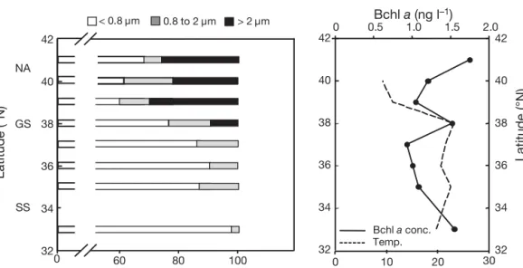
signal, which indicates that in the Sargasso Sea the majority of AAP bacterial cells were between 0.4 and 0.8 µm in size, and particle-attached AAP bacteria were absent. The size-fractionation measurements were also performed along the transect from the Sar-gasso Sea (Stn 16, 33° N 65° W) to the coast (Woods Hole, MA). In 3 samples collected in the Sargasso Sea, the < 0.8 µm fraction varied between 86 and 98% of the total, and > 2 µm fraction was absent. In the Gulf Stream (38° N, 69.5° W) the < 0.8 µm fraction decreased to 77%, whereas the > 2 µm fraction was 9% of the total Bchl a signal. In the productive waters of the Mid-0 20 40 60 80 100 0 1 2 3 4 Date (month/day) > 2 µm < 0.8 µm 03/26 04/10 04/16 04/23 05/02 05/09 Fraction of Bchl a fluor escence (%) Bchl a concentration (ng l –1) 0.8 to 2 µm
Fig. 3. Temporal variation of size fractionation of Bchl a
fluorescence (bars) and variations of Bchl a concentrations
(––s––) in the coastal Bay of Banyuls-sur-Mer, France, in spring 2007 0 10 20 30 40 50 60 70 80 90 100
Lagoon SOLA POLA MOLA Sargasso
0.8 to 2 µm > 2 µm < 0.8 µm Fraction of Bchl a fluor escence (%)
Fig. 4. Size fractionation of Bchl a fluorescence in a coastal
Mediterranean lagoon, in Mediterranean coastal (bay, Stn SOLA; shelf, Stn POLA) and offshore (Stn MOLA) waters
Aquat Microb Ecol 55: 31–38, 2009
dle Atlantic Bight, the small fraction further decreased to 50 to 68%, while the large > 2 µm fraction increased to 22 to 30% of the total Bchl a signal (Fig. 6).
DISCUSSION
We investigated the distribution of particle-attached and free-living AAP bacteria by the detection of Bchl a
fluorescence after size fractionation of seawater in
var-ious oceanographic contexts. Data on free-living ver-sus particle-attached AAP bacterial populations in marine waters are scarce. In their pioneering paper, Kolber et al. (2000) reported that most AAP bacteria in the subtropical Pacific pass through the GF/F filter, which suggests that the community is composed mostly of single cells < 0.7 µm. In contrast, large quan-tities of particle-attached AAP bacteria (31 to 94%) in estuarine waters were reported by Waidner & Kirch-man (2007). Our data clearly show that the proportion
36 Temp. Chl a conc. Particles AAP abund. Bchl a conc. Bchl a fluor. > 2 µm < 0.8 µm Fraction of Bchl a fluorescence (%) 13 14 15 16 17 18 19 0 20 40 60 80 100 0.0 0.2 0.4 0.6 0.8 0 40 80 120 0 20 40 60 80 0 10 20 30 40 50 0 2 4 6 8 0 50 100 150 200 250 0 20 40 60 80 100 100 0 20 40 60 80 100 0 20 40 60 80 100 0.8 to 2 µm Particulate matter (x103 µm3 ml–1) Bchl a fluorescence (mV) Temperature (ºC) Chl a concentrations (µg l–1) Bchl a concentrations (ng l–1) AAP abundance (x103 cells m l–1) Depth (m)
Fig. 5. Vertical distribution of temperature (temp.) and chlorophyll a concentrations (chl a), Bchl a concentrations (conc.), and
Bchla fluorescence (fluor.), aerobic anoxygenic phototrophic bacterial abundance (AAP abundance) and particulate matter, and
size fractionation of Bchl a fluorescence with depth. Data collected in Mediterranean offshore waters (Stn MOLA; May 23, 2007)
Temperature (ºC) 0 10 20 30 32 34 36 38 40 42 0 0.5 1.0 1.5 2.0 Fraction of Bchl a fluorescence (%) 0 60 80 100 32 34 36 38 40 42 NA SS GS 32 34 36 38 40 42 > 2 µm < 0.8 µm Latitude (°N) Latitude (°N) Bchl a conc. Temp. Bchl a (ng l–1) 0.8 to 2 µm
Fig. 6. Size fractionation of Bchl a fluorescence in the North Atlantic (NA) and Sargasso Sea (SS). Samples were taken during the
transect from SS (Stn 16, 33° N, 66° W) to Woods Hole, MA (41° 31.4’ N, 70° 40.3’ W) on April 10 to 13, 2008. The Gulf Stream (GS) was crossed at 38° N. The right-hand panel shows temperatures and Bchl a concentrations (estimated from Bchl a fluorescence)
of free-living and particle-attached AAP bacteria varies among the environments. In offshore and open-ocean environments, the majority of the AAP bacterial community were small (< 0.8 µm) free-living cells, which agrees with the findings of Kolber et al. (2000). Interestingly, our estimate of size ranges for the Sar-gasso Sea community (0.4 to 0.8 µm) agrees with AAP bacterial cell sizes (0.4 to 1.0 µm) determined by Sier-acki et al. (2006) in the same region using epi-fluorescence microscopy. Our observations are in con-trast with metagenomic analyses conducted in the oligotrophic Sargasso Sea suggesting that the majority of AAP bacteria are in the 3 to 20 µm fraction (Yutin et al. 2007).
We found a measurable percentage of Bchl a
fluores-cence in the 0.8 to 2 µm fraction in all environments. This signal likely comes from large but free-living AAP bacteria. It has previously been shown both in the North Atlantic and South Pacific Oceans that these microorganisms are larger than average heterotrophic bacterioplankton cells (Sieracki et al. 2006, Lami et al. 2007). It has been suggested that large cells may be the most active members of the bacterioplankton commu-nity (Bird & Kalff 1993, Gasol et al. 1995).
A different pattern was observed in a coastal lagoon and bay, where up to one-third of the AAP bacterial community was particle-attached. This is consistent with the large fraction of particle-attached AAP bacte-ria reported by Waidner & Kirchman (2007) from Chesapeake and Delaware estuaries. Interestingly, this pattern follows the usual observations of particle-attached versus free-living prokaryotes in the sea, with the highest percentages of attached bacteria in envi-ronments with high concentrations of particles (Simon et al. 2002). Collectively, the data presented in the pre-sent study show that AAP bacteria, in comparison with regular heterotrophic prokaryotes, do not exhibit an enhanced tendency to attach to particles or to form clumps. However, as the AAP bacterial community is composed of phylogenetically diverse organisms (Yutin et al. 2007, Salka et al. 2008), some AAP bacter-ial species might have an enhanced tendency to attach to particles as a part of their lifestyle.
We observed high AAP bacterial abundances at the DCM, consistent with high Bchl a concentrations. This
was previously found in other marine environments (Kolber et al. 2000, 2001, Sieracki et al. 2006). The slight decoupling between the peak of AAP bacterial cells and the peak of Bchl a may be due to regulation of
Bchl a content per cell. The interpretation of this peak
of photoheterotrophs is not completely clear. On the one hand, AAP bacteria and phytoplankton may require the same environmental conditions (light and nutrients) for their growth. Thus, they may occupy sim-ilar ecological niches and might strongly compete for
similar resources. On the other hand, AAP bacteria may find at the DCM specific organic compounds pro-vided by phytoplankton and required for their growth. Interestingly, we recorded a large fraction of AAP bac-teria attached to particles at the DCM, twice as much as in surface and deep layers. Attachment of AAP bac-terial populations also follows closely the distribution of particles in the water column as well as chl a
con-centrations. Thus, their attachment to particles proba-bly also depends on the concentrations of particles, including phytoplankton. The attached AAP bacteria may use a large spectrum of molecules, including diverse dissolved organic matter provided by various algal groups.
For pigment analyses, planktonic samples are rou-tinely collected onto glass fiber GF/F filters (What-man). Kolber et al. (2000) reported that in the tropical Pacific most of the Bchl a signal passes through GF/F.
In contrast, Goericke (2002) reported high retention of Bchl a on GF/F filters in his HPLC analyses. Based on
our data, it seems that the use of GF/F filters for pig-ment analyses in open-ocean environpig-ments might lead to a significant underestimation of true Bchl a
concen-trations, as GF/F filters did not trap 30 to 60% of the total Bchl a signal in our experiments. This is consistent
with earlier observations from the subtropical Atlantic, where 50 to 65% of the signal passed through GF/F filters (M. Koblí=ek unpubl. data). The GF/F filtration conducted at coastal Stn SOLA seemed to be more effective. However, in a previous study conducted in the Bay of Villfranche, France, the GF/F filters did not trap 30 to 50% of the signal (Koblí=ek et al. 2007).
The environmental variables that control AAP bacte-rial abundance and dynamics remain a matter of debate. Our data suggest that the characterization of ‘free-living’ versus ‘particle-attached’ is an important factor to explain the distribution and the ecology of AAP bacteria in marine environments. We have shown that AAP bacteria are mainly free living in the oceans. In addition, our data strongly suggest that in coastal bays and lagoons, large fractions of AAP bacterial cells have a particle-attached lifestyle. It has been shown that particle-attached bacteria can be the most active members of the prokaryotic community (Grossart et al. 2007). Thus, in coastal environments (lagoon, shelf) and at the DCM, some AAP bacteria may play signifi-cant roles in the mineralization of organic matter and particulate detritus degradation.
Acknowledgements. We thank L. Oriol, J. Caparros and I.
Obernosterer for the Mediterranean sample collection at Stn MOLA during the project MEDEA (INSU-LEFE-CYBER, P.I., I. Obernosterer), L. Zudaire for the CTD data and the captain and the crew of the RV ‘Nereis’ and RV ‘Tethys II’ for their support. The authors also thank the chief scientist B. Van Mooy for organizing the OC443 cruise and the captain and
Aquat Microb Ecol 55: 31–38, 2009
the crew of RV ‘Oceanus’ for their performance at sea. We thank J. J. Naudin at the Service d’Observation du Labora-toire Arago for oceanographic data and the French monitor-ing network SOMLIT for oceanographic coastal data. R.L.’s research was supported by a doctoral fellowship from the French Research and Education Ministry and by the Lavoisier postdoctoral fellowship from the French Foreign Office. M.K.’s stay in Banyuls was supported by a fellowship from the Université Pierre et Marie Curie – Paris 6. This research was also supported by GACR project 206/07/0241, GAAV project 1QS500200570 and the Inst. research concepts MSM6007-665808 and AV0Z50200510. We are grateful to D. L. Kirch-man and his group in the College of Marine and Earth Studies for helpful comments on an earlier version of this manuscript.
LITERATURE CITED
Allgaier M, Uphoff H, Felske A, Wagner-Döbler I (2003) Aerobic anoxygenic photosynthesis in Roseobacter clade
bacteria from diverse marine habitats. Appl Environ Microbiol 69:5051–5059
Béjà O, Aravind L, Koonin EV, Suzuki MT and others (2000) Bacterial rhodopsin: evidence for a new type of photo-trophy in the sea. Science 289:1902–1906
Bird DF, Kalff J (1993) Protozoan grazing and size-activity structure of limnetic bacterial communities. Can J Fish Aquat Sci 50:370–380
Cottrell MT, Mannino A, Kirchman DL (2006) Aerobic anoxy-genic phototrophic bacteria in the Mid-Atlantic Bight and the North Pacific Gyre. Appl Environ Microbiol 72: 557–564
Gasol JM, del Giorgio PA, Massana R, Duarte CM (1995) Active versus inactive bacteria: size-dependence in a coastal marine plankton community. Mar Ecol Prog Ser 128:91–97 Ghiglione JF, Mevel G, Pujo-Pay M, Mousseau L, Lebaron P,
Goutx M (2007) Diel and seasonal variations in abun-dance, activity and community structure of particle-attached and free-living bacteria in NW Mediterranean Sea. Microb Ecol 54:217–231
Goericke R (2002) Bacteriochlorophyll a in the ocean: Is
anoxygenic bacterial photosynthesis important? Limnol Oceanogr 47:290–295
Grossart HP, Tang KW, Kiøboe T, Ploug H (2007) Comparison of cell-specific activity between free-living and attached bacteria using isolates and natural assemblages. FEMS Microbiol Lett 266:194–200
Jiao N, Zhang Y, Zeng Y, Hong N, Liu R, Chen F, Wang P (2007) Distinct distribution pattern of abundance and diversity of aerobic anoxygenic phototrophic bacteria in the global ocean. Environ Microbiol 9:3091–3099 Koblí=ek M, Ston-Egiert J, Sagan S, Kolber ZS (2005) Diel
changes in bacteriochlorophyll a concentration suggest
rapid bacterioplankton cycling in the Baltic Sea. FEMS Microbiol Ecol 51:353–361
Koblí=ek M, Ma$ín M, Ras J, Poulton AJ, Prá$il O (2007) Rapid growth rates of aerobic anoxygenic phototrophs in the ocean. Environ Microbiol 9:2401–2406
Kolber ZS, Van Dover CL, Niederman RA, Falkowski PG (2000) Bacterial photosynthesis in surface waters of the open ocean. Nature 407:177–179
Kolber ZS, Plumley FG, Lang AS, Beatty JT and others (2001) Contribution of aerobic photoheterotrophic bacteria to the carbon cycle in the ocean. Science 292:2492–2495 Lami R, Cottrell MT, Ras J, Ulloa O, Obernosterer I,
Claus-tre H, Lebaron P (2007) High abundances of aerobic anoxygenic photosynthetic bacteria in the South Pacific Ocean. Appl Environ Microbiol 73:4198–4205
Ma$ín M, Zdun A, Sto´n-Egiert J, Nausch M, Labrenz M, Moulisová V, Koblí=ek M (2006) Seasonal changes and diversity of aerobic anoxygenic phototrophs in the Baltic Sea. Aquat Microb Ecol 45:247–254
Ma$ín M, Nedoma J, Pechar L, Koblí=ek M (2008) Distribution of aerobic anoxygenic phototrophs in temperate fresh-water systems. Environ Microbiol 10:1988–1996
Ras J, Claustre H, Uitz J (2008) Spatial variability of phyto-plankton pigment distributions in the subtropical South Pacific Ocean: comparison between in situ and predicted data. Biogeosciences 5:353–369
Salka I, Moulisová V, Koblí=ek M, Jost G, Jürgens K, Labrenz M (2008) Abundance, depth distribution, and composition of aerobic bacteriochlorophyll a-producing bacteria in
four basins of the central Baltic Sea. Appl Environ Micro-biol 74:4398–4404
Schwalbach MS, Fuhrman JA (2005) Wide-ranging abun-dances of aerobic anoxygenic phototrophic bacteria in the world ocean revealed by epifluorescence microscopy and quantitative PCR. Limnol Oceanogr 50:620–628
Sieracki ME, Gilg IC, Thier EC, Poulton NJ, Goericke R (2006) Distribution of planktonic aerobic anoxygenic photo-heterotrophic bacteria in the Northwest Atlantic. Limnol Oceanogr 51:38–46
Simon M, Grossart HP, Schweitzer B, Ploug H (2002) Micro-bial ecology of organic aggregates in aquatic ecosystems. Aquat Microb Ecol 28:175–211
Waidner LA, Kirchman DL (2007) Aerobic anoxygenic pho-totrophic bacteria attached to particles in turbid waters of the Delaware and Chesapeake estuaries. Appl Environ Microbiol 73:3936–3944
Yurkov VV, Beatty JT (1998) Aerobic anoxygenic photo-trophic bacteria. Microbiol Mol Biol Rev 62:695–724 Yutin N, Suzuki MT, Teeling H, Weber M, Venter JC, Rusch
DB, Béjà O (2007) Assessing diversity and biogeography of aerobic anoxygenic phototrophic bacteria in surface waters of the Atlantic and Pacific Oceans using the Global Ocean Sampling expedition metagenomes. Environ Microbiol 9:1464–1475
Zubkov MV, Tarran GA (2005) Amino acid uptake of
Prochlorococcus spp. in surface waters across the South
Atlantic Subtropical Front. Aquat Microb Ecol 40:241–249 38
Editorial responsibility: Jed Fuhrman, Los Angeles, California, USA
Submitted: July 1, 2008; Accepted: January 15, 2009 Proofs received from author(s): March 8, 2009


