HAL Id: tel-01431200
https://tel.archives-ouvertes.fr/tel-01431200
Submitted on 10 Jan 2017HAL is a multi-disciplinary open access archive for the deposit and dissemination of sci-entific research documents, whether they are pub-lished or not. The documents may come from teaching and research institutions in France or abroad, or from public or private research centers.
L’archive ouverte pluridisciplinaire HAL, est destinée au dépôt et à la diffusion de documents scientifiques de niveau recherche, publiés ou non, émanant des établissements d’enseignement et de recherche français ou étrangers, des laboratoires publics ou privés.
Study of ribonucleoprotein particle biogenesis and
quality control by a novel technique using bacterial Rho
factor as a tool
Mateja Remenaric Hajak
To cite this version:
Mateja Remenaric Hajak. Study of ribonucleoprotein particle biogenesis and quality control by a novel technique using bacterial Rho factor as a tool. Cellular Biology. Université d’Orléans, 2016. English. �NNT : 2016ORLE2013�. �tel-01431200�
UNIVERSITÉ D’ORLÉANS
ÉCOLE DOCTORALE
SANTE, SCIENCES BIOLOGIQUES ET CHIMIE DU VIVANT
Centre de Biophysique Moléculaire
THÈSE
présentée par :
Mateja REMENARIC HAJAK
soutenue le : 22 Avril 2016pour obtenir le grade de : Docteur de l’université d’Orléans Discipline/ Spécialité : Biologie moléculaire et cellulaire
Study of ribonucleoprotein particle
biogenesis and quality control by a novel
technique using bacterial Rho factor as a tool
THÈSE dirigée par :
Dr. A. Rachid RAHMOUNI Directeur de recherche, CNRS Orléans
RAPPORTEURS :
Dr. Lionel MINVIELLE-SEBASTIA Directeur de recherche, INSERM, Bordeaux Dr. Benoit PALANCADE Chargé de recherche, CNRS, Paris
____________________________________________________________________ JURY
Dr. Josette BANROQUES Chargé de recherche, CNRS, Paris
Dr. Lionel MINVIELLE-SEBASTIA Directeur de recherche, INSERM, Bordeaux Dr. Benoit PALANCADE Chargé de recherche, CNRS, Paris
Pr. Chantal PICHON Professeur d’Université, Orléans – présidente du jury
Acknowledgments
Research work done to obtain the results presented in thesis was carried out at the Center for Molecular Biophysics (CBM), CNRS Orléans, under supervision of Dr. A. Rachid Rahmouni. I would like to thank him for providing me with the opportunity to do my Master 2 internship in his laboratory and also for having me as his PhD student with the privilege to do my thesis as a part of his research group. It has been a priceless life experience throughout which I have benefited from his valuable advices, comments and suggestions.
I would also like to express my heartfelt gratitude to Christine Mosrin-Huaman for all her support, knowledge, advices, guidance and encouragement, to Igor Stuparevic for the discussions, encouragement and research- and life-related advices and to Nadegé Hervouet-Coste for her cooperation and help.
I thank all the colleagues and students who have spent more or less time as a part of our research group for their cooperation, especially Ivana Martinic for all the talks, her kindness and friendship.
I cannot imagine the years spent at CBM without the great times spent with fellow students, post-docs and other colleagues and staff from CBM and whole CNRS in Orléans, and their support at the most difficult periods, I will always cherish the fondest memories.
I would also like to thank other research groups at CBM for their collaboration and equipment indispensable for our research, especially to the groups of Dr. Claudine Kieda and Dr. Hélène Bénédetti.
I thank Dr. Visnja Besendorfer, Dr. Vladimir Mrsa and especially Dr. Daniel Hagege for creating the Master 2 program Bio-industrial techniques which opened the door of opportunities for me in France, for their support, time and energy invested in accompanying students through this program and I hope many more generations will have the opportunity to benefit from it.
I thank Dr. Lionel Minvielle-Sebastia and Dr. Benoit Palancade for their time and effort in checking this manuscript, for their knowledge and valuable suggestions and I thank Dr. Josette Banroques and Pr. Chantal Pichon for accepting to serve as members of my defense jury.
Most of all I would like to thank my parents, family and friends for their unconditional love and support.
To my husband Tomislav, for your continued and unfailing love, support, understanding and patience which made the completion of this thesis possible.
Table of Contents
1 INTRODUCTION - 4 -
1.1 MRNP BIOGENESIS IS CO-TRANSCRIPTIONAL IN SACCHAROMYCES CEREVISIAE -5-
1.1.1 RNAPOLYMERASE IICTD -6-
1.1.2 CO-TRANSCRIPTIONAL MRNA PROCESSING -8-
1.1.2.1 Coupling transcription with mRNA capping and splicing - 10 -
1.1.2.2 Coupling transcription with 3’ end processing - 12 -
1.1.2.2.1 Polyadenylation-dependent 3’ end processing - 12 -
1.1.2.2.2 Polyadenylation-independent 3’ end processing - 15 -
1.1.2.2.3 Integrator-dependent 3’ end processing - 16 -
1.1.3 CO-TRANSCRIPTIONAL MRNP ASSEMBLY COUPLED WITH EXPORT -18-
1.1.3.1 THO complex - 20 -
1.1.3.1.1 Transcription site recruitment - 20 -
1.1.3.1.2 THO function - 22 -
1.1.3.2 TREX complex - 23 -
1.1.3.2.1 Sub2 - 23 -
1.1.3.2.2 Yra1 - 24 -
1.1.3.2.3 TREX function - 27 -
1.1.3.2.4 Other THO/TREX interactions - 30 -
1.2 MRNP DECAY AND NUCLEAR QUALITY CONTROL IN YEAST -32-
1.2.1 MRNA DEGRADATION -32-
1.2.2 THE EXOSOME -34-
1.2.2.1 Exosome associated factors - 35 -
1.2.2.2 Exosome associated complexes - 36 -
1.2.3 NUCLEAR QUALITY CONTROL -38-
1.2.3.1 QC of mRNA processing - 39 -
1.2.3.2 QC of mRNP assembly - 39 -
1.2.3.3 Specific and/or competitive QC - 40 -
1.3 BACTERIAL FACTOR RHO AS A TOOL TO STUDY MRNP BIOGENESIS AND NUCLEAR QUALITY CONTROL IN
SACCHAROMYCES CEREVISIAE -42-
1.3.1 STRUCTURE AND FUNCTION OF RHO FACTOR -42-
1.4 THE RESEARCH QUESTION -49-
2 MATERIALS AND METHODS - 50 -
2.1 YEAST STRAINS AND PLASMIDS -51-
2.2 CELL GROWTH AND RHO INDUCTION -51-
2.3 SERIAL DILUTIONS TEST -52-
2.4 RNA ISOLATION AND NORTHERN BLOTTING -52-
2.5 RT-PCR AND RT-QPCR -53-
2.6 PROTEIN EXTRACTION AND WESTERN BLOTTING -54-
2.7 CHROMATIN IMMUNOPRECIPITATION -55-
2.8 RNA-FLUORESCENCE IN SITU HYBRIDIZATION (FISH) -56-
3 RESULTS - 60 -
3.1 NATURE OF RHO-INDUCED TRANSCRIPT DEGRADATION -61-
3.1.1 DEGRADATION OF TRANSCRIPTS BY RRP6 IN RHO EXPRESSION SYSTEM -62- 3.1.2 THE ROLE OF DIS3 AND THE EXOSOME IN RHO-INDUCED TRANSCRIPT DEGRADATION -65-
3.2 NATURE OF RHO-INDUCED TRANSCRIPT ABERRATION -68-
3.2.1 RHO AND THE THO-SUB2 COMPLEX -68-
3.2.1.1 Effect of Rho expression in strains with tagged THO-Sub2 members - 69 - 3.2.1.2 ChIP of THO-Sub2 tagged strains in Rho expression system - 71 - 3.2.1.3 RNase sensitivity of THO-Sub2 in Rho expression system - 73 -
3.2.2 THO-SUB2 COMPLEX AND THE DELETION OF MFT1 -76-
3.2.2.1 THO-Sub2 in strains with mft1Δ background - 76 -
3.2.2.2 Comparison between Rho expression system and mft1Δ background - 78 -
4 DISCUSSION - 83 -
4.1 RRP6 IS THE MAIN NUCLEASE DEGRADING THE RHO-INDUCED ABERRANT TRANSCRIPTS -84- 4.2 RHO ACTION REVEALS A SURPRISING BEHAVIOR OF MFT1 AND A “HIDDEN”RNA DEPENDENCE
OF THE THO COMPLEX -86-
- 1 -
LIST OF ABBREVIATIONS
7mG 7-methylguanosine Ala Alanine CBC Cap-binding complex CE Capping enzyme CFIA Cleavage factor IA CFIB Cleavage factor IBCPF Cleavage and polyadenylation factors CTD Carboxy-terminal domain
CUTs Cryptic unstable transcripts
DCF Differential chromatin fractionation DNA Deoxyribonucleic acid
eIF4F Eukaryotic initiation factor 4F EM Electron microscopy
FISH Fluorescent in situ hybridization GTFs General transcription factors heptad Heptapeptide
lncRNA Long noncoding ribonucleic acid miRNA Micro ribonucleic acid
mRNA Messenger ribonucleic acid ncRNA Noncoding ribonucleic acid NIM Nrd1 interacting motif NLS Nuclear localization signal NNS Nrd1-Nab3-Sen1 complex NPC Nuclear pore complex Nrd1C Nrd1 complex
PAR-CLIP Photoactivable-ribonucleoside-enhanced UV crosslinking and immunoprecipitation
PAS Poly(A) signal PBS Primary binding site PCR Polymerase chain reaction PIC Preinitiation complex
Pol I, II, III Ribonucleic acid polymerase I, II, III Pro Proline
Prp19C Prp19 complex QC Quality control
qPCR Quantitative polymerase chain reaction RBPs Ribonucleic acid -binding proteins
REF Ribonucleic acid and export factor binding protein RNA Ribonucleic acid
rRNA Ribosomal ribonucleic acid SAXS Small-Angle X-Ray Scattering SBS Secondary binding site
Ser Serine
snoRNA Small nucleolar ribonucleic acid snRNA Small nuclear ribonucleic acid
- 2 - TAP Tandem affinity purification Thr Threonine
TRAMP Trf4/Air2/Mtr4p Polyadenylation complex tRNA Transfer ribonucleic acid
Tyr Tyrosine
UBA ubiquitin-associated UTR untranslated region wt wild-type
- 3 -
PREFACE
The research for this thesis was performed in the laboratory group “RNA-proteins interactions and gene regulation” at the Center for Molecular Biophysics (CBM), CNRS Orléans, under supervision of Dr. A. Rachid Rahmouni. It presents new findings and ideas in the field of mRNP biogenesis and quality control in Saccharomyces cerevisiae. Thesis was funded with a fellowship from the Ministry of Higher Education and Research of France.
Transcription from DNA into RNA and the biogenesis of a mature messenger RNA particle suitable for export from the nucleus is a highly sophisticated process, far from being completely understood and described. The model developed so far postulates that after emerging from the transcribing polymerase, the nascent transcript is coated with numerous protein factors coupling transcription with mRNA processing events. They also secure transcript integrity, structure and quality for delivering an accurate and adequate message for translation in the cytoplasm. Aberrant transcripts are recognized and quickly degraded by the nuclear degradation machinery in the process of transcript quality control. In recent years the mechanisms of quality control during mRNP biogenesis have been highly researched and discovered for each step of the process. However, due to the complexity of these biogenesis and control events and the abundance of protein factors involved, the dynamic interactions between protein factors themselves and with the transcript still remain elusive.
In an effort to discover and describe new mechanisms of transcription quality control, we developed an innovative technique using a bacterial factor Rho as a tool to produce aberrant transcripts. Rho is a powerful molecular motor capable of removing protein factors off the transcript, thus interfering with the mRNA stability and processing and activating the quality control mechanisms. In our research so far using Rho factor, we have demonstrated a new nuclear quality control recognition mechanism of aberrant transcripts and identified many factors involved in mRNP biogenesis as suppressors of Rho-induced transcript defectiveness. In this thesis I will present additional findings regarding transcript degradation of Rho-induced aberrant transcripts and dissect the influence of Rho expression on a protein complex crucial for proper mRNP biogenesis, thus uncovering its new characteristics and possible roles in coupling transcription to mRNA packaging, processing and export.
- 4 -
- 5 -
1.1 mRNP biogenesis is co-transcriptional in Saccharomyces cerevisiae
Transcription, although being one of the processes most essential to life, is still far from being completely unraveled. While details of the process may differ among eukaryotes, its universal nature potentiates extensive research using model organisms, among them Saccharomyces cerevisiae. The complexity of transcription lies not only in the three major steps of initiation, elongation and termination, but also in the co-transcriptional coupling with maturation, packaging and export of messenger ribonucleic acids (mRNAs).
In eukaryotes synthesis of the ribonucleic acid (RNA) is confined to the cell nucleus and is carried out by three RNA polymerases. All three are structurally similar but are commonly regarded as having distinct roles and properties. RNA polymerase I (Pol I) transcribes the large precursor for 5.8S, 18S and 28S ribosomal RNAs (rRNAs). RNA polymerase II (Pol II) synthesizes all protein coding (mRNAs) and also some noncoding RNAs (ncRNAs), like small nuclear RNA (snRNA), small nucleolar RNA (snoRNA), micro RNA (miRNA), long noncoding RNA (lncRNA), cryptic unstable transcripts (CUTs) and others. RNA polymerase III (Pol III) catalyzes the transcription of other small ncRNAs, amongst them transfer RNA (tRNA) and 5S rRNA. In recent years evidence of common transcription units for both Pol II and III have emerged, which suggests RNA polymerase initiation specificity can vary according to surrounding conditions (Raha et al., 2010; Jamonnak et al., 2011; Duttke, 2014). Regulation of mRNA transcription is dependent upon promoter strength, availability of specific transcription factors and also the state of chromatin structure. Influence of the latter reaches all the way into productiveness of Pol II elongation, the ability to terminate transcription and may even effect mRNA processing (Alén et al., 2002; Morillon et al., 2003; Murawska and Brehm, 2011). In classic general story of mRNA biogenesis, pre-mRNA is firstly transcribed, then protected at both ends by addition of 7-methylguanosine (7mG) cap to the 5’end and polyadenylation (poly(A)) at the 3’end, and finalized by excision of introns and exon ligation. However, in living cells, transcription and most of the processing steps are physically and functionally coupled, thus enhancing the efficiency and accuracy of mRNA maturation. The newly formed transcript is co-transcriptionally assembled with specific protein factors, which ensure the production of a mature, export-competent messenger ribonucleoprotein particle (mRNP).
- 6 -
1.1.1 RNA Polymerase II CTD
The steps of mRNA processing, namely capping, splicing and polyadenylation, are tightly coupled to each other and to transcription. Co-transcriptional recruitment of transcription and processing factors is coordinated by the carboxy-terminal domain (CTD) of the largest subunit of Pol II.
The CTD of Pol II (reviewed in Corden, 2013; Eick and Geyer, 2013) is an unusual and unique, largely flexible domain (Meinhart et al., 2005). It comprises multiple heptapeptide (heptad) repeats, containing 4 different amino acids: serine (Ser), proline (Pro), tyrosine (Tyr) and threonine (Thr), with the consensus sequence Tyr1-Ser2-Pro3-Thr4-Ser5-Pro6-Ser7 (Y1S2P3T4S5P6S7) (Figure 1.1A) (Hall and Georgel, 2011). The CTD consensus
sequence is conserved in S. cerevisiae and mammals, yet it differs markedly in its length with 26 and 52 repeats, respectively. The heptads are said to come in tandems, as the insertion of an alanine (Ala) residue between two heptads in yeast is lethal, while the same insertion between two diheptads is tolerable, therefore it was believed to be a functional unit of the yeast CTD (Stiller and Cook, 2004). More recently, even smaller functional unit was defined, containing only the first 11 residues of a diheptad where the most essential are three serine-proline motifs and two tyrosine residues spaced at a heptad interval (Liu et al., 2010) (Figure 1.1B). However, the same study reveals that CTD length is more crucial for viability than the number of functional units contained within the amino acid sequence. For S. cerevisiae, minimal viable CTD length is 8 heptads containing 7 functional units (West and Corden, 1995).
Recruitment of different factors during transcription is achieved by extensive post-translational and conformational modifications of the CTD heptad repeats. These include phosphorylation of Tyr, Thr and Ser, glycosylation of Thr and Ser, and isomerization of Pro residues. Additional modifications are possible in non-consensus heptads at the end of mammalian CTD (Chapman et al., 2005; Napolitano et al., 2014), expanding even more the number of combinatorial possibilities orchestrating protein association and dissociation and defining the CTD code (Buratowski, 2003; Cassart et al., 2012; Schwer et al., 2014; reviewed in Egloff et al., 2012) (Figure 1.1C).
- 7 -
Figure 1.1: CTD modifications orchestrate binding and exchange of protein factors
and complexes involved in mRNA processing. (A) Heptapeptide consensus repeats of the CTD of the Pol II large subunit (Hall and Georgel, 2011). (B) CTD functional units: a diheptad proposed by Stiller and Cook (2004); a minimal functional unit determined by Liu et al. (2010). (C) Potential phosphorylation and proline isomerization combinations for CTD modification during transcription (adapted from Egloff et al., 2012). Phosphorylation is indicated by red circles and trans or cis isomerization of prolines by a blue t or c, respectively, below the amino acid.
B
C A
- 8 -
Among distinct CTD modifications, phosphorylation is the best characterized and is crucial in pre-mRNA maturation. The phosphorylation of Ser2 and Ser5 residues in a heptad sequence was firstly discovered more than 20 years ago (Zhang and Corden, 1991). It has remained in the focus of interest for the most part of that period, until the discovery of an in vivo Ser7 phosphorylation by Chapman et al., 2007. Although the potential for phosphorylation of Tyr and Thr residues in mammalian cells was described simultaneously as Ser2/5 phosphorylation (Zhang and Corden, 1991; Baskaran et al., 1993), their phosphorylation in the consensus sequence, in yeast, as well as their functions were discovered only recently (Sakurai and Ishihama, 2002; Hsin et al., 2011; Mayer et al., 2012), thus extending the CTD code.
1.1.2 Co-transcriptional mRNA processing
Phosphorylation code of the CTD has begun to be depicted in more detail (Figure 1.2) with the use of monoclonal antibodies in chromatin immunoprecipitation (ChIP) experiments (reviewed in Heidemann et al., 2013). They represent a powerful tool to study CTD modification patterns due to their specific recognition of the unphosphorylated CTD and the single or double phosphorylation marks (Ser-, Tyr-, Thr- or Ser2/Ser5) (Heidemann et al., 2013). Each antibody recognizes not only the phosphorylated mark but also the adjacent amino acids, which minimizes the recognition of the same phosphorylated amino acid residues on other proteins (Eick and Geyer, 2013).
- 9 -
Even though the use of monoclonal antibodies is common nowadays, the shortcomings of this technique, such as concealed phospho-epitopes and cross-reactivity, impede the unambiguous interpretation of the obtained data (Eick and Geyer, 2013). Future perspectives place hope in the use of mass spectrometry analysis in the research of CTD modifications. Nonetheless, ChIP results have led to important discoveries of CTD phosphorylation patterns, phosphorylation and dephosphorylation agents, as well as protein factor recruitment at distinct time-points during transcription corresponding to different phosphorylation marks (Figure 1.3).
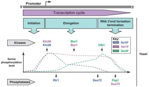
Figure 1.2: Schematic representation of the averaged CTD code profile, obtained by
ChIP experiments (Heidemann et al., 2013). Color gradients illustrate changing phosphorylation levels of Tyr1 (violet), Ser2 (green), Thr4 (orange), Ser5 (red) and Ser7 (blue) residues during different steps of transcription. The color of each noted transcription step corresponds to the color of the implicated phosphorylation mark. Ser5-P facilitates promoter escape and recruitment of the capping enzyme (CE) (see page 10). Ser7-P is involved in recruiting the Integrator complex in the 3’end processing pathway of snRNA-encoding genes in mammals, while its role in protein coding genes has not yet been determined (see pages 11 and 16). Tyr1 is phosphorylated in the middle of the transcription process and it seems to act in discriminating between elongation and termination since it impairs the recruitment of transcription termination factors (see page 16). Phosphorylation of Ser2 marks the beginning of the elongation phase. The level of Ser2-P increases during transcription and recruits many processing factors, among them 3’end processing and termination factors at the end of transcription (see pages 11 and 12).
- 10 -
1.1.2.1 Coupling transcription with mRNA capping and splicing
At the beginning of a transcription cycle, Pol II with hypo-phosphorylated CTD is recruited to promoter by general transcription factors (GTFs), along with gene-specific transcription factors and the Mediator complex, forming the preinitiation complex (PIC) (Zhang et al., 2012a; Murakami et al., 2013) (Figure 1.3B). Transcription passes into initiation phase with first CTD phosphorylation at Ser5 position by the Kin28 kinase, a component of general transcription factor TFIIH (Komarnitsky et al., 2000). This modification endorses promoter escape and stimulates dissociation of the Mediator complex (Wong et al., 2014). Another yeast kinase, Srb10 (homolog of CDK8 in mammals), has also been shown to phosphorylate CTD at Ser5 positions in vivo, but its actual contribution is still
Figure 1.3: Phosphorylation states and factors recruited during Pol II transcription
cycle (Eick and Geyer, 2013). (A) Free polymerase in a hypo-phosphorylated state. (B) General transcription factors (GTFs) recruit Pol II and the Mediator complex to the promoter, thus forming the preinitiation complex (PIC). (C) Ser5-P marks the intiation phase and recruits capping enzymes. (D) Dynamic phosphorylation and de-phosphorylation of the CTD during elongation phase recruits many elongation and processing factors. (E) Programming of the CTD for termination, gradual removal of CTD phosphorylation marks by phosphatases, and release of Pol II and transcripts from the template.
- 11 -
unclear (Galbraith et al., 2010). Likewise, phosphorylation of Ser5 (Ser5-P) promotes recruitment of RNA capping enzyme (CE), which protects the 5’end of the nascent transcript from degradation by adding a 7mG cap at the 5’end (Suh et al., 2010) (Figure 1.3C). In eukaryotes, capping is a three step enzymatic process: (i) the triphosphate 5’ end of the nascent transcript is hydrolyzed to a diphosphate; (ii) a guanosine monophosphate (GMP) is transferred to the diphosphate in a 5’-5’ linkage, forming a GpppN structure which is (iii) finally methylated, thus forming a complete 7mG cap (Schwer et al., 2000). The first two steps in yeast are performed by the CE made out of two tightly associated enzymes, an RNA triphosphatase (Cet1p) and a guanylyltransferase (Ceg1p), while in higher eukaryotes these two enzymatic functions are combined in a single bifunctional protein (Takase et al., 2000). The final step is carried out by a methyltransferase (Abd1 in yeast). The mature cap structure is then associated with other complexes, such as cap-binding complex (CBC) in the nucleus and eukaryotic initiation factor 4F (eIF4F) in the cytoplasm, which mediate further transcript processing, export and translation (Gonatopoulos-Pournatzis and Cowling, 2014).
The general elongation complex displaces PIC after successful promoter escape and Ser2 phosphorylation (Ser2-P) marks the beginning of the elongation phase (Mayer et al., 2010) (Figure 1.3D). Ser5-P recruits Bur1 kinase, which performs the initial Ser2 phosphorylation, while more extensive Ser2 phosphorylation is carried out by Ctk1, a catalytic subunit of CTD kinase-I, thus promoting transition into processive elongation (Jones et al., 2004; Bataille et al., 2012). Ser7 phosphorylation is suggested to be another signal of the CTD code important for promoting transcription elongation (Czudnochowski et al., 2012). This mark is set by Kin28 kinase at the promoter, but is maintained along the coding region by Bur1 kinase and is at high level until the very 3’end (Chapman et al., 2007; Tietjen et al., 2010). Nevertheless, the role of Ser7-P in transcription elongation has yet to be further investigated and confirmed.
Concurrent with the beginning of elongation, starts the removal of Ser5-P marks by Rtr1 phosphatase (Mosley et al., 2009). Consequent change in the ratio of Ser2/Ser5 phosphorylation signals the recruitment of processing complexes, which have yet to perform their functions. First in line is spliceosome, one of the largest macromolecular complexes found in living cells. It is composed of a minimum of ~100 proteins associated with five snRNAs (Hoskins et al., 2011). CTD has been shown to bind several splicing factors and they can sequentially recruit additional splicing machinery, i.e. the spliceosome activating Prp19
- 12 -
complex (Prp19C). This complex is also able to enhance transcription elongation (Hsin and Manley, 2012). Aside from its interaction with the CTD, splicing has been shown to be coupled with transcription by CBC dependent spliceosome assembly (Görnemann et al., 2005), it highly interacts with 3’end processing machinery in a reciprocal co-regulation (Li et al., 2001; Rigo and Martinson, 2009) and spliceosome could even be involved in regulation of gene expression (Volanakis et al., 2013).
1.1.2.2 Coupling transcription with 3’ end processing
As for splicing, phosphorylation of Ser2 residue is also important for 3’end processing events (Richard and Manley, 2009) (Figure 3E). These events can be carried out in three distinct pathways, depending on the type of RNA being transcribed: pre-mRNAs in poly(A)-dependent pathway, snoRNAs and CUTs in poly(A)-inpoly(A)-dependent pathway and snRNAs in Integrator-dependent processing-termination pathway (Eick and Geyer, 2013).
1.1.2.2.1 Polyadenylation-dependent 3’ end processing
The first pathway for 3’end processing is polyadenylation-dependent, thus it demands the presence of a specific poly(A) signal (PAS) in the nascent RNA, which is recognized by cleavage factors IA and IB (CFIA, CFIB), and cleavage and polyadenylation factors (CPF) (Dichtl and Keller, 2001; Mischo and Proudfoot, 2013) (Figure 1.4). Several representatives of these complexes have been shown to interact with phosphorylated CTD, a characteristic not essential for 3’end processing, but largely enhancing its efficiency (Rigo et al., 2005; Kuehner et al., 2011).
- 13 -
CFIA is a multisubunit complex which associates with Pol II and functions by selecting the cleavage site and assisting the recruitment of polyadenylation factors. Thus, 3’end formation is a two-step process: pre-mRNA is first cleaved and subsequently polyadenylated for protection from 3’ to 5’end exonucleolysis (Zhao et al., 1999). The cleavage and polyadenylation factor Pcf11, one of the CFIA members, contains a CTD Interacting Domain (CID) which preferentially interacts with Ser2-P (Barillà et al., 2001; Lunde et al., 2010; Gu et al., 2013). However, Pcf11-CID likewise binds the nascent RNA. This dual binding capability reflects double function of this factor during transcription. In addition to cleavage and polyadenylation, Pcf11 plays a role in transcription termination,
Figure 1.4: Polyadenylation-dependant 3'end processing signals and protein factors in
yeast (adapted from Mischo and Proudfoot, 2013). (A) CPF/CF specific binding sequences comprise the AU-rich efficiency element (EE), the A-rich positioning element (PE) and U-rich regions preceding the cleavage site. Low conservation of different sequence elements is exemplified by polyadenylation signals of CYC1 and ACT1 mRNAs. (B) Composition and suggested organisation of the CPF/CF complex on mRNAs. CPF members are represented in blue, pink and gray and CF members are in green and red.
- 14 -
together with the termination factor Rtt103 (Sadowski et al., 2003; Zhang, 2005; Hollingworth et al., 2006).
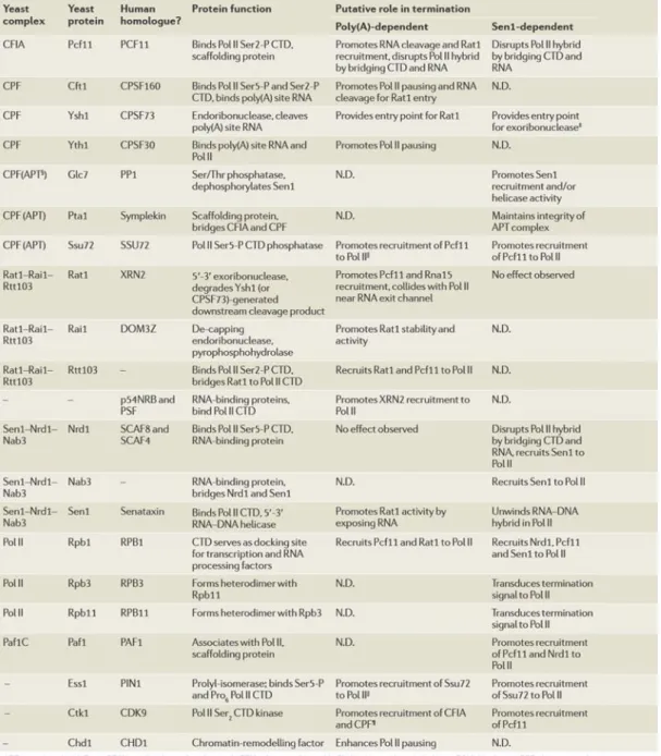
Table 1.1: Factors involved in Pol II 3’end processing and/or transcription termination (Kuehner et al., 2011).
- 15 -
Transcription termination is tightly connected to 3’end processing. It is hard to distinguish precisely where one begins and the other finishes, since many factors are involved in both processes (Table 1.1). Key event in termination is Pol II disengagement from the deoxyribonucleic acid (DNA) template. At first, two models were proposed to explain transcription termination of protein coding genes: the allosteric or anti-terminator model implies that transcription through PAS leads to a conformational change of elongation complex, while the torpedo model suggests that cleavage at the poly(A) site enables the 5’-3’ exonuclease Rat1 to load onto the unprotected 5’end of the RNA left emerging from the polymerase after the cut. Rat1 degrades the RNA and, in some still undefined way, promotes Pol II release after “catching up” with it (Richard and Manley, 2009). Lately, a unified model has mostly been advocated, since Rat1 action was shown insufficient for Pol II release from the template. Nevertheless, the main actor responsible for Pol II disengagement remains obscure (Luo et al., 2006; Richard and Manley, 2009; Schaughency et al., 2014).
1.1.2.2.2 Polyadenylation-independent 3’ end processing
The second 3’end processing and/or termination pathway is poly(A)-independent and directed by the Nrd1 complex (Nrd1C or Nrd1-Nab3-Sen1 complex (NNS)), which consists of RNA binding proteins Nrd1 and Nab3, and the RNA helicase Sen1 (Steinmetz et al., 2001) (also called Sen1-dependent termination, see Table 1.1). This complex interacts with CTD through Nrd1-CID, which preferentially binds at Ser5-P, thus explaining the Nrd1 crosslinking near 5’end of non-coding and coding genes, and it’s role in termination of short Pol II-transcribed genes (Vasiljeva et al., 2008; Kubicek et al., 2012). Sen1 also binds CTD but interacts with Ser2-P along the whole length of both non-coding and coding genes, and it was proposed to terminate transcription in a way similar to bacterial Rho helicase; this factor shall be presented in section 1.3 (Steinmetz et al., 2006; Creamer et al., 2011; Kuehner et al., 2011; Chinchilla et al., 2012; Porrua and Libri, 2013a). Nrd1 was originally believed to have a specific sequence binding site. However, its binding affinity has now been extended to AU-rich, GU-rich and G-rich sequences, thus extending the pool of possible RNA substrates targeted by Nrd1C (Creamer et al., 2011; Bacikova et al., 2014). Nevertheless, for functional termination by Nrd1C the mere presence of binding sequences is not enough. Their arrangement and association in supermotifs is crucial for termination (Porrua et al., 2012). Indeed, this is in accordance with recent findings of Nrd1C function in processing of even
- 16 -
mRNA coding genes and other types of RNAs. This function is mostly connected with nutrient response, suggesting a possible overlap of termination pathways which were before believed distinct (Jamonnak et al., 2011; Darby et al., 2012; Webb et al., 2014). Another interaction of Nrd1C is realized with the exosome, which leads to 3’end trimming or transcript degradation in a quality control process, which will also be described in more details later on (section 1.2.2.2) (Vasiljeva and Buratowski, 2006).
Another part of the CTD code is Tyr1 phosphorylation. The replacement of Tyr1 residue was proven to be lethal in yeast (West and Corden, 1995; Schwer and Shuman, 2011). This mark seems to aid in the discrimination between elongation and transcription termination, by preventing interaction of early (Nrd1) and late (Pcf11, Rtt103) termination factors with the CTD. Thus, Tyr1 is phosphorylated during transcription of the central region of genes (Figure 1.2), yet its presence does not impair CTD binding of the elongation factor Spt6 (Mayer et al., 2012). Although yeast Tyr1 kinase has not yet been found, very recently a CPF subunit Glc7 was shown to dephosphorylate Tyr1 in vitro and in vivo in S. cerevisiae. This finding, along with Glc7 role in recruiting termination factors Pcf11 and Rtt103, provides yet another connection between 3’end processing and termination (Schreieck et al., 2014).
1.1.2.2.3 Integrator-dependent 3’ end processing
Finally, the third 3’end processing pathway relies on Ser2 and Ser7 phosphorylation recruiting the Integrator complex to snRNA-encoding genes in mammals, while yeast homologs of this complex’s subunits have not been found (Baillat et al., 2005; Egloff et al., 2010). Contrary to the initial idea of a universal, genome-wide CTD code, Ser7 phosphorylation was first discovered as gene-specific, required for snRNA gene expression (Corden, 2007; Egloff et al., 2007). Thr4 phosphorylation was the next one, shown to be required for histone mRNA 3’end processing in chicken cells, and most recently, it was revealed as a regulator of expression of specific genes in S. cerevisiae (Hsin et al., 2011; Rosonina et al., 2014). Nevertheless, genome-wide studies performed until now have presented biased results regarding uniformity and/or specificity of CTD phosphorylation patterns, accentuating the need for further examination (Kim et al., 2010; Mayer et al., 2010; Tietjen et al., 2010; Bataille et al., 2012).
- 17 -
After successful transcription termination, by any of the three described pathways, Pol II CTD returns into its hypo-phosphorylated state (Figure 1.3A). Serine phosphatases have been best described so far (Figure 1.5), while a Thr4 phosphatase has not yet been determined. Ser5-P and Ser7-P are coupled by Kin28 phosphorylation at the beginning and Ssu72 de-phosphorylation at the end of transcription, while Ser5 phosphorylation is additionally removed from the start of elongation by an ill-defined Rtr1 phosphatase (Bataille et al., 2012), mentioned earlier (section 1.1.2.1). Ser2 phosphatase Fcp1 travels with Pol II and interplays with Ser2 kinase Ctk1, thus dynamically regulating Ser2 phosphorylation level (Cho et al., 2001). The same phosphatase performs complete Ser2-P de-phosphorylation after transcription termination (Bataille et al., 2012). In addition to the aforementioned importance of Tyr1 de-phosphorylation in transcription termination, the same was recently presented for Ser7 mark (Zhang et al., 2012b). The complete CTD de-phosphorylation allows the Pol II to be recycled and start another round of transcription (Cho et al., 1999).
Figure 1.5: Phosphorylation patterns of the Pol II CTD Ser marks during transcription
cycle of protein coding genes in yeast (Egloff et al., 2012). Characterized kinases and phosphatases which establish this patterns are represented in colors annotated to each Ser-P mark on which they act.
- 18 -
1.1.3 Co-transcriptional mRNP assembly coupled with export
mRNA has an important role of transferring information for protein synthesis from the coding DNA in the nucleus to ribosomal machinery in the cytoplasm. For production of a functional protein the newly formed transcript in the cell nucleus has to be properly processed and packaged with different proteins ensuring its integrity and directing it to export through the nuclear pore complex (NPC) (Aguilera, 2005; Luna et al., 2008) (Figure 1.6).
Nascent transcript is promptly associated with diverse protein factors as it emerges from the transcription machinery. Composition of this mRNP complex is not fixed but highly dynamic and interactive, as presented for processing factors in previous subchapter. Aside from members of the core transcription and processing machineries, mRNP is formed by many RNA-binding proteins (RBPs) and other proteins associated via protein-protein interactions (Müller-McNicoll and Neugebauer, 2013; Mitchell and Parker, 2014). Photoactivable-ribonucleoside-enhanced UV crosslinking and immunoprecipitation (PAR-CLIP) performed in human and yeast cells was used to identify ~800 and ~120 RBPs bound to a mature mRNA, respectively (Baltz et al., 2012; Mitchell et al., 2013). RBPs have an important role in proper packaging of mRNA to prevent excessive interactions between Figure 1.6: Simplified model of mRNP biogenesis steps producing export-competent transcripts (Aguilera, 2005).
- 19 -
nascent RNA and template DNA, which can lead to RNA:DNA hybrid formation. Hence, RPBs function in preventing genomic instability and at the same time they have to ensure enough flexibility to allow efficient processing steps (Müller-McNicoll and Neugebauer, 2013; Hamperl and Cimprich, 2014). After the release from transcription site, mRNP has to be perfectly compacted for diffusion through the nucleoplasm and towards the nuclear periphery (Mor et al., 2010; Oeffinger and Zenklusen, 2012). Electron microscopy (EM) analysis showed that mRNPs purified from budding yeast have an elongated, ribbon-like shape with lateral constrictions, are 5-7 nm thick and with length of 20-30 nm, increasing proportionally with the mRNA length (Batisse et al., 2009) (Figure 1.7).
In yeast, key components of this mRNP packaging process are the CTD of Pol II and a protein complex named THO, both implicated in recruitment of majority of RPBs and other mRNP protein factors (Tutucci and Stutz, 2011; Katahira, 2012; Oeffinger and Zenklusen, 2012). THO travels along with Pol II during transcription elongation and co-transcriptionally recruits mRNA export factors Yra1 and Sub2 in stoichiometric quantities (Strasser et al., 2002). Together they form TREX complex (TRanscription/EXport), which is conserved from yeast to humans and is believed indispensable for connection between transcription, mRNP assembly and export (Aguilera, 2005; Katahira, 2012).
Figure 1.7: Electron microscopy analysis of yeast mRNPs (Batisse et al., 2009). Visualization of mRNPs which were affinity purified by tandem affinity purification (TAP) tagged Nab2 factor. (A) An overview of one fraction from sucrose gradient. (B) A gallery of single mRNP particles.
- 20 - 1.1.3.1 THO complex
In Saccharomyces cerevisiae THO complex is characterized as a heteropentameric assembly, composed of Tho2 (184 kDa), Hpr1 (88 kDa), Tex1 (47 kDa), Mft1 (45 kDa) and Thp2 (33 kDa) (Chavez et al., 2000; Strasser et al., 2002). Recent studies with a focus on solving THO complex architecture confirm its five subunits structure (Pena et al., 2012; Poulsen et al., 2014) (Figure 1.8). However, not all members demonstrate the same relevance to THO complex integrity and function. In single-subunit THO-null mutants, the complex itself is destabilized and dissociated, with plausible subsequent degradation of other subunits, although mutant strains do not display a major growth defect below 37˚C. (Libri et al., 2002; Huertas et al., 2006).
1.1.3.1.1 Transcription site recruitment
As mentioned earlier, THO complex has been shown to travel with Pol II during transcription elongation. Still, the nature of this interaction has remained unresolved for a long time, just like the mechanism of THO recruitment to the transcribed genes (Strasser et al., 2002; Oeffinger and Zenklusen, 2012). Interaction with the aforementioned Prp19C splicing factor ensures THO occupancy on transcribed genes, but only at the 3’ end, whereas the upstream, initial recruitment is independent of this splicing associated complex (Chanarat
Figure 1.8: Model of the architectural organization of THO complex (Poulsen et al., 2014). This pentameric structure of THO complex is based on the combined EM and Small-Angle X-Ray Scattering (SAXS) data. Striped areas indicate predicted flexible areas.
- 21 -
et al., 2011). Recently, the highly disordered C-terminal region of Tho2 was characterized as the nucleic acid interacting domain, which facilitates but is not responsible for THO recruitment to chromatin (Pena et al., 2012).
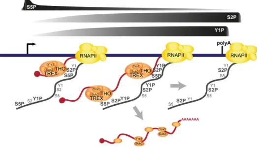
Finally, a novel study by Meinel et al. (2013) revealed that THO is recruited through direct binding to the phosphorylated Pol II CTD. THO complex exhibits the strongest interaction with the Ser2/Ser5 diphosphorylated CTD in vitro, while in vivo THO recruitment was shown to be dependent on Ser2-P and/or Tyr1-P. However, Ser2 and Tyr1 phosphorylations are most probably interdependent in vivo, and by mutating either, it is not possible to precisely determine which one is crucial for THO occupancy. Since THO does not bind Tyr1 phosphorylated CTD in vitro, and sn/snoRNA genes are low in both Ser2-P and THO occupancy but high in Tyr1P, one can suppose that Ser2P is the essential mark. Furthermore, THO complex presence increases from 5’ to 3’ end of a gene, as does Ser2-P, and the Ser2/Ser5 phosphorylation ratio of the CTD is presumed to be the molecular basis for this recruitment pattern (Figure 1.9) (Meinel et al., 2013; Katahira, 2015).
Figure 1.9: Model of TREX recruitment to transcription site (Meinel et al., 2013). TREX recruitment is dependent on Ser2/Ser5 phosphorylation ratio and is increased towards the 3' end as the Ser2 phosphorylation is increased. Soon after transcription termination, TREX complex dissociates from the transcription site and no chromatin recruitment can be observed. However, TREX members can still stay bound to the transcript during the process of export.
- 22 -
THO complex 5’ to 3’ increasing occupancy at transcribed genes is a distinctive characteristic differing this complex from other known transcription elongation factors and proteins interacting with the Ser2-P CTD mark (Abruzzi et al., 2004; Gomez-Gonzalez et al., 2011; Meinel et al., 2013). Since THO was shown to bind both DNA and RNA in vitro (Jimeno et al., 2002; Pena et al., 2012) the possibility that the observed increase in recruitment is due to THO binding along the nascent RNA has been investigated. ChIP assay coupled with RNase treatment demonstrates that THO chromatin recruitment does not depend on RNA binding (Abruzzi et al., 2004; Pena et al., 2012), while analysis of affinity-purified mRNPs show THO presence on the poly(A) mRNA (Batisse et al., 2009; Bretes et al., 2014). Aforementioned Meinel et al. (2013) research uses an RNA bearing a self-cleaving ribozyme sequence in ChIP assay. Cleavage of the nascent transcript leads to the observation that THO recruitment is RNA dependent, yet this dependency is not the cause for 5’ to 3’ increase in occupancy (Meinel et al., 2013). On the other hand, recent studies providing transcriptome maps of different mRNP biogenesis factors in S. cerevisiae, established THO binding across the whole mRNA length, with 5’ end enrichment and slight preference for longer transcripts (Tuck and Tollervey, 2013; Baejen et al., 2014). These results seem contradictory and further studies are necessary to link them into a joint recruitment/binding and function model for THO complex.
1.1.3.1.2 THO function
Members of the THO complex were first recognized for the observed transcription elongation impairment in null mutant strains, with the transcription-induced hyper-recombination as a specific characteristic (Aguilera and Klein, 1990; Piruat and Aguilera, 1998; Chavez et al., 2000). This latter phenomenon is caused by RNA:DNA hybrid formation (R-loops) behind the elongating Pol II, which leads to defective transcription elongation and blocks replication genome-wide, thus indicating that one of THO functional roles is to keep the nascent mRNA and the transcribed DNA apart (Huertas and Aguilera, 2003; Luna et al., 2005; Gomez-Gonzalez et al., 2011). Although genome-wide analyses demonstrate THO complex recruitment to all Pol II transcribed genes, the biggest impairment is exhibited in transcription of long, G + C rich, highly expressed genes and genes with internal repeats (Chávez et al., 2001; Voynov et al., 2006; Gomez-Gonzalez et al., 2011; Luna et al., 2012). Likewise, 5’ to 3’ increasing occupancy of THO was shown to have physiological importance
- 23 -
in expression of long transcripts (Meinel et al., 2013). This impairment is believed to be a consequence of reduced efficiency of transcription elongation (Rondon et al., 2003). Nonetheless, for the newest confirmed member of the THO complex, Tex1, no similar level of decreased gene expression or genome instability can be observed in a null mutant strain (Luna et al., 2005; Pena et al., 2012). Consistently, this mutant does not show the same importance for THO complex assembly nor its binding to nucleic acids (Pena et al., 2012).
Further insight into THO complex connection to proper assembly of the mRNP particle was obtained in Aguilera laboratory. An hpr1-101 point mutant, which does not compromise the THO complex stability or chromatin recruitment but hinders recruitment of TREX subunit Sub2, was found to confer transcription impairment at a null mutant level, without triggering hyper-recombination (Huertas et al., 2006). In another study, the same group has shown that in this point-mutant transcription impairment is independent of R-loops formation, thus adding more importance to THO complex relevance in transcription and mRNP biogenesis (Gómez-González and Aguilera, 2009).
1.1.3.2 TREX complex
THO complex functions in co-transcriptional loading of mRNA export proteins Sub2 and Yra1, together forming the TREX complex and playing a key role in mRNA transport from the nucleus to the cytoplasm (reviewed in Oeffinger and Zenklusen, 2012). These proteins interact with THO complex physically and genetically, and are likewise conserved (Strasser et al., 2002). Their interconnection is further strengthened by the fact that THO and Sub2/Yra1 mutants show similar effect on transcription and hyper-recombination as well as a defect in poly(A) mRNA export (Jimeno et al., 2002). However, deletions of Tho2, Hpr1 and Sub2 lead to the strongest impairment in growth, transcription and export, revealing a hierarchy among TREX members (Garcia-Rubio et al., 2008).
1.1.3.2.1 Sub2
Sub2, a DEAD box RNA-dependent helicase, is a conserved functional splicing and export factor (Fan et al., 2001; Jensen et al., 2001a; Libri et al., 2001; Sträßer and Hurt, 2001). Sub2p is involved in multiple stages of mRNA maturation and its inactivation leads to nonproductive spliceosome assembly, decreased polyadenylation efficiency and mRNA
- 24 -
instability, as well as nuclear accumulation of poly(A) RNA (Saguez et al., 2013). Sub2 acts in the process of mRNP export in interaction with Yra1. Sub2 was at first believed to be the recruitment mediator for Yra1 (Sträßer and Hurt, 2001). However, alternative recruitment pathways for Yra1 have been discovered and the involvement of Sub2 has been determined in taking on Yra1 from the Pcf11, thus allowing the normal CFIA complex assembly and 3’end processing/termination (see next section 1.1.3.2.2). Sub2 is also involved in chromatin maintenance by the fact that its overexpression suppresses the DNA instability in THO mutant strains, possibly due to its helicase activity acting in the unwinding of RNA:DNA hybrid structures (R-loops) formed during transcription in these strains (Chavez et al., 2000; Gomez-Gonzalez et al., 2011; Saguez et al., 2013).
As already stated, Sub2 is recruited to the transcribed genes through interaction with the THO complex. This is supported by direct in vitro interaction with Hpr1, along with reduced Sub2 recruitment in hpr1-101 and other THO mutant strains, as revealed by ChIP assay (Zenklusen et al., 2002; Huertas et al., 2006). Nevertheless, residual Sub2 recruitment and the fact that Sub2 overexpression suppresses phenotype defects of THO null mutant strains, argue the existence of an alternative, yet still undisclosed recruitment pathway (Fan et al., 2001; Zenklusen et al., 2002; Yu et al., 2012). Unlike THO complex, Sub2 recruitment was unambiguously shown to be RNA-dependent, which was proposed to reflect the 5’ to 3’ increasing ChIP profile of Sub2 (Abruzzi et al., 2004; Meinel et al., 2013).
Sub2 mutants convey most THO mutant phenotypes such as transcription and recombination defects, nuclear retention/degradation of aberrant transcripts and the formation of heavy-chromatin (Libri et al., 2002; Rougemaille et al., 2008; see page 27). Interestingly, both SUB2 deletion and overexpression under a strong promoter in wild-type (wt) background lead to mRNA export impairment (Sträßer and Hurt, 2001). This observation emphasizes the importance of THO complex in orchestrating the recruitment of protein factors and in transcript assembly into an export competent mRNP.
1.1.3.2.2 Yra1
Besides TREX connection to splicing through Sub2 function, this complex seems to be implicated in 3’end processing events by the action of Yra1, a member of evolutionary conserved family of RNA and export factor (REF) binding proteins (Zenklusen et al., 2002;
- 25 -
Johnson et al., 2009). Yra1 co-purifies as a part of the TREX complex, but the only direct contact was revealed with Sub2, which was believed to be the Yra1 transcription site recruitment mediator (Sträßer and Hurt, 2001; Zenklusen et al., 2002). However, this interaction was recently disproved as a cause for Yra1 recruitment, as Yra1 binding and recruitment reliance on the CFIA factor Pcf11 was discovered (Johnson et al., 2009). Indeed, the relevance of this interaction was reinforced by its implication in CFIA complex assembly, through competition for binding Pcf11 between Yra1 and CFIA member Clp1, with plausible influence on poly(A) site choice (Johnson et al., 2011; Haddad et al., 2012) (Figure 1.10). This model suggests Yra1 recruitment to transcription site through interaction with Pcf11, followed by Yra1 displacement by Clp1, facilitated through Yra1-Sub2 interactions and transfer onto mRNA (Johnson et al., 2009, 2011). Since complete CFIA complex is necessary for poly(A) site cleavage and mRNA release, this model can account for the aforementioned transcript cleavage/release defect when Sub2 is absent and not able to take on Yra1, thus preventing Clp1-Pcf11 binding and CFIA formation (Katahira, 2012; Oeffinger and Zenklusen, 2012). Another recent research demonstrated that Yra1 owns a CID domain through which it directly binds to Ser2/Ser5 diphosphorylated CTD in vitro. However, Yra1 CID also contains the nuclear localization signal (NLS), which makes it difficult to assess the importance of this interaction for Yra1 recruitment in vivo by simply deleting the CID domain (MacKellar and Greenleaf, 2011; Meinel et al., 2013).
- 26 -
Recently, Yra1 was shown to interact with another DEAD-box helicase, besides Sub2. Genetic and physical interactions were observed between Yra1 and Dbp2, with Yra1 inhibition effect on Dbp2 helicase activity in vitro. Furthermore, Dbp2 was shown to function in in vivo mRNP assembly, by enabling loading of Yra1, Nab2 and Mex67 on the poly(A) transcript. A model was proposed where Dbp2 action is necessary to unwind the nascent transcript for proper mRNP assembly, after which Yra1 binding prevents further rearrangements by this helicase (Ma et al., 2013).
Concurrent with the finding of Yra1 binding to Sub2, Yra1 was also shown to bind Mex67, thus making it a RNA-binding adaptor of Mex67-Mtr2 heterodimer mRNA export receptor (Sträßer and Hurt, 2001). Since Sub2 and Mex67 interact with the same domain within Yra1, it is believed to be handed over from Sub2 to Mex67 in the process of forming an export competent mRNP (Bonnet and Palancade, 2014) (Figure 1.10). Mex67 is recruited to the transcription site through interaction of its C-terminal ubiquitin-associated (UBA) domain with ubiquitylated Hpr1 subunit of THO complex, followed by a transfer to the transcript along with its RNA-binding adaptors Yra1, Npl3 and Nab2, (Gwizdek et al., 2006; Hobeika et al., 2009; Babour et al., 2012). The final contribution of Yra1 to the mRNA in
Figure 1.10: Suggested model for Yra1 influence on co-transcriptional 3' end
processing (Johnson et al., 2011). During transcription, Yra1 is recruited to transcription site through interaction with Pcf11. At the 3’ end of genes, Yra1 is transferred to Sub2 and thus clears the Pcf11 binding site for Clp1. Assembly of the complete CFIA complex enables poly(A) site cleavage (scissors) and mRNA release.
- 27 -
export is its dissociation from the mRNP prior to NPC passage, which is promoted by Yra1 ubiquitination and presents a possible crucial step in establishing an export-competent mRNP (Iglesias and Stutz, 2008; Iglesias et al., 2010).
1.1.3.2.3 TREX function
In addition to implication of THO complex in transcription elongation (see section 1.1.3.1.2), another class of transcripts was discovered in THO/Sub2 mutant strains, which are truncated at the 3’end and/or retained at the transcription site (Jensen et al., 2001a; Libri et al., 2002; Vinciguerra and Stutz, 2004) (Figure 1.11). Most of the research of this model was performed by observing heat shock mRNAs after a growth shift to 37˚C, namely the HSP104 transcript. Fluorescent in situ hybridization (FISH) assay enables the visualization of HSP104 retention dots in THO/Sub2 mutants. In Libri et al., 2002 the retention dots were detected even with the use of FISH probes targeting the HSP104 sequence just upstream of the stop codon. This suggests that the retained transcripts are complete or nearly complete. In the same strains and experimental conditions, the nuclear accumulation of poly(A) RNA can be detected. However, the same study determined another subset of HSP104 transcripts in THO/Sub2 mutants by Northern blot of total RNA from cells grown at 37˚C. These transcripts are truncated at the 3’end, which was determined to be the result of a 3’ to 5’ degradation by the Rrp6 exonuclease. Indeed, deletion of the RRP6 gene from the THO/Sub2 mutant strains leads to restoration of HSP104 3’end levels, but also results in disappearance of the HSP104 retention dots in the same double mutant strain. This is believed to be a result of transcription quality control step, which will be presented in more details in section 1.2.3.
The two proposed models explaining the origin of 3’ end truncated transcripts in TREX mutant strains are, however, not mutually exclusive. In the view of the literature regarding each of the two models, we can observe a major difference in experimental methods which could account for the discrepancy of the obtained results. Experiments supporting the transcriptional model (Figure 1.11, section 1.1.3.1.2) were carried out at optimal temperature for yeast growth. In this condition the function of THO in keeping the nascent transcript and the DNA apart is accentuated, as well as its importance in efficient transcription elongation (Jimeno et al., 2002; Rondon et al., 2003). However, the exosomal model, described in previous paragraph, focuses on the effect of THO mutations on a transcript induced in heat-shock conditions. In these conditions the transcription rate is upregulated, while RNAs exhibit
- 28 -
strong nuclear retention. A possible result of heat shock could be the polymerase overload on the transcribed gene (described in the next paragraph) which would diminish the necessity of THO complex in preventing RNA:DNA hybrid formation and stress its function in proper mRNP biogenesis. Consequently, production of defective mRNPs would activate nuclear surveillance machinery which would retain and degrade the affected transcripts. The fact that both phenotypes observed in THO mutant strains, hyper-recombination and/or transcript retention and degradation, can partially be alleviated by slowing down the rate of transcription, suggests THO function in securing optimal kinetics for mRNP assembly and contribution to transcription speed and efficiency (Jensen et al., 2004; Jimeno et al., 2008).
- 29 -
Additional importance of THO complex in promoting high pace transcription was determined by Libri group in collaboration with Jensen and Stutz groups. They found that under heat shock conditions THO null mutants exhibit accumulation of large protein-nucleic acid aggregates, referred to as the heavy chromatin. This formation contains stalled mRNP intermediates, along with nuclear pore components and polyadenylation factors associated with chromatin. During the chromatin extraction step in ChIP assay, the target heat shock sequences were absent from the THO mutant strains preparations. They were found to be sequestered in the pellet fraction during high speed centrifugation step, as a part of heavy chromatin complex. Hence, this phenomenon was termed differential chromatin fractionation (DCF). Heavy chromatin formation was found at the 3’end of ~400 genes, depending on the presence of a functional terminator. CPF and CFIA mutants defective for polyadenylation and transcription termination, respectively, abolished heavy chromatin formation. However, the most important determinant of heavy chromatin formation is the nature of the gene promoter. The high-power promoter firing leads to polymerase overload on the transcribed genes which are docked to the NPC. Consequently, this overwhelms the 3’end processing and transcript
Figure 1.11: Two proposed models explaining the origin of 3' end truncated transcripts
in TREX mutant strains (Vinciguerra and Stutz, 2004). The exosomal model (model 1) suggests that mutations in TREX lead to aberrant mRNP formation from fully synthesized transcripts, which are then retained at transcription site and degraded by the nuclear exosome. The transcriptional model (model 2) states that the lack of TREX complex leads to formation of DNA:RNA hybrids behind the elongation Pol II which impairs transcription and causes DNA hyper-recombination and genome instability. However, both models agree on the importance of TREX in mRNP formation and are not necessarily mutually exclusive.
- 30 -
release and leads to DCF (Figure 1.12). Taken together, these results affirm THO complex role in coordinating rate of transcription with the downstream processes, 3’end processing/termination and transcript release from the transcription site (Rougemaille et al., 2008; Mouaikel et al., 2013)
In accordance with these findings, mutation of THO complex was shown to impede 3’end processing factors’ release from the mRNP and lead to inefficient polyadenylation (Saguez et al., 2008; Qu et al., 2009). In addition to direct contribution of THO complex to transcription, mRNP assembly and possibly transcript processing events (reviewed in Luna et al., 2012), this complex also recruits and interacts with other protein factors, notably the ones essential for mRNA export, thus playing a major role in production of mature, export-competent mRNPs.
1.1.3.2.4 Other THO/TREX interactions
Two serine-arginine (SR) rich, poly(A) RNA-binding proteins, Gbp2 and Hrb1, co-purify with other members of the TREX complex, but they are not essential for the assembly
Figure 1.12: Model of heavy chromatin formation in THO-Sub2 mutants (Mouaikel et al., 2013). In wt strains THO-Sub2 complex coordinates different processing events and promotes optimal kinetics of transcription. In the absence of THO-Sub2, transcription kinetics is disturbed which leads to an overflow with transcribing polymerases reaching the end of a gene, piling up and docking to the NPC.
- 31 -
of this complex (Hurt et al., 2004). This interaction accounts for their co-transcriptional recruitment to active genes. Gbp2 and Hrb1 bind along the whole transcript length, with 5’end enrichment, as do other THO/TREX members, however, they do not show the same 5’ to 3’end increasing chromatin recruitment pattern (Reed and Cheng, 2005; Meinel et al., 2013; Tuck and Tollervey, 2013). To date, no research was published which would show whether Gbp2 or Hrb1 mutants share the phenotype indicated for other THO/TREX members. Recently, these two factors were shown to preferentially bind transcripts derived from intron-containing genes. They serve as splicing surveillance factors and stay bound to the export-competent transcript during its passage through the NPC and into the cytoplasm (Hackmann et al., 2014).
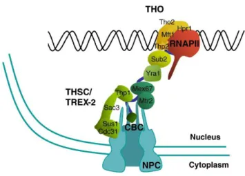
TREX maintains a functional interaction with another protein complex involved in coupling transcription elongation to mRNA export. This complex comprises Thp1-Sac3-Sus1-Cdc31, with Sem1 as the newest characterized member, together forming THSC, also named TREX-2 complex (Köhler and Hurt, 2007; Gonzalez-Aguilera et al., 2008; Faza et al., 2009). Similar to THO, transcription defect, genetic instability coupled with R-loops formation and impaired 3’end processing can be observed in TREX-2 mutants (Gonzalez-Aguilera et al., 2008; Rondón et al., 2010). However, only TREX-2 interacts with the NPC, and Sub2 overexpression is lethal in TREX-2 mutants (Rondón et al., 2010). It is proposed that the two complexes function at different steps of the same pathway, coupling transcription and export, and providing a feedback mechanism for control of transcription and genetic integrity (Gonzalez-Aguilera et al., 2008; Luna et al., 2012) (Figure 1.13).
Figure 1.13: THO/TREX and TREX-2 function in the same pathway from transcription
- 32 -
1.2 mRNP decay and nuclear quality control in yeast
Opposite from the enduring, information bearing DNA whose lifespan is paralleled with the life of the whole cell, RNA is a molecule with versatile functions and a regulated turnover. mRNA life-cycle starts with previously described steps of transcription, processing and export, followed by translation and decay in the cytoplasm. mRNA turnover defines cell growth, differentiation and environmental response (Ross, 2001; Haimovich et al., 2013). In addition to decay at the end of an RNA life-cycle, it can be degraded at earlier steps if it is found to be aberrant and not able to execute its function (reviewed in Parker, 2012).
1.2.1 mRNA degradation
After serving their purpose as information carriers for protein synthesis in the cytoplasm, mRNAs are degraded in a turnover process in that same cellular compartment. Possible degradation pathways in the turnover process are defined as general default decay pathways (Das and Das, 2013). These are initiated by mRNA deadenylation, leaving an unprotected oligo(A) 3’end (Norbury, 2013). Most transcripts are subsequently decapped and then degraded by Xrn1 nuclease in the 5’ to 3’ exonucleolytic pathway, while others are subjected to 3’ to 5’ exonucleolytic degradation pathway by cytoplasmic exosome (Das and Das, 2013).
Another kind of degradation pathways who function selectively to terminate transcripts recognized as aberrant are termed specialized mRNA decay pathways and they take place in the cell nucleus, as well as in the cytoplasm (Houseley and Tollervey, 2009) (Figure 1.14). Aberrant mRNPs are recognized during quality control (QC) surveillance of mRNP biogenesis. A QC check-point at each processing step and at the NPC in the nucleus of eukaryotes makes sure the mRNP is export-competent (Tutucci and Stutz, 2011; Eberle and Visa, 2014). Degradation of aberrant transcripts in the nucleus can be carried out by two pathways: the minor - 5’ to 3’ degradation by Rat1 exonuclease after decapping of the transcript; and the major pathway – carried out by the 3’ to 5’ exonucleolytic action of the nuclear exosome (Tutucci and Stutz, 2011). The nuclear subset of QC processes will be presented in further details in section 1.2.3.
- 33 -
At the cytoplasmic side quality control mechanisms act in response to difficulties encountered in the process of translation (Doma and Parker, 2007). Adaptor proteins interact with translation machinery and direct aberrant mRNPs into different degradation pathways: Nonsense-mediated, No-go and Non-stop decay pathways, which have been extensively reviewed by Parker, 2012. Nonsense-mediated decay acts in response to faulty translation termination, caused by a variety of events such as long 3’ untranslated region (UTR), alternative translation initiation sites, upstream open reading frames (ORFs), presence of introns with stop codons and translation frameshift. In this pathway, aberrant transcripts are decapped or deadenylated and degraded in 5’ to 3’ or 3’ to 5’ end manner, respectively, and coupled to repression of translation. In the No-go decay pathway, stalled translation leads to endonucleolytic transcript cleavage by a still unknown endonuclease. The remaining mRNA fragments are degraded in both 5’ to 3’ and 3’ to 5’ directions, by Xrn1 and exosome, respectively. Finally, if for different reasons transcript does not contain a stop codon, 3’ end
Figure 1.14: Decay systems in different nuclear and cytoplasmic QC mechanisms (Eberle and Visa, 2014). Most 3’ to 5’ exonucleolytic and endonucleolytic degradation in the nucleus is carried out by the nuclear exosome, while 5’ to 3’ decay is performed by Rat1 and DOX exonucleases. Their cytoplasmic counterparts are cytoplasmic exosome for 3’ to 5’ and Xrn1 for 5’ to 3’ decay.
- 34 -
ribosome stalling triggers its rapid degradation from 3’ to 5’ by the exosome in the Non-stop decay pathway (Parker, 2012).
1.2.2 The exosome
The eukaryotic RNA exosome is a multisubunit complex with a highly conserved core structure. Indeed, the core structure homologues reach to Archaea and Eubacteria (Lykke-Andersen et al., 2011). 9 subunits of a barrel-like core form a two-layered ring: the bottom hexamer (Rrp41, Rrp42, Rrp43, Rrp45, Rrp46 and Mtr3), and the upper RNA binding cap (Rrp4, Rrp40 and Csl4) (Chlebowski et al., 2013; Schneider and Tollervey, 2013) (Figure 1.15). However, in yeast and humans the exosome core itself is catalytically inactive. Still the core is indispensable for the nuclease activity carried out by two associated components, Dis3/Rrp44 and Rrp6 (Wasmuth and Lima, 2012) (Figure 1.15). In budding yeast, Dis3 subunit accompanies the core in both the nucleus and the cytoplasm, while Rrp6 is confined to provide catalytic activity only in the nucleus. Hence, the exosome exists in two isoforms: cytoplasmic (core + Dis3) and nuclear (core + Dis3 +Rrp6) (Chlebowski et al., 2013).
Dis3 possesses two nuclease activities: endonuclease, which may act on substrates arriving through the central channel of the exosome core or directly from the surroundings, and the 3' to 5' exonuclease activity, whose active site is placed at the very bottom of the
Figure 1.15: Schematic representation of the eukaryotic nuclear exosome complex (Chlebowski et al., 2013). The barrel-like core is depicted in blue (hexamer) and magenta (cap). The two catalytical components are represented in yellow (Rrp6) and green (Dis3) with the active sites denoted in red.