HAL Id: hal-02326835
https://hal.archives-ouvertes.fr/hal-02326835
Submitted on 15 Dec 2020
HAL is a multi-disciplinary open access
archive for the deposit and dissemination of
sci-entific research documents, whether they are
pub-lished or not. The documents may come from
teaching and research institutions in France or
abroad, or from public or private research centers.
L’archive ouverte pluridisciplinaire HAL, est
destinée au dépôt et à la diffusion de documents
scientifiques de niveau recherche, publiés ou non,
émanant des établissements d’enseignement et de
recherche français ou étrangers, des laboratoires
publics ou privés.
Convertible and conformationally constrained nucleic
acids (C 2 NAs)
Jean-Marc Escudier, Corinne Payrastre, Béatrice Gerland, Nathalie Tarrat
To cite this version:
Jean-Marc Escudier, Corinne Payrastre, Béatrice Gerland, Nathalie Tarrat. Convertible and
confor-mationally constrained nucleic acids (C 2 NAs). Organic and Biomolecular Chemistry, Royal Society
of Chemistry, 2019, 17 (26), pp.6386-6397. �10.1039/C9OB01150A�. �hal-02326835�
Convertible and Conformationally Constrained Nucleic
Acids (C
2NAs)
Escudier Jean-Marc,*a Payrastre Corinne,a Gerland Béatrice a and TarratNathalie b
We introduce the concept of Convertible and Constrained Nucleic Acid (C2NA). By means of the synthesis of a stereocontrolled N-propargyl dioxo-1,3,2-oxaza-phosphorinane as internucleotidic linkage, the torsional angles and can adopt either the canonical (g-, t) set of values able to increase DNA duplex stability or the non-canonical (g+, t) set that stabilized hairpin structure when installed within the loop moiety. With an appended propargyl function on the nitrogen atom of the six-membered ring, the copper catalysed Huisgen’s cycloaddition (CuAAC click chemistry) allows for the introduction of new functionalities at any location on the nucleic acid chain while maintaining the properties brought by the geometrical constrain and the neutral internucleotidic linkage.
Introduction
Nucleic acid molecules exhibit an increasing importance in the context of therapeutics, molecular diagnostics and
nanotechnologies.1 For these purposes, chemists have
developed modifications of their inner nucleotide components in order to improve their bio-stability, their target affinity or their ability to be decorated by new functionalities such as fluorescent labels or any other molecules able to explore new
applications.2 In that context, the most efficient modification
was the restriction of the conformational space of the sugar moiety in its C3'-endo conformation in order to mimic the
geometry adopted in RNA duplex.3 Locked Nucleic Acid (LNA) in
which the torsional angle (Figure 1) is completely frozen, is to date the best element of this class of conformationally
constrained nucleotides.4 Concerning the decoration of
oligonucleotides (ODNs) by extra functionalities, the copper catalysed azide-alkyne cycloaddition (CuAAC) appeared to be a
very powerful approach.5 These considerations taken together,
the Wengel’s group proposed an amino-LNA with an appended alkyne function. This RNA nucleotide mimic could be denoted as a convertible and locked nucleotide that exhibited extensive
properties and applications.6
Based on the concepts of preorganization7 and of convertible
approach,8 we engaged in two parallel programs. The first was
directed towards the development of nucleotides in which the sugar/phosphate backbone geometry was constrained (Constrained Nucleic Acids: CNAs) in order to propose nucleotides analogues able to fit either with the B-DNA canonical structure or others non-helical relevant disparate
secondary structures.9 The second research axis was oriented
towards the design of convertible nucleotides where an appended arm at the C5' position was bearing an orthogonally
reactive function such as an alkyl halide or alkyne.10 Here we
present our effort to merge these two approaches in the development of a combination of the two properties into one family of dinucleotides: the Constrained and Convertible
Nucleic Acid (C2NA).
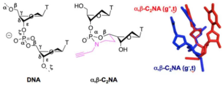
The most prominent members of the CNA family are the -D-CNA in which the conformational control of the torsional angle and of the nucleic acid sugar/phosphate backbone was achieved by introduction of a 1,3,2-dioxa-phosphorinane ring connecting one oxygen atom of the phosphate moiety to
the 5'-carbon of the deoxyribose.11 When featuring the
canonical set of values gauche(-)/trans, -D-CNA are perfect B-DNA nucleotide mimics and induce a strong stabilization of
duplex structure12 whereas stability of hairpin or bulge
structures can be modulated by using -D-CNA featuring a gauche(+) conformation of the angle, located in the unpaired
moiety of the secondary nucleic acid structure.13
Since we wanted to keep the D-CNA’s general scaffold for their
interesting stabilization effects for the newly designed C2NA and
because the C5'-carbon was no longer available for further functionalization, we chose to investigate the replacement of one oxygen atom within the dioxa-phosphorinane ring by a nitrogen atom properly alkylated by a propargyl function in order to bring the convertible ability. It was also of importance to keep the natural C5'-O5'-P-O3' bonds unchanged to prevent any change into the internucleotidic length and local geometry. Therefore, it turned out that one of the non-bridging oxygen of the phosphate was formally suitable to be replaced by the propargyl amino group to design
dioxo-1,3,2-oxaza-phosphorinane as scaffold for the development of C2NA (Figure
1).
Fig. 1. Left: the six backbone torsion angles (labelled to of nucleic acids. Middle: the constrained and convertible dinucleotide in which and are stereocontrolled by a dioxo-1,3,2-oxaza-phosphorinane ring structure bearing a clickable propargyl function (-C2NA). Right: superimposition of minimized structures of (SC5', SP) -C2NA TT (red) a.Laboratoire de Synthèse et Physico-Chimie de Molécules d’Intérêt Biologique,
UMR CNRS 5068, Université Paul Sabatier, 118 route de Narbonne, 31062 Toulouse, France.
E-mail: [email protected].
b.CEMES, Université de Toulouse, CNRS, 29 rue Jeanne Marvig, Toulouse 31055, France.
Electronic Supplementary Information (ESI) available: NMR spectra of described compounds, mass spectrometry data of ODNs, thermal denaturation curves, typical HPLC profile for CuAAC conjugation. See DOI: 10.1039/x0xx00000x
and (RC5', RP) -C2NA TT (blue) with featuring a non-canonical gauche(+) and canonical gauche(-) configuration, respectively.
In this paper, we present the in silico calculations that ascertain the synthesis of the appropriate regio-isomer of the
oxaza-phosphorinane towards the best -C2NA design. The rapid
synthesis of both diastereoisomers of -C2NA
phosphoramidites by H-phosphonate chemistry and their structural determination by means of NMR spectroscopy are
described. Next, we show that -C2NAs can be efficiently
incorporated within oligonucleotides and exhibit stabilizing properties of nucleic acid secondary structures (duplex, hairpin and bulges). Eventually, as a proof of concept, we report the
conjugation of oligonucleotides including -C2NAs with
fluorescein through copper catalysed Huisgen’s cycloaddition.
Results and discussion
In silico -C2NA structure conformational study
As previously observed with -CNAs,9, 12, 14 among the four
possible -C2NAs isomers, only two of them can have all the
substituents of the phosphorinane ring in the orientation that favours their formation, stability and conformational interest: the upper nucleoside in the apical position, the exocyclic oxygen and the lower nucleoside in equatorial position. Moreover in line with the results obtained with their -CNA analogues, a simple molecular model examination indicated that this couple
should mainly differ on the torsional angle conformation.11
Therefore we focused the in silico conformational investigation
on these two potentially major diastereoisomers of -C2NAs,
i.e. the (SC5', SP) -C2NA (3 in its 5'-O and 3'-O unprotected
form, Scheme 1) and (RC5', RP) -C2NA (11 in its 5'-O and 3'-O
unprotected form, Scheme 2). The accessible conformation space of the two compounds was explored through ab initio molecular dynamics simulations (AIMD) followed by quenches. AIMD were done starting from different initial conformations.
The total AIMD trajectory length was 18 ps for (SC5', SP) -C2NA
and 12 ps for (RC5', RP) -C2NA (with a time step of 1.5 fs), the
associated average temperatures of the trajectories being respectively 543K and 545K. Thirty five structures were extracted by dividing the trajectories into equal intervals. After their geometry optimizations, the energy of the structures was located within a range of 11.0 1 and 9.6
kcal.mol-1 for (SC5', SP) -C2NA and (RC5', RP) -C2NA, respectively. The
most stable conformation of the (RC5', RP) isomer was found to
be 1.4 kcal.mol-1 below the one of the (SC5’, SP) isomer (Figure
2).
In both optimized dinucleotides, the puckering of the upper sugar unit is C2'-endo and the oxaza-phosphorinane ring exhibits a chair conformation. However, the two structures differ in the conformation of the lower sugar unit: C2'-endo for
(RC5', RP) -C2NA and C3'-endo for (SC5', SP) -C2NA. A
W-shaped P-O5'-C5'-C4'-H4' structure is observed for the two compounds, a geometrical feature that will be confirmed by the NMR study (in agreement with the observed long range 4JH/P
coupling constants). Interestingly, the distances between the branching points of the dinucleotide (5'-O of the upper
nucleotide and 3'-O of the lower) differ by 2.1Å (8.5Å for (SC5',
SP) -C2NA and 10.6Å for (RC5', RP) -C2NA), and those
between the bases centres by 1.3Å (4.9Å and 3.6Å, respectively). The latter certainly explains in large part the energy difference between the two diastereoisomers by the
loss of the stabilizing stacking interaction for (SC5', SP) -C2NA
whereas the conformation of (RC5', RP) -C2NA is highly
favourable. Another important structural point is that
compound (SC5', SP) -C2NA exhibits an orientation flip of its
upper sugar ring, a phenomenon that appears in loop moiety of hairpin structure. All these features outlined by the simulation
indicate that (RC5', RP) -C2NA should be well adapted to fit
with a duplex structure whereas (SC5', SP) -C2NA should be
better integrated within unpaired structure such as loop. Concerning the clickable propargyl function, one can notice that it is easily accessible by an external molecule in both cases. The partial charges of the propargyl carbon atoms are similar in both isomers: -0.3 on the carbon bound to the nitrogen atom, 0.0 on the central carbon and -0.2 on the final one. It indicates that if a reactivity difference would be observed once introduced into extended oligonucleotides, it would belong to the accessibility and not to the charges of the function.
Fig. 2. Most stable conformations issued from the in silico protocol. Left: Dinucleotide (SC5', SP) -C2NA (3 in its unprotected form). Right: Dinucleotide (RC5', RP) -C2NA (11 in its unprotected form).
Synthesis of -C2NA
The key intermediates in the synthetic pathway of -C2NAs
are the 5'-C-propargylaminoethyl thymidines 2 and 10 that are epimers at the 5’-carbon of the sugar moiety of the nucleoside (Scheme 1 and 2). They are both obtained in moderate to good yield by displacement by the propargylamine of a tosylate group of tosyloxyethyl thymidines 1 and 9, respectively, both
previously described in the preparation ofD-CNAs.14
Thanks to the methodology elaborated by A. Kraszewski and collaborators in the early 90’s, preactivation of the thymidine H-phosphonate monoester by pivaloyl chloride led to a dipivaloyl phosphite that can react with the amino alcohol derivative to form a cyclic phosphoramidite intermediate, further oxidized in situ by water after addition of iodine that provided the
oxaza-phosphorinane.15 This procedure led in average yield of 80%
from 5'-C(S)-propargylaminoethyl thymidine 2 to a 2:1 ratio of diastereoisomeric oxaza-phosphorinane dinucleotides
-C2NAs 3 and 4, determined by 31P NMR (= 2.4 and 4.7 ppm for
3 and 4, respectively, Scheme 1).
Scheme 1. Synthesis of (SC5', SP) -C2NA and (SC5', RP) -C2NA. a- Propargylamine, iPr2NEt, DMF, 65%. b- 5'-O-DMTr Thymidine H-phosphonate triethylammonium salt, PivCl, Pyr then I2, 80%. c- TBAF, THF, 85-90%. d- 2-cyanoethoxydiisopropylamino chlorophosphine, iPr2NEt, THF, 87-95%.
A similar result was obtained from the epimeric propargylaminoethyl thymidine 10 with a slightly higher
diastereoselectivity of 3:1 in favour of -C2NA 11 (31P NMR
= 2.7 ppm) over 12 (31P NMR = 3.2 ppm) that appeared to be
rather unstable under chromatographic conditions, explaining the modest overall yield of 60% (Scheme 2). In both cases, the major compounds formed were depicted at lower chemical shift
in 31P NMR with respect to their corresponding isomer
(, which suggested the formation of the most stable chair conformation of the oxaza-phosphorinane as expected and previously observed
during -D-CNAs synthesis.16
Scheme 2. Synthesis of (RC5', RP) -C2NA and (RC5', SP) -C2NA. a- Propargylamine, iPr2NEt, DMF, quant. b- 5'-O-DMTr Thymidine H-phosphonate triethylammonium salt, PivCl, Pyr then I2, 60%. c- TBAF, THF, 85-87%. d- 2-cyanoethoxydiisopropylamino chlorophosphine, iPr2NEt, THF, 72-94%.
NMR structural study of C2NA
When compared with their analogues D-CNAs, the
-C2NAs differ by the replacement of an oxygen atom by a
nitrogen within the phosphorinane ring and obviously, by the appended propargyl arm in between the two nucleosides. These differences could induce a dramatic change of conformational behaviour. Therefore, the analysis of the conformation of this
new family of constrained nucleotides was carried out using 1H,
13C, 31P and 2D NMR techniques, in order to determine the sugar
puckering preference of each deoxyribose and the geometry of the oxaza-phosphorinane ring.
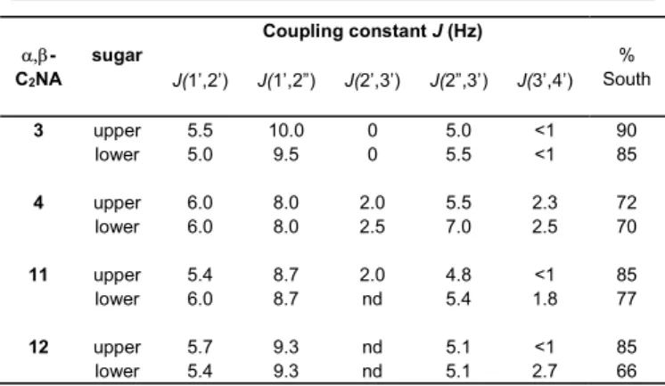
Sugar Puckering
By measuring the coupling constants exhibited by the protons
of the 2’-deoxyribose moieties in 1H NMR spectra of -C2NAs
3, 4, 11 and 12, a rough evaluation of the sugar puckering can be estimated by the Altona and Sudaralingam’s equation:
%South = [JH1',H2'/(JH1',H2' + JH3',H4')] x 100,17 with the results
reported in Table 1.
All the 2’-deoxyribose moieties of -C2NAs exhibited the
South conformation (C2'-endo) as expected for DNA constituents. However, it is noteworthy that the upper sugars of the major isomers 3 and 11 were pushed strongly towards the south conformation in comparison with the lowers units and with the standard puckering adopted within DNA. This favoured conformation can be associated with the neutral
internucleotidic linkage featured by C2NA that is known to
enforce the C2'-endo puckering even in the case of ribose
appended propargyl arm within -C2NAs have modified the
sugar geometry with respect to their -D-CNAs analogues.
Table 1. H/H coupling constants (Hz) of 2'-deoxyribose moieties in the 1H NMR spectra (500 MHz) of -C2NA dinucleotides (SC5', SP) 3, (SC5', RP) 4 and (RC5', RP) 11, (RC5', SP) 12. -C2NA sugar Coupling constant J (Hz) % South J(1’,2’) J(1’,2”) J(2’,3’) J(2”,3’) J(3’,4’) 3 upper 5.5 10.0 0 5.0 <1 90 lower 5.0 9.5 0 5.5 <1 85 4 upper 6.0 8.0 2.0 5.5 2.3 72 lower 6.0 8.0 2.5 7.0 2.5 70 11 upper 5.4 8.7 2.0 4.8 <1 85 lower 6.0 8.7 nd 5.4 1.8 77 12 upper 5.7 9.3 nd 5.1 <1 85 lower 5.4 9.3 nd 5.1 2.7 66
Oxaza-phosphorinane ring conformation
The conformation of six membered ring including phosphorus and two oxygen or nitrogen (phosphorinane) can be
determined by examining the JH/P coupling constants in 1H NMR
spectra of the proton within the cyclic structure. Gorenstein
showed that when 3JH/P < 3 Hz, the considered proton can be
assigned to be in an axial position and when 3JH/P > 20 Hz the
proton adopts an equatorial position. This taken into
consideration and together with the relative 31P NMR chemical
shift depicted for the phosphorus in each couple of diastereoisomers (the lower chemical shift belongs to a phosphorus with the oxygen in equatorial position on the phosphorinane ring), the absolute configuration of phosphorus
can be assigned accordingly.19
On the other hand, Altona proposed that the torsional angle
was correlated with 3JH/P of the 3'-proton according to the
relation: = - -120°, where is obtained from 3J3’-H/P = 15.3
cos2 .20
Therefore, we collected and reported in Table 2 all the
measurable JH/P coupling constant values in 1H NMR spectra for
-C2NAs 3, 4, 11 and 12.
It is clear that the major isomers (SC5', SP) -C2NA 3 and (RC5',
RP) -C2NA 11 present the oxaza-phosphorinane ring in a pure
chair conformation with the 5'b- and 7'b-protons exhibiting constant values in agreement with axial positions whereas the 7''b-protons were depicted with large constants of 24 and 22 Hz, respectively, evidencing their equatorial positions. With
average 3JH/P constant values, the minor isomers 4 and 12
appeared to fit with an oxaza-phosphorinane structure in slightly twisted chair conformation. This less stable conformation could explain the relative instability that became
apparent upon standing for the minor isomer (RC5', SP)
-C2NAs 12. Knowing the geometry adopted by the
internucleotidic linkage gave an access to the torsional angle values of and (Table 3) by a simple molecular models
examination. The (SC5', SP) -C2NA 3 featured a non-canonical
(gauche(+), trans) combination whereas (RC5', RP) -C2NA 11
fitted with the conformation (gauche(-), trans) observed in B-DNA.
The observed 3JH3'a/P coupling constants were in the same range
of value (7.5 ± 0.5 Hz, Table 3) and the calculated torsional angles were found to be in the trans conformation for all the
-C2NAs isomers which was very similar to the one found in
the corresponding -D-CNAs.
Table 2. H/P coupling constants (Hz) in the 1H NMR spectra (500 MHz) of α,β-C2NA dinucleotides 3, 4 and 11, 12. Coupling constant J (Hz) -C2NA J(5'b/P) J(7'b/P) J(7''b/P) J(3'a/P) J(4'b/P)[a] 3 <1 <1 24.0 8.0 5.5 4 2.0 8.5 12.0 7.2 4.0 11 <1 <1 22.0 6.9 3.6 12 2.1 nd 15.0 7.2 5.4
[a] Protons denoted as “a” belong to the upper nucleoside whereas those denoted as “b” belong to the lower nucleoside including the phosphorinane moiety.<w
Eventually, long range 4JH/P coupling constants were observed
for all the -C2NAs isomers between the 4'-H of the
downstream nucleoside and the phosphorus atom. A W-shaped P-O5'-C5'-C4'-H4' arrangement can explain these observations and provided an access to the value of the torsional angle , considering the sugar south conformation and each oxaza-phosphorinane chair or twist-chair geometry (Table 3). The main isomers 3 and 11 adopted a gauche(+) conformation for , when the minors were slightly distorted to the cis(+). Therefore
(RC5', RP) -C2NA 11 appeared to be an interesting
conformationally locked B-type analogue with four of its torsional angles in the same range than those observed for
nucleotide involved in B-duplex.21
Table 3. Estimated torsional angle values of -C2NA dinucleotides 3, 4 and 11, 12.
-C2NA
Torsional angle[a]
[b]
3 (SC5', SP) g+ t g+ t
4 (SC5', RP) t t c+ t
11 (RC5', RP) g- t g+ t
[a] The following staggered pattern of the torsional angles is used: cis= 0 ±30° (c), gauche(+)= 60 ±30° (g+), trans= 180±30° (t), gauche(-)= 300 ±30° (g-). [b] angle
(C5'-O5'-P-O3'), angle (C4'-C5'-O5'-P), and angle (C3'-C4'-C5'-O5') estimated from NMR data, angle (C4'-C3'-O3'-P) calculated from the Altona relation.
Oppositely, (SC5', SP) -C2NA 3 differed from the canonical set
of torsional angle value with that was pushed by 120° from gauche(-) to gauche(+) conformation, this distortion was outlined at the turning phosphate position within loop moiety of hairpin structure and also appeared within bended DNA
involved in protein/DNA complexes.22
Thermostabilty of -C2NA within duplex, hairpin and bulge
structures
In order to evaluate the impact on DNA secondary structures formation ability of the newly synthesized convertible and
constrained dinucleotide units -C2NAs featuring either the
non-canonical or the canonical set of torsional angle conformation, the corresponding phosphoramidites derivatives 7 and 15 (Schemes 1 and 2) were used during automated synthesis of oligonucleotide according to the phosphoramidite
technology.23
The following oligonucleotide sequences have been chosen to have a direct comparison of the melting temperature with those previously measured with the same constraint brought by the -CNA analogues in order to have an insight of the influence of the appended propargyl function. Duplex 5'-GCGCTTGCCG/3'-CGCGAACGGC (ODN 1) has been modified on the TT step with the application of the gauche(-) or the gauche(+) constraint to the alpha torsional angle (Table 4, ODN
2 and 7).24 Duplexes 5'-CGTTTTTTGCT/3'-GCAAAAAACGA (ODN
3) have been designed to study the influence of the gauche(-)
constraint location by moving the modified -C2NA TT along
the chain (Table 4, ODN 4, 5 and 6).25 A four thymidine-looped
hairpin (ODN 8) has been modified with -C2NA TT featuring
the gauche(+) constraint embedded at all the possible positions within the unpaired loop moiety (Table 4, ODN 9, 10, 11 and 12).26
Eventually, bulges have been constructed with the gauche(+) constraint within the loop or opposite to the loop using the
-C2NA TT modified sequence ODN 13
5'-GATTTGCATATTCATGAG.13 Restricting the complementary
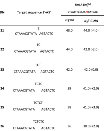
strand generated a constrained loop on the modified strand (Table 5, bulges made with ODN 13 and ODN 15 to 20) whereas expanding the complementary strand with unpaired moiety from one to six nucleotides led to bulged structures facing the constraint (Table 6, bulges made with ODN 13 and ODN 21 to 26).
Phosphoramidites 7 and 15 were incorporated with similar yields than those obtained for unmodified nucleotide phosphoramidite with no change either in concentration nor in synthetic cycle time during automated oligonucleotide synthesis. An important point that need to be outlined was that
the oxaza-phosphorinane structure of -C2NA was stable
towards the ammonia treatment and even to the use of
ammonia/methyl amine (AMA) at 65°C, with no detectable ODN degradation by ring opening, an issue encountered with their dioxa-phosphorinane-CNA analogues during synthesis of
chimeric antisens oligonucleotides.28 All the modified
oligonucleotides were characterized by mass spectroscopy in MALDI Tof mode and showed an increased mass of 64 Da with respect to their unmodified analogues.
According to the preorganization concept, -C2NA featuring
the B-DNA canonical gauche(-) constraint induced a neat thermal stabilization of duplex with Tm around +3°C (±1°C) independently of the composition (ODN 2 and ODN 4, Table 4) and the location (ODN 4 compared to ODN 5 and 6, Table 4) of the constraint within the sequence. Despite this interesting
impact on the duplex stability, the effect of -C2NA was
lowered compared with the one exhibited by its -D-CNA
analogues (+5°C).25 According to the in silico study in the (RC5',
RP) -C2NA, the appended hydrophobic propargyl substituent
should be oriented towards the solvent and therefore could provide unfavourable interactions affecting the overall duplex stability.
Table 4. Sequences and thermal melting temperatures [°C] of (RC5', RP) -C2NA TT featuring B-type canonical value ( = gauche(-), = trans) containing duplexes and of (SC5', SP) -C2NA TT featuring non-canonical value ( = gauche(+), = trans) containing duplexes and hairpins.
ODN Sequences[a] Tm[b] Tm
1 5'-GCGCTTGCCG 3'-CGCGAACGGC 57.0 - 2 5'-GCGCTTGCCG 3'- CGCGAACGGC 60.0 +3.0 3 5'-GCGTTTTTTGCT 3'-CGCAAAAAACGA 51.0 - 4 5'-GCGTTTTTTGCT 3'-CGCAAAAAACGA 55.0 +4.0 5 5'-GCGTTTTTTGCT 3'-CGCAAAAAACGA 54.0 +3.0 6 5'-GCGTTTTTTGCT 3'-CGCAAAAAACGA 54.0 +3.0 7 5'-GCGCTTGCCG 3'- CGCGAACGGC 48.0 -9.0 8 5'-ATCCTATTTTTAGGAT 52.0 - 9 5'-ATCCTATTTTTAGGAT 50.0 -2.0 10 5'-ATCCTATTTTTAGGAT 58.0 +6.0 11 5'-ATCCTATTTTTAGGAT 54.0 +4.0 12 5'-ATCCTATTTTTAGGAT 53.0 +1.0
[a] TT = (RC5', RP) - C2NA TT (g-,t) or TT = (SC5', SP) - C2NA TT (g+,t) within the
strand. T in italic indicates an unpaired thymidine within the structure [b] UV melting experiments were carried out in sodium phosphate buffer (10 mM, pH 7.0) containing NaCl (100 mM) and EDTA (1 mM). Melting temperatures (Tm) were measured as the maximum of the first derivate of the UV melting curve (OD260 vs temperature, 20-90 °C, 0.5 °C/min) which was recorded at concentration of 5 µM in sodium phosphate buffer (10 mM, pH 7.0) containing NaCl (100 mM) and EDTA (1 mM).
The Tm measured for ODN 7 (Table 4) and ODN 14 (Table 5)
showed that the gauche(+) constraint imposed by (SC5', SP)
-C2NA destabilized the duplex by -9°C and -7°C, respectively
which reached the same level displayed by the corresponding -D-CNA for the former. It confirmed that in this configuration there was again an additive negative effect brought by the propargyl function that should be oriented towards the minor groove of the duplex structure.
On the other hand, aside from the introduction of the constrain at the junction between the loop and the stem (ODN 9, Table 4) that appeared to be badly accommodated (Tm = -2°C), an
improved thermal stability was depicted for (SC5', SP) -C2NA
modified hairpins (ODN 10, 11 and 12, Table 4) that reached up to +6°C when the gauche(+) constraint was located at the described turning phosphate position (ODN 10) and was superior to that induced by the -D-CNA analogue. It can be
proposed that the four thymidines composing the loop provided a favourable surrounding for the hydrophobic propargyl arm that could participate to stacking and therefore improved the overall structure thermal stability.
Table 5. Thermal melting temperatures [°C] of bulges constructed with(GATTTGCATATTCATGAG) where TT= -C2NA featuring (g+,t) conformation is within the loop.
ODN Target sequence 3'→5'
Tm(Tm)[a] 5'-GATTTGCATATTCATGAG wtTT[b] -C2NA 14 CTAAACGTATAAGTACTC 55.0 48.0 (-7.0) 15 CTAAACGTAT AGTACTC 47 45.0 (-2.0) 16 CTAAACGTAT GTACTC 42 42.0 (0.0) 17 CTAAACGTA GTACTC 39.0 39.0 (0.0) 18 CTAAACGTA TACTC 35.0 34.0 (-1.0) 19 CTAAACGT TACTC 27.0 26.0 (-1.0) 20 CTAAACG TACTC <25 26.0 (+1.0)
[a] Melting temperatures were measured as the maximum of the first derivate of the UV melting curve (OD260 vs temperature, 20-90 °C, 0.5 °C/min) which was recorded at concentration of 5 µM in sodium phosphate buffer (10 mM, pH 7.0) containing NaCl (100 mM) and EDTA (1 mM). [b] wt: unmodified phosphodiester internucleotidic linkage.
When inserted within bulged structures, (SC5', SP) -C2NA
exhibited two different behaviours (Tables 5 and 6).
When the gauche(+) constraint was installed within the loop it slightly destabilized the bulge (ODN 15, 18, and 19, Table 5) or can have no effect (ODN 16 and 17, Table 5) for loop size varying from one to five nucleotides. Only the rather unstable 6-nucleotides looped bulge made with ODN 20 exhibited a small stability improvement. In that context, unlike its behaviour into
T4-hairpin, the (SC5', SP) -C2NA modification was not well
tolerated and did not behave as its -CNA analogue that improved, in the same conditions, the bulge thermal
stabilities.13
In the other bulge construction, where the (SC5', SP) -C2NA
faced the growing loop (Table 6), it appeared that this modification conferred to the structures roughly the same thermal stability around 40 ±2°C regardless to the loop size. While for the one to three nucleotides loop the gauche(+) constraint was unfavourable or neutral (Table 6, bulges made with ODN 21, 22 or 23), it turned out that it became stabilizing for loop made with at least four nucleotides (Table 6, bulges made with ODN 24, 25 or 26) similarly to what has been
Table 6. Thermal melting temperatures [°C] of bulges constructed with(GATTTGCATATTCATGAG) where TT= -C2NA featuring (g+,t) conformation is opposite to the loop.
ODN Target sequence 3'→5'
Tm(Tm)[a] 5'-GATTTGCATATTCATGAG wtTT[b] -C2NA 21 T CTAAACGTATA AGTACTC 48.0 44.0 (-4.0) 22 CTAAACGTATA AGTACTC TC 44.0 42.0 (-2.0) 23 CTAAACGTATA AGTACTC TCT 42.0 42.0 (0.0) 24 CTAAACGTATA AGTACTC TCTC 39 41.0 (+2.0) 25 CTAAACGTATA AGTACTC TCTCT 38 41.0 (+3.0) 26 CTAAACGTATA AGTACTC TCTCTC 36 38.0 (+2.0)
[a] Melting temperatures were measured as the maximum of the first derivate of the UV melting curve (OD260 vs temperature, 20-90°C, 0.5 °C/min) which was recorded at concentration of 5 µM in sodium phosphate buffer (10 mM, pH 7.0) containing NaCl (100 mM) and EDTA (1 mM). [b] wt: unmodified phosphodiester internucleotidic linkage
FAM decoration of -C2NA modified oligonucleotides
In order to validate the concept of constrained and convertible
nucleotides, we had to show that -C2NA can be easily
conjugated through the Huisgen’s cycloaddition (CuAAC). We
choose to decorate two (RC5', RP) -C2NA modified ODN (ODN
2 and 4 converted into f-ODN 2 and f-ODN 4, respectively) and
two (SC5', SP) -C2NA modified ODN (ODN 10 and 13 converted
into f-ODN 10 and f-ODN 13, respectively) with fluorescein by mean of its commercially available azide derivative (6-FAM-Azide, Figure 3).
Each conjugation proceeded cleanly in 1h in standard conditions
(cat. CuSO4, ascorbic acid) as indicated by reverse phase
analytical HPLC monitoring. All the conjugates were characterized by mass spectroscopy in MALDI Tof mode and showed an increase of mass of 460 Da with respect to their precursors. Another common characteristic of the conjugates was depicted on the UV spectra where two maxima of absorption were observed at 260 and 497 nm that can be easily attributed to the nucleic bases for the former and to the fluorescein moiety for the latter. Therefore, the propargyl arm
featured by -C2NA appeared to be a very good partner for
CuAAC decoration of oligonucleotides.28
Fig. 3. : Conjugation with fluorescein of -C2NA modified ODN by CuAAC.
The thermal stability of the duplex made with conjugates were measured to get insight of the dye impact on the probe-target duplex when the conjugate was directed towards the solvent within f-ODN 2 and f-ODN 4 with a canonical gauche(-) constraint or towards the minor groove within f-ODN 13 with a non-canonical gauche(+) constraint. Duplex formed with f-ODN 2 and f-f-ODN 4 and their respective complementary strand exhibited the same Tm value of 52°C and appeared to be less stable than those formed with their constrained precursor by - 8 and -3°C, respectively. In the case of f-ODN 2, the gauche(-) constraint did not compensate the dye negative effect when compared with the corresponding unmodified ODN 1 (Tm = -5°C) but with f-ODN 4 the constraint restored the thermal stability to the level of the unmodified ODN 3 (Tm = +1°C). Therefore, it looked like the gauche(-) constraint was able to compensate the negative effect on thermal stability induced by the dye depending on the sequence composition.
With a measured Tm of 44°C, combination of the gauche(+) constraint and the hydrophobic fluorescein were additive and decreased the Tm value of the duplex formed with f-ODN 13 by -4°C relatively to its constrained precursor ODN 13 and reached -11°C when compared to the wild type.
Interestingly, even if the conjugation was destabilizing (Tm = -4°C) compared with ODN 10, hairpin structure f-ODN 10 (Tm = 54°C) accommodated the dye decoration within the loop when compared with the unmodified hairpin ODN 8 with a Tm of +2°C and therefore ensure an overall stabilization of the highly modified structure.
Conclusions
In silico simulations showed us that the choice of dioxo-1,3,2-oxaza-phosphorinane structure with an appended propargyl function on the nitrogen atom for modifying the sugar/phosphate backbone within dinucleotide was perfectly adapted for the design of this new concept of convertible and
constrained nucleic acid (C2NA). The two major isomers (RC5’, RP)
and (SC5’, SP) -C2NA in which two torsional angle an are
constrained within the cyclic system appeared to fit with a duplex or hairpin structure, respectively while keeping the propargyl arm accessible for further conjugation.
H-phosphonate chemistry provided a rapid access to the target
compounds -C2NA starting from stereocontrolled
5'-C-propargylaminoethyl thymidine. The structural study by NMR
means of (RC5', RP) and (SC5', SP) -C2NAs outlined that and
were locked in gauche(-)/trans and gauche(+)/trans conformations, respectively. Accordingly with the calculation results and as expected for torsional angle constrained into a
B-type canonical conformation, incorporation of the (RC5', RP)
-C2NA isomer into oligonucleotides led to stabilization of
DNA-duplex by an average increase of the melting temperature of +3°C.
On the other hand the (SC5', SP) isomer featuring the
non-canonical gauche(+) conformation of , was poorly effective towards bulges structures but exhibited the highest level of hairpin structure thermal stability ever reached when installed within the loop moiety (Tm = +6°C).
As a proof of concept -C2NA constrained ODNs have been
efficiently decorated with fluorescein through CuAAC and the thermal denaturation study showed that the benefit brought by the constraint when properly installed can compensate the destabilizing effect of the dye, generating ODNs with similar stability than unmodified ones but with enhanced properties.
These -C2NA represent the first elements of a new family of
convertible and constrained nucleotides that will be extended
to and -C2NA to have access to a large range of
geometrical features. These neutral backbone-modified oligonucleotides could be used in lot of biological applications such as exon skipping, gene interference, antisense therapeutics applications and for the development of DNA based therapeutic agents relying on bioconjugation with
increased stability and affinity for the target.29
Convertible and constrained nucleic acids should also found biotechnological applications such as SNP (Single Nucleotide Polymorphism) detection through fluorescence change upon hybridization, decoration and stabilisation of small branched DNA nanostructures and DNA origami staples modification for
regio-controlled installation of new functionalities.30
Experimental
Compound 5’-C(S)-tosyloxyethyl-3’-O-tert-butyldiphenylsilyl-thymidine 1 and compound 5’-C(R)-tosyloxyethyl-3’-O-tert-butyldiphenylsilyl-thymidine 9 were prepared according procedures described in ref 14.
Compound 2:
5’-C(S)-Propargylaminoethyl-3’-O-tert-butyldiphenylsilyl-thymidine.
To a solution of 5’-C(S)-tosyloxyethyl-thymidine 1 (500 mg, 0.74 mmol) in anhydrous acetonitrile (4 mL), were added ethyldiisopropylamine (1.28 mL, 7.4 mmol) and propargyl amine (0.473 mL, 7.4 mmol). After stirring at 45°C for 12h, the solvent and volatile reactants were removed under vacuo. The crude material was extracted with ethyl acetate and washed with a
saturated aqueous solution of NaHCO3 and brine. 2 (270 mg,
65%) was isolated after silica gel chromatography with ethyl
acetate as solvent. 1H NMR (300 MHz, CDCl3): = 7.84 (d,
1H, J = 1.2 Hz, H6), 7.64-7.60 (m, 4H, Ph), 7.42-7.36 (m, 6H, Ph),
6.47 (dd, 1H, J = 5.4 and 9.0 Hz, H1'), 4.47 (dt, 1H, J = 1.5 and 5.7
Hz, H3), 3.71 (t, 1H, J = 1.5 Hz, H4'), 3.34 and 3.33 (AB part of an
ABX syst, 2H, J = 2.4 and 16.8 Hz, H8'), 3.30 (dt, 1H, J = 1.5 and
10 Hz, H5'), 3.12 (dt, 1H, J = 3.8 and 12.3 Hz, H7'), 2.58 (td, 1H, J
= 2.7 and 12.3 Hz, H7'), 2.20 (t, 1H, J = 2.4 Hz, H9'), 2.19 (A part
of an ABX syst, 1H, J = 1.5, 5.4 and 13.2 Hz, H2'), 2.01 (B part of
an ABX syst, 1H, J = 5.4, 8.4 and 13.2 Hz, H2'), 1.82 (d, 3H, J = 1.2
Hz, Me), 1.63 (m, 1H, H6'), 1.30 (m, 1H, H6'), 1.06 (s, 9H, tBu). 13C
NMR (75 MHz, CDCl3): ppm = 164.1, 151.0, 141.3, 140.5, 137.3,
135.8, 133.4, 133.1, 130.1, 128.8, 127.9, 125.8, 111.0, 90.3, 86.6, 75.4, 74.9, 70.9, 45.9, 39.7, 37.0, 30.4, 26.9, 19.0, 12.5
ppm. HRMS (ESI):cald. 562.2737 for C31H40N3O5Si; found
562.2718.
Compound 3: (SP,SC5'
)-5'-O-Dimethoxytrityl-3'-O-tert-butylldiphenylsilyl--C2NA TT and compound 4:(RP,SC5'
)-5'-O-Dimethoxytrityl-3'-O-tert-butylldiphenylsilyl--C2NA TT.
To a solution of 5'-O-dimethoxy-tritylthymidine 3'-H-phosphonate (312 mg, 0.44 mmol, 1eq.) in anhydrous pyridine (3 mL) was added pivaloyl chloride (162 L, 3eq). After 15 min this solution was transferred to a solution of 2 (247 mg, 0.44 mmol) in anhydrous pyridine (1.5 mL) at room temperature. After 30 min of stirring, a solution of iodine (0.02 M in
THF/pyr/H2O, 7:2:1) was added drop wise until a dark brown
colour persists. The reaction medium was diluted with ethyl acetate and an aqueous solution of 10% of sodium thiosulfate was added until the brown colour disappeared. The organic layer was washed with brine and dried over magnesium sulphate and the solvent removed under vacuum. Compound 3 and 4 were separated by silica gel column chromatography
eluting with a mixture of EtOAc/CH2Cl2 (2:3 to 4:1) and obtained
as white foam in 80% yield (238 mg and 125 mg, respectively).
Compound 3: 1H NMR (500 MHz, CDCl3): ppm = 9.13 (m, 1H,
NH), 8.76 (m, 1H, NH), 7.59-7.17 (m, 21H, Ph and H6), 6.83-6.78
(m, 4H, Ph), 6.58 (dd, 1H, J = 5.0 and 9.5 Hz, H1'b), 6.46 (dd, 1H,
J = 10.0 and 5.5 Hz, H1'a), 5.07 (t, 1H, J = 5.0 and JH/P = 8.0 Hz,
H3'a), 4.15 (d, 1H, J = 5.5 Hz, H3'b), 3.94 (ddd, 1H, J = 2.5, 18.0 and
JH/P = 9.0 Hz, H8'b), 3.86 (bs, 1H, H4'a), 3.77 and 3.76 (s, 6H, OMe),
3.70 (d, 1H, JH/P = 5.5 Hz, H4'b), 3,63 (ddd, 1H, J = 2.5, 18.0 and
JH/P = 16.5 Hz, H8'b), 3.32 (bd, 1H, J = 11.5 and JH/P < 1 Hz, H5'b),
3.27 (A part of an ABX syst, 1H, J = 2.0 and 10.5 Hz, H5'a), 3.14
(B part of an ABX syst, 1H, J = 2.5 and 10.5 Hz, H5'a), 3.12 (m, 1H,
J = 3.0, 12.5 and JH/P < 1 Hz, H7'b), 3.07 (m, 1H, J = 2.5, 5.0, 12.5
and JH/P = 24.0 Hz, H7'b), 2.51 (A part of an ABX syst, 1H, J = 5.0
and 14.0 Hz, H2'a), 2.37 (t, 1H, J = 2.5 Hz, H10'b), 2.36 (m, 1H, J =
6.0 and 14.0 Hz, H2'a), 2.20 (A part of an ABX syst, 1H, J = 5.0 and
12.5 Hz, H2'b), 2.07 (m, 1H, H6'b), 1.91 (d, 3H, J = 1.1 Hz, Me),
1.87 (B part of an ABX syst, 1H, J = 5.5, 9.0 and 12.5 Hz, H2'b),
1.34 (d, 3H, J = 1.1 Hz, Me), 1.29 (m, 1H, H6'b), 1.03 (s, 9H, tBu). 13C NMR (125 MHz, CDCl3): ppm = 163.7, 163.6, 158.9, 158.8, 150.7, 150.6, 144.0, 135.8, 135.7, 135.5, 135.4, 135.0, 134.9, 133.2, 132.4, 130.5, 130.3, 130.2, 130.1, 128.2, 128.1, 128.0, 127.9, 127.4, 113.4, 112.1, 111.9, 85.2, 84.9, 84.5, 80.1, 78.5, 78.3, 75.1, 73.8, 63.5, 55.3, 45.7, 39.9, 39.4, 37.8, 29.7, 27.4, 26.9, 19.0, 12.4, 11.6. 31P NMR (202 MHz, CDCl3): = 2.4 ppm.
HRMS (ESI): cald. 1172.4218 for C62H68N5O13PSiNa; found 1172.4219. Compound 4: 1H NMR (500 MHz, CDCl3): ppm = 9.00 (m, 1H, NH), 8.94 (m, 1H, NH), 7.65-7.58 (m, 4H, Ph), 7.47 (bd, 1H, J = 1.1 Hz, H6), 7.45-7.19 (m, 15H, Ph), 7.10 (bd, 1H, J = 1.1 Hz, H6), 6.81-6.79 (m, 4H, Ph), 6.39 (dd, 1H, J = 6.0 and 8.0 Hz, H1'b), 6.28
(dd, 1H, J = 6.0 and 8.0 Hz, H1'a), 5.04 (m, 1H, JH/P = 7.2 Hz, H3'a),
4.44 (m, 1H, H3'b), 4.07 (bd, 1H, J = 2.5 Hz, H4'a), 3.89 (ddd, 1H, J
= 2.5, 18.0 Hz and JH/P = 9.5 Hz, H8'b), 3.76 (s, 6H, OMe), 3.69 (q,
1H, J = 2.5, 2.5 Hz and JH/P = 4.0 Hz, H4'b), 3.56 (dt, 1H, J = 1.8,
10.5 Hz and JH/P = 2.0 Hz, H5'b), 3.52 (ddd, 1H, J = 2.5, 18.0 Hz and
JH/P = 11.5 Hz, H8'b), 3.51 and 3.32 (AB part of an ABX syst, 2H, J
= 2.5, 2.5 and 1.0 Hz, H5'a), 3.10 (m, 1H, J = 3.0, 12.5 Hz and JH/P
= 8.5 Hz, H7'b), 2.86 (m, 1H, J = 2.5, 5.0, 12.5 Hz and JH/P = 12.0
Hz, H7'b), 2.47 (A part of an ABX syst, 1H, J = 2.0, 8.0 and 14.0 Hz,
H2'a), 2.34 (m, 1H, J = 2.5, 6.0 and 13.5 Hz, H2'a), 2.32 (m, 1H,
H2'b), 1.99 (t, 1H, J = 2.3 Hz, H10'b), 1.90 (m, 1H, H6'b), 1.83 (d, 3H, J = 1.1 Hz, Me), 1.80 (m, 1H, H2'b), 1.52 (m, 1H, H6'b), 1.31 (d, 3H, J = 1.1 Hz, Me), 1.06 (s, 9H, tBu). 13C NMR (125 MHz, CDCl3): ppm = 163.7, 163.6, 158.9, 158.8, 150.4, 150.2, 144.0, 135.9, 135.8, 135.7, 135.3, 135.2, 135.1, 135.0, 133.3, 132.6, 130.2, 130.1, 128.3, 128.1, 127.3, 113.3, 111.5, 111.4, 84.6, 84.4, 84.3, 77.7, 77.6, 73.2, 73.0, 63.3, 55.3, 44.6, 40.1, 39.2, 37.7, 27.1, 26.9, 19.0, 12.8, 11.6. 31P NMR (202 MHz, CDCl3): = 4.67 ppm.
HRMS (ESI): cald. 1172.4218 for C62H68N5O13PSiNa; found
1172.4244.
Compound 5: (SP,SC5')-5'-O-Dimethoxytrityl--C2NA TT.
To a solution of compound 3 (300 mg, 0.26 mmol) in anhydrous
THF (4 mL) was added at room temperature nBu4NF (1 M sol in
THF, 300 L, 0.3 mmol, 1.15 eq.). After 1h of stirring the reaction medium was concentrated under vacuum and submitted to silica gel column chromatography eluting with ethyl acetate. Compound 5 was isolated as a white foam in 85% yield (200 mg). Probably due to the formation of aggregates in solution, the
NMR spectra were recorded with a very poor resolution for 1H
and with a lack of detection of several aliphatic carbon for 13C.
This phenomenon has already been observed for -D-CNA (see supporting information of Le Clézio et al. Org. Lett. 2003, 5, 161-164, ref 16). 1H NMR (300 MHz, CDCl3): ppm = 9.52 (m, 1H, NH), 9.19 (m, 1H, NH), 7.52 (bd, 1H, J = 1.2 Hz, H6), 7.47 (bd, 1H, J = 1.2 Hz, H6), 7.35-7.21 (m, 9H, Ph), 6.82-6.79 (m, 4H, Ph), 6.46 (dd, 1H, J = 5.4 and 8.4 Hz, H1'), 6.39 (t, 1H, J = 6.6 Hz, H1'), 5.09 (bt, 1H, J = 6.2 Hz, H3'a), 4.51 (bd, 1H, J = 11.0 Hz), 4.32-4.27 (m, 2H), 4.02 (ddd, 1H, J = 2.1, 7.8 and 17.4 Hz, H8'), 3.87 (m, 1H), 3.76 (s, 6H, OMe), 3.72-3.64 (m, 2H), 3.52-3.32 (m, 4H), 3.16 (m, 1H, H8'), 2.58 (A
part of an ABX syst, 1H, J = 5.1 and 13.8 Hz, H2'), 2.46 (m, 1H),
2.36 (t, 1H, J = 2.3 Hz, H10'), 2.29-2.05 (m, 3H), 1.91 (s, 3H, Me),
1.70 (m, 1H), 1.39 (s, 3H, Me). 13C NMR (75 MHz, CDCl3): ppm
= 164.4, 164.2, 158.9, 151.3, 151.1, 144.2, 135.6, 135.2, 130.3, 128.2, 127.4, 113.6, 112.2, 112.0, 85.0, 84.9, 84.7, 78.4, 73.9,
72.4, 68.1, 67.2, 63.9, 55.4, 38.0, 37.9, 29.8, 12.6, 11.8. 31P NMR
(121 MHz, CDCl3): = 2.7 ppm. HRMS (ESI): cald. 934.3040 for
C46H50N5O13PNa; found 934.3037.
Compound 6: (RP,SC5')-5'-O-Dimethoxytrityl--C2NA TT.
Compound 6 (110 mg, 0.12 mmol) was obtained as a white foam in 90% yield from 4 (153 mg, 0.13 mmol) by the same procedure as described for 5 and the same problem in NMR analysis was
encountered. 1H NMR (300 MHz, CDCl3): ppm = 9.83 (bs, 2H,
NH), 7.53 (bs, 1H, H6), 7.34-7.20 (m, 10H, Ph), 6.81 (d, 2H, Ph);
6.33 (dd, 1H, J = 5.1 and 8.7 Hz, H1'), 6.24 (t, 1H, J = 6.9 Hz, H1'),
5.15 (bt, 1H, J = 4.8 Hz, H3'a), 4.61 (m, 1H), 4.47 (m, 1H), 4.19 (s,
1H), 3.94 (m, 1H), 3.88 (m, 1H), 3.75 (s, 6H, OMe), 3.60 (m, 1H), 3.46 (m, 1H), 3.34-3.17 (m, 3H), 2.65 (A part of an ABX syst, 1H,
J = 5.7 and 13.5 Hz, H2'), 2.37 (m, 2H), 2.20 (m, 2H), 2.08 (t, 1H, J = 2.3 Hz, H10'), 1.95 (m, 1H), 1.86 (s, 3H, Me), 1.30 (s, 3H, Me), 1.22 (m, 1H). 13C NMR (75 MHz, CDCl3): ppm = 164.2, 158.7, 150.9, 144.0, 135.0, 130.2, 128.2, 128.1,113.4, 111.7, 111.3, 86.3, 85.1, 84.6, 84.4, 78.8, 73.1, 70.8, 68.1, 63.5, 55.3, 45.0, 39.9, 37.6, 29.7, 12.7, 11.7. 31P NMR (121 MHz, CDCl3): = 4.5
ppm. HRMS (ESI): cald. 934.3040 for C46H50N5O13PNa; found
934.3030.
Compound 7: (SP,SC5'
)-5'-O-Dimethoxytrityl-3'-O-(cyanoethyl-diisopropylamino-phosphoramidite)--C2NA TT.
Compound 5 (215 mg, 0.23 mmol) was dissolved under argon in dry THF (2 mL) at room temperature. N-Ethyldiisopropylamine (0.164 mL, 0.94 mmol) and then 2-cyanoethyl-N,N-diisopropylchlorophosphoramidite (111 mg, 0.471 mmol) were added. The reaction mixture was stirred for 2h, the white precipitate was filtered and the filtrate diluted in EtOAc saturated with argon. The organic layer was washed with a cold aqueous solution of potassium carbonate (10%), dried over
MgSO4, filtered and concentrated with care under reduced
pressure. The crude was then purified by silica gel column
chromatography eluting with a mixture of EtOAc/Et3N (10:0.02)
to give 7 (95%, 248 mg) contaminated with inseparable small amount of residual H-phosphonate as a white foam. Because
the -C2NA TT phosphoramidites are isolated as a mixture of
diastereomers, the 1H and 13C NMR spectra were highly
complicated and therefore are not presented even if some
characteristic signals can be recognized. 31P NMR (121 MHz,
CDCl3): = 150.1, 147.7, 3.1, 2.7 ppm. HRMS (ESI): cald.
1134.4119 for C55H68N7O14P2Na; found 1134.4139.
Compound 8: (RP,SC5'
)-5'-O-Dimethoxytrityl-3'-O-(cyanoethyl-diisopropylamino-phosphoramidite)--C2NA TT.
Compound 8 (150 mg, 0.135 mmol) was obtained as a white foam in 87% yield from 6 (142 mg, 0.155 mmol) by the same procedure as described for 7 and the same problem in NMR
analysis was encountered. 31P NMR (121 MHz, CDCl3): = 149.6,
148.6, 4.9, 4.5 ppm. HRMS (ESI): cald. 1112.4299 for
C55H68N7O14P2; found 1112. 4326.
Compound 10:
5'-C(R)-Propargylaminoethyl-3'-O-tert-butyldiphenylsilyl-thymidine.
To a solution of tosyloxyethyl-thymidine 9 (934 mg, 1.37 mmol) in anhydrous dimethylformamide (6 mL), were added ethyldiisopropyl amine (1.20 mL, 6.85 mmol) and propargyl amine (0.881 mL, 13.7 mmol). After stirring at 30°C for 24h, the solvent and volatile reactants were removed under vacuo. The
crude material was extracted with ethyl acetate and washed
with a 10% aqueous solution of Na2CO3 and brine. 10 (762 mg,
quant) was isolated after silica gel chromatography with ethyl
acetate/methanol (95:5) as solvent. 1H NMR (300 MHz, CDCl3): = 7.75 (d, 1H, J = 1.2 Hz, H6), 7.68-7.63 (m, 4H, Ph), 7.44-7.35 (m, 6H, Ph), 6.48 (dd, 1H, J = 5.4 and 9.1 Hz, H1'), 4.45 (bd, 1H, J = 4.8 Hz, H3'), 3.85-3.80 (m, 2H, H5' and H4'), 3.26 (d, 2H, J = 2.3 Hz, H8'), 3.12 (dt, 1H, J = 3.7 and 12.4 Hz, H7'), 2.56 (td, 1H, J = 2.8 and 11.9 Hz, H7'), 2.21 (m, 1H, H2'), 2.20 (t, 1H, J = 2.4 Hz,
H10'), 2.02 (B part of an ABX syst, 1H, J = 5.0, 9.0 and 14.2 Hz,
H2'), 1.85 (d, 3H, J = 1.2 Hz, Me), 1.06 (s, 9H, tBu), 1.02 (m, 1H,
H6'), 0.76 (m, 1H, H6'). 13C NMR (75 MHz, CDCl3): ppm = 164.4,
150.9, 136.9, 136.1, 136.0, 133.4, 130.1, 127.9, 111.0, 91.3, 85.2, 81.0, 77.9, 73.4, 47.1, 41.1, 37.4, 29.9, 27.0, 19.2, 12.7.
HRMS (ESI): cald. 562.2737 for C31H40N3O5Si; found 562.2727.
Compound 11: (RP,RC5'
)-5'-O-Dimethoxytrityl-3'-O-tert-butyldiphenylsilyl--C2NA TT and compound 12:(SP,RC5'
)-5'-O-Dimethoxytrityl-3'-O-tert-butyldiphenylsilyl--C2NA TT.
Starting from 10 (526 mg, 0.937 mmol) and following the
procedure described for -C2NA TT 3 and 4, compound 11 and
12 were isolated as white foam in 60% overall yield (592 mg and 50 mg, respectively). Whereas the diastereoisomeric ratio
measured by 31P NMR on the crude was 3:1 in favour of 11, it
appeared that 12 was sensitive to the chromatographic
conditions and partially decomposed. Compound 11: 1H NMR
(500 MHz, CDCl3): ppm = 9.16 (m, 1H, NH), 9.08 (m, 1H, NH),
7.67-7.18 (m, 21H, Ph and H6), 6.83-6.79 (m, 4H, Ph), 6.40 (dd,
1H, J = 6.0 and 8.7 Hz, H1'b), 6.34 (dd, 1H, J = 5.4 and 8.7 Hz, H1'a),
5.07 (t, 1H, J = 4.8 and JH/P = 6.9 Hz, H3'a), 4.53 (d, 1H, J = 5.4 Hz,
H3'b), 4.32 (m, 1H, J = 11.7, 4.0, 3.0 and JH/P < 1 Hz, H5'b), 4.19 (bs,
1H, H4'a), 3.96 (m, 1H, J = 3.9, 4.0, and JH/P = 3.6 Hz, H4'b), 3.84
(ddd, 1H, J = 2.5, 18.0 and JH/P = 7.5 Hz, H8'b), 3.77 (s, 6H, OMe),
3,56 (ddd, 1H, J = 2.4, 18.0 and JH/P = 17.7 Hz, H8'b), 3.15 (A part
of an ABX syst, 1H, J = 2.4 and 10.5 Hz, H5'a), 3.34 (B part of an
ABX syst, 1H, J = 2.7 and 10.5 Hz, H5'a), 3.15 (m, 1H, J = 3.6, 12.3
and JH/P < 1 Hz, H7'b), 2.87 (m, 1H, JH/P = 22.0 Hz, H7'b), 2.29 (A
part of an ABX syst, 1H, J = 2.0, 4.8 and 14.1 Hz, H2'a), 2.19 (m,
1H, H2'a), 2.14 (m, 1H, H2'b), 2.05 (t, 1H, J = 2.4 Hz, H10'b), 2.03 (m, 1H, H2'b), 1.81 (d, 3H, J = 1.1 Hz, Me), 1.42 (m, 1H, H6'b), 1.34 (m, 1H, H6'b), 1.29 (d, 3H, J = 1.1 Hz, Me), 1.05 (s, 9H, tBu). 13C NMR (125 MHz, CDCl3): = 164.1, 163.9, 158.8, 150.9, 150.8, 144.0, 136.0, 135.8, 35.0, 132.9, 132.8, 130.1, 128.3, 128.0, 113.4, 111,8, 111.3, 88.2, 88.0, 87.3, 84.7, 84.6, 84.2, 80.3, 80.2, 78.2, 77.7, 73.3, 72.6, 63.4, 55.3, 46.1, 39.7, 38.6, 37.6, 29.7, 26.9, 19.1, 12.6, 11.6. 31P NMR (202 MHz, CDCl3): = 2.7 ppm.
HRMS (ESI): cald. 1172.4218 for C62H68N5O13PSiNa; found
1172.4210.
Compound 12: 1H NMR (500 MHz, CDCl3): ppm = 8.99 (m, 1H,
NH), 8.72 (m, 1H, NH), 7.68-7.21 (m, 21H, Ph and H6), 6.85-6.81
(m, 4H, Ph), 6.40 (dd, 1H, J = 5.4 and 9.3 Hz, H1'b), 6.35 (dd, 1H,
J = 5.7 and 9.3 Hz, H1'a), 5.03 (t, 1H, J = 5.1 and JH/P = 7.5 Hz, H3'a),
4.36 (d, 1H, J = 5.1 Hz, H3'b), 4.30 (m, 1H, J = 11.1, 2.7, 2.7 Hz and
JH/P = 2.1 Hz, H5'b), 4.06 (ddd, 1H, J = 2.7, 18.0 and JH/P = 8.1 Hz,
H8'b), 3.94 (bs, 1H, H4'a), 3.90 (bt, 1H, J = 1.8 and JH/P = 5.4 Hz,
H4'b), 3.80 (s, 6H, OMe), 3.57 (ddd, 1H, J = 2.4, 18.0 and JH/P =
15.3 Hz, H8'b), 3.34 (A part of an ABX syst, 1H, J = 1.8 and 10.8
Hz, H5'a), 3.17 (B part of an ABX syst, 1H, J = 2.4 and 10.8 Hz,
H5'a), 3.14 (m, 1H, H7'b), 2.93 (m, 1H, JH/P = 15.0 Hz, H7'b), 2.48 (A
part of an ABX syst, 1H, J = 5.4 and 14.0 Hz, H2'a), 2.36 (t, 1H, J =
2.3 Hz, H10'b), 2.33 (m, 1H, H2'a), 2.14 (m, 1H, H2'b), 1.80 (m, 1H, H2'b), 1.88 (d, 3H, J = 1.1 Hz, Me), 1.37-1.22 (m, 2H, H6'b), 1.34 (d, 3H, J = 1.1 Hz, Me), 1.02 (s, 9H, tBu). 13C NMR (125 MHz, CDCl3): = 164.0, 163.9, 159.0, 150.8, 150.5, 144.1, 136.0, 135.9, 135.2, 135.1, 132.9, 132.8, 130.5, 130.4, 130.2, 128.4, 128.3, 128.2, 113.5, 111.9, 88.6, 88.5, 87.4, 85.5, 85.3, 85.1, 84.6, 79.9, 79.8, 78.6, 73.8, 73.1, 67.2, 63.7, 54.4, 44.7, 40.2, 37.6, 31.1, 27.0, 19.3, 12.7, 11.7. 31P NMR (202 MHz, CDCl3): = 3.2 ppm.
HRMS (ESI): cald. 1172.4218 for C62H68N5O13PSiNa; found
1172.4219.
Compound 13: (RP,RC5')-5'-O-Dimethoxytrityl--C2NA TT
Compound 13 (310 mg, 0.34 mmol) was obtained as a white foam in 87% yield from 11 (450 mg, 0.39 mmol) by the same procedure as described for 5 and the same problem in NMR
analysis was encountered. 1H NMR (300 MHz, CDCl3): ppm =
7.55 (ds, 1H, H6), 7.34-7.20 (m, 10H, H6 and Ph), 6.80 (d, 4H, Ph), 6.44 (dd, 1H, J = 5.1 and 9.6 Hz, H1'), 6.27 (dd, 1H, J= 6.3 and 8.1 Hz, H1'), 5.08 (bt, 1H, J = 4.2 and JH/P = 6.7 Hz, H3'a), 4.65-4.58 (m, 2H), 4.38 (bs, 1H), 4.12 (ddd, 1H, J = 2.4, 17.7 and JH/P = 9.0 Hz, H8'b), 3.94 (m, 1H), 3.76 (s, 6H, OMe), 3.72 (m, H), 3.63 (ddd, 1H, J = 2.4, 17.7 and JH/P = 13.5 Hz, H8'b), 3.43 (m, 3H), 3.26-3.15 (m, 1H), 2.55-2.31 (m, 3H, H2'), 2.25-2.08 (m, 3H), 2.21 (t, 1H, J = 2.4 Hz, H10'b), 1.83 (d, 3H, J = 1.2 Hz, Me), 1.36 (d, 3H, J = 1.2 Hz, Me). 13C NMR (75 MHz, CDCl3): ppm = 164.1, 163.9, 158.8, 151.4, 150.6, 144.2, 135.2, 135.1, 130.1, 128.3, 128.2, 127.5, 113.4, 112.2, 111.6, 87.3, 85.4, 84.9, 84.5, 73.2, 70.5, 67.9, 67.0, 63.7, 55.3, 40.2, 39.1, 37.7, 26.4, 12.6, 11.7. 31P NMR (121 MHz,
CDCl3): = 2.2 ppm. HRMS (ESI): cald. 934.3040 for
C46H50N5O13PNa; found 934.3030.
Compound 14: (SP,RC5')-5'-O-Dimethoxytrityl--C2NA TT.
Compound 14 (34 mg, 0.037 mmol) was obtained as a white foam in 85% yield from 12 (50 mg, 0.044 mmol) by the same procedure as described for 5 and the same problem in NMR
analysis was encountered. 1H NMR (300 MHz, CDCl3): ppm =
7.47 (s, 1H, H6), 7.33-7.17 (m, 10H, H6 and Ph), 6.79 (d, 4H, Ph), 6.35 (t, 1H, J = 6.7 Hz, H1'), 6.24 (t, 1H, J = 6.6 Hz, H1'), 5.14 (m, 1H, H3'a), 4.59 (m, 2H), 4.21 (bs, 1H), 3.90-3.83 (m, 2H), 3.73 (s, 6H, OMe), 3.68-3.24 (m, 6H), 3.13-3.01 (m, 2H), 2.58 (m, 1H, H2'), 2.28 (m, 3H), 1.86 (m, 1H), 1.80 (s, 3H, Me), 1.28 (s, 3H, Me). 13C NMR (75 MHz, CDCl3): ppm = 164.2, 158.9, 151.1, 144.2, 136.5, 135.2, 130.3, 128.3, 128.2, 127.4, 113.5, 112.0, 111.4, 87.4, 87.2, 85.1, 84.9, 84.8, 84.5, 80.5, 80.4, 78.2, 73.4, 70.5, 63.6, 55.4, 46.4, 39.6, 38.9, 37.7, 27.7, 12.7, 11.8. 31P NMR
(121 MHz, CDCl3): = 3.2 ppm. HRMS (ESI): cald. 934.3040 for
C46H50N5O13PNa; found 934.3036.
Compound 15: (RP,RC5'
)-5'-O-Dimethoxytrityl-3'-O-(cyanoethyl-diisopropylamino-phosphoramidite)--C2NA TT.
Compound 15 (454 mg, 0.37 mmol) was obtained as a white foam in 94% yield from 13 (397 mg, 0.44 mmol) by the same procedure as described for 7 and the same problem in NMR
analysis was encountered. 31P NMR (121 MHz, CDCl3): = 149.7,
148.9, 2.85, 2.83 ppm. HRMS (ESI): cald. 1112.4299 for
C55H68N7O14P2; found 1112.4304.
Compound 16: (SP,RC5'
)-5'-O-Dimethoxytrityl-3'-O-(cyanoethyl-diisopropylamino-phosphoramidite)--C2NA TT.
Compound 16 (29 mg, 0.027 mmol) was obtained as a white foam in 72% yield from 14 (34 mg, 0.037 mmol) by the same procedure as described for 7 and the same problem in NMR
analysis encountered. 31P NMR (121 MHz, CDCl3): = 149.1,
148.7, 3.7, 3.3 ppm. HRMS (ESI): cald. 1112.4299 for
C55H68N7O14P2; found 1112.4302.
Computational details
To investigate the dinucleotides conformation space, Langevin dynamics were performed (with a damping coefficient set to 2
ps−1) using the Vienna Ab initio Simulation Package VASP.31 In
the VASP scheme, pseudo-potentials within the projector
augmented wave method were employed together32 with one
of the dispersion-corrected functionals implemented in the VASP package, namely optB86b-vdw (dispersion correction is
missing in conventional DFT functionals).33 The wave function
was expanded on a plane-waves basis set up to a kinetic energy cut-off of 300 eV and the simulation box size was large enough to avoid any interaction between the dinucleotide and its periodic images. A k-point sampling restricted to a single -point was sufficient to ensure the good convergence of the total energy. This method was chosen to explore the conformation space because of its high computationally efficiency. All subsequent geometry optimizations were performed using the
Gaussian 09 set of programs.34 The Grimme’s B97-D DFT
functional35, which includes an explicit dispersion correction
term, was used together with the Def2-TZVP basis set. This combination was chosen for the minimization step of the dinucleotides on the basis of a benchmark covering
thermochemistry, kinetics and non-covalent interactions.36
Structural parameters result from full geometry optimization in the gas phase, with no imposed constraints, and default SCF and geometry optimization criteria were used. Vibrational frequencies were calculated in order to verify that the localized stationary points coincide with energy minima. The partial charges were calculated using the Natural Bond Orbital method (NBO) implemented in the Gaussian 09 program.
Oligonucleotides synthesis
The oligonucleotides were assembled on polystyrene support (0.2 mol scale) on a ABI 394 using the standard phosphoramidite chemistry. After complete assembly of the
oligonucleotide chain, deprotection was achieved with NH4OH
(33%) at 25°C for 24 h or with AMA 10 min at 65°C. The crude product was analysed and purified by reversed phase HPLC
(Waters X Bridge OST C18, 2.5 µm, 50 x 4.6 mm for analysis or 50
x 10 mm for purification scale) on Waters apparatus (Alliance system and a 996 photodiode array detector), using a gradient from 95% of A to 85% of A in B (A: TEAA buffer 0.05 M, pH 7.0;
B: CH3CN). Analyses of the oligonucleotides were performed by
mass spectrometry in MALDI TOF mode on a PerSeptive Biosystems Voyager Spectrometer with THAP, 10% ammonium citrate as matrix.
Thermal denaturation studies
Absorbance versus temperature profiles were recorded at 260 nm in fused quartz cuvettes on a Carry 300 Bio spectrophotometer equipped with a Peltier temperature control device. Each sample was heated to 90°C and then slowly cooled before measurements. The temperature is increased by 0.5°C/min from 15 to 90°C. The two complementary strands were in 2 to 5 M range concentration (10 mM phosphate buffer, pH 7.00, 100 mM NaCl, 1 mM EDTA) assuming identical
extinction coefficient for the -D-C2NA including
oligonucleotide and the corresponding unmodified ones. Melting temperatures were calculated by use of the Carry software.
Conflicts of interest
There are no conflicts to declare.
Acknowledgements
This work was granted access to the HPC resources of the CALMIP supercomputing centre (allocation p18001).
Notes and references
1 a) O. Khakshoor and E. T. Kool, Chem. Commun. 2011, 47, 7018. b) Y. Morita, M. Leslie, H. Kameyama, D. E. Volk and T. Tanaka, Cancers. 2018, 10, 80, 1. c) A. Rajendran, E. Nakata, S. Nakano and T. Morii, ChemBioChem 2017, 18, 696. d) P. Chidchob and H. F. Sleiman, Curr. Opin. Chem. Biol., 2018, 46, 63. e) S. Li, Q. Jiang, S. Liu, Y. Zhang, Y. Tian, C. Song, J. Wang, Y. Zou, G. J. Anderson, J.-Y. Han, Y. Chang, Y. Liu, C. Zhang, L. Chen, G. Zhou, G. Nie, H. Yan, B. Ding and Y. Zhao, Nat. Biotechnol. 2018, 36, 258.
2 a) A. Khvorova and J. K. Watts, Nat. Biotechnol. 2017, 35 (3), 238. b) N. M. Bell and J. Micklefield, ChemBioChem, 2009, 10, 2691. c) Chemical synthesis of nucleoside analogues Ed.: P. Merino J. Wiley & Sons, Hoboken, 2013. d) W. R. Algar, D. E. Prasuhn, M. H. Stewart, T. L. Jennings, J. B. Blanco-Canosa, P. E. Dawson and I. L. Medintz Bioconjugate Chem. 2011, 22, 825. e) Y. Singh, P. Murat and E. Defrancq, Chem. Soc. Rev. 2010, 39, 2054.
3 J. Lebreton, J.-M. Escudier, L. Arzel and C. Len, Chem. Rev. 2010, 110, 3371.
4 H. Kaur, B. R. Babu and S. Maiti, Chem. Rev. 2007, 107, 4672. 5 a) P. M. E. Gramlich, C. T. Wirges, A. Manetto and T. Carell,
Angew. Chem. Int. Ed. 2008, 47, 8350. b) C. J. Pickens, S. N. Johnson, M. M. Pressnall, M. A. Leon and C. J. Berkland Bioconjugate Chem. 2017, 29, 686. c) J. Matyašovský, R. Pohl and M. Hocek, Chem. Eur. J. 2018, 24, 14938.
6 a) I. K. Astakhova and J. Wengel, Acc. Chem. Res. 2014, 47, 1768. b) M. Ejlersen, N. J. Christensen, K. K. Sørensen, K. J. Jensen, J. Wengel and C. Lou, Bioconjugate Chem. 2018, 29, 1025.
7 a) D. J. Cram, Angew. Chem. Int. Ed. 1988, 27, 1009. b) M. Tarköy and C. J. Leumann, Angew. Chem. Int. Ed. 1993, 32, 1432.
8 a) C. R. Allerson, S. L. Chen and G. L. Verdine, J. Am. Chem. Soc, 1997, 119, 7423. b) J.-M. Escudier, I. Tworkowski, L. Bouziani and L. Gorrichon, Tetrahedron Lett. 1996, 37, 4689.
9 D.-A. Catana, B.-L. Renard, M. Maturano, C. Payrastre, N. Tarrat and J.-M. Escudier, Journal of Nucleic Acids 2012, 215876.
10 a) V. Banuls, J.-M. Escudier, C. Zedde, C. Claparols, B. Donnadieu and H. Plaisencié, Eur. J. Org. Chem, 2001, 4693. b) J. Schulz, D. Vimont, T. Bordenave, D. James, J.-M. Escudier, M. Allard, M. Szlosek-Pinaud and E. Fouquet Chem. Eur. J. 2011, 17, 3096. c) C. Addiamiano, B. Gerland, C. Payrastre and J.-M. Escudier Molecules, 2016, 21, 1082.
11 C. Dupouy, I. Le Clézio, P. Lavedan, H. Gornitzka, J.-M. Escudier and A. Vigroux, Eur. J. Org. Chem. 2006, 5515.
12 A. Boissonnet, C. Dupouy, P. Millard, M.-P. Durrieu, N. Tarrat and J.-M. Escudier, New J. Chem. 2011, 35, 1528.
13 B. Gerland, P. Millard, C. Dupouy, B.-L. Renard and J.-M. Escudier, RSC Advances 2014, 4, 48821.
14 B. Gerland, C. Addiamiano, B.-L. Renard, C. Payrastre, D. Gopaul and J.-M. Escudier, Eur. J. Org. Chem. 2017, 1450. 15 A. Kraszewski, M. Sobkowski and J. Stawinski, J. Chem. Soc.
Perkin Trans 1, 1993, 1699.
16 I. Le Clézio, J.-M. Escudier and A. Vigroux, Org. Lett. 2003, 5, 161.
17 C. Altona and M. Sundaralingam, J. Am. Chem. Soc. 1973, 95, 2333.
18 M. Ikehara, Heterocycles 1984, 21, 75.
19 a) D. G. Gorenstein, R. Rowell and J. Findlay, J. Am. Chem. Soc. 1980, 102, 5077. b) J. M. Harrison, T. D. Inch and G. L. Lewis, J. Chem. Soc. Perkin Trans 1, 1975, 1892. c) J. P. Majoral and J. Navech, Bull. Soc. Chim. France, 1971, 95, 1331.
20 P. P. Lankhorst, C. A. G. Haasnoot, C. Erkelens and C. Altona, J. Biomol. Struct. Dyn. 1984, 1, 1387.
21 B. Schneider, S. Neidle and H. M. Berman, Biopolymers, 1997, 42, 113.
22 A. Polyanichko and H. Wieser, Biopolymers, 2005, 78, 329. 23 M. H. Caruthers, Acc. Chem. Res. 1991, 24, 278.
24 C. Dupouy, N. Iché-Tarrat, M. P. Durrieu, F. Rodriguez, J.-M. Escudier and A. Vigroux, Angew. Chem. Int. Ed. 2006, 46, 3623. 25 C. Dupouy, N. Iché-Tarrat, M. P. Durrieu, A. Vigroux and J.-M.
Escudier, Org. Biomol. Chem. 2008, 6, 2849.
26 C. Dupouy, P. Millard, A. Boissonnet and J.-M. Escudier, Chem. Commun, 2010, 46, 5142.
27 M. E Østergaard, B. Gerland, J.-M. Escudier, E. E. Swayze and P. P. Seth, ACS Chem. Biol. 2014, 9, 1975.
28 a) T. Efthymiou, W. Gong and J.-P. Desaulniers, Molecules, 2012, 17, 12665. b) J. Dadová, M. Vrábel, M. Adámik, M. Brázdová, R. Pohl, M. Fojta and M. Hocek, Chem. Eur. J. 2015, 21, 16091. c) C. Ligeour, L. Dupin, A. Marra, G. Vergoten, A. Meyer, A. Dondoni, E. Souteyrand, J.-J. Vasseur, Y. Chevelot and F. Morvan, Eur. J. Org. Chem, 2014, 7621.
29 A. Pusuluri, V. Krishnan, V. Lensch, A. Sarode, E. Bunyan, D. R. Vogus, S. Menegatti, H. T. Soh and S. Mitragotri, Angew. Chem. Int. Ed. 2019, 58, 1437.
30 a) G. T. Hwang, Molecules, 2018, 23, 124. b) V. Valsangkar, A. R. Chandrasekaran, R. Wand, P. Haruehanroengra, O. Levchenko, K. Halvorsen and J. Sheng, J. Mater. Chem. B, 2017, 5, 2074.
31 a) G. Kresse and J. Hafner, Phys. Rev. B, 1993, 47, 558. b) G. Kresse and J. Furthmüller, Comput. Mater. Sci., 1996, 6, 15. c) G. Kresse and J. Furthmüller, Phys. Rev. B, 1996, 54, 11169. 32 a) P. E. Blöchl, Phys. Rev. B, 1994, 50, 17953. b) G. Kresse and
D. Joubert, Phys. Rev. B, 1999, 59, 1758.
33 J. Klimes, D. R. Bowler and A. Michaelides, J. Phys. Condens. Matter, 2010, 22, 022201.
34 Gaussian 09, Revision D.01, M. J. Frisch, G. W. Trucks, H. B. Schlegel, G. E. Scuseria, M. A. Robb, J. R. Cheeseman, G. Scalmani, V. Barone, G. A. Petersson, H. Nakatsuji, X. Li, M. Caricato, A. Marenich, J. Bloino, B. G. Janesko, R. Gomperts, B. Mennucci, H. P. Hratchian, J. V. Ortiz, A. F. Izmaylov, J. L. Sonnenberg, D. Williams-Young, F. Ding, F. Lipparini, F. Egidi, J. Goings, B. Peng, A. Petrone, T. Henderson, D. Ranasinghe, V. G. Zakrzewski, J. Gao, N. Rega, G. Zheng, W. Liang, M. Hada, M. Ehara, K. Toyota, R. Fukuda, J. Hasegawa, M. Ishida, T. Nakajima, Y. Honda, O. Kitao, H. Nakai, T. Vreven, K. Throssell, J. A. Montgomery Jr., J. E. Peralta, F. Ogliaro, M. Bearpark, J. J. Heyd, E. Brothers, K. N. Kudin, V. N. Staroverov, T. Keith, R. Kobayashi, J. Normand, K. Raghavachari, A. Rendell, J. C. Burant, S. S. Iyengar, J. Tomasi, M. Cossi, J. M. Millam, M. Klene, C. Adamo, R. Cammi, J. W. Ochterski, R. L. Martin, K. Morokuma, O. Farkas, J. B. Foresman, and D. J. Fox, Gaussian Inc., Wallingford CT, 2016.
35 S. Grimme, J. Comp. Chem. 2006, 27, 1787.
36 A. Carvalho, M. L. Gouveia, C. Raju Kanna, S. Wärmländer, J. Platts and S. Kamerlin, F1000 Research 2015, 4, 52.

![Table 4. Sequences and thermal melting temperatures [°C] of (R C5' , R P ) -C 2 NA TT featuring B-type canonical value ( = gauche(-), = trans) containing duplexes and of (S C5' , S P ) -C 2 NA TT featuring non-canonical value ( = gauc](https://thumb-eu.123doks.com/thumbv2/123doknet/13627717.426190/7.892.82.422.177.663/sequences-temperatures-featuring-canonical-containing-duplexes-featuring-canonical.webp)