HAL Id: hal-02266280
https://hal.archives-ouvertes.fr/hal-02266280
Submitted on 13 Aug 2019
HAL is a multi-disciplinary open access
archive for the deposit and dissemination of
sci-entific research documents, whether they are
pub-lished or not. The documents may come from
teaching and research institutions in France or
abroad, or from public or private research centers.
L’archive ouverte pluridisciplinaire HAL, est
destinée au dépôt et à la diffusion de documents
scientifiques de niveau recherche, publiés ou non,
émanant des établissements d’enseignement et de
recherche français ou étrangers, des laboratoires
publics ou privés.
Brain Tolerance
Nicolas Blondeau, Catherine Widmann, Michel Lazdunski, Catherine
Heurteaux
To cite this version:
Nicolas Blondeau, Catherine Widmann, Michel Lazdunski, Catherine Heurteaux. Activation of the
Nuclear Factor-κB Is a Key Event in Brain Tolerance. Journal of Neuroscience, Society for
Neuro-science, 2001, 21 (13), pp.4668-4677. �10.1523/JNEUROSCI.21-13-04668.2001�. �hal-02266280�
Activation of the Nuclear Factor-
B Is a Key Event in
Brain Tolerance
Nicolas Blondeau, Catherine Widmann, Michel Lazdunski, and Catherine Heurteaux
Institut de Pharmacologie Mole´culaire et Cellulaire, Centre National de la Recherche Scientifique, Unite´ Mixte de
Recherche 6097, Sophia Antipolis, 06560 Valbonne, France
The transcription factor nuclear factor-B (NFB) is an ubiqui-tously expressed inducible regulator of a broad range of genes and plays a pivotal role in cell death and survival pathways. Three models of brain tolerance (ischemic, epileptic, and poly-unsaturated fatty acid-induced preconditioning), known to con-fer resistance to neurons against ischemia or status epilepticus, were used to determine whether NFB mediated the late pre-conditioning. A sublethal 3 min ischemia, a dose of 5 mg/kg kainic acid (KA5) or 500 nmol of linolenic acid (LIN500) led to a rapid increase of NFB DNA-binding activity and nuclear trans-location of p65 and p50 subunits of NFB in neurons. Pretreat-ment with the NFB inhibitor diethyldithiocarbamate or B decoy DNA blocked the increased DNA-binding activity and the nuclear translocation of NFB and abolished the
neuroprotec-tive effects of different delayed preconditionings against severe ischemia or epilepsy. The inhibition of NFB observed in rats preconditioned with 3 min ischemia, KA5 or LIN500 treatments compared with ischemic or epileptic controls was correlated with the prevention of the inducible degradation of the inhibitory protein IB␣. Preconditioning probably inhibits the activation of NFB by interfering with a pathway that leads to the direct transcriptional activation of IB␣ by NFB itself. The present work provides evidence that activation of NFB is a crucial step in the signal transduction pathway that underlies the develop-ment of brain tolerance and may open new strategies in the prevention of cerebral diseases, such as ischemia or epilepsy.
Key words: NFB; brain preconditioning; ischemia; kainic acid; excitotoxicity; polyunsaturated fatty acids
Noxious stimuli applied at doses close to but below the threshold of cell injury induce adaptative responses that protect the brain against additional stress from the same (tolerance) or other (cross-tolerance) stimuli. Among different stresses, hypoxia (Gid-day et al., 1994), ischemia (Kitagawa et al., 1990; Kirino et al., 1991; Liu et al., 1992; Simon et al., 1993; Glazier et al., 1994; Heurteaux et al., 1995; Matsushima and Hakim, 1995; Toyoda et al., 1997), seizures (Plamondon et al., 1999), anoxia (Perez-Pinzon et al., 1996), spreading depression (Kawahara et al., 1994; Matsushima et al., 1996), heat (Chopp et al., 1989; Kitagawa et al., 1991b), oxidative stress (Ohtsuki et al., 1992), polyunsaturated fatty acids (PUFAs) treatment (our unpublished data), and in-hibitors of oxidative phosphorylation (Riepe et al., 1997) induce tolerance to subsequent cerebral (focal or global) ischemia. Sim-ilarly, kainic acid (KA)- or bicuculline-induced seizures and hippocampal kindling reduce the injurious impact of a second epileptic challenge (Kelly and McIntyre, 1994; Sasahira et al., 1995; Plamondon et al., 1999). The existence of early and delayed phases of preconditioning against brain injuries is now as well established as it is in cardiac preconditioning (Millar et al., 1996). Clearly, the first phase occurs within minutes and is transient (Perez-Pinzon et al., 1997), whereas the delayed phase takes hours to become apparent and lasts for days (Kitagawa et al.,
1990; Kirino et al., 1991). Recent studies show that the initial signals responsible for triggering the development of both pre-conditionings involve the opening of ATP-sensitive K⫹channels
(KATP channels) via the activation of adenosine A1 receptors
(Heurteaux et al., 1995; Reshef et al., 1998a,b; Plamondon et al., 1999; Blondeau et al., 2000). However, delayed preconditioning requires de novo synthesis of proteins, promoting neuronal sur-vival, including heat shock protein 70 (Kitagawa et al., 1990, 1991a; Liu et al., 1993; Nishi et al., 1993; Simon et al., 1993; Blondeau et al., 2000), Bcl-2 (Shimazaki et al., 1994), and super-oxide dismutase (MnSOD) (Toyoda et al., 1997). It seems likely that the signaling pathway of late preconditioning would include the activation of transcription factors that drive the expression of proteins responsible for neuroprotection.
One of the transcription factors that could activate gene ex-pression in response to epileptic or ischemic preconditioning is the nuclear factor-B (NFB). This oxidative responsive tran-scription factor plays a pivotal role in neuronal survival and plasticity (for review, see Mattson et al., 2000). NFB is activated by various intercellular signals, including cytokines, neurotrophic factors, and neurotransmitters. Oxidative stress and elevation of intracellular calcium levels are particularly important inducers of NFB activation. Stimulation of glutamate receptors and mem-brane depolarization lead to activation of NFB in hippocampal pyramidal neurons and cerebellar granule neurons (Guerrini et al., 1995; Kaltschmidt et al., 1995). NFB activity is increased in neurons and glial cells in both neurodegenerative disorders (Hunot et al., 1997; Kaltschmidt et al., 1997; Lukiw and Bazan, 1998) and models of stroke, cardiac arrest, or epilepsy (Rong and Baudry, 1996; Clemens et al., 1997; Gabriel et al., 1999). The involvement of NFB in the inhibition of apoptosis is now well established (Mattson et al., 2000). NFB plays a central role in
Received March 19, 2001; revised April 12, 2001; accepted April 13, 2001. This work was supported by the Centre National de la Recherche Scientifique, the Association Franc¸aise contre les Myopathies, and the Conseil Regional (Provence-Alpes-Cote-d’Azur). We are grateful to G. Jarretou, F. Aguila, and V. Lopez for technical assistance.
Correspondence should be addressed to Prof. M. Lazdunski, Institut de Pharma-cologie Mole´culaire et Cellulaire, Centre National de la Recherche Scientifique, Unite´ Mixte de Recherche 6097, 660 route des Lucioles, Sophia Antipolis, 06560 Valbonne, France. E-mail: [email protected].
Copyright © 2001 Society for Neuroscience 0270-6474/01/214668-10$15.00/0
the induction of neuroprotective antiapoptotic gene products, such as MnSOD and Bcl-2 that are known to contribute to ischemic tolerance (Toyoda et al., 1997). NFB is made up of two prototypical subunits of 50 kDa (p50) and 65 kDa (p65; RelA) that belong to the Rel family. The most usual form of NFB is a heterodimer of p65 and p50, which normally exists in the cyto-plasm in a dormant form bound to one of a member of inhibitory proteins called IB␣, IB, IB⑀, p105, and p100 (for review, see Verma et al., 1995; Baeuerle and Baltimore, 1996). During acti-vation, NFB dissociates from I and translocates as a p50/p65 dimer to the nucleus as a result of the complete proteolytic degradation of I proteins or the partial degradation of p105 and p100 precursors. Phosphorylation by a protein kinase com-plex I kinase and ubiquitination of I are necessary for dissociation of I from the transcription dimer, which binds to consensus sequences in the enhancer region of -responsive genes and then can initiate gene transcription (Chen et al., 1995; Verma et al., 1995; Baeuerle and Baltimore, 1996).
The purpose of the present study is to test the hypothesis that the delayed preconditioning is mediated by NFB and to charac-terize the mechanisms that trigger the translocation of NFB during preconditioning in three models of brain tolerance (isch-emic, epileptic, and polyunsaturated fatty acid-induced precondi-tioning) known to confer resistance to hippocampal neurons against neuronal injury associated with ischemia or status epilepticus.
MATERIALS AND METHODS
Animals. Experiments were performed on male Wistar rats weighing
250 –300 gm (Charles River Laboratories, St. Aubin, France). The ani-mals, maintained on a 12 hr light/dark cycle, were given food and water
ad libitum. They were acclimatized for at least 1 week before drug
treatments or surgery. All animal experiments were performed in accor-dance with NIH Guide for the Care and Use of Laboratory Animals. All efforts were made to minimize the number of animals used and their suffering.
Preconditioning procedures. Two different paradigms of brain injury
were used: the four-vessel occlusion model of transient global forebrain ischemia (Pulsinelli and Brierley, 1979) and KA-induced (seizure-mediated) excitotoxic damage (Lothman and Collins, 1981) to hip-pocampal neurons. Vehicle-injected or sham-operated rats were used as negative controls for ischemia and epilepsy. Preconditioned groups were as follows. Ischemic preconditioned rats received a 3 min sublethal global ischemia (I3) before severe 6 min ischemia (I6); KA5-preconditioned animals were intraperitoneally injected with a 5 mg/kg dose of KA 3 d before a second KA7.5 challenge (7.5 mg/kg). PUFA-preconditioned rats were intravenously injected with linolenic acid (LIN500, 500 nmol/kg body weight) 3 d before KA7.5 injection or 6 min ischemia. Vehicle-injected or sham-operated animals treated 3 d before KA7.5 injection or 6 min ischemia were used as controls for the preconditioned groups. Rats were killed at 1, 24, and 72 hr after the last treatment (n⫽ 6 per time per group).
Transient complete forebrain ischemia. Transient complete forebrain
ischemia was performed by four-vessel occlusion as described previously (Pulsinelli and Brierley, 1979; Heurteaux et al., 1995). Briefly, rats were deeply anesthetized by inhalation of 2% halothane mixed with 30% oxygen and 70% nitrous oxide. The vertebral arteries were irreversibly occluded by electrocoagulation, and a small-diameter silicon tubing was looped around the carotid arteries to facilitate subsequent occlusion. On the following day, both carotid arteries were clamped with microvascular clamps for 3 or 6 min in awake and spontaneously ventilating animals. Rats lost their righting reflex within 1 min of carotid clamping. Cessation of electroencephalographic activity was confirmed during ischemic in-sults. Heartbeat, arterial blood pressure, and core temperature were continuously monitored during surgery, ischemia, and drug administra-tion. The body temperature of rats was supported with a heating blanket during and in the hours after surgery, ischemia, and reperfusion. No difference in body temperature, heartbeat, and arterial blood pressure has been seen among the different groups.
Drug treatments. All drugs were freshly mixed on the day of
experi-mentation. KA (Sigma, St. Louis, MO) was dissolved in NaCl 0.9% solution and injected intraperitoneally (5 and 7.5 mg/kg, i.p.). All rats that were treated with KA exhibited seizures within the first hour after injection. LIN was first dissolved in ethanol at a molar concentration and then diluted in NaCl 0.9% solution to reach a final concentration of 500 M. LIN500 (500 nmol/kg body weight, i.v.) was injected 3 d before KA
(7.5 mg/kg, i.p.) injection or 6 min ischemia. The pH of the different solutions was adjusted to 7.0. The diethyldithiocarbamate (DTTC) (Sig-ma) was dissolved in NaCl 0.9% solution and injected intraperitoneally (150 mg/kg, i.p.) 15 min before LIN500 or KA5 administration or sublethal 3 min ischemia.
Administration of B decoy DNA. As reported previously (Smith-Swintosky et al., 1994; Yu et al., 1999), double-strandedB decoy DNA was prepared by annealing complementary single-stranded oligonucleo-tides of the following sequences: 5⬘-GAGGGGACTTTCCCT-3⬘ and 5⬘-AGGGAAAGTCCCCTC-3⬘. Control DNA with a scrambled se-quence was prepared by annealing oligonucleotides of the following sequences: 5⬘-GATGCGTCTGTCGCA-3⬘ and 5⬘-TGCGACAGACG-C1ATC-3⬘. Stocks of double-stranded DNA were prepared at a concen-tration of 2 mMin saline.B decoy and control scrambled DNA (60 g)
were infused intracerebroventricularly at a rate of 0.5l/min for 20 min (two injections at 24 and 2 hr before sublethal 3 min ischemia or KA or LIN500 treatment) via a stainless steel cannula (23 gauge) stereotaxically implanted with a Kopf stereotaxic apparatus (David Kopf Instruments, Tujunga, CA) into the right lateral ventricle (dorsoventral,⫺ 3.6 mm below the cortical surface; mediolateral, ⫹1.4 mm from bregma; and anteroposterior,⫺ 0.8 mm from bregma) (Smith-Swintosky et al., 1994; Yu et al., 1999).
Histological procedures. At the end of the experiment, animals were
killed at designated time points, and brains were frozen quickly in isopentane at ⫺40°C. Coronal sections (10m) were cut on cryostat (Leica, Nussloch, Germany) and post-fixed by immersion in 4% parafor-maldehyde–10⫺2 M PBS for 30 min. Slides were then dehydrated in
ethanol baths (50, 70, and 100%), air dried, and stored at⫺70°C until use. For each brain studied (n⫽ 6 per time point and treatment), two sections were placed on 3-aminopropylethoxysilane-coated slides, and 10 slides (randomly chosen) per rat were stained with cresyl violet. The neuronal density of hippocampal CA1 subfield, known to be the most vulnerable to ischemia, was determined by the method of Kirino (1982) on coronal sections of the dorsal hippocampus corresponding to brain sections located between 3.14 and 4.16 mm posterior to bregma (Paxinos and Watson, 1986). The total linear length of the CA1 sector was measured using a digitizer. The number of living neurons in the stratum pyramidale within the CA1 subfield was counted using a Leica Aris-toplan photomicroscope at a magnification of 400⫻. Neurons that had shrunken cell bodies with surrounding empty spaces were excluded. The neuronal density of CA1 sector, i.e., the number of intact pyramidal cells per 1 mm linear length of the CA1 stratum pyramidale observed in each 10m section, was quantified. Thus, a mean value for each hippocampal CA1 substructure was obtained from 10 bilateral measurements on two sections per slide and 10 slides per rat, for the six animals in each of experimental group. The neuronal density for a given animal represents the average of both right and left hippocampal neuronal cell densities. Neuronal density values were expressed as mean⫾ SEM. Data analysis was performed by two-factor (experimental condition and brain region) ANOVA, followed by Tukey’s w test for multiple comparisons. A prob-ability⬍5% was considered statistically significant.
Preparation of nuclear and cytosolic extracts. Nuclear and cytosolic
extracts from hippocampi were prepared according to the method re-ported previously with some modifications (Dignam et al., 1983). Briefly, after being removed from storage at ⫺70°C, hippocampi (n ⫽ 6 per experimental group) were homogenized in four volumes of ice-cold lysis buffer containing 10 mMHEPES, pH 7.9, 1.5 mMMgCl2, 10 mMKCl, 1%
NP-40, 0.5 mMdithiothreitol, and protease inhibitor cocktail on ice using
a Dounce homogenizer. After 10 min at room temperature, homogenates were centrifuged at 6000⫻ g for 5 min at 4°C. The supernatant was taken as the crude cytosolic fraction. The pellet was resuspended in 10 vol of cold washing buffer (20 mMHEPES, pH 7.9, 25% glycerol, 420 mMNaCl, 1.5 mM MgCl2, 0.2 mM EDTA, 0.5 mM dithiothreitol, and protease
inhibitor cocktail). After 30 min at 4°C, under continuous gentle mixing, the extracted nuclear proteins were collected by centrifugation at 12,500⫻ g for 30 min at 4°C. The supernatant was dialyzed 1 hr at 4°C against 10 mMTris, 10 mMMgCl2, 100 mMKCl, 1 mMEDTA, 1 mM
was again centrifuged at 50,000 ⫻ g for 30 min at 4°C. The pellet resuspended in 20 mMHEPES, pH 7.9, 50 mMKCl, 0.2 mMEDTA, 0.2
mM dithiothreitol, and protease inhibitor cocktail was aliquoted and stored at⫺70°C until use.
The crude cytosolic fractions were mixed with1⁄10vol cytosolic
extrac-tion buffer (0.3MHEPES, pH 7.9, 0.03 mMMgCl2, 1.4 M KCl, 0.5 mM
dithiothreitol, and protease inhibitor cocktail) and centrifuged at 100,000⫻ g for 1 hr at 4°C. The supernatant was dialyzed, centrifuged, aliquoted, and stored at⫺70°C as described above. Protein concentra-tions of the final nuclear and cytosolic fracconcentra-tions were determined by using Bradford’s method (Bradford, 1976). Aliquots of the nuclear and cyto-solic extracts were stored at⫺70°C for gel shifts, supershift assays, and Western blottings.
Electrophoretic mobility shift assay. The gel-shift assay was performed
using a commercial DNA-binding protein detection system (Promega, Charbonnie´res, France), as described by the manufacturer. Briefly, double-stranded oligonucleotide containing the B consensus sequence (5⬘-AGTTGAGGGGACTTTCCCAGGC-3⬘) was end-labeled using [␥-32P]ATP (6000 Ci/mmol; ICN Biochemicals, Orsay, France) and T4
polynucleotide kinase and was purified by centrifugation through a G-25 spin column (Amersham Pharmacia Biotech, Saclay, France). DNA binding was performed in 20l reaction containing a 20 g aliquot of extracted nuclear protein, 10 mMHEPES, pH 7.9, 50 mMKCl, 0.2 mM
EDTA, 2.5 mMDTT, 10% glycerol, and 0.05% NP-40. After 10 min of
incubation on ice, 3⫻ 10⫺4 cpm of labeled probe was added to the
reaction, and the mixture was incubated for 20 min at 37°C. Then, protein–DNA complexes were resolved by electrophoresis through 4 or 7% native polyacrylamide gels (37.5:1 acrylamide/bisacrylamide) in buffer containing 5 mMTris, pH 8.3, and 38 mMglycine for 4 hr at 150 V.
Gels were dried, autoradiographed on a Bas-1500 phosphorimager (Fu-jifilm, Tokyo, Japan) for 4 hr, and exposed to x-ray film (X-Omat; Eastman Kodak, Le Pontet, France) using intensifier screens at room temperature for 3 d. The specificity of the identified-binding proteins in the nuclear extracts was determined by adding a 50-fold excess of unla-beled competitor to 20g of nuclear extract before incubation with the binding mixture containing buffer and labeled probe. The transcription factor AP2 consensus oligonucleotide (5⬘-GATCGAACTGACCGCCCG CGGCCCGT-3⬘) was used as the unlabeled nonspecific competitor. To ensure consistency in the data analysis, the nuclear and cytosolic extracts of different experimental groups were run on the same gel.
To measure total NFB activity in the cytosol, cytosolic extracts (20 g) were treated after the DNA-binding reaction with deoxycholate (DOC) at a final concentration of 0.5% for 5 min on ice, followed by incubation in 1% NP-40 for 30 min at room temperature to release NFB from IB.
For gel supershift experiments, the oligonucleotide–nuclear protein binding reaction was followed by incubating the mixture overnight at 4°C with 1g of either anti-p50 or anti-p65 antibodies (Santa Cruz Biotech-nology, Tebu, France) separately or together. Electrophoretic mobility shift assay (EMSA) was then performed as described above.
All radioactive gels were quantified by densitometry using a Bas-1500 phosphorimaging system (Fujifilm) and TINA software. Data are ex-pressed as mean ⫾ SEM (n ⫽ 6). Data analysis was performed by two-factor ANOVA, followed by Tukey’s w test for multiple compari-sons. A probability⬍5% was considered statistically significant.
Western blotting. Proteins in the nuclear and cytosolic extracts (20g) were separated by SDS-PAGE on 10% SDS-PAGE gels for 1 hr at 100 mA. Proteins were transferred onto nitrocellulose membrane (Hybond-C) in blotting buffer (156 mMTris, 1Mglycine, and PBS) for 2
hr at 50 mA and blocked with 4% skim milk (Regilait) in PBS for 2 hr at room temperature. The blotted membrane was incubated for 4 hr at room temperature with the rabbit polyclonal anti-NFB p65 antibody (AB1604, diluted 1:2000; Chemicon, Euromedex, Mundolsheim, France) and overnight at 4°C with the mouse monoclonal antibody raised against the active form of NFB (MAB3026, diluted 1:150; Chemicon) or the rabbit polyclonal anti-IB␣ antibody (sc-847, diluted 1:500; Santa Cruz Biotechnology). After washing with 0.1% Tween 20–PBS (four times, 15 min each), the blots were incubated for 1 hr at room temperature with goat anti-rabbit IgG coupled to horseradish peroxidase (diluted 1:15,000; Jackson ImmunoResearch, Interchim, Montluson, France) and then washed again in 0.1% Tween 20 –PBS. Fluorography was performed on Kodak X-Omat AR film using Western blotting detection reagents (Pierce, Rockford, IL) following the enhanced chemiluminescence tech-nique. To ensure consistency in the data analysis, the nuclear and cytosolic extracts of the same sample were run on the same gel (n⫽ 6).
In each blot,␣-tubulin was used as an internal control for the loading of protein level (data not shown).
Immunohistochemistry. Frozen sections (10m) were post-fixed with acetone for 10 min at ⫺20°C. Sections were then immersed in 0.3% H2O2–methanol for 30 min, permeabilized in 0.3% Tween 20 –PBS for 15
min, treated for 10 min in a microwave oven in 0.1Mcitrate buffer, pH
6.0, and blocked with 1% horse–goat serum (Vector Laboratories, Bur-lingame, CA) for 4 hr at room temperature. Sections were then incubated with the mouse monoclonal antibody raised against the active form of NFB (MAB3026, diluted 1:150; Chemicon) overnight at 4°C. After the primary incubation and three rinses in 1⫻ PBS, sections were then incubated in biotinylated horse anti-mouse IgG or biotinylated goat anti-rabbit IgG (diluted 1:1000; Jackson ImmunoResearch) for 2 hr. NFB labeling was compared with immunohistochemical stainings ob-tained with the monoclonal mouse antibody against the neuron-specific nuclear protein NeuN (neuronal nuclei) (MAB377, diluted 1:250; Chemicon), the rabbit anti-cow glial fibrillary acidic protein (GFAP) marker of astrocytes (clone V9, diluted 1:250; Dako, Trappes, France), the monoclonal mouse anti-vimentin (Z0334, diluted 1:50; Dako), and the monoclonal mouse anti-RT1B directed against the Class II major histocompatibility complex (MRC OX6, diluted 1:50; Biosource Interna-tional, Cliniscience Montrouge, France), markers of activated microglia. Immunohistochemical expressions were visualized by DAB–DAB-Ni staining using the VectaStain ABC kit (Vector Laboratories). All sec-tions were washed in distilled water and mounted with Entellan. For double labeling, the monoclonal mouse antibody against NeuN (MAB377, diluted 1:250; Chemicon) and the rabbit polyclonal anti-NF-B p65 subunit antibody (AB1604, diluted 1:150; Chemicon) were used and detected with the Alexa Fluor 488 goat–horse anti-rabbit–anti-mouse IgG antibodies (diluted 1:1000; Molecular Probes, Interchim, Montluson, France). Sections were analyzed using a laser-scanning con-focal microscope (Leitz, Wetzlar, Germany).
RESULTS
Effect of preconditioning on NF
B DNA-binding activity
and subcellular localization of p65 and p50 subunits
(Western blotting and immunohistochemistry)
Figure 1 demonstrates that the three different preconditioning stimuli (sublethal 3 min ischemia, KA5 treatment, or LIN500 treatment) were associated with activation of NFB. The gel-shift analysis (EMSA) of NFB DNA-binding proteins at 1, 24, and 72 hr after ischemia or after treatment in hippocampal nuclear extracts from rats subjected to 3 min ischemia or injected with KA5 or LIN500 was compared with that obtained from controls (sham-operated or vehicle-injected). As illustrated in Figure 1, A and B, each preconditioning stimulus induced an intense NFB DNA-binding activity as early as 1 hr after sublethal ischemia, KA5 treatment, or LIN500 treatment in pooled nuclear hip-pocampal tissue. Figure 1B summarizes the quantification of NFB binding activity in nuclear extracts from the complete set of rats. For the three preconditioning stimuli, the increase in NFB DNA-binding activity in the nuclear fraction varied from 5.4- to 9.7-fold compared with respective control groups. The specificity of the NFB binding was demonstrated by a compet-itive experiment using unlabeled NFB oligonucleotide probe and nonspecific transcription factor AP2 oligoprobe and per-formed on nuclear extracts from rats submitted to 3 min ischemia or treated with LIN500 or KA5. Competition assays using excess of nonradiolabeled NFB-specific oligoprobe extinguished the specific retarded NFB band, whereas the nonspecific competitor AP2 oligoprobe has no effect. Furthermore, supershift assays showed that the NFB complex was composed predominantly of p65/p50 heterodimers under all conditions. Figure 1C gives one example of supershift assay obtained with a preconditioning in-duced by a sublethal 3 min ischemia. Figure 1C shows the marked slowing down of the migration of the NFB–oligonucleotide complex (supershift) after the addition of specific anti-p50 and p65 antibodies to the EMSA reaction mixture. A combination of
anti-p65 and anti-p50 antibodies caused additional gel retardation and reduced the NFB DNA band (Fig. 1C). Similar results were obtained with KA5 or LIN500 treatment preconditioning stimu-lus (data not shown).
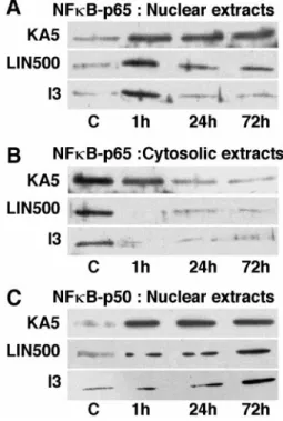
Figure 2 depicts the induction of NFB protein in the hip-pocampus after the three types of preconditioning assessed using Western blot analysis (Fig. 2A–C) and immunohistochemistry (Fig. 3). Figure 2A–C shows representative Western blotting analysis of the p65 (Fig. 2A,B) and p50 (Fig. 2C) content in cytosolic and nuclear extracts from rats subjected to 3 min isch-emia or injected with 5 mg/kg KA or 500 nmol/kg LIN taken at serial times after sublethal or drug treatments. The p65 and p50 subunits of NFB were rich in the cytosolic fractions from sham-operated or vehicle-injected rats but undetectable in the nuclear extracts. In contrast, the protein levels of p65 and p50 in the three preconditioned groups were significantly enhanced in the nuclear fraction and concurrently decreased in the cytosol as early as 1 hr, indicating a rapid translocation of these NFB subunits from the cytosol to the nucleus.
Immunostaining using a monoclonal antibody raised against the active form of NFB performed at 24 hr after each of different preconditioning triggers (short ischemia, KA5 treat-ment, or LIN500 treatment) was performed to determine the cellular identity of the increase in NFB activity associated with preconditioning visualized above by gel shift and Western blot-ting analysis (Figs. 1, 2A–C). Immunoreactivity for the activated form of NFB was not detectable in either sham-operated (Fig. 3Ba,Bb) or vehicle-injected (data not shown) hippocampus. Sub-lethal 3 min ischemia (Fig. 3Ac,Ad), KA5 treatment (Fig. 3Ca,Cb), or LIN500 treatment (Fig. 3Da,Db) induced a strong immunoreactivity for NFB in CA1 and CA3 pyramidal cell layers (Fig. 3Ac,Ad,Ca,Cb,Da,Db), hilar neurons, and dentate gyrus (data not shown) 24 hr after preconditioning stimulus. The extent and intensity of immunoreactivity appeared similar in
three treatments. According to morphological criteria, the posi-tive cells seemed to be neurons. Immunohistochemical observa-tions with GFAP and vimentin antibodies excluded astrocytes and microglia as main sources of NFB activation in the three models of preconditioning tested (data not shown).
Immunostain-Figure 2. Representative Western blotting analysis of p65 and p50
sub-units of NFB in nuclear (A, C) and cytosolic (B) extracts from hip-pocampi of control rats ( C) or rats submitted to 3 min ischemia (I3) or treated with KA5 or LIN500 and obtained at 1, 24, and 72 hr after the different preconditionings.
Figure 1. EMSA showing the time
course of increased NFB DNA-binding activity induced by the three different preconditionings (3 min isch-emia, KA5 treatment, and LIN500 treatment). A, Representative gel shifts analysis showing NFB DNA-binding activity in nuclear protein extracts from hippocampi of control rats (lane 1) or rats submitted to 3 min ischemia (lanes
5–9) or treated with KA5 (lanes 2–4 ) or
LIN500 (lanes 10–12) and obtained at 1, 24, and 72 hr after the different precon-ditionings. NFB DNA-binding activity was assayed as described in Materials and Methods. Competition assays of NFB DNA-binding activity was per-formed in the presence of 50-fold excess of unlabeled competitor NFB (lane 9) and nonspecific competitor AP2 (lane 8) consensus oligonucleotides. The shifted bands of specific NFB DNA complexes are indicated by the arrowheads. The
right and left gels correspond to 4 and
7% polyacrylamide gels, respectively. B,
Quantification of NFB DNA-binding activity in the different experimental groups. The specific shifted bands were quantified using a phosphorimaging system as described in Materials and Methods. The values are expressed as a percentage of control. No significant differences were found between vehicle-injected and sham-operated rats, and values of these groups were pooled and termed control. Data represent the mean⫾ SEM values. Data are representative of six separate experiments in each group (n⫽ 6). *p ⬍ 0.05 indicates statistical significance when compared with control. C, Supershift analysis of NFB binding proteins present in nuclear extracts 24 hr after 3 min ischemia. The binding activity assay was performed in the presence of anti-p65 antibodies, anti-p50 antibodies, a combination of both, or no antibody. The arrowheads indicate the bands of specific NFB–DNA complexes, which were supershifted by anti-p65 antibodies, anti-p50 antibodies, or by their combination.
ings obtained with the antibody raised against the neuron-specific nuclear protein NeuN (Fig. 3Aa,Ab) provided evidence that the immunoreactivity for the active form of NFB was predominant in the nucleus. Double labeling with antibodies against the neuron-specific nuclear protein NeuN and the NFB-p65 subunit indicated nuclear colocalization (Fig. 3Ea,Eb).
Effects of
B decoy DNA and DTTC on NFB
DNA-binding activity and subcellular localization of
p65 subunit
B decoy DNA and DTTC have been shown to block NFB DNA-binding activity in different systems (Schreck et al., 1992; Smith-Swintosky et al., 1994; Yu et al., 1999). In the present study, double-stranded B decoy DNA or control double-stranded DNA with a scrambled sequence was injected intracere-broventricularly (two injections at 24 and 2 hr before 3 min ischemia, KA5 treatment, or LIN500 treatment), and rats were killed 1 hr after the preconditioning stimulus. As illustrated in Figure 4, A and B, the gel-shift analysis of NFB shows that the increase in NFB DNA-binding activity induced by each precon-ditioning was suppressed in hippocampus of rats injected withB decoy DNA but was unaffected in animals injected with control scrambled DNA. Figure 4B summarizes the quantification of NFB DNA-binding activity in nuclear extracts from the com-plete set of rats. For the three preconditioning stimuli, the de-crease in NFB DNA-binding activity in the nuclear fraction varied from 4.8- to 6.0-fold compared with respective control
groups (KA5, LIN500, or I3). The remaining NFB DNA-binding activity corresponded to that of negative controls (vehicle-injected or sham-operated rats). Administration of B decoy DNA prevented the decrease in p65 content in the cytosolic extracts (data not shown) and its increase induced by sublethal ischemia (I3) or LIN500 treatment in the nuclear extracts ana-lyzed 24 hr after the preconditioning stimulus (Fig. 4C). A similar result was obtained with KA5 treatment preconditioning stimulus (data not shown).
In the same manner, administration of DTTC (150 mg/kg, i.p.) 15 min before 3 min ischemia, KA5 treatment, or LIN500 treat-ment significantly blocked the increase in NFB DNA-binding activity in the nuclear fraction of hippocampi analyzed 24 hr after sublethal ischemia or drug treatment (KA5 or LIN500) (Fig. 4A,B). Analysis of extracts from rats pretreated with DTTC and then exposed to the different preconditioning stimuli showed a greatly reduced level of p65 content in nuclear fraction compared with hippocampi submitted to 3 min ischemia or injected with LIN500 in the absence of DTTC (Fig. 4C). A similar result was obtained with KA5 treatment preconditioning stimulus (data not shown).
Effects of
B decoy DNA and DTTC treatment on the
hippocampal ischemic tolerance induced by the
different preconditionings
The effects of B decoy DNA and DTTC treatment on brain tolerance were assessed by analysis of the neuronal degeneration in CA1 substructure of rats killed 7 d after the second ischemia. Figure 5, A and C, shows representative photomicrographs of the CA1 sector in the different groups, and Figure 5, B and D, reports the quantitative analysis of neuronal cell density in the respective experimental groups. As expected, control (sham-operated or vehicle-injected) rats did not show neurodegeneration (Fig. 5Aa). In contrast, typical cell death appeared in pyramidal neurons of CA1 substructure after 6 min ischemia (Fig. 5Ab,Cb). Compared with control rats (Fig. 5Ba,Da), 21% of CA1 pyramidal cells only survived (Fig. 5Bb,Db). Preconditioning with sublethal 3 min ischemia (Fig. 5Ac) or LIN500 injection (Fig. 5Cc) 3 d before 6 min ischemia totally prevented neuronal death induced by severe ischemia. In these preconditioned groups, 99 and 95% of CA1 cells were preserved, respectively (Fig. 5Bc,Dc). However, the intracerebroventricular administration ofB decoy DNA before each preconditioning stimulus (3 min ischemia or LIN500 treat-ment) abolished the ischemic tolerance and markedly enhanced the hippocampal neurodegeneration (Fig. 5Ad,Cd) compared with rats that were injected with scrambled control DNA before preconditioning (Fig. 5Ae,Ce). The injection ofB decoy DNA alone did not induce any damage in hippocampus (Fig. 5Cg,Dg). In the same manner, the intraperitoneal injection of DTTC be-fore 3 min ischemia (Fig. 5Af) or LIN500 treatment (Fig. 5Cf) blocked the ischemic tolerance induced by each preconditioning. Quantitative analysis of neuronal damage in CA1 substructure confirmed the marked enhancement of CA1 pyramidal cell de-generation in rats injected with B decoy DNA before each preconditioning stimulus (Fig. 5Bd,Dd) compared with rats sub-mitted to 6 min ischemia (Fig. 5Bb,Db). Seventy and 75% of CA1 pyramidal neurons were destroyed in rats injected withB decoy DNA before sublethal 3 min ischemia (Fig. 5Bd) or LIN500 injection (Fig. 5Dd), respectively. Similar results were obtained with epileptic tolerance (data not shown). Together, these data show that the treatment withB decoy DNA or DTTC prevented the neuroprotective effects of late preconditionings (sublethal
Figure 3. Representative immunohistochemical staining of the active
form of NFB protein in hippocampal CA1 and CA3 substructures of rats killed 24 hr after each of preconditioning stimuli. Aa–Ad, 3 min ischemia.
Ba, Bb, Sham-surgery. Ca, Cb, KA5 treatment. Da, Db, LIN500
treat-ment. Aa, Ab, Immunohistochemical staining of NeuN protein within CA1 and CA3 pyramidal cells after sublethal ischemia. E, Colocalization of NFB-p65 and the neuron-specific nuclear protein NeuN (neuronal nuclei) within CA1 (Ea) and CA3 (Eb) pyramidal cells. Brain sections were immunostained with antibodies against the p65 subunit of NFB ( green) and NeuN (red) for double-labeling. Scale bars, 20m.
ischemia, LIN500 treatment, or KA5 treatment) against ischemia or epilepsy and that the blockade of NFB activation was dele-terious in the three models of brain tolerance used in that work.
Changes in NF
B DNA-binding activity and subcellular
localization of I
B␣ and the active form of p65 in the
different groups of preconditioned brains
Electrophoretic mobility shift assays revealed that NFB DNA-binding activity was decreased in nuclear extracts of rats precon-ditioned with 3 min ischemia, KA5 treatment, or LIN500 treat-ment compared with rats submitted to 6 min ischemia or treated with 7.5 mg/kg KA (Fig. 6A). Treatment with DOC, which dissociates the cytosolic NFB–IB complex, allowing the detec-tion of inactive NFB, restored NFB DNA-binding activity in preconditioned hippocampi (Fig. 6A). This indicated that NFB was present but not active in preconditioned rats. Figure 6B summarizes the quantification of NFB DNA-binding activity in nuclear and cytosolic extracts from the complete set of rats. For the three preconditioned groups, the NFB DNA-binding activ-ity in nuclear extracts decreased 44–65% compared with respec-tive posirespec-tive controls. The restoring of the NFB DNA-binding activity after DOC treatment in cytosolic extracts reached 90% compared with positive controls (Fig. 6B). A possible mediator of the inhibitory action of preconditioning on NFB activation is IB, encoded by the immediate early gene MAD-3 (Haskill et al., 1991). IB degradation is enhanced by various NFB inducers, which also cause new synthesis of IB␣ by an inducible autoreg-ulatory pathway (Sun et al., 1993). The effect of preconditioning on IB␣ and NFB metabolism was examined by immunoblotting with antibodies to IB␣ and active form of NFB, which recog-nizes the heterotetrameric protein, consisting of the two p50 and two p65 subunits. Analysis of nuclear and cytosolic extracts of control, ischemic, or epileptic rats revealed that the loss of IB␣
correlated with translocation of NFB from the cytoplasm to the nucleus (Fig. 6C). Severe ischemia and KA treatment (7.5 mg/kg) led to the disappearance of IB␣ and to the presence of the active form of NFB in cytosolic extracts. In contrast, the amount of IB␣ protein was notably increased in nuclear extracts of rats preconditioned with LIN500 treatment (I6 or LIN500-KA7.5) and submitted to severe 6 min ischemia or KA7.5 treat-ment 3 d later (Fig. 6C). A similar result was obtained with rats preconditioned with 3 min ischemia or KA5 treatment (data not shown). The profile of preconditioning-induced IB␣ and of the accumulation of the active form of NFB paralleled that of the inhibition of NFB DNA-binding activity. Preconditioning with 3 min ischemia, KA5 or LIN500 treatment resulted in retention of p65 in cytoplasm (data not shown).
DISCUSSION
Many different mechanisms have been proposed to be involved in the development of brain tolerance, including adenosine receptor stimulation, KATP channel opening, and delayed expression of
neuroprotective genes. However, little is known on the nature of events occurring between the early opening of KATP channels
through adenosine receptors and the induction of neuroprotec-tive genes within the time window of protection-promoting neu-ronal survival. The switching “on–off” of gene expression is the province of transcription factors, which operate singly or in asso-ciation with other proteins. The present work provides the first demonstration of the key role of the NFB transcription factor in the signal transduction cascade of brain tolerance. The NFB proteins are a family of inducible transcription factors that acti-vate a variety of cellular genes involved in control of the inflam-matory response and in regulating cellular growth (Mattson et al., 2000). The NFB signaling pathway is a major determinant in the
Figure 4. Electrophoretic mobility shift
as-say (A, B) and Western blotting analysis of p65 subunit of NFB (C) showing the effect of intracerebroventricular administration of B decoy DNA or intraperitoneal injection of DTTC on the increased NFB DNA-binding activity induced by the three differ-ent preconditionings (3 min ischemia, KA5 treatment, and LIN500 treatments). A, Representative gel shift analysis showing NFB DNA-binding activity in hippocam-pal nuclear extracts isolated from rats that had received intracerebroventricular injec-tions of vehicle, 60g of B decoy DNA (Bdecoy), 60 g of scrambled control DNA (ScDNA), or intraperitoneal injection of DTTC (150 mg/kg) before sublethal isch-emia (I3) or administration of either KA5 or LIN500. Rats were killed 1 hr (forB decoy or scrambled control DNA) or 24 hr (for DTTC) after each type of precondi-tioning. NFB activity was assayed as de-scribed in Materials and Methods. The shifted band of specific NFB DNA com-plexes is indicated by the arrowhead. B, Quantification of NFB DNA-binding
ac-tivity in the different experimental groups. The specific shifted bands were quantified using a phosphorimaging system as described in Materials and Methods. Values are expressed as a percentage of control. Results are expressed as mean⫾ SEM. Data are representative of six separate experiments in each group (n⫽ 6). *p ⬍ 0.05 indicates statistical significance when compared with vehicle-injected rats. C, Representative Western blotting analysis of p65 subunit of NFB in hippocampal nuclear extracts isolated from rats that had received intracerebroventricular injections of vehicle, 60 g ofB decoy DNA (Bdecoy), 60 g of scrambled control DNA (ScDNA) or intraperitoneal DTTC injection (150 mg/kg) before I3 or LIN500. Rats were killed 24 hr after the conditioning stimulus. The position of p65 is indicated by the arrowhead. 1, Vehicle; 2, DTTC–LIN500; 3, LIN500; 4, Sham-operated; 5, I3; 6, DTTC–I3; 7, scrambled control DNA–LIN500; 8,B decoy DNA–LIN500; 9, B decoy DNA–I3; 10, scrambled control DNA–I3.
control of neuronal death and/or cell survival. Although NFB activation occurs in neurons after brain injury, its role in the injury outcome remains unclear. The assumption that NFB is contributing to neuronal cell death is documented by many pa-pers reporting increased NFB activity under pathological con-ditions in which neurons were dying (Grilli et al., 1996; Clemens et al., 1997). However, subsequent in vitro studies provide
evi-dence that the activation of NFB can protect neurons against amyloid-peptide toxicity (Barger et al., 1995) and excitotoxic or oxidative stress (Goodman and Mattson, 1996; Mattson et al., 1997). Previous studies, in which injury-induced NFB activity was suppressed by gene deletion of the p50 subunit (Yu et al., 1999) or administration ofB decoy DNA (Mattson et al., 1997; Yu et al., 1999) or pharmacological agents (Taglialatela et al., 1997; Maggirwar et al., 1998), strongly support a neuroprotective role for NFB activation. NFB consists of a p50 and p65/RelA complex that is trapped in the cytoplasm by an inhibitory protein IB␣ (Verma et al., 1995; Baeuerle and Baltimore, 1996). As long as IB␣ is bound to the p50/p65 complex, its translocation to the nucleus is prevented. Activation of NFB consists of phosphor-ylation or degradation of IB␣ from the complex in the cyto-plasm, and during activation NFB is translocated to the nucleus of the cell (Chen et al., 1995; Verma et al., 1995; Baeuerle and Baltimore, 1996).
Results reported in this paper demonstrate that, in the three models of brain tolerance studied (ischemic, epileptic, and poly-unsaturated fatty acid-induced preconditionings), the activation of NFB was required for the development of late cerebral preconditioning against severe ischemia or epilepsy. The three different inducers of preconditioning (sublethal 3 min ischemia, KA5 treatment, or LIN500 treatment) induced rapid activation of NFB, as evidenced by its increased DNA-binding activity and nuclear translocation. The gel-shift analyses revealed that DNA-binding activity increased as early as 1 hr after sublethal ischemia, KA5 treatment, or LIN500 treatment, and the shifted band con-sisted of p50/p65 heterodimers. The presence of active NFB was confirmed by nuclear localization of p50 and p65 subunits in Western blots of hippocampal extracts as early as 1 hr after each preconditioning stimulus, indicating a rapid translocation of NFB subunits from cytosol to the nucleus. Immunohistochem-ical analyses using p65 antibodies revealed that, 24 hr after each of preconditioning stimuli, NFB activation occurred in the py-ramidal neurons and not in the glial cells. Pretreatment with the NFB inhibitor DTTC or B decoy DNA blocked the increased DNA-binding activity and the nuclear translocation of NFB and, at the same time, abolished the neuroprotective effects of different delayed preconditionings against severe ischemia or epilepsy. Previous studies have shown that B decoy DNA can block activation of B-responsive genes, greatly increase the vulnerability of neurons to different insults, and promote neuro-nal cell death (Mattson et al., 1997; Yu et al., 1999). Dithiocar-bamates, such as DTTC, have also been reported to be potent inhibitors of NFB activation in various cell types (Schreck et al., 1992; Sherman et al., 1993; Ziegler-Heitbrock et al., 1993). To-gether, these results indicate that activation of NFB is an essen-tial mechanism whereby sublethal ischemia, KA5 treatment, or LIN500 treatment results in delayed neuroprotection.
The present work also provides evidence that the inhibition of NFB observed in rats preconditioned with 3 min ischemia, KA5 treatment, or LIN500 treatment compared with ischemic or epi-leptic controls was correlated with the prevention of the inducible degradation of IB␣. Treatment of preconditioned extracts with the detergent DOC, which revealed as much DNA-binding activ-ity in the different preconditioned hippocampi as in the controls, indicated that NFB was present but not active in the precondi-tioned extracts. The preconditioning also prevented the release of NFB from IB␣ after a severe ischemic or epileptic insult. The phosphorylation and degradation of IB␣ is necessary for the activation of NFB and its subsequent appearance in the nucleus
Figure 5. Effect ofB decoy DNA, scrambled control DNA, and DTTC injections on the ischemic tolerance induced by brief ischemia or LIN500 treatment. A, C, Representative photomicrographs highlighting morpho-logical changes in CA1 subfield of cresyl violet-stained hippocampal sections 7 d after severe 6 min ischemia in the different experimental groups. I6 corresponds to rats submitted to 6 min ischemia. I3-I6 corre-sponds to rats submitted to 3 min ischemia 3 d before 6 min ischemia. decoy I3-I6 or ScDNA I3-I6 corresponds to rats that, respectively, had received intracerebroventricular injections of 60g of B decoy DNA or scrambled control DNA at 24 and 2 hr before 3 min ischemia. DTTC I3-I6 corresponds to rats that had received an intraperitoneal injection of DTTC (150 mg/kg) 30 min before 3 min ischemia. Rats were killed 7 d after 6 min ischemia. Scale bar, 100m. B, D, Quantification of neuronal density in the hippocampal CA1 pyramidal layer of different experimental groups. Results are expressed as mean⫾ SEM (n ⫽ 6) and represent neuronal densities assessed in cresyl violet-stained sections per 1 mm linear length of CA1 pyramidal layer. A mean value for each CA1 substructure was obtained from 10 bilateral measurements on two sections per slide and 10 slides per animal (n⫽ 6) in each of the experimental groups. Differences were considered statistically significant when p⬍ 0.05 (Tukey’s test). * indicates significantly different from control (sham-operated animals). # indicates significantly different from ischemic ani-mals (6 min).
(Chen et al., 1995; Verma et al., 1995; Baeuerle and Baltimore, 1996). Therefore, on activation of NFB, IB␣ concentrations decrease. Immunoblotting with an antibody raised against IB␣ reveals that the amount of IB␣ protein increased in the preconditioned hippocampi compared with ischemic or epilep-tic cytosolic extracts. An interesting observation is the pres-ence of IB␣ protein and the active form of NFB in the nuclear hippocampi of rats preconditioned with 3 min isch-emia, KA5 treatment, or LIN500 treatment compared with ischemic or epileptic controls, in which IB␣ protein was absent. The increase in IB␣ abundance in preconditioned rats is probably the result of increased IB␣ synthesis. This result strongly suggests that the inhibition of NFB activation in-duced by preconditioning is mediated by induction of the IB␣ inhibitory protein, which traps activated NFB in inactive cytoplasmic complexes. In accordance with previous work, demonstrating the direct transcriptional activation of IB␣ by NFB itself (Scott et al., 1993; Sun et al., 1993; Chiao et al., 1994), IB␣ probably newly synthetized after the activation of NFB by preconditioning stimulus will bind free cytoplasmic NFB and inactivate its potential nuclear translocation. Fur-thermore, excess IB␣ translocates to the nucleus by an un-known mechanism, in which, as shown in vitro, it can sequester free NFB (p65), promote net dissociation of DNA-bound NFB, and thereby terminate its activity (Arenzana-Seisdedos et al., 1995). This nuclear NFB–IB␣ complex may be trans-ported back to the cytoplasm or degraded in the nucleus. Recently, it has been shown that immunosuppression by glu-cocorticoids may be the direct outcome of the inhibition of NFB activity through induction of IB␣ synthesis (Auphan et al., 1995; Scheinman et al., 1995). It is not excluded that such a mechanism is also involved in the anti-inflammatory
re-sponse and neuroprotection induced by sodium salicylate and aspirin (Kopp and Ghosh, 1994; Grilli et al., 1996).
In conclusion, these findings demonstrate that the transcription factor NFB is indeed at the crossing of neuronal cell death and survival pathways and is a crucial component of the signal trans-duction cascade of cerebral preconditioning. This paper shows that (1) the three preconditioning protocols induce a rapid neu-roprotective activation of NFB in hippocampus, (2) the in-creases in NFB DNA-activity determined by gel shift and West-ern blot analysis specifically reflect increases in hippocampal neurons, (3) preconditioning the brain with sublethal ischemia, KA5 treatment, or LIN500 treatment inhibits the activation of NFB after the second injury and renders the brain more resis-tant to a subsequent potentially lethal ischemic or epileptic insult, and (4) the inhibition of NFB is mediated by induction of the IB␣ inhibitory protein, which traps activated NFB in inactive cytoplasmic complexes. The cellular mechanisms whereby the three preconditioning treatments activate NFB remains to be identified. Nitric oxide and free hydroxyl radicals are potential candidates for the role of NFB activators. Not only they are known to activate NFB but they are also known to be involved in brain and heart tolerance (Bolli et al., 1997; Centeno et al., 1999; Xuan et al., 1999; Rauca et al., 2000). The neuroprotection induced by activation of NFB in hippocampal neurons related to induction of MnSOD (Mattson et al., 1997) and the increase of MnSOD activity in brain tolerance (Toyoda et al., 1997) strongly support the link between NFB and oxidative stress in cerebral preconditioning. Together, these results strongly suggest that, whatever the type of preconditioning, the initial signals elicited by the conditioning stimulus are transduced into protective changes via an NFB-dependent mechanism. The crucial involvement of
Figure 6. Changes in NFB DNA-binding activity and subcellular localization of IB␣ and active form of NFB in the different groups of preconditioned brains. A, Repre-sentative gel shifts analysis showing NFB DNA-binding activity in hippocampal nu-clear and cytosolic extracts isolated from rats preconditioned with 3 min ischemia (I3-I6 ), KA5 treatment (K A5-K A7.5), or LIN500 treatment (I6 or
LIN500-K A7.5) and submitted to 6 min ischemia or
KA7.5 treatment 3 d later. Rats were killed 24 hr after the last treatment. Cytosolic extracts were analyzed by EMSA with DOC treatment after binding as described in Ma-terials and Methods. The shifted band of specific NFB–DNA complexes is indi-cated by the arrowhead. B, Quantification of NFB DNA-binding activity in the differ-ent experimdiffer-ental groups. The specific shifted bands were quantified using a phos-phorimaging system as described in Mate-rials and Methods. The values are expressed as a percentage of control. No significant differences were found between saline-injected and sham-operated rats, and values of these groups were pooled and termed control. Results are expressed as mean⫾ SEM. Data are representative of six sepa-rate experiments in each group (n ⫽ 6).
Differences were considered statistically significant when p⬍ 0.05 (Tukey’s test). * indicates significantly different from control (saline-injected or sham-operated animals). # indicates significantly different from KA7.5-injected animals. $ indicates significantly different from ischemic animals (6 min).
C, Representative Western blotting analysis of IB␣ and active form of NFB in hippocampal nuclear and cytosolic extracts isolated from ischemic (I6) and epileptic (K A7.5) rats and rats preconditioned with LIN500 treatment (LIN500-I6 or LIN500-K A7.5) and submitted to severe 6 min ischemia or KA7.5 treatment 3 d later. Rats were killed 24 hr after the last treatment. The positions of IB␣ and active form of NFB are indicated by the arrowheads.
this transcription factor in brain tolerance may open new ways in the search of therapeutic strategies.
REFERENCES
Arenzana-Seisdedos F, Thompson J, Rodriguez MS, Bachelerie F, Thomas D, Hay RT (1995) Inducible nuclear expression of newly synthesized I kappa B alpha negatively regulates DNA-binding and transcriptional activities of NF-kappa B. Mol Cell Biol 15:2689–2696. Auphan N, DiDonato JA, Rosette C, Helmberg A, Karin M (1995) Immunosuppression by glucocorticoids: inhibition of NF-kappa B ac-tivity through induction of I kappa B synthesis. Science 270:286–290. Baeuerle PA, Baltimore D (1996) NF-kappa B: ten years after. Cell
87:13–20.
Barger SW, Horster D, Furukawa K, Goodman Y, Krieglstein J, Mattson MP (1995) Tumor necrosis factors alpha and beta protect neurons against amyloid beta-peptide toxicity: evidence for involvement of a kappa B-binding factor and attenuation of peroxide and Ca2⫹
accu-mulation. Proc Natl Acad Sci USA 92:9328–9332.
Blondeau N, Plamondon H, Richelme C, Heurteaux C, Lazdunski M (2000) KATPchannel openers, adenosine agonists and epileptic
precon-ditioning are stress signals inducing hippocampal neuroprotection. Neuroscience 100:465–474.
Bolli R, Manchikalapudi S, Tang XL, Takano H, Qiu Y, Guo Y, Zhang Q, Jadoon AK (1997) The protective effect of late preconditioning against myocardial stunning in conscious rabbits is mediated by nitric oxide synthase. Evidence that nitric oxide acts both as a trigger and as a mediator of the late phase of ischemic preconditioning. Circ Res 81:1094–1107.
Bradford MM (1976) A rapid and sensitive method for the quantitation of microgram quantities of protein utilizing the principle of protein-dye binding. Anal Biochem 72:248–254.
Centeno JM, Orti M, Salom JB, Sick TJ, Perez-Pinzon MA (1999) Nitric oxide is involved in anoxic preconditioning neuroprotection in rat hippocampal slices. Brain Res 836:62–69.
Chen Z, Hagler J, Palombella VJ, Melandri F, Scherer D, Ballard D, Maniatis T (1995) Signal-induced site-specific phosphorylation targets I kappa B alpha to the ubiquitin-proteasome pathway. Genes Dev 9:1586–1597.
Chiao PJ, Miyamoto S, Verma IM (1994) Autoregulation of I kappa B alpha activity. Proc Natl Acad Sci USA 91:28–32.
Chopp M, Chen H, Ho KL, Dereski MO, Brown E, Hetzel FW, Welch KMA (1989) Transient hyperthermia protects against subsequent forebrain ischemic cell damage in the rat. Neurology 39:1396–1398. Clemens JA, Stephenson DT, Smalstig EB, Dixon EP, Little SP (1997)
Global ischemia activates nuclear factor-kappa B in forebrain neurons of rats. Stroke 28:1073–1081.
Dignam JD, Lebovitz RM, Roeder RG (1983) Accurate transcription initiation by RNA polymerase II in a soluble extract from isolated mammalian nuclei. Nucleic Acids Res 11:1475–1489.
Gabriel C, Justicia C, Camins A, Planas AM (1999) Activation of nu-clear factor-kappa B in the rat brain after transient focal ischemia. Brain Res Mol Brain Res 65:61–69.
Gidday JM, Fitzgibbons JC, Shah AR, Park TS (1994) Neuroprotection from ischemic brain injury by hypoxic preconditioning in the neonatal rat. Neurosci Lett 168:221–224.
Glazier SS, O’Rourke DM, Graham DI, Welsh FA (1994) Induction of ischemic tolerance following brief focal ischemia in rat brain. J Cereb Blood Flow Metab 14:545–553.
Goodman Y, Mattson MP (1996) Ceramide protects hippocampal neu-rons against excitotoxic and oxidative insults, and amyloid beta-peptide toxicity. J Neurochem 66:869–872.
Grilli M, Pizzi M, Memo M, Spano P (1996) Neuroprotection by aspirin and sodium salicylate through blockade of NF-kappa B activation. Science 274:1383–1385.
Guerrini L, Blasi F, Denis-Donini S (1995) Synaptic activation of NF-kappa B by glutamate in cerebellar granule neurons in vitro. Proc Natl Acad Sci USA 92:9077–9081.
Haskill, S, Beg AA, Tompkins SM, Morris JS, Yurochko AD, Sampson-Johannes A, Mondal K, Ralph P, Baldwin AS Jr (1991) Characteriza-tion of an immediate-early gene induced in adherent monocytes that encodes I-like activity. Cell 65:1281–1289.
Heurteaux C, Lauritzen I, Widmann C, Lazdunski M (1995) Essential role of adenosine, adenosine A1receptors and KATPchannels in
cere-bral ischemic preconditioning. Proc Natl Acad Sci USA 92:4666–4670. Hunot S, Brugg B, Ricard D, Michel PP, Muriel MP, Ruberg M, Fau-cheux BA, Agid Y, Hirsch EC (1997) Nuclear translocation of NF-kappa B is increased in dopaminergic neurons of patients with Parkin-son disease. Proc Natl Acad Sci USA 94:7531–7536.
Kaltschmidt B, Uherek M, Volk B, Baeuerle PA, Kaltschmidt C (1997) Transcription factor NF-kappa B is activated in primary neurons by amyloid beta peptides and in neurons surrounding early plaques from patients with Alzheimer disease. Proc Natl Acad Sci USA 94:2642–2647.
Kaltschmidt C, Kaltschmidt B, Baeuerle PA (1995) Stimulation of iono-tropic glutamate receptors activates transcription factor NF-kappa B in primary neurons. Proc Natl Acad Sci USA 92:9618–9622.
Kawahara N, Reutzler CA, Klatzo I (1994) A delayed effect of spread-ing depression on brain tissue associated with protection against isch-emic neuronal damage. Brain Pathol [Suppl] 4:5098.
Kelly ME, McIntyre DC (1994) Hippocampal kindling protects several structures from the neuronal damage from kainic acid-induced status epilepticus. Brain Res 634:245–256.
Kirino T (1982) Delayed neuronal death in the gerbil hippocampus following ischemia. Brain Res 239:57–69.
Kirino T, Tsujita Y, Tamura A (1991) Induced tolerance to ischemia in gerbil hippocampal neurons. J Cereb Blood Flow Metab 11:299–307. Kitagawa K, Matsumoto M, Tagaya M, Hata R, Ueda H, Niinobe M,
Handa N, Fukunaga R, Kimura K, Mikoshiba K, Kamada T (1990) “Ischemic tolerance” phenomenon found in the brain. Brain Res 528:21–24.
Kitagawa K, Matsumoto M, Kuwabara K, Tagaya M, Ohtsuki T, Hata R, Ueda H, Handa N, Kimura K, Kamada T (1991a) “Ischemic toler-ance” phenomenon detected in various brain regions. Brain Res 561:203–211.
Kitagawa K, Matsumoto M, Tagaya M, Kuwabara K, Hata R, Handa N, Fukunaga R, Kimura K, Kamada T (1991b) Hyperthermia-induced neuronal protection against ischemic injury in gerbils. J Cereb Blood Flow Metab 11:449–452.
Kopp E, Ghosh S (1994) Inhibition of NF-kappa B by sodium salicylate and aspirin. Science 265:956–959.
Liu Y, Kato H, Nakata N, Kogure K (1992) Protection of rat hippocam-pus against ischemic neuronal damage by pretreatment with sublethal ischemia. Brain Res 586:121–124.
Liu Y, Kato H, Nakata N, Kogure K (1993) Temporal profile of heat shock protein 70 synthesis in ischemic tolerance induced by precondi-tioning ischemia in rat hippocampus. Neuroscience 56:921–927. Lothman EW, Collins RC (1981) Kainic acid induced limbic seizures:
metabolic, behavioral, electroencephalographic and neuropathological correlates. Brain Res 218:299–318.
Lukiw WJ, Bazan NG (1998) Strong nuclear factor-kappa B-DNA bind-ing parallels cyclooxygenase-2 gene transcription in agbind-ing and in spo-radic Alzheimer’s disease superior temporal lobe neocortex. J Neurosci Res 53:583–592.
Maggirwar SB, Sarmiere PD, Dewhurst S, Freeman RS (1998) Nerve growth factor-dependent activation of NF-kappa B contributes to sur-vival of sympathetic neurons. J Neurosci 18:10356–10365.
Matsushima K, Hakim AM (1995) Transient forebrain ischemia protects against subsequent focal cerebral ischemia without changing cerebral perfusion. Stroke 26:1047–1052.
Matsushima K, Hogan MJ, Hakim AM (1996) Cortical spreading de-pression protects against subsequent focal cerebral ischemia in rats. J Cereb Blood Flow Metab 16:221–226.
Mattson MP, Goodman Y, Luo H, Fu W, Furukawa K (1997) Activation of NF-kappa B protects hippocampal neurons against oxidative stress-induced apoptosis: evidence for induction of manganese superoxide dismutase and suppression of peroxynitrite production and protein tyrosine nitration. J Neurosci Res 49:681–697.
Mattson MP, Culmsee C, Yu Z, Camandola S (2000) Roles of nuclear factor kappa B in neuronal survival and plasticity. J Neurochem 74:443–456.
Millar CGM, Baxter GF, Thiemermann C (1996) Protection of the myo-cardium by ischaemic preconditioning: mechanisms and therapeutic implications. Pharmacol Ther 69:143–151.
Nishi S, Taki W, Uemura Y, Higashi T, Kikuchi H, Kudoh H, Satoh M, Nagata K (1993) Ischemic tolerance due to the induction of HSP70 in a rat ischemic recirculation model. Brain Res 615:281–288.
Ohtsuki T, Matsumoto M, Kuwabara K, Kitagawa K, Suzuki K, Tanigu-chi N, Kamada T (1992) Influence of oxidative stress on induced tolerance to ischemia in gerbil hippocampal neurons. Brain Res 599:246–252.
Paxinos G, Watson C (1986) The rat brain in stereotaxic coordinates. San Diego: Academic.
Perez-Pinzon MA, Mumford PL, Rosenthal M, Sick TJ (1996) Anoxic preconditioning in hippocampal slices: role of adenosine. Neuroscience 75:687–694.
Perez-Pinzon MA, Xu GP, Dietrich WD, Rosenthal M, Sick TJ (1997) Rapid preconditioning protects rats against ischemic neuronal damage after 3 but not 7 days of reperfusion following global cerebral ischemia. J Cereb Blood Flow Metab 17:175–182.
Plamondon H, Blondeau N, Heurteaux C, Lazdunski M (1999) Mutually protective actions of kainic acid epileptic preconditioning and sublethal global ischemia on hippocampal neuronal death: involvement of aden-osine A1receptors and KATPchannels. J Cereb Blood Flow Metab
19:1296–1308.
Pulsinelli WA, Brierley JB (1979) A new model of bilateral hemispheric ischemia in the unanesthetized rat. Stroke 10:267–272.
Rauca C, Zerbe R, Jantze H, Krug M (2000) The importance of free hydroxyl radicals to hypoxia preconditioning. Brain Res 868:147–149.
Reshef A, Sperling O, Zoref-Shani E (1998a) Adenosine-induced pre-conditioning of rat neuronal cultures against ischemia-reperfusion in-jury. Adv Exp Med Biol 431:365–368.
Reshef A, Sperling O, Zoref-Shani E (1998b) Opening of ATP-sensitive potassium channels by cromakalim confers tolerance against chemical ischemia in rat neuronal cultures. Neurosci Lett 250:111–114. Riepe MW, Esclaire F, Kasischke K, Schreiber S, Nakase H, Kempski O,
Ludolph AC, Dirnagl U, Hugon J (1997) Increased hypoxic tolerance by chemical inhibition of oxidative phosphorylation: “chemical precon-ditioning.” J Cereb Blood Flow Metab 17:257–264.
Rong Y, Baudry M (1996) Seizure activity results in a rapid induction of nuclear factor-kappa B in adult but not juvenile rat limbic structures. J Neurochem 67:662–668.
Sasahira M, Lowry T, Simon RP, Greenberg DA (1995) Epileptic toler-ance: previous seizures protect against seizure-induced neuronal in-jury. Neurosci Lett 185:95–98.
Scheinman RI, Cogswell PC, Lofquist AK, Baldwin Jr AS (1995) Role of transcriptional activation of I kappa B alpha in mediation of immu-nosuppression by glucocorticoids. Science 270:283–286.
Schreck R, Meier B, Mannel DN, Droge W, Baeuerle PA (1992) Dithio-carbamates as potent inhibitors of nuclear factor kappa B activation in intact cells. J Exp Med 175:1181–1194.
Scott ML, Fujita T, Liou HC, Nolan GP, Baltimore D (1993) The p65 subunit of NF-kappa B regulates I kappa B by two distinct mechanisms. Genes Dev 7:1266–1276.
Sherman MP, Aeberhard EE, Wong VZ, Griscavage JM, Ignarro LJ (1993) Pyrrolidine dithiocarbamate inhibits induction of nitric oxide synthase activity in rat alveolar macrophages. Biochem Biophys Res Commun 191:1301–1308.
Shimazaki K, Ishida A, Kawai N (1994) Increase in bcl-2 oncoprotein
and the tolerance to ischemia-induced neuronal death in the gerbil hippocampus. Neurosci Res 20:95–99.
Simon RP, Niiro M, Gwinn R (1993) Prior ischemic stress protects against experimental stroke. Neurosci Lett 163:135–137.
Smith-Swintosky VL, Pettigrew LC, Craddock SD, Culwell AR, Rydel RE, Mattson MP (1994) Secreted forms of beta-amyloid precursor protein protect against ischemic brain injury. J Neurochem 63:781–784. Sun SC, Ganchi PA, Ballard DW, Greene WC (1993) NF-kappa B controls expression of inhibitor I kappa B alpha: evidence for an inducible autoregulatory pathway. Science 259:1912–1915.
Taglialatela G, Robinson R, Perez-Polo JR (1997) Inhibition of nuclear factor kappa B (NFkappa B) activity induces nerve growth factor-resistant apoptosis in PC12 cells. J Neurosci Res 47:155–162. Toyoda T, Kassell NF, Lee KS (1997) Induction of ischemic tolerance
and antioxidant activity by brief focal ischemia. NeuroReport 8:847–851.
Verma IM, Stevenson JK, Schwarz EM, Van Antwerp D, Miyamoto S (1995) Rel/NF-kappa B/I kappa B family: intimate tales of association and dissociation. Genes Dev 9:2723–2735.
Xuan YT, Tang XL, Banerjee S, Takano H, Li RC, Han H, Qiu Y, Li JJ, Bolli R (1999) Nuclear factor-kappa B plays an essential role in the late phase of ischemic preconditioning in conscious rabbits. Circ Res 84:1095–1109.
Yu Z, Zhou D, Bruce-Keller AJ, Kindy MS, Mattson MP (1999) Lack of the p50-subunit of nuclear factor-kappa B increases the vulnerability of hippocampal neurons to excitotoxic injury. J Neurosci 19:8856–8865. Ziegler-Heitbrock HW, Sternsdorf T, Liese J, Belohradsky B, Weber C,
Wedel A, Schreck R, Bauerle P, Strobel M (1993) Pyrrolidine dithio-carbamate inhibits NF-kappa B mobilization and TNF production in human monocytes. J Immunol 151:6986–6993.