HAL Id: hal-00982135
https://hal.archives-ouvertes.fr/hal-00982135
Submitted on 26 Nov 2018
HAL is a multi-disciplinary open access
archive for the deposit and dissemination of
sci-entific research documents, whether they are
pub-lished or not. The documents may come from
teaching and research institutions in France or
abroad, or from public or private research centers.
L’archive ouverte pluridisciplinaire HAL, est
destinée au dépôt et à la diffusion de documents
scientifiques de niveau recherche, publiés ou non,
émanant des établissements d’enseignement et de
recherche français ou étrangers, des laboratoires
publics ou privés.
Distributed under a Creative Commons Attribution| 4.0 International License
Viruses in a 14th-century coprolite
Sandra Appelt, Laura Fancello, Matthieu Le Bailly, Didier Raoult, Michel
Drancourt, Christelle Desnues
To cite this version:
Sandra Appelt, Laura Fancello, Matthieu Le Bailly, Didier Raoult, Michel Drancourt, et al.. Viruses
in a 14th-century coprolite. Applied and Environmental Microbiology, American Society for
Microbi-ology, 2014, 80 (9), pp.2648-55. �10.1128/AEM.03242-13�. �hal-00982135�
Viruses in a 14th-Century Coprolite
Sandra Appelt,aLaura Fancello,aMatthieu Le Bailly,bDidier Raoult,aMichel Drancourt,aChristelle Desnuesa
Aix Marseille University, URMITE, UM63, CNRS 7278, IRD 198, INSERM 1095, Marseille, Francea
; Franche-Comté University, CNRS UMR 6249 Chrono-Environment, Besançon, Franceb
Coprolites are fossilized fecal material that can reveal information about ancient intestinal and environmental microbiota. Viral metagenomics has allowed systematic characterization of viral diversity in environmental and human-associated specimens, but little is known about the viral diversity in fossil remains. Here, we analyzed the viral community of a 14th-century coprolite from a closed barrel in a Middle Ages site in Belgium using electron microscopy and metagenomics. Viruses that infect eukaryotes, bacteria, and archaea were detected, and we confirmed the presence of some of them by ad hoc suicide PCR. The coprolite DNA viral metagenome was dominated by sequences showing homologies to phages commonly found in modern stools and soil. Al-though their phylogenetic compositions differed, the metabolic functions of the viral communities have remained conserved across centuries. Antibiotic resistance was one of the reconstructed metabolic functions detected.
V
iral metagenomics is a sequencing-based analysis of all of viral genomes isolated from a sample. It has promoted the charac-terization of viral community diversity. Viral metagenomics has already been successfully applied to the exploration of modern environmental specimens sampled from marine water, freshwa-ter, stromatolites and thrombolites, and soil (1–4) and to modern human-associated specimens collected from the liver, blood, na-sopharyngeal aspirates, and stool (5–9). The DNA viromes gener-ated from modern stools have been demonstrgener-ated to be domi-nated by bacteriophages (10, 11) and to be less diverse than environmental samples (8,12).Viral metagenomics does not require culturing viruses or a
priori knowledge of the sequences that will be targeted, which
allows for the identification of new, unknown or unexpected viruses and for the global assessment of the virome. Viral met-agenomics is thus particularly suitable for paleomicrobiologi-cal studies, as little is known about which viruses are charac-teristic of ancient specimens. Indeed, the majority of ancient DNA (aDNA) studies are based on the analysis of human and bacterial aDNA (13–15), and viral persistence and its detect-ability in ancient specimens remain unclear. Electron micros-copy has previously revealed that viral particles can persist for over 400 years, but their viability was lost (16). Moreover, PCR amplifications yielded positive results for viral aDNA in an-cient specimens, such as mummified soft tissues, bones, and teeth. The amplification products varied between 100 and 570 bp in size, which indicated that viral aDNA can be detected for at least 1,500 years (17–20).
Here, we used electron microscopy and, for what may be the first time, viral metagenomics to characterize the viral community of an ancient stool specimen. A viral DNA metagenome was gen-erated from a 14th-century coprolite sample that was recovered from a Middle Ages site in Namur, Belgium.
MATERIALS AND METHODS
VLP isolation, TEM, and DNA extraction. First, 5.8 g of the interior of
the coprolite was aseptically removed and solubilized overnight at 4°C under continuous rotation in 40 ml of phosphate-buffered saline (PBS), pH 7.4 (bioMérieux, Marcy l’Etoile, France), which had previ-ously been passed through a 0.02-m-pore filter. The coprolite solu-tion was centrifuged for 10 min at 500⫻ g, and then the upper layer was removed and filtered in stages using sterile Whatman filters (pore
sizes of 0.8, 0.45, and 0.22m [Whatman division of GE Healthcare, Dassel, Germany]). Twenty-five milliliters of the coprolite filtrate was used to precipitate and purify viral particles onto a cesium chloride density gradient using ultracentrifugation, and DNase treatment was then performed (21). A 40-l aliquot of the purified viral particles was
stained with 1.5% ammonium molybdate (Euromedex) and observed by transmission electron microscopy (TEM) using a Philips Morgagni 268D electron microscope (FEI Co., Eindhoven, Netherlands). To iso-late the nucleic acids from the purified viral particles, the formamide procedure previously described by Thurber et al. (21) was used. A standard 18S ribosomal DNA (rDNA) PCR was performed to verify the absence of human DNA contamination.
Viral metagenomic library preparation and sequencing. Nucleic
ac-ids were amplified in duplicate reactions using the Illustra GenomiPhi V2 DNA amplification kit (GE Healthcare Life Sciences, Freiburg, Germany). Amplification products were pooled and ethanol purified.
A shotgun strategy was chosen for high-throughput pyrosequencing on a 454 Life Sciences Genome FLX sequencer using titanium chemistry (Genome Sequencer RLX; Roche). Sequencing was performed using 1/16 of a picotiter plate.
Preprocessing of sequencing data. The reads were screened for
qual-ity using mothur (22). Only reads longer than 50 bp and with an average quality score greater than 21 were kept. Reads with more than two ambig-uous base calls and/or reads with homopolymers longer than 10 bases were eliminated. Identical sequences artificially generated by the pyrose-quencing technology were also excluded using the “unique.seqs” mothur command. The preprocessed viral metagenome is publicly available on the Metavir server (http://metavir-meb.univ-bpclermont.fr) with the identifier “NAMUR_viral” under the project “HumanCoprolite” and on the NCBI Sequence Read Archive (http://www.ncbi.nlm.nih.gov/sra) un-der accession no. SRP033437.
Address correspondence to Christelle Desnues, [email protected]. Supplemental material for this article may be found at http://
Annotation of reads. A BLASTN search against the nonredundant
NCBI database (E value,⬍1e⫺5) was performed. Reads with no signifi-cant similarity to sequences stored in the NCBI database were classified as “unknown reads.” The virome taxonomic composition was estimated us-ing GAAS (23), which is based on a BLASTX search against the RefSeq Viral Genomes database (E value,⬍1e⫺5) and normalizes the number of reads matching each viral genotype by the length of the genome.
Functional annotation was performed on the MG-RAST server (24) using the nonredundant SEED database (E value,⬍1e⫺5). A stringent search of virulence factors was also performed using BLASTX on the Vir-ulence Factor Database (25), with 60% as the minimum identity and a cutoff E value of⬍1e⫺5.
Assembly and contig annotation. The reads were assembled into
con-tigs using the Newbler de novo assembler (Roche) with at least 98% iden-tity and 35 bp of overlap. Only contigs longer than 400 bp were used in subsequent analyses.
Known and unknown contigs were identified on the basis of the BLASTN search against the nonredundant NCBI database (E value, ⬍1e⫺5). The taxonomic and functional contig classification was based on a BLASTX search against the nonredundant NCBI database (E value, ⬍1⫺5). A specific search for contigs encoding antibiotic resistance genes was also performed using BLAST on the ARDB (Antibiotic Resistance Genes Database) with an E value of⬍1e⫺5 (26). Significant hits were manually verified.
target ORFs identified in some viral contigs assembled de novo from the virome and matching cyanophages, Mycobacterium phages, Bacillus phages, Burkholderia phages, Celeribacter phages, and Clostridium phages (see Table S1 and Section 4 in the supplemental material).
RESULTS
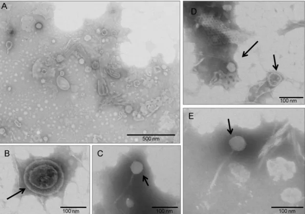
The specimen was excavated in 1996 and collected from the interior of a closed barrel, which was commonly used during this period as a pit or latrine (36). The barrel was buried at a depth of 3.80 m. The 121.4-g coprolite specimen was dark brown and well preserved under anaerobic taphonomic condi-tions. Extensive precautions were undertaken to avoid contam-inating the coprolite specimen in our laboratory environment: no positive control was used (15), and suicide PCR protocols were applied (35). All negative controls, used in a 1:4 control/ specimen ratio, were consistent with current recommenda-tions for paleomicrobiological and paleoparasitological studies (13, 15, 37, 38) and remained negative. Virus-like particles (VLPs) purified from the internal region of the coprolite, after the external layer was removed, were morphologically diverse and varied in size and shape. Oval particles of different lengths (up to 200 nm) and diameters (up to 100 nm), as well as rod-shaped structures (up to 250 nm in length), were observed (Fig. 1A). We identified a VLP with a dense core and a diameter of approximately 150 nm, apparently surrounded by an envelope-like structure (Fig. 1B). Viral particles exhibiting characteris-tics typical of the Siphoviridae bacteriophage family (icosahedral head, long tail) were also observed (Fig. 1CtoE).
High-throughput sequencing generated 30,654 reads corre-sponding to approximately 10.8 million bp. After quality trim-ming and duplicate removal, 29,811 reads remained (see Table S2 in the supplemental material). The preprocessed read lengths ranged between 77 bp and 574 bp and had an average GC content of 47% (see Fig. S2 in the supplemental material). Finally, 41.93% of the reads were assembled into 1,464 contigs that ranged from 421 to 12,500 bp (see Table S2). In total, 22.15% of all reads and 17.28% of all contigs were significantly similar to known se-quences from public databases (Fig. 2A; also see Table S2). Ge-nome-length normalized counts of viral reads showed that about 85.21% and 0.81% of viral similarities were to double-stranded DNA (dsDNA) viruses and single-stranded DNA (ssDNA) vi-ruses, respectively (Fig. 2B). Among the double-stranded DNA viral reads, we mostly observed Siphoviridae (58.89%), Myoviridae (8.79%), and Podoviridae (5.95%). Overall, we found reads to viral families that can infect eukaryotes (Ascoviridae, Poxviridae,
Iridoviridae, Adenoviridae, Mimiviridae, Herpesviridae, Baculoviri-dae, PolydnaviriBaculoviri-dae, and Phycodnaviridae), archaea (Lipothrixi-viridae, Tecti(Lipothrixi-viridae, and Bicaudaviridae), and bacteria (Siphoviri-dae, Myoviri(Siphoviri-dae, and Podoviridae) (Fig. 2B).
Few reads showed similarities to eukaryotic viruses, and among them, those belonging to Phycodnaviridae were the most abundant (0.81%) (Fig. 2B). We also identified a contig encoding a hypothetical protein of invertebrate iridescent virus 3 (IIV-3). IIV-3 is a member of the Iridoviridae family, genus Chloriridovirus, which has a large particle size (180 nm) and infects mosquitoes (see Table S3 in the supplemental material). Metagenomic results were confirmed by ad hoc suicide PCR (35). In the presence of negative controls, a 167-bp fragment of a Mimiviridae-like non-functional B-family DNA polymerase was amplified and
se-Phylogenetic trees. When possible, phylogenetic trees of the contigs
encoding antibiotic resistance genes were built. The program Prodigal was used to search for open reading frames (ORFs) in these contigs (27).
Homologs to the translated ORFs were searched against the nonredun-dant NCBI database using BLASTP. A multiple alignment was con-structed using MUSCLE (28) and curated using Gblocks (29). The
phylo-genetic tree was then built using the PhyML algorithm (30) with a
bootstrap value of 100. These tasks were all performed using the pipeline freely available at www.phylogeny.fr(31). The trees were visualized using MEGA v.4 (32).
Evidence of temperate bacteriophages. Contigs generated from the
assembly were analyzed to search for indicators of temperate bacterio-phages, as previously described (33). We searched for three indicators: (i)
nucleotide identity to bacterial genomes (BLASTN; E value, ⬍1e⫺5; 90% minimum identity; 90% minimum query coverage), (ii) the presence of integrase-encoding genes using annotations from the COG and PFAM databases (E value, ⬍1e⫺5), and (iii) significant similarity to prophage proteins available on the ACLAME database (BLASTX on the ACLAME prophages database; E value, ⬍1e⫺5). Data were graphically represented using the R package “VennDiagram.”
Comparative metagenomics. The coprolite-associated virome was
taxonomically and functionally compared to 21 published viromes of modern stools from healthy adult humans (12, 33), which had been
gen-erated using multiple-displacement amplification (MDA), as was the case with the coprolite virome. All viromes were taxonomically (GenBank da-tabase; E value, ⬍1e⫺5) and functionally annotated (SEED dada-tabase; E value, ⬍1e⫺5) using MG-RAST. Annotations were performed on reads using amino-acid-level comparisons. The taxonomic and functional vi-rome profiles were compared using principal component analysis on the MG-RAST server (normalized data, Bray-Curtis measure of distance). Species richness estimations were obtained from the MG-RAST server. Functional diversity (measured by the Shannon-Wiener index) was cal-culated using the “estimateDiversity” function of the ShotGunFunction-alizeR package on the SEED-based functional metagenome annotations (E value, ⬍1e⫺5) (34).
Specific PCR amplifications and sequencing. Suicide PCR
amplifica-tions were performed to confirm high-throughput pyrosequencing re-sults. To perform suicide PCR, the primer pairs were used only once in working areas, and no positive controls were used (35). For giant virus detection, primer pairs targeting the nonfunctional B-family DNA poly-merase were used. Additional primer pairs were designed to specifically
was further supported by contig reconstruction, ad hoc PCR am-plification, and sequencing (see Tables S1 and S3 in the supple-mental material). Moreover, contigs were found to harbor ORFs with similarity to genes from bacteriophages that are likely to in-fect hosts known to live in aquatic environments. In particular, we detected contigs matching the tail fiber protein coding gene of cyanophage S-TIM5, the tape measure protein coding gene of
Planctomyces limnophilus DSM 3776, the gene coding for an
un-named protein product of Synechococcus phage S-CB53, and a hypothetical protein coding gene from an uncultured phage iden-tified in a viral metagenomic study of water from the Mediterra-nean Sea. An ORF encoding a putative phage tail fiber protein of
Celeribacter phage P12053L was also identified on one of these
contigs, amplified by specific PCR, and the 280-bp amplicon was verified by Sanger sequencing. However, the weak similarities shown to some of these highly shared bacteriophage genes and the database bias toward genomes of marine viruses make it difficult to state if these specific aquatic bacteriophages or other popula-tions of bacteriophages are present in the sample. At last, a 1,939-bp contig matched an unidentified phage previously de-scribed in a viral metagenomic study performed on modern hu-man stools (33) (see Table S3). Only a scaffold is available for the unidentified phage, and the matched protein is annotated as a hypothetical protein; however, this hypothetical protein is pre-dicted to contain a conserved domain corresponding to an N-acetylmuramoyl-L-alanine amidase. This domain is characteristic of autolysins that degrade peptidoglycans and is typically observed in bacteriophage, prophage, and bacterial genomes.
FIG 1 Transmission electron microscopy of negatively stained viral particles. (A) Overview of stained viral particles, which vary in size and shape, isolated from
the Middle Ages coprolite. (B to E) A representative virion (B) and virus-like particles (C to E) with icosahedral nucleocapsids and a long filament tail characteristic of Siphoviridae bacteriophages.
quenced, revealing 84% identity to that of the Moumouvirus of the Mimiviridae family (GenBank accession no. GU265560.1).
Only a small proportion of viral reads were related to viral families infecting archaea. These families corresponded to
Lipo-thrixiviridae (0.04%), Tectiviridae (0.11%), and Bicaudaviridae
(0.02%) (Fig. 2B). One contig was found to have similarity to an environmental halophage, eHP-6, an unclassified bacteriophage that infects Haloarchaea (see Table S3 in the supplemental mate-rial).
In contrast, the majority of the identifiable viral reads showed homology to genomes of viruses infecting bacteria (bacterio-phages), especially those of the genus Bacillus (14.08%). We iden-tified reads with homology to genomes of bacteriophages infect-ing as many as 37 different bacterial genera, includinfect-ing bacterial genera commonly associated with the human gut, such as
En-terobacterium phages (11.54%), Lactobacillus phages (2.23%),
and Lactococcus phages (2.14%) (Fig. 3). Other findings included reads with similarity to bacteriophages that infect typical soil-dwelling bacteria: Geobacillus phages (7.53%), Streptomyces phages (3.98%), and Delftia phages (0.11%). Several reads were found to show homology to bacteriophages whose bacterial hosts belong to genera that also include human pathogens, such as
My-cobacterium phages (7.89%), Vibrio phages (0.29%), Pseudomonas
phages (4.01%), Streptococcus phages (5.06%), Staphylococcus phages (5.07%), Listeria phages (3.48%), Burkholderia phages (3.38%), and Clostridium phages (3.83%) (Fig. 3). The presence of sequences homologous to genes of some of these bacteriophages (Bacillus, Clostridium, Mycobacterium, and Burkholderia phages)
genes involved in DNA metabolism (n⫽ 80), as is typical of vi-romes, and virulence genes (n⫽ 87). The most abundant viru-lence genes were those involved in resistance to antibiotics and toxic compounds. In particular, a contig encoding a chloram-phenicol O-acetyltransferase gene that mediates chloramchloram-phenicol resistance was observed. This gene was found to belong to
Chryseobacterium sp., and the BLAST-based annotation was
fur-ther confirmed by a phylogenetic tree constructed from the ORF of this contig (see Fig. S3 in the supplemental material). To further investigate the presence of virulence genes, virome reads were ex-amined using the Virulence Factor Database, which includes both conventional factors directly involved in pathogenesis and factors important to establishing infection. A stringent search allowed the identification of 166 reads encoding virulence factors. In particu-lar, virulence factors of the bacterial genera Escherichia (n⫽ 42),
Salmonella (n⫽ 39), and Shigella (n ⫽ 34) were observed (see
Table S5 in the supplemental material). A pathway-centric analy-sis based on COG annotation revealed that virulence (defense mechanisms) was overrepresented in the coprolite compared to modern stools. Other differences included an overrepresentation of lipid transport and metabolism, fatty acid biosynthesis, and amino acid transport and metabolism (see Table S6 in the supple-mental material). Indeed, 12 ORFs on annotated contigs were found to contain genes involved in lipid metabolism, in particular fatty acid biosynthesis (n⫽ 3), glycerolipid and glycerophospho-lipid metabolism (n⫽ 3), isoprenoid metabolism (n ⫽ 3), and polyhydroxybutyrate metabolism (n⫽ 3). Annotated contigs also contained 36 ORFs encoding functions related to the metabolism
FIG 2 (A) The proportion of known and unknown reads (in percent). Reads were defined as “unknown” if they lacked homology to the nonredundant NCBI
database according to a BLASTN search (E value,⬍1e⫺5) and as “known” otherwise. (B) Relative abundance of viral families. The relative abundance of identified viral families was estimated using the GAAS software.
Evidence of a temperate life cycle for the detected bacterio-phage sequences was observed using three indicators (33): (i)
nu-cleotide identity to bacterial genomes (an indicator of prophage formation), (ii) the presence of integrase-encoding genes (mark-ers of temperate bacteriophages), and (iii) similarity to prophage sequences available in the ACLAME database (see Materials and Methods). We observed that 329 contigs (22.47%) significantly matched prophage proteins in the ACLAME database, 52 contigs (3.55%) had significant nucleotide identity to bacterial genomes (especially genomes of Escherichia coli strains), and 32 contigs (2.18%) harbored integrase-encoding genes (see Fig. S2 in the supplemental material). This strategy provides only a minimal estimate of the number of temperate bacteriophages, as stated by Minot et al. (33). Overall, 375 contigs (25.61%) presented at least
one of the three indicators and could be tentatively classified as temperate bacteriophages (see Fig. S2 in the supplemental mate-rial).
The coprolite-associated DNA virome was compared to the viromes of 21 modern human stool specimens (Fig. 4). At the taxonomic level, the coprolite virome did not group with mod-ern stool viromes, whereas it was functionally more similar to some of the modern stool samples (Fig. 4). Overall, the copro-lite virome displayed higher species richness (315.279) and seemed to be more functionally diverse (average Shannon-Wiener index of 4.8693) than modern stool viromes (average species richness, 77.824; average Shannon-Wiener index, 4.1264) (see Table S4 in the supplemental material). A more ex-tensive functional analysis of the assembled contigs revealed that most of the identifiable ORFs harbored by these contigs carried
gle-stranded DNA (12). To potentially minimize this bias, a du-plicate amplification reaction was performed as previously sug-gested (21).
The majority of the generated metagenomic sequences were of unknown origin. The taxonomic composition of the gener-ated virome was estimgener-ated on the basis of the identifiable viral sequences (known sequences) of the DNA virome. The known sequences corresponded to DNA viruses that infect eukaryotes, bacteria, and archaea. Eukaryotic and archaeal viral sequences were detected only at low abundances, and their presence was supported by contig recovery or confirmed by ad hoc suicide PCR. The majority of the identifiable sequences recovered from the coprolite corresponded to bacteriophages, especially
Sipho-viridae. Contigs with significant similarity to characteristic
bacte-riophage genes were identified, such as genes coding for structural proteins (tail fiber proteins and capsid proteins) as well as proteins involved in replication (DNA polymerase) or DNA packaging (terminase). We could also identify reads with significant similar-ity to virulence factors associated with pathogenic bacteria and genes from bacteriophages that infect bacteria belonging to genera that include mammalian pathogens. These findings are consistent with those obtained for the previously generated bacterial metag-enome associated with the coprolite (S. Appelt, F. Armougom, M. Le Bailly, C. Robert, and M. Drancourt, unpublished data).
Com-FIG 3 Relative abundance of hits to known bacteriophages. The relative abundance of hits to known bacteriophages was estimated using the GAAS software. The
hosts of the bacteriophages that were also identified in a previous study on the bacterial community associated with this specimen (Appelt et al., unpublished data) are noted to the right with dots.
of amino acids, especially lysine, threonine, methionine, and cys-teine (n ⫽ 11) and arginine, urea, and polyamines (n ⫽ 10).
DISCUSSION
We report what we believe to be the first metagenomic analysis of an ancient human DNA virome. The use of viral metagenomics allowed us to perform a systematic research of known and un-known viruses without a priori targeting of expected viruses.
Because minimizing contamination is vital in paleomicrobiol-ogy, extensive precautions established by previously published recommended protocols were implemented to avoid contamina-tion of the coprolite specimen (13, 15, 37, 38). The coprolite
stud-ied here was recovered from a sealed barrel that was still intact at the time it was found, suggesting that the coprolite was protected from contamination by environmental material for centuries. Only the internal region of the coprolite was used in our experi-ments. We ascertained the presence of viruses by three indepen-dent approaches: i.e., electron microscopy, metagenomics, and suicide PCR. The PCR amplification product sequences were orig-inal (i.e., they had not been previously observed in our labora-tory), and all negative controls remained negative.
The viral metagenome was generated using a multiple-dis-placement amplification of viral genomes via the 29 polymerase. This method is known to preferentially amplify circular and
sin-logenetic studies have demonstrated that the evolution and dis-semination of resistance genes started well before the use of anti-biotics (43–45). Accordingly, direct evidence for the presence of antibiotic resistance genes in pre-antibiotic-era specimens was provided by ad hoc PCR amplifications using DNA extracted from 30,000-year-old permafrost sediments in Canada (46). Here, we demonstrate that bacteriophages are an ancient reservoir of resis-tance genes associated with human samples that date back as far as the Middle Ages. Moreover, we provide evidence for the lysogenic lifestyle of these bacteriophages, which may support their role in the mobilization and lateral transfer of genes in bacterial commu-nities.
Overall, this study furthers our understanding of past viral di-versity and distribution and promotes the further exploration of ancient viral communities using coprolite specimens.
ACKNOWLEDGMENTS
We thank Sonia Monteil Bouchard and Catherina Robert for technical assistance.
C.D. and L.F. were funded by starting grant no. 242729 from the Eu-ropean Research Council to C.D.
The authors declare no competing interests.
REFERENCES
1. Angly FE, Felts B, Breitbart M, Salamon P, Edwards RA, Carlson C,
Chan AM, Haynes M, Kelley S, Liu H, Mahaffy JM, Mueller JE, Nulton J, Olson R, Parsons R, Rayhawk S, Suttle CA, Rohwer F. 2006. The
marine viromes of four oceanic regions. PLoS Biol. 4:e368.http://dx.doi .org/10.1371/journal.pbio.0040368.
2. Lopez-Bueno A, Tamames J, Velazquez D, Moya A, Quesada A, Alcami
A. 2009. High diversity of the viral community from an Antarctic lake.
Science 326:858 – 861.http://dx.doi.org/10.1126/science.1179287. 3. Desnues C, Rodriguez-Brito B, Rayhawk S, Kelley S, Tran T, Haynes M,
Liu H, Furlan M, Wegley L, Chau B, Ruan Y, Hall D, Angly FE, Edwards RA, Li L, Thurber RV, Reid RP, Siefert J, Souza V, Valentine FIG 4 Comparison between the modern human stool viromes and the coprolite virome. Principal component (PCO) analysis was used to compare the viral
metagenomes associated with the coprolite (highlighted in red) to those associated with modern human stool samples (S1 to S21) at the taxonomic (A) and functional (B) levels.
parative analyses to previously published viromes show that mod-ern human stool viromes do not group with the coprolite virome at the taxonomic level. All previous works on viral communities associated with the stool from healthy individuals showed a high prevalence of bacteriophages, in particular double-stranded DNA bacteriophages of the Siphoviridae family (8, 11, 33) or
single-stranded DNA bacteriophages of the Microviridae family (10, 12),
with high interindividual variability. Accordingly, the coprolite virome shows a high prevalence of Siphoviridae (8, 11, 33).
More-over, as in modern stools, we found evidence for temperate bac-teriophages (10, 33). However, we did not observe significant
abundance of single-stranded DNA viruses (10, 12) or the same
most abundant prophages identified in other modern stool vi-romes (33). At the functional level, no clear separation can be
observed between the coprolite virome and modern stool vi-romes, and functions might be conserved between the coprolite and some modern stool samples. This finding is consistent with those of a recent study that demonstrated that despite interindi-vidual taxonomic variability, the metabolic profile was signifi-cantly conserved within viromes from the same ecological niche (39). This persistence of metabolic functionalities across centuries
may reinforce the crucial role of the viral community in the hu-man gastrointestinal tract.
Finally, the coprolite virome is more functionally diverse and rich in virulence genes than modern stool sample viromes. One contig containing a gene for chloramphenicol resistance (the chloramphenicol O-acetyltransferase gene), a broad-spectrum antibiotic that inhibits bacterial protein synthesis, was identified. The presence of antibiotic resistance genes in viral metagenomes has been reported in modern human stools (33). Indeed,
bacte-riophages constitute a reservoir of resistance genes (40–42), and
bacteriophage transduction represents one important mode of lateral transfer of resistance genes between bacterial species.
Phy-DL, Swan BK, Breitbart M, Rohwer F. 2008. Biodiversity and
biogeog-raphy of phages in modern stromatolites and thrombolites. Nature 452: 340 –343.http://dx.doi.org/10.1038/nature06735.
4. Kim KH, Chang HW, Nam YD, Roh SW, Kim MS, Sung Y, Jeon CO,
Oh HM, Bae JW. 2008. Amplification of uncultured single-stranded
DNA viruses from rice paddy soil. Appl. Environ. Microbiol. 74:5975– 5985.http://dx.doi.org/10.1128/AEM.01275-08.
5. Briese T, Paweska JT, McMullan LK, Hutchison SK, Street C, Palacios
G, Khristova ML, Weyer J, Swanepoel R, Egholm M, Nichol ST, Lipkin WI. 2009. Genetic detection and characterization of Lujo virus, a new
hemorrhagic fever-associated arenavirus from southern Africa. PLoS Pat-hog. 5:e1000455.http://dx.doi.org/10.1371/journal.ppat.1000455. 6. Anderson NG, Gerin JL, Anderson NL. 2003. Global screening for
hu-man viral pathogens. Emerg. Infect. Dis. 9:768 –774.http://dx.doi.org/10 .3201/eid0907.030004.
7. Nakamura S, Yang CS, Sakon N, Ueda M, Tougan T, Yamashita A, Goto N,
Takahashi K, Yasunaga T, Ikuta K, Mizutani T, Okamoto Y, Tagami M, Morita R, Maeda N, Kawai J, Hayashizaki Y, Nagai Y, Horii T, Iida T, Nakaya T. 2009. Direct metagenomic detection of viral pathogens in nasal and fecal
spec-imens using an unbiased high-throughput sequencing approach. PLoS One
4:e4219.http://dx.doi.org/10.1371/journal.pone.0004219.
8. Breitbart M, Hewson I, Felts B, Mahaffy JM, Nulton J, Salamon P,
Rohwer F. 2003. Metagenomic analyses of an uncultured viral
commu-nity from human feces. J. Bacteriol. 185:6220 – 6223.http://dx.doi.org/10 .1128/JB.185.20.6220-6223.2003.
9. Breitbart M, Rohwer F. 2005. Method for discovering novel DNA viruses in blood using viral particle selection and shotgun sequencing. Biotech-niques 39:729 –736.http://dx.doi.org/10.2144/000112019.
10. Reyes A, Haynes M, Hanson N, Angly FE, Heath AC, Rohwer F, Gordon JI. 2010. Viruses in the faecal microbiota of monozygotic twins and their mothers. Nature 466:334–338.http://dx.doi.org/10.1038/nature09199.
11. Breitbart M, Haynes M, Kelley S, Angly F, Edwards RA, Felts B,
Mahaffy JM, Mueller J, Nulton J, Rayhawk S, Rodriguez-Brito B, Salamon P, Rohwer F. 2008. Viral diversity and dynamics in an infant gut.
Res. Microbiol. 159:367–373.http://dx.doi.org/10.1016/j.resmic.2008.04 .006.
12. Kim MS, Park EJ, Roh SW, Bae JW. 2011. Diversity and abundance of single-stranded DNA viruses in human feces. Appl. Environ. Microbiol.
77:8062– 8070.http://dx.doi.org/10.1128/AEM.06331-11.
13. Pääbo S, Poinar H, Serre D, Jaenicke-Despres V, Hebler J, Rohland N,
Kuch M, Krause J, Vigilant L, Hofreiter M. 2004. Genetic analyses from
ancient DNA. Annu. Rev. Genet. 38:645– 679.http://dx.doi.org/10.1146 /annurev.genet.37.110801.143214.
14. Willerslev E, Cooper A. 2005. Ancient DNA. Proc. Biol. Sci. 272:3–16.
http://dx.doi.org/10.1098/rspb.2004.2813.
15. Drancourt M, Raoult D. 2005. Palaeomicrobiology: current issues and perspectives. Nat. Rev. Microbiol. 3:23–35. http://dx.doi.org/10.1038 /nrmicro1063.
16. Marennikova SS, Shelukhina EM, Zhukova OA, Yanova NN, Loparev
VN. 1990. Smallpox diagnosed 400 years later: results of skin lesions
ex-amination of 16th century Italian mummy. J. Hyg. Epidemiol. Microbiol. Immunol. 34:227–231.
17. Bedarida S, Dutour O, Buzhilova AP, de Micco P, Biagini P. 2011. Identification of viral DNA (Anelloviridae) in a 200-year-old dental pulp sample (Napoleon’s Great Army, Kaliningrad, 1812). Infect. Genet. Evol.
11:358 –362.http://dx.doi.org/10.1016/j.meegid.2010.11.007.
18. Biagini P, Theves C, Balaresque P, Geraut A, Cannet C, Keyser C,
Nikolaeva D, Gerard P, Duchesne S, Orlando L, Willerslev E, Alekseev AN, de Micco P, Ludes B, Crubezy E. 2012. Variola virus in a
300-year-old Siberian mummy. N. Engl. J. Med. 367:2057–2059.http://dx.doi.org /10.1056/NEJMc1208124.
19. Li HC, Fujiyoshi T, Lou H, Yashiki S, Sonoda S, Cartier L, Nunez L,
Munoz I, Horai S, Tajima K. 1999. The presence of ancient human T-cell
lymphotropic virus type I provirus DNA in an Andean mummy. Nat. Med. 5:1428 –1432.http://dx.doi.org/10.1038/71006.
20. Sonoda S, Li HC, Cartier L, Nunez L, Tajima K. 2000. Ancient HTLV type 1 provirus DNA of Andean mummy. AIDS Res. Hum. Retroviruses
16:1753–1756.http://dx.doi.org/10.1089/08892220050193263. 21. Thurber RV, Haynes M, Breitbart M, Wegley L, Rohwer F. 2009.
Laboratory procedures to generate viral metagenomes. Nat. Protoc.
4:470 – 483.http://dx.doi.org/10.1038/nprot.2009.10.
22. Schloss PD, Westcott SL, Ryabin T, Hall JR, Hartmann M, Hollister EB,
Lesniewski RA, Oakley BB, Parks DH, Robinson CJ, Sahl JW, Stres B,
Thallinger GG, Van Horn DJ, Weber CF. 2009. Introducing mothur:
open-source, platform-independent, community-supported software for describing and comparing microbial communities. Appl. Environ. Micro-biol. 75:7537–7541.http://dx.doi.org/10.1128/AEM.01541-09.
23. Angly FE, Willner D, Prieto-Davo A, Edwards RA, Schmieder R,
Vega-Thurber R, Antonopoulos DA, Barott K, Cottrell MT, Desnues C, Dinsdale EA, Furlan M, Haynes M, Henn MR, Hu Y, Kirchman DL, McDole T, McPherson JD, Meyer F, Miller RM, Mundt E, Naviaux RK, Rodriguez-Mueller B, Stevens R, Wegley L, Zhang L, Zhu B, Rohwer F. 2009. The GAAS metagenomic tool and its
estima-tions of viral and microbial average genome size in four major biomes. PLoS Comput. Biol. 5:e1000593. http://dx.doi.org/10.1371/journal .pcbi.1000593.
24. Meyer F, Paarmann D, D’Souza M, Olson R, Glass EM, Kubal M,
Paczian T, Rodriguez A, Stevens R, Wilke A, Wilkening J, Edwards RA.
2008. The metagenomics RAST server—a public resource for the auto-matic phylogenetic and functional analysis of metagenomes. BMC Bioin-formatics 9:386.http://dx.doi.org/10.1186/1471-2105-9-386.
25. Chen L, Xiong Z, Sun L, Yang J, Jin Q. 2012. VFDB 2012 update: toward the genetic diversity and molecular evolution of bacterial virulence fac-tors. Nucleic Acids Res. 40:D641–D645.http://dx.doi.org/10.1093/nar /gkr989.
26. Liu B, Pop M. 2009. ARDB—Antibiotic Resistance Genes Database. Nu-cleic Acids Res. 37:D443–D447.http://dx.doi.org/10.1093/nar/gkn656. 27. Hyatt D, Chen GL, Locascio PF, Land ML, Larimer FW, Hauser LJ.
2010. Prodigal: prokaryotic gene recognition and translation initiation site identification. BMC Bioinformatics 11:119. http://dx.doi.org/10.1186 /1471-2105-11-119.
28. Edgar RC. 2004. MUSCLE: multiple sequence alignment with high accu-racy and high throughput. Nucleic Acids Res. 32:1792–1797.http://dx.doi .org/10.1093/nar/gkh340.
29. Talavera G, Castresana J. 2007. Improvement of phylogenies after removing divergent and ambiguously aligned blocks from protein sequence alignments. Syst. Biol. 56:564–577.http://dx.doi.org/10.1080/10635150701472164. 30. Guindon S, Delsuc F, Dufayard JF, Gascuel O. 2009. Estimating
maxi-mum likelihood phylogenies with PhyML. Methods Mol. Biol. 537:113– 137.http://dx.doi.org/10.1007/978-1-59745-251-9_6.
31. Dereeper A, Guignon V, Blanc G, Audic S, Buffet S, Chevenet F, Dufayard JF,
Guindon S, Lefort V, Lescot M, Claverie JM, Gascuel O. 2008. Phylogeny.fr:
robust phylogenetic analysis for the non-specialist. Nucleic Acids Res. 36:W465– W469.http://dx.doi.org/10.1093/nar/gkn180.
32. Tamura K, Dudley J, Nei M, Kumar S. 2007. MEGA4: Molecular Evo-lutionary Genetics Analysis (MEGA) software version 4.0. Mol. Biol. Evol.
24:1596 –1599.http://dx.doi.org/10.1093/molbev/msm092.
33. Minot S, Sinha R, Chen J, Li H, Keilbaugh SA, Wu GD, Lewis JD,
Bushman FD. 2011. The human gut virome: inter-individual variation
and dynamic response to diet. Genome Res. 21:1616 –1625.http://dx.doi .org/10.1101/gr.122705.111.
34. Kristiansson E, Hugenholtz P, Dalevi D. 2009. ShotgunFunctionalizeR: an R-package for functional comparison of metagenomes. Bioinformatics
25:2737–2738.http://dx.doi.org/10.1093/bioinformatics/btp508. 35. Raoult D, Aboudharam G, Crubezy E, Larrouy G, Ludes B, Drancourt
M. 2000. Molecular identification by “suicide PCR” of Yersinia pestis as the
agent of medieval black death. Proc. Natl. Acad. Sci. U. S. A. 97:12800 – 12803.http://dx.doi.org/10.1073/pnas.220225197.
36. Rocha GCd. 2003. Praça das Armas, Namur, Bélgica. Contribuição de um estudo paleoparasitológico. Escola Nacional de Saúde Pública-Fiocruz, Rio de Janeiro, Rio de Janeiro, Brazil.
37. Cooper A, Poinar HN. 2000. Ancient DNA: do it right or not at all. Science 289:1139.http://dx.doi.org/10.1126/science.289.5482.1139b. 38. Hofreiter M, Serre D, Poinar HN, Kuch M, Paabo S. 2001. Ancient
DNA. Nat. Rev. Genet. 2:353–359.http://dx.doi.org/10.1038/35072071. 39. Willner D, Furlan M, Haynes M, Schmieder R, Angly FE, Silva J,
Tammadoni S, Nosrat B, Conrad D, Rohwer F. 2009. Metagenomic
analysis of respiratory tract DNA viral communities in cystic fibrosis and non-cystic fibrosis individuals. PLoS One 4:e7370.http://dx.doi.org/10 .1371/journal.pone.0007370.
40. Muniesa M, Garcia A, Miro E, Mirelis B, Prats G, Jofre J, Navarro F. 2004. Bacteriophages and diffusion of-lactamase genes. Emerg. Infect. Dis. 10:1134 –1137.http://dx.doi.org/10.3201/eid1006.030472.
41. Colomer-Lluch M, Jofre J, Muniesa M. 2011. Antibiotic resistance genes in the bacteriophage DNA fraction of environmental samples. PLoS One
6:e17549.http://dx.doi.org/10.1371/journal.pone.0017549.
42. Mazaheri Nezhad Fard R, Barton MD, Heuzenroeder MW. 2011. Bacte-riophage-mediated transduction of antibiotic resistance in enterococci. Lett. Appl. Microbiol. 52:559 –564. http://dx.doi.org/10.1111/j.1472-765X.2011 .03043.x.
43. Hall BG, Barlow M. 2004. Evolution of the serine-lactamases: past, present and future. Drug Resist. Updates 7:111–123.http://dx.doi.org/10 .1016/j.drup.2004.02.003.
44. Garau G, Di Guilmi AM, Hall BG. 2005. Structure-based phylogeny of the metallo--lactamases. Antimicrob. Agents Chemother. 49:2778 – 2784.http://dx.doi.org/10.1128/AAC.49.7.2778-2784.2005.
45. Aminov RI, Mackie RI. 2007. Evolution and ecology of antibiotic resis-tance genes. FEMS Microbiol. Lett. 271:147–161.http://dx.doi.org/10 .1111/j.1574-6968.2007.00757.x.
46. D’Costa VM, King CE, Kalan L, Morar M, Sung WW, Schwarz C,
Froese D, Zazula G, Calmels F, Debruyne R, Golding GB, Poinar HN, Wright GD. 2011. Antibiotic resistance is ancient. Nature 477:457– 461.