Publisher’s version / Version de l'éditeur:
Vous avez des questions? Nous pouvons vous aider. Pour communiquer directement avec un auteur, consultez la première page de la revue dans laquelle son article a été publié afin de trouver ses coordonnées. Si vous n’arrivez pas à les repérer, communiquez avec nous à PublicationsArchive-ArchivesPublications@nrc-cnrc.gc.ca.
Questions? Contact the NRC Publications Archive team at
PublicationsArchive-ArchivesPublications@nrc-cnrc.gc.ca. If you wish to email the authors directly, please see the first page of the publication for their contact information.
https://publications-cnrc.canada.ca/fra/droits
L’accès à ce site Web et l’utilisation de son contenu sont assujettis aux conditions présentées dans le site LISEZ CES CONDITIONS ATTENTIVEMENT AVANT D’UTILISER CE SITE WEB.
The Journal of Biological Chemistry, 258, 3, pp. 1431-1434, 1983-02-10
READ THESE TERMS AND CONDITIONS CAREFULLY BEFORE USING THIS WEBSITE. https://nrc-publications.canada.ca/eng/copyright
NRC Publications Archive Record / Notice des Archives des publications du CNRC :
https://nrc-publications.canada.ca/eng/view/object/?id=725aa6f4-dc22-4c9e-99ea-3e63fe5429e4
https://publications-cnrc.canada.ca/fra/voir/objet/?id=725aa6f4-dc22-4c9e-99ea-3e63fe5429e4
NRC Publications Archive
Archives des publications du CNRC
This publication could be one of several versions: author’s original, accepted manuscript or the publisher’s version. / La version de cette publication peut être l’une des suivantes : la version prépublication de l’auteur, la version acceptée du manuscrit ou la version de l’éditeur.
Access and use of this website and the material on it are subject to the Terms and Conditions set forth at
Sequence determination for N-linked oligosaccharides through the use
of the nuclear Overhauser effect
Communication
zyxwvutsrqponmlkjihgfedcbaZYXWVUTSRQPONMLKJIHGFEDCBA
Sequence Determination
for
N-linked Oligosaccharides through
the Use of the Nuclear Overhauser
Effect*
zyxwvutsrqponmlkjihgfedcbaZYXWVUTSRQPONMLKJIHGFEDCBA
(Received for publication, September 27, 1982)
Jean-Robert BrissonS and Jeremy P. Carver8
From the Departments
zyxwvutsrqponmlkjihgfedcbaZYXWVUTSRQPONMLKJIHGFEDCBA
of Medical Genetics and MedicalBiophysics, University of Toronto,
Toronto, Ontario, Canada, M5S 1A8
A nuclear magnetic resonance technique, the nuclear Overhauser effect, has been used to confirm the pres-
ence of Manal-2, GlcNAcPl-2, and Manal-3 residues
and to establish the branching pattern and sequences in four classes of N-linked oligosaccharides. This method offers a rapid and nondestructive approach to the elucidation of sequences in carbohydrate chains, in
contrast to enzymatic and chemical methods.
High resolution NMR is now a well established method for the determination of the primary structure of asparagine- linked oligosaccharides (1, 2). Its success is based on the fact that the chemical shifts of the H1 and H2 of mannose residues and of the H1 of other hexoses can be correlated with specific sequences and branching patterns obtained from studies of model compounds (1). However, due to the complexity of the spectrum and the similarity of the various components in the branches of glycopeptides, the determination of the sequence in those branches is often difficult and one must resort to enzymatic degradation, methylation analysis, or acetolysis to obtain an unequivocal assignment (3). Since the latter meth- ods are time consuming and destructive, we present in this paper a rapid, nondestructive approach which can be used to confirm the sequences deduced solely from ’H NMR chemical shift data. This method, based on the nuclear Overhauser effect, is applied to four types of N-linked oligosaccharides.
The nuclear Overhauser effect has recently been applied to the determination of sequences in glycosphingolipids (4, 5) and to the assessment of oligosaccharide solution conforma- tions (6-8).
EXPERIMENTAL PROCEDURES
All glycopeptides were generous gifts from Dr. H. Schachter (Hos- pital for Sick Children, Toronto) (9, 10). Samples were prepared in 99.968
b o ,
a t concentrations of 10-30 mM, as previously described(1). All experiments were performed on a Nicolet 360 MHz spectrom- eter located at the Toronto Biomedical NMR Center. All spectra were recorded a t 23 2 “C and chemical shifts were calculated
relative to internal acetone set a t 2.225 ppm. All NOE’ spectra were
zyxwvutsrqponmlkjihgfedcbaZYXWVUTSRQPONMLKJIHGFEDCBA
* This work was supported by Grants MT-3732 and MT-6499 (to the Toronto Biomedical NMR Centre) from the Medical Research Council of Canada. The costs of publication of this article were defrayed in part by the payment of page charges. This article must therefore be hereby marked “advertisement” in accordance with 18
U.S.C. Section 1734 solely to indicate this fact.
Canada.
$ Recipient of a studentship from the Medical Research Council of
8
To whom correspondence should be addressed.I The abbreviation used is: NOE, nuclear Overhauser enhancement.
obtained by difference spectroscopy (11). Usually, 512 difference free induction decays of 8K points, using quadrature phase detection, were accumulated and Fourier transformed with an exponential line broad- ening factor of 1.0 in order to improve the signal to noise ratio.
The residual HDO resonance was always a problem because it occurred a t 4.8 ppm a t 23 “C and overlapped with signals from the oligosaccharide. In addition, the signal intensity was usually too large compared to the glycopeptide signals to get an adequate signal to noise ratio in the NOE spectra. In order to reduce the intensity of the HDO signal, it was selectively inverted by a long soft pulse before each accumulation and the acquisition was initiated when the HI>O signal was at a null.
RESULTS AND DISCUSSION
The nuclear Overhauser effect is defined as the fractional change in the integrated intensity of a signal brought about by the saturation of another signal in the spectrum under well defined experimental conditions. Of the many factors which influence the magnitude of this effect, the most important is the r-’ dependence of the NOE on a proton resonance, where
r is the distance between this proton and the proton whose signal has been saturated. The sign and magnitude of the NOE also depends on the relative orientation of neighboring protons (“three spin effects”), on the Larmor frequency (wo) of the hydrogen nuclei and on the tumbling time ( T ~ ) of the
molecules (12, 13). Since the products of the latter two factors were similar for all the present compounds (i.e. W”T?
-
2 to2.5), negative NOEs of similar magnitude were observed in all cases. As an example, when saturating the H1 resonance of an a-D-mannopyranoside residue at 5.115 ppm in the glycopep- tide GGN (Fig. 2c, see Fig. 1 for nomenclature), a negative NOE of 11% is observed on the H2 signal of the same residue at 4.118 ppm. In a-D-mannopyranose, the H2-Hl distance, which is known from crystallographic data, is approximately 2.6 A (14). NOEs on other protons ( r > 3.5 A) of this residue
are less than 5%.
In asparagine-linked oligosaccharides, one can use this tech- nique to sequence sugars because of the nature of their struc- ture and their IH NMR spectra. The different classes of N -
linked carbohydrates are classified according to the pattern of substitution of their core mannotrioside (1, 2). Depending on the pattern of substitution, the H1 and H2 resonances of the mannoses along with the anomeric resonances of other resi- dues have characteristic chemical shifts which occur in an isolated region of the spectrum from 5.4 to 4.0 ppm. For the structures considered here, these resonances have all been assigned previously (2, 3, 9). The H1 and H2 signals of man- nose residues are particulary amenable to NOE experiments since their multiplet patterns are narrow. Thus, less power is required to saturate these resonances and better selectivity can be achieved.
In general, when saturating the resonance of a certain proton of a residue (the glycon), NOEs can be observed on proton resonances of the aglycon as well as on the glycon. Sometimes, depending on the linkage, enhancements are also
observed on proton resonances
zyxwvutsrqponmlkjihgfedcbaZYXWVUTSRQPONMLKJIHGFEDCBA
of residues linked to the gly-con. Valuable sequence information can be obtained from these inter-residue NOEs. For al-2, Dl-2, and al-3 linkages to mannose, inter-residue NOEs are observed in the 5.4 t o 4.0 ppm region. Since all the resonances have been assigned in that region of the spectrum, one can easily deduce which residues are linked together.
Only a qualitative analysis of the NOEs in the 5.4 to 4.0
1432
zyxwvutsrqponmlkjihgfedcbaZYXWVUTSRQPONMLKJIHGFEDCBA
Oligosaccharide Sequencing
6 6 6GNZ 6 izyxwvutsrqponmlkjihgfedcbaZYXWVUTSRQPONMLKJIHGFEDCBA
G ~ l ~ l - 4 G l ~ N & p l - w l m d - 6 F C O W L E X Facml-6zyxwvutsrqponmlkjihgfedcbaZYXWVUTSRQPONMLKJIHGFEDCBA
\I ~ l ~ l - 4 B l a N & C l - I Q l a N & ~ l - ~ ~ OGN
zyxwvutsrqponmlkjihgfedcbaZYXWVUTSRQPONMLKJIHGFEDCBA
( a )zyxwvutsrqponmlkjihgfedcbaZYXWVUTSRQPONMLKJIHGFEDCBA
I 4 i c o r e ASLI \ D l o N & p l - Z h m d - J 3 G N 2 3 i ON GN 6 t hid-6 3 t Ya~d-3Yamml-6
b i r GN G ~ ~ N & ~ ~ - ~ I ~ ~ ~ ~ - ~ G I E N & ~ ~ - ~ G I ~ N & ~ ~ - ~ ~ A3 ( b )
B I S E a E D HYBRID \ 6i \ I 4 i core Asn GNZ Q l o N A o p l - Z h * d - 3 GN GN I 3 i Galel-4GIcNAopl-4 G ON4
FIG.
zyxwvutsrqponmlkjihgfedcbaZYXWVUTSRQPONMLKJIHGFEDCBA
1. Nomenclature for Asn-linked oligosaccharides. Onlya complex structure ( a ) and a bisected hybrid structure ( b ) are shown,
since the other structures presented in this paper can be derived from these. For complex structures, to indicate whether a residue is located
on the 3-arm or the 6-arm of the core mannotriose unit, an italicized
number ( 3 or
zyxwvutsrqponmlkjihgfedcbaZYXWVUTSRQPONMLKJIHGFEDCBA
6 ) precedes the name of the residue.ppm region of the spectrum will be presented here, since this is the only information necessary to deduce the sequence of residues. The quantitative analysis, which has been used to determine the solution conformation of N-linked oligosaccha- rides will be presented elsewhere.'
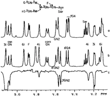
In a biantennary complex structure, the mannotriose unit is substituted by Gal/31-4GlcNAc/31- arms (Fig. l a ) . Units linked to the Mancul-6 residue of the mannotrioside are said to be located on the &arm, while those units linked to the Manal-3 of the core mannotrioside are located on the 3-arm. The unequivocal assignment of the resonances arising from the residues in those arms is difficult because the branches have similar composition. In GGN, the chemical shift of the H1 resonances of the GlcNAc residues are different because only one is substituted by Gal. Since the H1 and H2 reso- nances of the Man residues are not appreciably affected by the presence of a terminal Gal residue in the structure (9), it is difficult from chemical shift data alone to tell on which arm the Gal residue is situated. However, the location of the Gal residue in GGN and the unequivocal assignment of the GlcNAc and Gal H1 resonances can be deduced from NOE experiments. Saturation of the H1 resonance of the 3i residue (see Fig. l a for nomenclature) yields enhancements on the following signals: 3i HZ, 4i H2,4i H1, and GN2 H1 (4.553 ppm) (Fig. PC). Since the intensity of the latter is similar to that of the 3i H2 NOE
(-lo%),
the GN2 H1 must be within 2.6 A of the 3i H1. The close proximity'between these two atoms can only arise if these two residues are linked. The NOEs on the 4i H2 and H1 signals are a consequence of the al-3 linkage between the two Man residues in the GGN structure. Al- though 4i H1 and HZ are more than 4A
away from 3i H1, small negative NOEs of 2 and 4%, respectively, are observed as a consequence of the three spin effect (13). The 4i H1 and H2 relax through mutual dipolar interactions with the 4i H3 which exhibits a large negative NOE of -20% (not shown) because of its proximity to the 3i H1. Hence, this very specific chain of NOEs on the 4i H3, H2, and H1 resonances is highly useful in establishing the sequence Manal-3Manpl-. The above enhancements thus confirm the sequence GlcNAc/31- 2Mancul-SManpl- and permit the assignment of the 4.553 ppm resonance to H1 of 3GN2 and the 4.578 ppm resonance to the H1 of 6GN2 (Fig. 26). By comparing the spectra of GG and GGN (Fig. 2, a and 6), one can deduce that a substitution a t C4 on GN2 shifts its H1 resonance downfield by 0.025 ppm (9). Since the two equivalent GN2 H I resonances of GG have a similar chemical shift with that of the 6GN2 H1 resonance'
Brisson, J.-R., and Carver, J. P., manuscript in preparation.in GGN, the Gal residue in GGN must be located on the 6- arm and the Gal resonance a t 4.471 ppm in GGN can be assigned as 6G4 HI.
In GG, although the Gal H1 resonances have slightly differ- ent chemical shifts, their assignment cannot be determined a
priori. However, the lowfield component of the G4 H1 reso- nances in GG can be tentatively assigned as 6G4 H1 since it has the same chemical shift as the 6G4 H1 resonance in GGN (4.471 ppm). The highfield component of the Gal H1 reso- nances in GG (4.466 ppm) then corresponds to 3G4 H1.
These assignments are identical to those reported for a similar compound with no Fuccul-6 present in the core (2). The latter were deduced from the corresponding oligosaccha- ride terminating as Man/?l-4GlcNAc(a,/3) and from com- pounds in which the Gal residues were substituted by sialic acid. Thus, the NOE technique permits an alternative way of assigning resonances when appropriately modified compounds are not available.
For the bisected biantennary hybrid C3B, saturation of the 3i H2 resonance (Fig. 36) and saturation of the 3i H1 signal (Fig. 3c) can be used to establish the sequence for this com- pound. Saturation of the H2 resonance a t 4.255 ppm yields
NOEs on the H1 signal (5.055 ppm) and the GN2 H1 reso- nance. Upon saturation of the H1 signal a t 5.055 ppm, the same NOE pattern as in GGN was observed (inter-residue NOEs on GN2 H1 and 4i H2). The ratio of the enhancements on the 4i HZ to 3i H2 resonances is the same as that found for GGN (Fig. 2c uersus Fig. 3 c ) . If the GN2 residue was linked
to the Manal-3 residue on the 6-arm, one would expect to see an enhancement on the 6i H2 resonance which is comparable to the NOE on the 3i H2 signal. Since this is not the case, the GN2 residue must be located on the 3-arm of the core, thus establishing the sequence GlcNAc/31-2Mana1-3Man/31-. The small NOE on the 3t H2 resonance (4.061 ppm, Fig. 3c) is caused by the partial saturation (50%) of the 3t H1 resonance (see Fig. l b for nomenclature).
I t should be noted that the H1 resonances for the 3i and 3t residues, were interchanged in the original work (3). These assignments were based on decoupling experiments where
a
GN2
U
1 " ' 1 " ' 1 " ' 1 " ' 1 "
5 .o Y . e Y . 6 Y . Y Y .2 PPV FIG. 2. Sequence determination for the biantennary com- plex glycopeptides GG and GGN. a, Spectrum of GG. The residual HDO signal at 4.8 ppm has been saturated. b, Spectrum of GGN. c,
NOE difference spectrum of GGN: saturation of the 3i H1 resonance
at 5.115 ppm. The HDO signal (4.8 ppm) has been removed. In all
these figures, the biggest NOE, which is about 10% of the saturated
Oligosaccharide Sequencing
zyxwvutsrqponmlkjihgfedcbaZYXWVUTSRQPONMLKJIHGFEDCBA
1433
zyxwvutsrqponmlkjihgfedcbaZYXWVUTSRQPONMLKJIHGFEDCBA
changes in signal intensities brought about by the loss of a coupling between protons are often difficult to detect when the coupling constants involved are of the same order as the line widths of the resonances (1-2 Hz). This is usually the case for the H1 resonances of mannose residues in these high molecular weight compounds. Thus, NOE experiments, which are not affected by these restraints, can be extremely useful in establishing assignments in such situations.
As can be observed in the NMR spectrum of the bisected triantennary hybrid A3, the only isolated resonances are the 4i H1 and the 3i H2 (Fig. 4a). Saturation of the former (Fig. 4b) confirms the sequence Manal-3Manpl-, since small inter- residue enhancements are observed on the 3i H1 and H2 signals (5.048 and 4.290 ppm, respectively). As explained above, these NOEs are a result of a three spin effect between 4i H1,4i H2, and 3i H1 which arises because they are adjacent in space and relax mutually with each other (13). There is no significant relaxation pathway between 6i H1 and 4i H1 which would allow, upon saturation of the 4i H1 resonance, NOEs to
3i 31
61
zyxwvutsrqponmlkjihgfedcbaZYXWVUTSRQPONMLKJIHGFEDCBA
61zyxwvutsrqponmlkjihgfedcbaZYXWVUTSRQPONMLKJIHGFEDCBA
6i 4 1 corezyxwvutsrqponmlkjihgfedcbaZYXWVUTSRQPONMLKJIHGFEDCBA
brs 3i 4i 61 31GN
zyxwvutsrqponmlkjihgfedcbaZYXWVUTSRQPONMLKJIHGFEDCBA
GN2 GN0
b
5 .o Y . B ' 4 . 6 Y .Y Y
zyxwvutsrqponmlkjihgfedcbaZYXWVUTSRQPONMLKJIHGFEDCBA
.2 Y . a PPMFIG. 3. Sequence determination for the bisected biantennary hybrid glycopeptide C3B. a, Spectrum of C3B. T h e broad line shape of the core GN H I resonance is due to virtual coupling (15). T h e HDO signal (4.8 ppm) has been saturated. NOE difference spectra: b, saturation of the 3i HZ resonance a t 4.255 ppm; c, saturation of the 3i H1 resonance a t 5.055 ppm. The 3t H1 signal is also partially saturated (50%). The HDO signal (4.8 ppm) has been removed.
61 6i 61 4i.6i 31 4i core 64 bis a - , . . l . . , l , , . , , ,
, . .
, - ,
5 . o Y .B Y . 6 Y . Y Y . 2 Y . a PPMFIG. 4. Sequence determination for the bisected trianten- nary hybrid glycopeptide A3. a, Spectrum of A3. T h e line shape of core GN H I resonance is also affected by virtual coupling (see Fig.
3). NOE difference spectra: b, saturation of the 4i H I resonance a t 4.730 ppm; c, saturation of the 3i HZ resonance a t 4.290 ppm, d , saturation of the 3i H3, 3t H2, 6t HZ resonances around 4.02 ppm. In
b and c, the large HDO signal (4.8 pprn), which in b probably obscured
a small NOE (1%) on 4i H1, has been removed.
31 21 HDO
zyxwvutsrqponmlkjihgfedcbaZYXWVUTSRQPONMLKJIHGFEDCBA
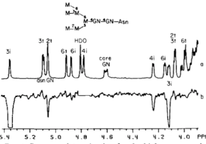
21 3i a 3i U Y l " ' l " ' l " ' l " ' l " ' l " ' l ' r 5 . Y 5 . 2 5 . 0 Y . B Y . 6 Y . Y Y . 2 Y . 0 PPMFIG. 5. Sequence determination for the high mannose gly-
copeptide D3.
zyxwvutsrqponmlkjihgfedcbaZYXWVUTSRQPONMLKJIHGFEDCBA
a, Spectrum of D3, the line shape of the core GN H1signal is also due to virtual coupling (see Fig. 3). b, NOE difference spectrum with saturation of the 3i H1 signal at 5.348 ppm. The large HDO signal (4.8 ppm), which probably obscured a small NOE on the 4i H1 resonance, has been removed.
TABLE I
Nuclear Overhauser enhancements that can be used to confirm
some sequences in Asn-linked oligosaccharides
The saturated signal is denoted by *. An observable NOE is denoted by
+
and the absence of an NOE is denoted by -. a indicatesa signal that is partially saturated due to its proximity to t.he saturated signal and b indicates a probable NOE which is obscured by the large HDO signal.
Sequence
Residues
A H C
Manpl- Manul-3 "I
GlcNAo/j1-2 C R A H1 H2 H1 H2 H I H2 ~" Manal-ZManotl-3Manfil- *
+
+
-+
*
+ a -+
*
+
+
-b a+
*
+ a+
+
* ++
*
" " " GlcNAcfil-2Manal-3Man~l- b+
*+
+
*
+
+
+
* "occur on the H1 and H2 signals of the Manal-3 residue on the 6-arm. The small intensity on the core GN H1 is probably due to partial saturation, while the signals near 4.88 ppm probably arise from a spinning sideband and partial saturation of the 6i H1 resonance. T h e small NOE on the bis GN H1 (4.413 ppm) is probably a consequence of mutual dipolar interactions between protons in the GlcNAcpl-4Manpl- unit.
As in C3B, saturation of the 3i H2 resonance in A3 (Fig. 4c) confirms the sequence GlcNAcpl-2Man. However, a new NOE appears at 4.037 ppm. From its coupling constants and intensity, it can be easily assigned as 3i H3. In C3B, this proton resonates a t 3.88 ppm.' This shift of 0.16 ppm is characteristic of a substitution a t C4 (15). Hence, the 3i residue must be doubly substituted at its C2 and C4 positions by GlcNAc. Further evidence supporting the C4-substitution is obtained upon saturation of the resonances (3i H3,3t H2,6t H2) near 4.03 ppm (Fig. 4 4 , since a small NOE (1%) is observed on the GN4 H1 at 4.535 ppm. This NOE can only arise through mutual dipolar relaxation with protons involved in the GlcNAcpl-4Manal- unit located on the 3-arm, since the 3t and 6t residues are too far removed from GN4. In Fig. 4 4 the other enhancements (3t H1, 6t H1, and 3i H2) are all intra-residue NOEs.
1434
zyxwvutsrqponmlkjihgfedcbaZYXWVUTSRQPONMLKJIHGFEDCBA
zyxwvutsrqponmlkjihgfedcbaZYXWVUTSRQPONMLKJIHGFEDCBA
Oligosaccharide Sequencing
For the high mannose compound D3, the position of the Manal-2 residue (2t) in the structure cannot be readily deter- mined solely from chemical shift data (3). However, upon saturation of the 3i H1 resonance (Fig. 5 6 ) NOEs are detected on the 3i H2, on the 4i H2, and on the 2t H1 signals, thus establishing the sequence Manal-ZManal-3Manfil-. Once again, the NOE on the 4i H2 arises from a three spin effect.
Thus, NOEs can be used to establish sequences that contain Manal-PMan, Manal-3Man, and GlcNAcpl-2Man units. The H1 resonances of hexoses and the H2 resonances of mannoses can be saturated to obtain sequencing information. The vari- ations that are possible for the above linkages are listed in Table I. The availability of several approaches permits con- siderable flexibility in choosing suitably isolated resonances for the NOE experiment. Also, since this technique permits the observation of only those protons that are in close prox- imity to the saturated proton, its use as a tool for assigning new resonances should be quite general.
REFERENCES
1. Carver, J . P., and Grey, A. A. (1981) Biochemistry 20, 6607-6616 2. Vliegenthart, J . F., van Halbeek, H., and Dorland, L. (1981) Pure
Appl. Chem. 53,45-77
3. Carver, J . P., Grey, A. A., Winnik, F. M., Hakimi, J., Ceccarini, C. C., and Atkinson, P. H. (1981) Biochemistry 20, 6600-6606 4. Hanfland, P., Egge, H., Dabrowski, U., Kuhn, S., Hoelcke, D., and
Dabrowski, J. (1981) Biochemistry 20, 5310-5319
5. Dabrowski, J., Hanfland, P., Egge, H., and Dabrowski,
zyxwvutsrqponmlkjihgfedcbaZYXWVUTSRQPONMLKJIHGFEDCBA
U. (1982)Arch. Biochem. Biophys. 210, 405-411
zyxwvutsrqponmlkjihgfedcbaZYXWVUTSRQPONMLKJIHGFEDCBA
zyxwvutsrqponmlkjihgfedcbaZYXWVUTSRQPONMLKJIHGFEDCBA
6. Lemieux, R. U., Bock, K., Delbaere, L. T. J., Koto, S., and Rao,
V. S. (1980) Can. J . Chem. 58,
zyxwvutsrqponmlkjihgfedcbaZYXWVUTSRQPONMLKJIHGFEDCBA
631-6537. Bock, K., Josephson, S., and Bundle, D.
zyxwvutsrqponmlkjihgfedcbaZYXWVUTSRQPONMLKJIHGFEDCBA
R. (1982) J . Chem. Soc.Perkin Trans. 2,59-70
8. Brisson, J.-R., and Carver, J. P. (1982) Biochemistry, in press 9. Grey, A. A., Narasimhan, S., Brisson, J.-R., Schachter, H., and
Carver, J . P. (1982) Can. J. Biochem., in press
10. Narasimhan, S., Harpaz, N., Longmore, G., Carver, J. P., Grey, A. A,, and Schachter, H. (1980) J. Biol. Chem. 255,4876-4884
11. Kicharz, R., and Wuthrich, K. (1978) J . Magn. Reson. 30, 147-
150
12. Bothner-By, A. A. (1979) in Biological Applications ofMagnetic
Resonance (Shulman, R. G., ed) pp. 177-219, Academic Press,
NY
13. Noggle, J . H., and Schirmer, R. E. (1971) The Nuclear Ouerhau-
ser Effect, Academic Press, NY
14. Jeffrey, G. A,, McMullan, R. K., and Takagi,
zyxwvutsrqponmlkjihgfedcbaZYXWVUTSRQPONMLKJIHGFEDCBA
S.
(1977) ActaCrystallogr. Sect. B . Struct. Crystallogr. Cryst. Chem. 33, 728-
737
15. Brisson, J.-R., and Carver, J. P. (1982) J . Biol. Chem. 257,11207-