Sebastian Kos Rolf Huegli Georg M. Bongartz Augustinus L. Jacob Deniz Bilecen Received: 10 February 2007 Revised: 1 October 2007 Accepted: 30 October 2007
Published online: 11 December 2007 # European Society of Radiology 2007
MR-guided endovascular interventions:
a comprehensive review on techniques
and applications
Abstract The magnetic resonance (MR) guidance of endovascular inter-ventions is probably one of the greatest challenges of clinical MR research. MR angiography is not only an imaging tool for the vasculature but can also simultaneously depict high tissue contrast, including the differ-entiation of the vascular wall and perivascular tissues, as well as vas-cular function. Several hurdles had to be overcome to allow MR guidance
for endovascular interventions. MR hardware and sequence design had to be developed to achieve acceptable patient access and to allow real-time or near real-time imaging. The devel-opment of interventional devices, both applicable and safe for MR imaging (MRI), was also mandatory. The subject of this review is to summarize the latest developments in real-time MRI hardware, MRI, visualization tools, interventional devices, endo-vascular tracking techniques, actual applications and safety issues.
Keywords Device tracking . Endovascular intervention . MR angiography . Real-time MRI
Introduction
An increasing demand for secure, minimally invasive interventional procedures has occurred in recent years. This is especially true for minimally invasive endovascular techniques developed for vessel stenosis and occlusion by percutaneous transluminal angioplasty (PTA), stenting or stent grafting. So far endovascular interventions are mostly performed under X-ray fluoroscopic guidance, where patients and staff are exposed to accumulating doses of radiation. Additionally, for sufficient intra-luminal contrast iodinated contrast agents are applied at cumulative doses, which may reveal relevant nephrotoxic effects. In contrast, interventional magnetic resonance imaging (iMRI) has no known harmful effects as it avoids radiation exposure. Additionally, the injection of gadolinium-based contrast agents is normally restricted to much lower doses with
respective lower nephrotoxicity. MRI also provides supe-rior soft tissue contrast, sufficient high spatial resolution and allows multiplanar three-dimensional reconstructions or maximum intensity projections. In addition, functional parameters like flow, temperature, tissue oxygenation, perfusion and diffusion can also be visualized and might influence the end-point of an endovascular intervention in the near future [1]. Because of this, MRI is a valuable tool for endovascular procedures [2–7].
The first part of this review article addresses technical developments. The MRI hardware and MR visualization tools and the essentials of MR angiography are presented. Interventional devices and endovascular tracking tech-niques are summarized. In the second part, an overview on the actual applications is given and tools for intravascular interventions and a preview on future developments are described. Resulting safety issues are addressed.
S. Kos (*) . A. L. Jacob . D. Bilecen Institute of Radiology,
Division of Interventional Radiology, University Hospital Basel,
Petersgraben 4, 4031 Basel, Switzerland e-mail: skos@gmx.de Tel.: +41-61-3286326 Fax: +41-61-26555383 R. Huegli Institute of Radiology, Kantonsspital Bruderholz, Bruderholz, Switzerland G. M. Bongartz Institute of Radiology, University Hospital Basel, Petersgraben 4,
MRI hardware and visualization tools
The optimal configuration of an MR system for interven-tional procedures provides three key features. (1) Free patient access, in combination with (2) fast MR data acquisition and (3) a high image quality. In reality inter-ventional imaging is a trade-off between patient access, field of view, signal-to-noise ratio (SNR) and costs.
Since both the spatial and temporal resolution of MR images depend directly or indirectly on the magnetic field strength, higher B0 fields are more suited; but, on the other hand, the large magnets necessary disturb the accessibility of the patient. Also, the gradient systems need to become stronger and faster to increase the data acquisition and SNR. Limitations of this development might be physiological effects, like tissue heating and nerval stimulation [8].
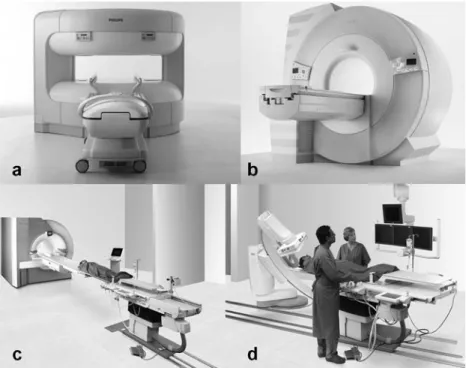
Different MR scanners are in use for interventional procedures, as (1) open scanner (Fig. 1a), (2) short-bore scanner (Fig. 1b) and (3) hybrid systems (Fig. 1c,d). Regarding the open MR systems, variant designs are presently available in order to gain better patient access compared with standard diagnostic“closed” MR scanners (Fig.1). For example C-shaped systems with a vertically oriented magnetic field are frequently used for biopsies and thermal ablations (Fig. 1a) [9]. These MR systems allow better patient access (e.g. lateral, foot, head). However, they lack a sufficient SNR and imaging speed as they use weaker magnetic fields and lower gradient strength (B0≤ 1 T, slew rate 50 T/m/s, Gmax≤ 25 mT/m) compared with standard MRI (B0 ≥ 1.5 T, slew rate 100 T/m/s, Gmax ≥ 30 mT/m). For exemple, the use of a 1-T closed-bore
scanner halved the procedure time when guiding percuta-neous angioplasty in swines compared with a 0.2-T open-bore system [10]. As SNR and speed are essential for MR endovascular procedure control and steering, those open systems are presently not well suited for these procedures. In contrast to percutaneous biopsies and thermal ablations, routine endovascular interventions allow a distance between the puncture site and the target region. Therefore, common MR scanners may be acceptable for endovascular interventions in certain cases with the puncture site outside the scanner and the area of interest in the iso-centre of the magnet. However, to serve practical interventional needs, such systems have already been modified, now providing shorter magnets (125 cm, commonly 160 cm) with a larger gantry diameter (70 cm, commonly 60 cm) (Fig. 1b). Thereby patient access is further improved at the expense of a smaller field of view (35 cm, commonly 50 cm in z-direction).
Keeping the peri-interventional switch to conventional fluoroscopy guidance an option, XMR systems have been developed and are in use, combining MR system and conventional angiography [11, 12]. Different designs are available on the market, with either the X-ray source between two halves of a magnet (so-called double dough-nut) or keeping both systems separate connected by a patient transport lane (Fig. 1c,d) [13–15]. The first alternative avoids delays by patient transport, the latter keeps the option to use both methods independently.
Beside the innovations in the MR design, it is crucial to increase the interactive capabilities of the system. The interventionalist needs tableside control to trigger sequences without a relevant delay. Real-time availability with
inter-Fig. 1 Different designs of interventional MR systems. a A 1.0-T vertically oriented magnet, allowing patient access from head, foot, and from the side. b A short (125 cm) 1.5-T magnet with a 70-cm opening allowing transfemoral vessel access. c, d Hybrid system connecting MRI (c) and DSA (d) via a transport lane. (Courtesy of Philips, Hamburg and Siemens, Munich)
active data transfer of acquired MR images is mandatory, with in-room projection devices already in use [16].
Essentials of MR angiography in endovascular interventions
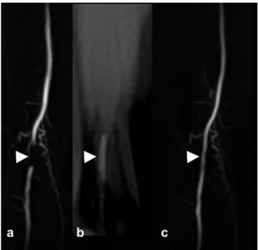
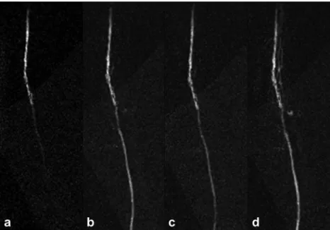
In combination with X-ray fluoroscopy-guided interven-tions, intra-arterial digital subtraction angiography (DSA) is still the “gold standard” of diagnostic angiography. Lacking exposure to iodinated contrast media and ionizing radiation, MR angiography has nevertheless been shown to accurately depict the anatomy of vessels and its pathologies as seen in peripheral artery occlusion disease (Fig.2) [17, 18]. Additional to projective images, MR angiography offers three-dimensional (3D) imaging that exceeds the capabilities of the standard method DSA. By this, not only various reconstruction methods (MPR) can be applied, like maximum intensity projection (MIP), and surface render-ing options but it also offers direct access to the morphol-ogy of the vascular wall [19]. Thereby the status of the vessel wall can be assessed and furthermore even the balloon can be depicted during the PTA (Fig.3).
Of course, vessel depiction and access is possible with native MR sequences (e.g. SSFP sequences) only, which was widely used in interventional animal studies [20,21]. However, for most clinical procedures, the application of
MR contrast media is helpful [22]. This increases the blood contrast and provides excellent angiograms, but may also assist precise device tracking. Furthermore, contrast media can provide grading of stenotic lesions and might depict active extravasation of contrast media during acute bleed-ing. Contrast images (3D MR angiography) may even be used as a template for later contrast-enhanced roadmap and/or overlay techniques [18].
Compared with intravenous MR angiography, a sub-stantial reduction of the injected gadolinium dosage during interventional procedures can be achieved by intra-arterial injection using the side port of the introducer sheath or a selective catheter in place, without loss of image quality [17]. Presently the intra-arterial application of gadolinium is an off-label use, which may be applied as there are no serious side effects documented so far. Due to the short half-life of gadolinium chelates and the consecutive need for repetitive applications, a low-dose gadolinium injection protocol is required [18,23]. Herein, multiple applications of gadolinium chelates during interventions should not exceed the daily dose limit (0.3 mmol/l/kg body weight) for gadolinium, as given by the U.S. Food and Drug Administration (FDA). The previously suggested injection protocol, depending on factors like injection rate, blood flow rate of the target vessel and gadolinium concentration, furthermore keeps the gadolinium blood concentration in a target range during MR data acquisition [23]. This is important because the paramagnetic effects of gadolinium Fig. 2 Comparison between MR angiography and DSA in a
65-year-old woman with known peripheral arterial occlusive disease and left-sided lower leg claudication. Angiographies reveal a significant stenosis at the origin of peroneal (arrowhead) and posterior tibial (straight arrow) arteries. Collateralization extends from the tibioperoneal trunk to the peroneal artery and fades with vessel occlusion in the descending part of the anterior tibial artery (curved arrow). a Coronal MIP of a mask-subtracted 3D gradient-echo image of the proximal calf arteries acquired with intra-arterial MR angiography (3.5/1.3; flip angle, 20°; matrix, 448×307; field of view, 380×326; voxel size, 0.8×1.1×1.0 mm; acquisition time, 33 s). b, c Corresponding intra-arterial DSA images obtained in the early (b) and late (c) phases of the examination. (Reproduced from Huegli et al. [17])
Fig. 3 Intra-arterial MR angiography in a patient with peripheral artery occlusive disease (PAOD) before (a), during (b) and after (c) dilatation of the femoro-popliteal artery. Arrowheads mark a short occlusion of the distal superficial femoral artery (a), the endovas-cular balloon-catheter in place (b) and the result after dilatation (c)
would otherwise lead to fast spin-dephasing with T2/T2* signal reduction (gadolinium concentration above target range) or signal loss due to insufficient T1-shortening (gadolinium concentration below target range) [24].
Blood pool contrast agents like Gadomer-17 allow a longer vessel detection and prevent flow artefacts during catheter manipulation as the steady state is reached earlier. For catheter detection additive catheter injections of a negative contrast medium (CO2) as a double-contrast technique were applied. Resulting replacement of the Gadomer-enhanced blood at the catheter tip and consecu-tive immediate temporal signal loss in the vessel lumen improved catheter visibility in one study [25]. The same might be true for Vasovist as the presently only certified blood-pool contrast agent on the market [26].
In vivo, MR angiography-guided angioplasty in swine was also performed with intra-arterial contrast medium application. Omary et al. [27] depicted the aortorenal vasculature prior to the procedure by injecting gadopen-tate dimeglumine at 4 ml/s using an aortic catheter. After dilatation of the surgically induced renal artery stenosis, the same group documented the technical success with another catheter-directed intra-arterial contrast-enhanced MR angiography. Overall, the definite indication for contrast media application is presently not clear but it certainly may play a major role in endovascular inter-ventions in humans.
In contrast to standard diagnostic MR imaging, iMRI shifts the balance from spatial to temporal resolution. For near real-time imaging the lower limits of the image quality are defined by the ability to track endovascular devices [28].
Conventional contrast-enhanced MR angiography se-quences are based on repetitive application of a fast
T1-weighted FLASH sequence, well usable for 3D angiog-raphies, but its use for device tracking is limited by partial volume effects along the slice direction [29–31].
Better suited for device tracking in endovascular inter-ventions are gradient imaging techniques [steady-state fast precession (SSFP) techniques, TrueFISP, FIESTA or balanced fast field echo], which in combination with contrast enhancement provide a high SNR and increased speed with a short TR, allow improved projection of vessels and its surrounding tissue with high signal and contrast [32, 33].
Therefore intra-arterial gadolinium-enhanced images are subtracted from a native background mask gained by averaged consecutive SSFP measurements (Fig. 4). In contrast, non-balanced SSFP techniques show a slightly lower signal intensity than balanced SSFPs but are more robust and less vulnerable to influences like flow or motion artefacts, eddy currents and imperfect shim [34]. After acquisition of native mask images, used for background suppression, intra-arterial filling images are added to create a road map image to guide further interventions. Acceler-ation of the image acquisition is achieved by parallel acquisition techniques with generalized autocalibrating partially parallel acquisition (GRAPPA) [35], and key-hole imaging [36]. GRAPPA algorithm with an acceleration factor of 2 was able to reduce the acquisition time per image by 34% in an approach with active catheter racking and automated slice alignement [21]. Thereby the catheter was steered from the inferior vena cava through the beating heart into the pulmonary vasculature. Recently Potthast et al. used parallel acquisition techniques (GRAPPA) in MR aortography [37]. Being initially developed to monitor the effect of contrast agents, by 1997 the keyhole technique had speeded up MR catheter tracking by a factor of 4 [20,36].
Fig. 4 Complex subtracted intra-arterial near real-time contrast-enhanced MRI of the superficial femoral artery in a patient with peripheral artery occlusive disease. This MR fluoroscopic technique allows data acquisition times of 700 ms per frame
MRI device-tracking techniques
Respecting the interventionalist’s needs when performing endovascular procedures, the techniques applied should be maximally robust, as simple as possible, and cost-effective. In iMRI the tools used for conventional X-ray fluoroscopy-guided interventions are either almost invisible (e.g. plastic catheters), cause severe artefacts like metallic implants or metal-enforced catheters, or are unsafe (e.g. metallic guidewires may induce heat). Today two principle strate-gies in iMRI device development are under evaluation and seem to be valuable. These strategies can be subdivided into a “passive” and an “active” visualization approach, each having its specific advantages and limitations (Fig.5) [6,38].
Concerning the“passive” approach susceptibility markers (negative markers) for guidewires or signal enhancing markers (positive markers) for guidewires and catheters are linked to the device, thereby gaining detectability as a key feature without using further hardware [6,7].
As positive markers, paramagnetic contrast media have been applied either on the surface of the catheter or within its lumen, in combination with T1-weighted sequences [39]. As negative markers, creating local field distortions, paramagnetic rings or ferrite-mixtures are in use. Small markers can be mounted on non-conducting (e.g. plastic) guidewires and catheters, allowing sufficient negative contrast under in vivo conditions [20, 40]. As a dis-advantage, those negative markers creating susceptibility artefacts may be masked by other signal losses within the excited slice, e.g. by metallic implants, calcifications or bowel air.
Endovascular devices must provide a high flexibility to adapt to the individual vessel configuration during naviga-tion, as many arteries, such as the iliac arteries, have kinks and curves. This means that in most of the cases they are not depictable in a single thin slice and thick slabs are needed to depict vessel anatomy. But as a dilemma, to increase the contrast between the susceptibility artefact and the surrounding tissue it is necessary to keep the slice-thickness as thin as possible and to use image subtraction from a baseline image to highlight the signal changes invoked by the device [41]. Those subtracted images can be overlaid on a contrast-enhanced roadmap MR angiogram, as mentioned above. As a limitation, the susceptibility-induced passive tracking schemes using image subtraction showed a lack of consistency in device visualization, due to the influence of different field-strengths, motion, device orientation, device material and sequence parameters on the artefact [42–44]. The use of an adaptive subtraction technique, which automatically selects the most suitable reference image from a dynamic series for subtraction, was described by Bakker et al. in 2004 [45]. This leads to a reduced image vulnerability to slow periodic motion (e.g. breathing), only transient degradation by gross subject motion or alternation of the scan parameters and thereby a more robust and consistent depiction of the artefact. To allow monitoring of the catheter tip and shaft simulta-neously, projection techniques in combination with a background suppression technique have been suggested [45–47].
Omary et al. [46] applied mask-mode subtraction to increase catheter visibility and a projection dephaser in the slice-selection direction to suppress background signal. Thereby they increased the contrast between background and catheter and were able to depict the catheter on its entire length when showing the feasibility of renal artery angioplasty in a pig model.
A relevant drawback of passive tracking versus the active approach may be found in the fact that passive tracking is image-based and thus, automated slice-repositioning not yet feasible. Therefore, a manual repositioning of the slice might be more time consuming. This may be compensated as in combination with the previously described fast sequences and applying projection techniques, key-hole imaging, and parallel imaging, near real-time imaging can be achieved with appropriate image quality, spatial resolution and field of view.
However, a major and crucial advantage is that, unlike the active tracking techniques in use, passive tracking techniques are much more simple to use and more robust, thereby fulfilling key features for interventional proce-dures [6].
Active tracking was introduced by Dumoulin et al. in 1993 [48]. The active tracking approaches available as (1) micro coils, (2) electrified wire loops and (3) self-resonant radiofrequency circuits, use active signal detection or extinction by the device as the common feature [49]. Fig. 5 Synopsis of different active and passive tracking techniques,
As the underlying principle using micro coils, a non-selective radiofrequency pulse emitted by a transmitting coil (e.g. surface body coil) leads to a peak in Fourier transformed signals, when detected by radiofrequency micro-receiver coils mounted on the device. Using low flip angle amplification, the signal of spins in close proximity to the catheter coil appears bright due to the resonance in the coils, whereas the surrounding tissue has a low signal intensity [50].
By gradient readouts along a single axis, the position can be fast determined [49, 51, 52]. By repetition along the other axes, the precise 3D position of the device can be determined within three repetition times. As an advantage, image acquisition is not necessary and the detected device position can be superimposed on a prior generated, e.g. contrast-enhanced roadmap image or a real-time image acquired intermittently [53,54]. Consecutively, automated scan plane and parameter adjustments based on the current device position are possible [55,56]. This means that active tracking is less time consuming regarding device tracking in complex vessel configurations, compared with the passive approach.
The coils are mounted either only on the tip of a catheter or along the whole catheter axis to track both tip and shaft, with each coil connected to a separate receiver channel [51]. Arrays with a single receiver coil display a single point of the catheter on the background image, giving no information on the orientation and configuration of the device, as a major drawback for endovascular interven-tions. Multiple coils along the device axis allow separate visualization of the different coils, which can be encoded in different colours on the roadmap image, but again increase the technical effort significantly. Variant configurations, to gain detectability of the whole device, with a coil at the tip and a guidewire antenna in the core of the catheter (so-called profiling) have been described [48,57].
As major disadvantages of active tracking with micro-coils, (1) sophisticated synchronization with external tuning, matching and decoupling electronics is needed, consecutively raising (2) the product costs. Therefore a direct cable connection (e.g. coaxial cable or laser fibres) between these electronics and the device is used. With the long conducting wire in situ, the radiofrequency energy which is transmitted into the body might get locally amplified and thus (3) may lead to rapid radiofrequency heating in vivo [57]. The latter risk can be reduced by integrating, for example, transformers or coaxial chokes into the cable between scanner and device, but this again increases the technical effort significantly [58,59].
The use of micro-coils under in vivo conditions reveals robust tracking of the catheter tip and has been successfully demonstrated in animal models (Fig.6). Wacker et al. [56] presented a so-named catheter-driven MRI scanner, which collects the tracking data within 40 ms, needs another 10– 20 ms for localization and image updates, and is thereby able to localize a motionless catheter equipped with
micro-coils in the aorta in 100% of the cases and a moving catheter in 98% of the attempts. Automated consecutive real-time adjustments of spatial resolution and field of view respecting the catheter velocity allowed successful cathe-terization of renal arteries in two pigs. Bücker et al. [60] were able to guide and position a balloon catheter into a surgically created porcine arterial stenosis using a micro-coil on a catheter tip and simultaneously acquire real-time (20 frames/s) vascular visualization with a radial gradient echo technique. Bock et al. [54] used 5-French catheters equipped with three separate radiofrequency coils at a distance of 3 cm, for successful navigation of a catheter into the aorta and renal arteries of a pig.
As another active tracking approach, controlled focal distorsions of the magnetic field have been induced by a small direct current (10–150 mA) applied through an electrified wire loop in a catheter. This is comparable to the passive tracking methods leading to intravoxel dephasing and consecutive signal loss in the vicinity of the device [57]. Until today with this technique device localization in vivo is not feasible.
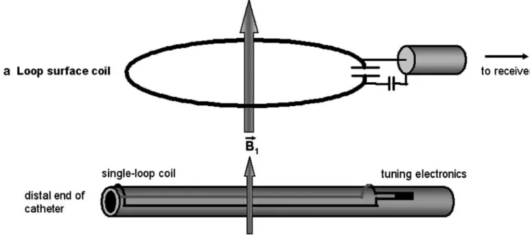
Trying to minimize the risk of rapid heating in vivo, a strategy based on self-resonant radiofrequency circuits, which in earlier MR research were successfully employed as high-contrast localization markers, was developed [50]. For this purpose, a coil or antenna tuned to the Lamor frequency of the scanner is attached to the catheter, connecting the MR signal to the surface coil via induction and making a direct cable connection unnecessary (Fig.7) [61]. Thereby, Quick et al. [61] demonstrated high contrast visualization of a long catheter segment including curva-ture and tip in the aorta of a swine (Fig.8). As a drawback, this method does not allow automated slice-repositioning and is rather sensitive to detuning. Furthermore, the lack of a direct connection to the MR system, decreasing the risk of rapid heating, leads to a constant flip angle amplification even in intermittent imaging, which may cause artefacts in the vicinity of the device. This again can be overcome by adding a photo-resistance, changing the resonance-circuit using light from an optical fibre [62], thereby again increasing the technical effort.
As prototypes, the detection of gradients-fields using Hall probes or a Farraday sensor were presented and are mentioned here only for the sake of completeness, with in vivo test not being performed yet [63,64].
We must emphasize that the active and passive track-ing techniques may well be used simultaneously. If radiofrequency heating is not crucial or can be limited as described above, a combination of tracking techniques may allow distinction of different instruments used during endovascular procedures. In an animal study, an actively tracked 0.030-inch diameter loopless antenna guidewire coil, in combination with a passively tracked 5-French aortic cathteter filled with dilute 4% gadopen-tate dimeglumine, allowed catheterization and stenting of renal arteries [27].
New interventional devices
A crucial step in the future evolution of iMRI will be the further development and evolution of diagnostic catheters, guidewires, stents and other interventional tools, which need to be compatible, safe, visible, robust and as simple and cheap as possible.
In 1997, Bakker et al. [20] were the first to use the passive tracking technique for catheter localization and steering in the basilic vein of a human. At this time, dysprosium markers mounted on a 3-French non-braided catheter were 1-mm thick and coated by a 0.1-mm thick nylon sleeve, thereby increasing the catheter diameter and reducing potential access to smaller vessels siginificantly. This clearly demonstrates that tracking techniques and
devices must be developed in parallel if they should not inhibit the others progression.
A huge step has been made by presenting MR-compatible 0.035-inch diameter guidewires using fibreglass [65] or a polyetherketone (PEEK) (Fig. 9). This was necessary as standard guidewires with a stainless steel core may be drawn into the magnet, nitinol guidewires may lead to rapid heating as discussed below and simple plastic guidewires are too floppy and lack satisfactory torque control [6].
The PEEK-guidewire presented by Mekle et al. [40], consists of a PEEK polymer core, with a conic shape at its flexible distal end, where it’s coated with small iron particles. Even with the additional outer polymer coating, the diameter is only 0.035 inch over the entire length of the wire, thereby reducing influences on the mechanical Fig. 6 Real-time TrueFisp
ac-quired with five imaging coils and an active imaging coil at the catheter tip. The tracking coli is either visible as a bright spot (open arrows) or the coil posi-tion is marked by a dotted cross (a, d, g). The catheter is ad-vanced through the vena cava (a, b) into the right atrium (c, d) and right ventricle (e–g), and further into the pulmonary artery (h). (Reproduced from Bock et al. [21])
properties and vessel accessibility. It was tracked using signal peaks at the side bands of an SSFP sequence with very low flip angles [40]. This passive approach on the one hand seems superior to those passive concepts tracking
pure signal voids, as a signal void may be averaged in a larger imaging slice and as other conditions may cause signal loss in MRI, as mentioned above Furthermore, compared with active devices, much less technical and regulatory requirements exist, as shown above.
In general, the critical features of guidewires are steerability, robustness and torque control. Fibreglass, for example, offers a good pushability, but is less resistant to torque and thus may fracture during an intervention. PEEK, on the other hand, is less rigid, thereby minimizing the risk of core fracture but offering less pushability. In our opinion, a compound solution with a fibreglass core and a PEEK surface may be the solution.
To our knowledge, the loopless antenna guidewire coil (Intercept; Surgi-Vision, Gaithersburg, Md.) used by Omary et al. [27], being 0.030 inch in diameter is the smallest guidewire for MR interventions available so far. As a mechanical hurdle until today, it was not shown that smaller MR guidwire diameters, such as 0.014 inch and Fig. 8 a MIP of an MR angiogram acquired in the abdominal aorta
of a pig following intra-venous contrast injection gives a com-prehensive view of the vascular morphology. b The active wireless catheter has successfully been guided with real-time projection reconstruction TrueFISP imaging (one frame shown) into the celiac trunk. Images (c) and (d) show MIPs of selective time-resolved high-resolution 3D FLASH contrast-enhanced MRA of the celiac trunk with the SMA in a coronal (c) and in a sagittal (d) projection. Contrast agent was administered through the tip of the wireless active catheter. The distal active end of the catheter was coloured in red. Note excellent correlation between the intra-venous MR angiography in (a) and the intra-arterial MRA in (c) and (d). (Reproduced from Quick et al. [61])
Fig. 9 Newly developed polyetherketone (PEEK) MR-compatible guidewire reinforced by a fibreglass compound. Metal rings are mounted on the tip. The total diameter is 0.035 in/0.85 mm. This allows the use of conventional catheters over the wire
Fig. 7 Principle of wireless signal coupling between two radio-frequency coils and its application to wireless active catheter visualization. a Loop surface coil that is connected to the radiofrequency receiver of the scanner. b Distal end of a catheter that is equipped with a closed-loop coil that is tuned to resonance with a capacitor. In the body coil (not shown) radiofrequency
transmit mode, the resonant catheter coil locally multiplies the excitation flip angle. In the radiofrequency receive mode, the resonant catheter coil picks up the MR signal in its immediate vicinity, resulting in a B1 field vector that can be inductively coupled to that of the loop surface coil (a). (Reproduced from Quick et al. [61])
0.018 inch, as used in cardiac and cerebrovascular inter-ventions, may be produced with the attachments needed for MR tracking.
Safety aspects
Following the ALARA principle (as low as reasonable achievable) for the use of ionizing radiation, the use of MR guidance for interventional procedures should be favoured over X-ray fluoroscopy and CT. However, today’s distribu-tion of MR systems and the resulting costs make a reducdistribu-tion to special indications necessary. Primarily such patients should be in the focus for MR-guided interventions who undergo complex and long-lasting or redundant procedures, thereby increasing the risk for patient and staff to suffer from deterministic X-ray damage like erythema or hair loss. Also, young patients carrying the stochastic events of radiation exposure throughout their lifetimes should benefit from the use of MRI in procedure guidance.
Rapid heating of electrically conductive foreign bodies and instruments under radiofrequency energy excitation is a well-known problem. In 1.5-T fields, temperature
changes of up to 44°C were observed [66, 67]. In vitro experiments with standard nitinol guidewires, which are not ferromegnatic but electrical conductors, showed substantial heating around the guidewire tips. The degree of heating is proportional to the power of the radio-frequency pulse. It also varies between different sequences and changes with the flip angle. Wildermuth et al. [68] found sequences with higher duty cyles, as fast spin echo sequences, and high flip angles (90°) in gradient-recall echo sequences to cause most wire heating (18–20°C). However, studies using nitinol guidewires “off-label” did not show any harmful side effects caused by this heating in vivo, so its use is controversial [69]. The shown potential risks enforce the need for new MR-suitable instruments, e.g. the above-mentioned guidewires, replacing nitinol-based components, to make further discussion about the potential effects in vivo unnecessary. These tools will need compar-able rigidity and torque responsiveness as their conventional counterparts, to prevent device fracture in vivo.
Further research will be needed to investigate the postulated effects of gadolinium-based contrast media on the pathogenesis of nephrogenic systemic fibrosis in patients with end-stage renal insufficiency [70].
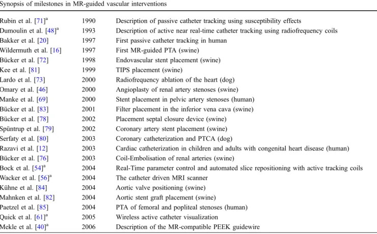
Table 1 Synopsis of hallmarks in endovascular interventional MR, mentioning animal experiments and clinical studies Synopsis of milestones in MR-guided vascular interventions
Rubin et al. [71]a 1990 Description of passive catheter tracking using susceptibility effects
Dumoulin et al. [48]a 1993 Description of active near real-time catheter tracking using radiofrequency coils Bakker et al. [20] 1997 First passive catheter tracking in human
Wildermuth et al. [16] 1997 First MR-guided PTA (swine) Bücker et al. [72] 1998 Endovascular stent placement (swine) Kee et al. [81] 1999 TIPS placement (swine)
Lardo et al. [73] 2000 Radiofrequency ablation of the heart (dog) Omary et al. [46] 2000 Angioplasty of renal artery stenoses (swine) Manke et al. [69] 2000 Stent placement in pelvic artery stenoses (human) Bücker et al. [83] 2001 Filter placement in the inferior vena cava (swine) Bücker et al. [78] 2002 Placement septal closure device (swine)
Spüntrup et al. [79] 2002 Coronary artery stent placement (swine) Serfaty et al. [80] 2003 Coronary catheterization and PTCA (dog)
Razavi et al. [12] 2003 Cardiac catheterization in children and adults with congenital heart disease (human) Bücker et al. [76] 2003 Coil-Embolisation of renal arteries (swine)
Bock et al. [54]a 2004 Real-Time parameter control and automated slice repositioning with active tracking coils Wacker et al. [56]a 2004 The catheter driven MRI scanner
Kühne et al. [84] 2004 Aortic valve positioning (swine) Mahnken et al. [82] 2004 Aortic stent graft placement (swine)
Paetzel et al. [85] 2004 PTA of femoral and popliteal stenoses (human) Quick et al. [61]a 2005 Wireless active catheter visualization
Mekle et al. [40]a 2006 Description of the MR-compatible PEEK guidewire a
In vivo applications
Since the description of MR tracking techniques suitable for endovascular interventions, research has been performed showing feasibility in animal studies and consecutively demonstrating the possible clinical transfer (Table1) [48,71]. In 1997, the first human experiment described excellent MR visualization of a solid catheter with five integrated dysprosium rings [20]. Simple movements of the catheter in the cephalic vein were possible without a guidewire. In 1998, Bücker et al. [72] first showed the feasibility of MR-guided stent placement. They successfully placed stents in porcine iliac arteries using radial scanning with a gradient-echo sequence for guidance. In the meantime, it has been shown in other animal models that angioplasty, TIPS placement, stenting of aortic aneurysms, aortic dissections, pelvic, coronary and renal arteries and the inferior vena cava is as feasible as selective renal artery embolisation and occlusion of cardiac septal defects, prosthetic valve implan-tation and radiofrequency ablation under MR guidance (Fig. 10) [27, 46, 72–84]. First interventional procedures have been demonstrated in humans with conventional devices [85]. As pioneers, in 2000 Manke et al. [69] successfully performed MR-guided stent angioplasty of iliac artery stenoses using the passive tracking approach. In 2003, Razavi et. al. [12] were furthermore able to guide diagnostic or therapeutic cardiac catheterization in 14 patients with congenital heart disease. Krombach et al. [86]
even used MRI to document the distribution of intramyo-cardially injected extracellular MR contrast medium (gadolinium-DTPA), which may be of use in the con-trolled and focal delivery of cardiac therapy under MR guidance, as described by Saeed et al. [87]. However, there is still a lack of certified MR tools, especially guidewires, that allow secure endovascular applications. After further development and certification, clinical trials on human application are recommended to demonstrate the benefit and possible superiority of MR-guided interven-tions over X-ray-guided intervention for clinical routine. In principle, there is no concern why vascular intervention, being conventionally performed so far, should not be feasible under MR guidance. It is also likely that the MR capability to even visualize functional parameters like flow, temperature, perfusion and diffusion will be used to directly monitor the outcome of interventions.
Summary
Within the last decade, tremendously increasing numbers of minimally invasive interventional procedures have been performed throughout the world. To broadly implement a novel image modality, like in this case MRI as a guidance procedure in daily practice, it will need further research to show its superiority towards existing modalities. This is especially demanding as the existing conventional systems
Fig. 10 Safe advancement of the stent-graft delivery system (arrows) up to the level of a dissection (arrowheads) under real-time MRI guidance (a–e). The correct stent-graft position (arrows) is confirmed immediately after stent-graft deployment showing complete coverage of the dissection (f). Real-time imaging was
performed with an interactive projection reconstruction real-time TrueFISP sequence providing a reconstructed imaging rate of 7 frames/s at an imaging matrix of 192×192 pixel. (Reproduced from Eggebrecht et al. [75])
and techniques have been optimized for years, leading to a stable, robust, and for the interventionalist, convenient set-up. IMRI therefore faces big challenges to guarantee the same comfort (e.g. patient accessibility, speed, interactive procedure control, less noise) and stability as conventional systems already guidance today.
It is very likely that unique features of MRI—like the use of non-nephrotoxic doses of contrast agents, the lack of ionizing radiation and its high contrast resolution, multi-planar 3D capabilities with arbitrary plane selection—and the ability to depict tissue composition and functional
parameters—like flow, temperature, perfusion and diffu-sion—will allow this proof. To achieve this goal, interdisciplinary research and development regarding faster MR sequences, better MR systems, interactive visualiza-tion tools and new intervenvisualiza-tional and tracking devices, have to be performed. By that approach the still existing technical limitations and problems, as also discussed in this review, will certainly be resolved. Big steps have been made and a few devices are already close to certification, but in a second step the therapeutic impact and patient benefit must be proven, to justify the expectable costs.
References
1. Rhee TK, Larson AC, Prasad PV, Santos E, Sato KT, Salem R, Deng J, Paunesku T, Woloschak GE, Mulcahy MF, Li D, Omary RA (2005) Feasibility of blood oxygenation level-dependent MR imaging to monitor hepatic trans-catheter arterial embolization in rabbits. J Vasc Interv Radiol 16:1523–1528 2. Wacker FK (2004) [Interventionel
MRT: current inventory and preview]. Rofo 176:941–943
3. Ozturk C, Guttman M, McVeigh ER, Lederman RJ (2005) Magnetic reso-nance imaging-guided vascular inter-ventions. Top Magn Reson Imaging 16:369–381
4. Wacker FK, Hillenbrand CM, Duerk JL, Lewin JS (2005) MR-guided en-dovascular interventions: device visu-alization, tracking, navigation, clinical applications, and safety aspects. Magn Reson Imaging Clin N Am 13:431–439 5. Schulz T, Puccini S, Schneider JP,
Kahn T (2004) Interventional and in-traoperative MR: review and update of techniques and clinical experience. Eur Radiol 14:2212–2227
6. Smits HF, Bos C, van der Weide R, Bakker CJ (1999) Interventional MR: vascular applications. Eur Radiol 9:1488–1495
7. Wacker FK, Bock M (2007) [Magnetic resonance imaging-guided endovascu-lar interventions]. Rofo 179:355–364 8. Shellock FG, Crues JV (2004) MR
procedures: biologic effects, safety, and patient care. Radiology 232:635–652 9. Silverman SG, Tuncali K, Morrison PR
(2005) MR imaging-guided percutane-ous tumor ablation. Acad Radiol 12:1100–1109
10. Wacker FK, Hillenbrand C, Elgort DR, Zhang S, Duerk JL, Lewin JS (2005) MR imaging-guided percutaneous angioplasty and stent placement in a swine model comparison of open- and closed-bore scanners. Acad Radiol 12:1085–1088
11. Buecker A, Spuentrup E, Schmitz-Rode T, Kinzel S, Pfeffer J, Hohl C, van Vaals JJ, Gunther RW (2004) Use of a nonmetallic guide wire for mag-netic resonance-guided coronary artery catheterization. Invest Radiol 39:656– 660
12. Razavi R, Hill DL, Keevil SF, Miquel ME, Muthurangu V, Hegde S, Rhode K, Barnett M, van Vaals J, Hawkes DJ, Baker E (2003) Cardiac catheterisation guided by MRI in children and adults with congenital heart disease. Lancet 362:1877–1882
13. Dick AJ, Raman VK, Raval AN, Guttman MA, Thompson RB, Ozturk C, Peters DC, Stine AM, Wright VJ, Schenke WH, Lederman RJ (2005) Invasive human magnetic resonance imaging: feasibility during revascular-ization in a combined XMR suite. Catheter Cardiovasc Interv 64:265–274 14. Vogl TJ, Balzer JO, Mack MG, Bett G, Oppelt A (2002) Hybrid MR interven-tional imaging system: combined MR and angiography suites with single interactive table. Feasibility study in vascular liver tumor procedures. Eur Radiol 12:1394–1400 15. Fahrig R, Butts K, Rowlands JA,
Saunders R, Stanton J, Stevens GM, Daniel BL, Wen Z, Ergun DL, Pelc NJ (2001) A truly hybrid interventional MR/X-ray system: feasibility demon-stration. J Magn Reson Imaging 13:294–300
16. Wildermuth S, Debatin JF, Leung DA, Dumoulin CL, Darrow RD, Uhlschmid G, Hofmann E, Thyregod J, von Schulthess GK (1997) MR imaging-guided intravascular procedures: initial demonstration in a pig model. Radiology 202:578–583
17. Huegli RW, Aschwanden M,
Bongartz G, Jaeger K, Heidecker HG, Thalhammer C, Schulte AC, Hashagen C, Jacob AL, Bilecen D (2006) Intraarterial MR angiography and DSA in patients with peripheral arterial occlusive disease: prospective comparison. Radiology 239:901–908 18. Huegli RW, Aschwanden M, Scheffler
K, Bilecen D (2006) Fluoroscopic contrast-enhanced MR angiography with a magnetization-prepared steady-state free precession technique in peripheral arterial occlusive disease. AJR Am J Roentgenol 187:242–247 19. Wyttenbach R, Gallino A, Alerci M, Mahler F, Cozzi L, Di Valentino M, Badimon JJ, Fuster V, Corti R (2004) Effects of percutaneous transluminal angioplasty and endovascular brachy-therapy on vascular remodeling of human femoropopliteal artery by non-invasive magnetic resonance imaging. Circulation 110:1156–1161
20. Bakker CJ, Hoogeveen RM, Hurtak WF, van Vaals JJ, Viergever MA, Mali WP (1997) MR-guided endovascular interventions: susceptibility-based catheter and near-real-time imaging technique. Radiology 202:273–276 21. Bock M, Muller S, Zuehlsdorff S,
Speier P, Fink C, Hallscheidt P, Umathum R, Semmler W (2006) Ac-tive catheter tracking using parallel MRI and real-time image reconstruc-tion. Magn Reson Med 55:1454–1459 22. Zhang H, Maki JH, Prince MR (2007) 3D contrast-enhanced MR angiogra-phy. J Magn Reson Imaging 25:13–25 23. Potthast S, Schulte AC, Bongartz GM,
Hugli R, Aschwanden M, Bilecen D (2005) Low-dose intra-arterial contrast-enhanced MR aortography in patients based on a theoretically derived injec-tion protocol. Eur Radiol 15:2347–2353 24. Lin SP, Brown JJ (2007) MR contrast
agents: physical and pharmacologic ba-sics. J Magn Reson Imaging 25:884–899
25. Maes RM, Lewin JS, Duerk JL, Wacker FK (2005) Combined use of the intra-vascular blood-pool agent, gadomer, and carbon dioxide: a novel type of double-contrast magnetic resonance angiography (MRA). J Magn Reson Imaging 21:645–649
26. Goyen M, Shamsi K, Schoenberg SO (2006) Vasovist-enhanced MR angiog-raphy. Eur Radiol 16(Suppl 2):B9–14 27. Omary RA, Gehl JA, Schirf BE, Green
JD, Lu B, Pereles FS, Huang J, Larson AC, Li D (2006) MR imaging- versus conventional X-ray fluoroscopy-guided renal angioplasty in swine: prospective randomized comparison. Radiology 238:489–496
28. Duerk JL, Butts K, Hwang KP, Lewin JS (2000) Pulse sequences for inter-ventional magnetic resonance imaging. Top Magn Reson Imaging 11:147–162 29. Hennig J, Scheffler K, Laubenberger J, Strecker R (1997) Time-resolved pro-jection angiography after bolus injec-tion of contrast agent. Magn Reson Med 37:341–345
30. Wang Y, Johnston DL, Breen JF, Huston J 3rd, Jack CR, Julsrud PR, Kiely MJ, King BF, Riederer SL, Ehman RL (1996) Dynamic MR digital subtraction angiography using contrast enhancement, fast data acquisition, and complex subtraction. Magn Reson Med 36:551–556
31. Bos C, Bakker CJ, Viergever MA (2001) Background suppression using magnetization preparation for contrast-enhanced MR projection angiography. Magn Reson Med 46:78–87
32. Quick HH, Kuehl H, Kaiser G, Hornscheidt D, Mikolajczyk KP, Aker S, Debatin JF, Ladd ME (2003) Inter-ventional MRA using actively visua-lized catheters, TrueFISP, and real-time image fusion. Magn Reson Med 49:129–137
33. Kaul MG, Stork A, Bansmann PM, Nolte-Ernsting C, Lund GK, Weber C, Adam G (2004) Evaluation of balanced steady-state free precession (TrueFISP) and K-space segmented gradient echo sequences for 3D coronary MR angiography with navigator gating at 3 Tesla. Rofo 176:1560–1565 34. Bieri O, Markl M, Scheffler K (2005)
Analysis and compensation of eddy currents in balanced SSFP. Magn Reson Med 54:129–137
35. Griswold MA, Jakob PM, Heidemann RM, Nittka M, Jellus V, Wang J, Kiefer B, Haase A (2002) Generalized auto-calibrating partially parallel acquisi-tions (GRAPPA). Magn Reson Med 47:1202–1210
36. van Vaals JJ, Brummer ME, Dixon WT, Tuithof HH, Engels H, Nelson RC, Gerety BM, Chezmar JL, den Boer JA (1993)“Keyhole” method for acceler-ating imaging of contrast agent uptake. J Magn Reson Imaging 3:671–675 37. Potthast S, Bongartz GM, Huegli R,
Schulte AC, Schwarz JG, Aschwanden M, Bilecen D (2007) Intraarterial con-trast-enhanced MR aortography with and without parallel acquisition tech-nique in patients with peripheral arterial occlusive disease. AJR Am J
Roentgenol 188:823–829 38. Araki T, Aoki S, Ishigame K,
Kumagai H, Nanbu A, Hori M, Ueki J, Komiyama T, Araki T (2002) MR-guided intravascular catheter manipu-lation: feasibility of both active and passive tracking in experimental study and initial clinical applications. Radiat Med 20:1–8
39. Unal O, Li J, Cheng W, Yu H, Strother CM (2006) MR-visible coatings for endovascular device visualization. J Magn Reson Imaging 23:763–769 40. Mekle R, Hofmann E, Scheffler K,
Bilecen D (2006) A polymer-based MR-compatible guidewire: a study to explore new prospects for interven-tional peripheral magnetic resonance angiography (ipMRA). J Magn Reson Imaging 23:145–155
41. Bakker CJ, Hoogeveen RM, Weber J, van Vaals JJ, Viergever MA, Mali WP (1996) Visualization of dedicated cath-eters using fast scanning techniques with potential for MR-guided vascular interventions. Magn Reson Med 36:816–820
42. Bakker CJ, Bos C, Weinmann HJ (2001) Passive tracking of catheters and guidewires by contrast-enhanced MR fluoroscopy. Magn Reson Med 45:17– 23
43. Godart F, Beregi JP, Nicol L, Occelli B, Vincentelli A, Daanen V, Rey C, Rousseau J (2000) MR-guided balloon angioplasty of stenosed aorta: in vivo evaluation using near-standard instru-ments and a passive tracking technique. J Magn Reson Imaging 12:639–644 44. Ladd ME, Quick HH, Debatin JF
(2000) Interventional MRA and intra-vascular imaging. J Magn Reson Imaging 12:534–546
45. Bakker CJ, Seppenwoolde JH, Bartels LW, van der Weide R (2004) Adaptive subtraction as an aid in MR-guided placement of catheters and guidewires. J Magn Reson Imaging 20:470–474 46. Omary RA, Frayne R, Unal O, Warner
T, Korosec FR, Mistretta CA, Strother CM, Grist TM (2000) MR-guided angioplasty of renal artery stenosis in a pig model: a feasibility study. J Vasc Interv Radiol 11:373–381
47. Serfaty JM, Atalar E, Declerck J, Karmakar P, Quick HH, Shunk KA, Heldman AW, Yang X (2000) Real-time projection MR angiography: feasibility study. Radiology 217:290–295 48. Dumoulin CL, Souza SP, Darrow RD
(1993) Real-time position monitoring of invasive devices using magnetic reso-nance. Magn Reson Med 29:411–415 49. Flask C, Elgort D, Wong E,
Shankaranarayanan A, Lewin J, Wendt M, Duerk JL (2001) A method for fast 3D tracking using tuned fiducial mar-kers and a limited projection recon-struction FISP (LPR-FISP) sequence. J Magn Reson Imaging 14:617–627 50. Burl M, Coutts GA, Young IR (1996)
Tuned fiducial markers to identify body locations with minimal perturbation of tissue magnetization. Magn Reson Med 36:491–493
51. Wendt M, Busch M, Wetzler R, Zhang Q, Melzer A, Wacker F, Duerk JL, Lewin JS (1998) Shifted rotated key-hole imaging and active tip-tracking for interventional procedure guidance. J Magn Reson Imaging 8:258–261 52. Zuehlsdorff S, Umathum R, Volz S,
Hallscheidt P, Fink C, Semmler W, Bock M (2004) MR coil design for simultaneous tip tracking and curvature delineation of a catheter. Magn Reson Med 52:214–218
53. Weiss S, Kuehne T, Brinkert F, Krombach G, Katoh M, Schaeffter T, Guenther RW, Buecker A (2004) In vivo safe catheter visualization and slice tracking using an optically detun-able resonant marker. Magn Reson Med 52:860–868
54. Bock M, Volz S, Zuhlsdorff S, Umathum R, Fink C, Hallscheidt P, Semmler W (2004) MR-guided intra-vascular procedures: real-time parameter control and automated slice positioning with active tracking coils. J Magn Reson Imaging 19:580–589 55. Elgort DR, Wong EY, Hillenbrand CM,
Wacker FK, Lewin JS, Duerk JL (2003) Real-time catheter tracking and adap-tive imaging. J Magn Reson Imaging 18:621–626
56. Wacker FK, Elgort D, Hillenbrand CM, Duerk JL, Lewin JS (2004) The catheter-driven MRI scanner: a new approach to intravascular catheter tracking and imaging-parameter ad-justment for interventional MRI. AJR Am J Roentgenol 183:391–395 57. Glowinski A, Adam G, Bucker A,
Neuerburg J, van Vaals JJ, Gunther RW (1997) Catheter visualization using locally induced, actively controlled field inhomogeneities. Magn Reson Med 38:253–258
58. Weiss S, Vernickel P, Schaeffter T, Schulz V, Gleich B (2005) Transmis-sion line for improved RF safety of interventional devices. Magn Reson Med 54:182–189
59. Ladd ME, Quick HH (2000) Reduction of resonant RF heating in intravascular catheters using coaxial chokes. Magn Reson Med 43:615–619
60. Buecker A, Adam GB, Neuerburg JM, Kinzel S, Glowinski A, Schaeffter T, Rasche V, van Vaals JJ, Guenther RW (2002) Simultaneous real-time visual-ization of the catheter tip and vascular anatomy for MR-guided PTA of iliac arteries in an animal model. J Magn Reson Imaging 16:201–208 61. Quick HH, Zenge MO, Kuehl H,
Kaiser G, Aker S, Massing S, Bosk S, Ladd ME (2005) Interventional mag-netic resonance angiography with no strings attached: wireless active cathe-ter visualization. Magn Reson Med 53:446–455
62. Wong EY, Zhang Q, Duerk JL, Lewin JS, Wendt M (2000) An optical system for wireless detuning of parallel reso-nant circuits. J Magn Reson Imaging 12:632–638
63. Bock M, Umathum R, Sikora J, Brenner S, Aguor EN, Semmler W (2006) A Faraday effect position sensor for interventional magnetic resonance imaging. Phys Med Biol 51:999–1009 64. Scheffler K, Korvink JG (2004)
Navi-gation with Hall sensor device for interventional MRI. In: Proceedings of the 12th Annual Meeting of ISMRM, Kyoto,, 2004, p 950
65. Bakker CJ, Smits HF, Bos C, van der Weide R, Zuiderveld KJ, van Vaals JJ, Hurtak WF, Viergever MA, Mali WP (1998) MR-guided balloon angioplasty: in vitro demonstration of the potential of MRI for guiding, monitoring, and evaluating endovascular interventions. J Magn Reson Imaging 8:245–250 66. Liu CY, Farahani K, Lu DS, Duckwiler
G, Oppelt A (2000) Safety of MRI-guided endovascular guidewire applications. J Magn Reson Imaging 12:75–78
67. Nitz WR, Oppelt A, Renz W, Manke C, Lenhart M, Link J (2001) On the heating of linear conductive structures as guide wires and catheters in interventional MRI. J Magn Reson Imaging 13:105–114 68. Wildermuth S, Dumoulin CL,
Pfammatter T, Maier SE, Hofmann E, Debatin JF (1998) MR-guided percu-taneous angioplasty: assessment of tracking safety, catheter handling and functionality. Cardiovasc Intervent Radiol 21:404–410
69. Manke C, Nitz WR, Lenhart M, Volk M, Geissler A, Djavidani B, Strotzer M, Kasprzak P, Feuerbach S, Link J (2000) [Stent angioplasty of pelvic artery ste-nosis with MRI control: initial clinical results]. Rofo 172:92–97
70. Thomsen HS (2006) Nephrogenic sys-temic fibrosis: a serious late adverse reaction to gadodiamide. Eur Radiol 16:2619–2621
71. Rubin DL, Ratner AV, Young SW (1990) Magnetic susceptibility effects and their application in the develop-ment of new ferromagnetic catheters for magnetic resonance imaging. Invest Radiol 25:1325–1332
72. Bucker A, Neuerburg JM, Adam G, Schurmann K, Rasche V, van Vaals JJ, Molgaard-Nielsen A, Gunther RW (1998) [Stent placement with real time MRI guidance: initial animal experi-ment experiences]. Rofo 169:655–657 73. Lardo AC, McVeigh ER, Jumrussirikul
P, Berger RD, Calkins H, Lima J, Halperin HR (2000) Visualization and temporal/spatial characterization of cardiac radiofrequency ablation lesions using magnetic resonance imaging. Circulation 102:698–705
74. Wacker FK, Reither K, Ebert W, Wendt M, Lewin JS, Wolf KJ (2003) MR image-guided endovascular procedures with the ultrasmall superparamagnetic iron oxide SH U 555 C as an intravas-cular contrast agent: study in pigs. Radiology 226:459–464
75. Eggebrecht H, Kuhl H, Kaiser GM, Aker S, Zenge MO, Stock F,
Breuckmann F, Grabellus F, Ladd ME, Mehta RH, Erbel R, Quick HH (2006) Feasibility of real-time magnetic resonance-guided stent-graft
placement in a swine model of de-scending aortic dissection. Eur Heart J 27:613–620
76. Bucker A, Neuerburg JM, Adam G, Glowinski A, van Vaals JJ, Gunther RW (2003) [MR-guided coil embolisa-tion of renal arteries in an animal model]. Rofo 175:271–274
77. Fink C, Bock M, Umathum R, Volz S, Zuehlsdorff S, Grobholz R, Kauczor HU, Hallscheidt P (2004) Renal embolization: feasibility of magnetic resonance-guidance using active cathe-ter tracking and intraarcathe-terial magnetic resonance angiography. Invest Radiol 39:111–119
78. Buecker A, Spuentrup E, Grabitz R, Freudenthal F, Muehler EG, Schaeffter T, van Vaals JJ, Gunther RW (2002) Magnetic resonance-guided placement of atrial septal closure device in animal model of patent foramen ovale. Circulation 106:511–515
79. Spuentrup E, Ruebben A, Schaeffter T, Manning WJ, Gunther RW, Buecker A (2002) Magnetic resonance-guided coronary artery stent placement in a swine model. Circulation 105:874–879 80. Serfaty JM, Yang X, Foo TK, Kumar
A, Derbyshire A, Atalar E (2003) MRI-guided coronary catheterization and PTCA: a feasibility study on a dog model. Magn Reson Med 49:258–263 81. Kee ST, Rhee JS, Butts K, Daniel B,
Pauly J, Kerr A, O’Sullivan GJ, Sze DY, Razavi MK, Semba CP, Herfkens RJ, Dake MD (1999) 1999 Gary J. Becker Young Investigator Award. MR-guided transjugular portosystemic shunt placement in a swine model. J Vasc Interv Radiol 10:529–535 82. Mahnken AH, Chalabi K, Jalali F,
Gunther RW, Buecker A (2004) Mag-netic resonance-guided placement of aortic stents grafts: feasibility with real-time magnetic resonance fluoroscopy. J Vasc Interv Radiol 15:189–195 83. Bucker A, Neuerburg JM, Adam GB,
Glowinski A, Schaeffter T, Rasche V, van Vaals JJ, Gunther RW (2001) Real-time MR Guidance for inferior vena cava filter placement in an animal model. J Vasc Interv Radiol 12: 753–756
84. Kuehne T, Yilmaz S, Meinus C, Moore P, Saeed M, Weber O, Higgins CB, Blank T, Elsaesser E, Schnackenburg B, Ewert P, Lange PE, Nagel E (2004) Magnetic resonance imaging-guided transcatheter implantation of a pros-thetic valve in aortic valve position: feasibility study in swine. J Am Coll Cardiol 44:2247–2249
85. Paetzel C, Zorger N, Bachthaler M, Hamer OW, Stehr A, Feuerbach S, Lenhart M, Volk M, Herold T, Kasprzak P, Nitz WR (2005) Magnetic resonance-guided percutaneous angioplasty of femoral and popliteal artery stenoses using real-time imaging and intra-arterial contrast-enhanced magnetic resonance angiography. Invest Radiol 40:257–262
86. Krombach GA, Baireuther R, Higgins CB, Saeed M (2004) Distribution of intramyocardially injected extracellular MR contrast medium: effects of con-centration and volume. Eur Radiol 14:334–340
87. Saeed M, Saloner D, Weber O, Martin A, Henk C, Higgins C (2005) MRI in guiding and assessing intramyocardial therapy. Eur Radiol 15:851–863