A comparison of apical root resorption after orthodontic
treatment with surgical exposure and traction of maxillary
impacted canines versus that without impactions
Evangelia Lempesi
*
, Nikolaos Pandis
**
,***
, Padhraig S. Fleming
****
and
Maria Mavragani
*
*Orthodontic Department, School of Dentistry, University of Athens, Greece, **Private Practice, Corfu, Greece, ***Department of Orthodontics and Dentofacial Orthopedics, Dental School/Medical Faculty, University of Bern, Switzerland, ****Institute of Dentistry, Queen Mary University of London, UK
Correspondence to: Evangelia Lempesi, Aristeidou 37, 15234 Chalandri, Athens, Greece. E-mail: evalempesi@yahoo.gr
SUMMAry
BACKGrOUND/OBjECtIvES: Orthodontic management of maxillary canine impaction (MCI), including forced
eruption, may result in significant root resorption; however, the association between MCI and orthodonti-cally induced root resorption (OIrr) is not yet sufficiently established. the purpose of this retrospective cohort study was to comparatively evaluate the severity of OIrr of maxillary incisors in orthodontically treated patients with MCI. Additionally, impaction characteristics were associated with OIrr severity.
SUBjECtS AND MEthODS: the sample comprised 48 patients undergoing fixed-appliance treatment—24
with unilateral/bilateral MCI and 24 matched controls without impaction. OIrr was calculated using pre- and post-operative panoramic tomograms. the orientation of eruption path, height, sector location, and follicle/tooth ratio of the impacted canine were also recorded. Mann–Whitney U-test and univariate and multivariate linear mixed models were used to test for the associations of interest.
rESULtS: Maxillary central left incisor underwent more OIrr in the impaction group (mean
differ-ence = 0.58 mm, P = 0.04). Overall, the impaction group had 0.38 mm more OIrr compared to the control (95% confidence interval, CI: 0.03, 0.74; P = 0.04). however, multivariate analysis demonstrated no differ-ence in the amount of OIrr between impaction and non-impaction groups overall. A positive association between OIrr and initial root length was observed (95% CI: 0.08, 0.27; P < 0.001). the severity of canine impaction was not found to be a significant predictor of OIrr.
LIMItAtIONS: this study was a retrospective study and used panoramic tomograms for OIrr measurements.
CONCLUSIONS: this study indicates that MCI is a weak OIrr predictor. Interpretation of the results needs
caution due to the observational nature of the present study.
Introduction
With the exception of the third molars, maxillary canines are most likely to be impacted, with the reported frequency of ectopic canines typically ranging from 0.8 to 3% (Ericson and Kurol, 1986). Maxillary impacted canines can manifest in a range of bucco-lingual, vertical, and antero-posterior locations with the complexity and duration of treatment believed to relate to the degree of displacement (Stewart et al., 2001; Zuccatti et al., 2006; Fleming et al., 2009).
A degree of root resorption is an inevitable conse-quence of orthodontic treatment, with the maxillary lateral and central incisors typically mostly affected (Linge and Linge, 1983, 1991; Levander and Malmgren, 1988, 2000;
Mirabella and Årtun, 1995). While typically orthodontically induced root resorption (OIRR) is inconsequential and an incidental radiographic finding, resorption of an incisor by 5 mm or more has been estimated to occur in 5% of ortho-dontic patients (Killiany, 1999). Both morphological and
genetic factors are thought to have a bearing on the sever-ity of resorption, with allelic associations (Hartsfield et al., 2004) and various morphological characteristics, includ-ing blunt, pipette-shaped roots and thin roots, shown to be more prone to resorption (Levander and Malmgren, 1988;
Sameshima and Sinclair, 2001, 2004).
Furthermore, both the total treatment time and the mag-nitude of tooth movement are implicated in the develop-ment of OIRR (Segal et al., 2004; Weltman et al., 2010). Treatment time with ectopic canines is known to be consid-erably extended (Mavreas and Athanasiou, 2008); similarly, significant movement is necessary to correct the position of grossly displaced teeth. Additionally, anchorage demands on adjacent incisors are increased because they are subject to considerable reactionary forces during mechanical erup-tion of the canine tooth, predisposing them to resorperup-tion (Woloshyn et al., 1994; Blake et al., 1995).
The primary aim of this retrospective cohort study was to compare the severity of root resorption of the maxillary incisors during orthodontic treatment in patients under-going mechanical eruption of surgically exposed canines versus resorption in patients without impactions. A sec-ondary aim was to gauge the influence of radiographic position of the canine on severity of treatment-induced resorption.
Subjects and methods
The study protocol was approved by the ethical committee of the School of Dentistry, University of Athens (decision number 189/01.11.12).
The present study adopted a retrospective cohort design. A sample size calculation determined that a minimum of 22 subjects per group would be sufficient to detect a differ-ence of at least 0.85 mm in root resorption (standard devia-tion, SD = 1 mm) between the impaction and non-impaction groups (Pandis et al., 2008), with 0.05 significance level and 80% power (β = 0.2).
The sample comprised 48 patients undergoing treat-ment in the Postgraduate Clinic, Orthodontic Departtreat-ment, School of Dentistry, University of Athens in Greece. In the experimental group, 24 patients having maxillary canines impacted unilaterally or bilaterally, either palatally or buc-cally, were included. The control group was selected to match the experimental group in respect of age, gender, and angle classification.
Complete orthodontic records, including pre-treatment and post-treatment panoramic tomograms (DPTs), were pre-sent for all subjects. All subjects underwent comprehensive treatment with fixed appliances. Surgical exposure of one or both maxillary canines was undertaken to facilitate forced eruption of the canine(s) in the impacted canine group. Exclusion criteria included craniofacial deformities or syn-dromes and reshaping of the incisal edge of the maxillary incisors before final radiographic examination. A range of demographic and clinical data including gender, age, over-jet, overbite, angle classification, trauma, habits, agenesis, oral habits, extraction, elastics, treatment duration, contrac-tion duracontrac-tion, and general factors were obtained from the records of each participant.
Data analysis
All preoperative panoramic radiographs were scanned (Epson Scanner, Expression 1680 Pro) with eight-bit-gray-scale analysis at 150 dpi (dots per inch) and stored in uncompressed JPEG format. Following digitization, the radiographs were viewed randomly with 600% zoom by one examiner (EL). The dhal software Viewbox, version 4.0.0.105 (Kifissia, Greece), was used for measurement of data. The position of the impacted canines as depicted on initial panoramic views was evaluated quantitatively.
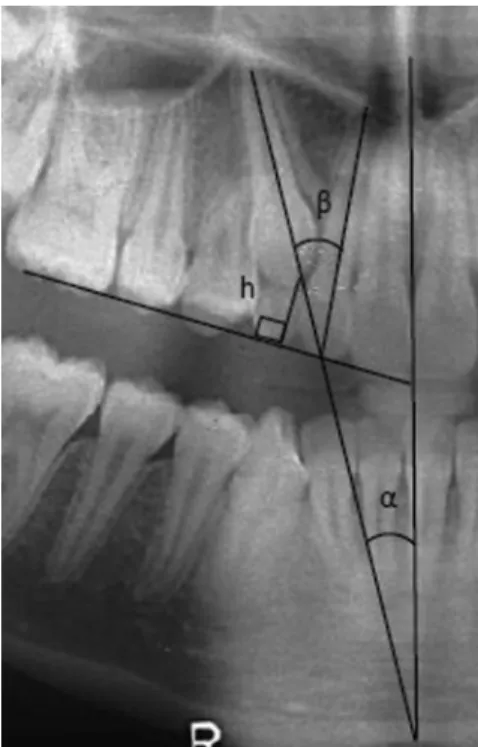
To assess the angulation of the impacted canine, the angle between the long axis of the impacted canine and the upper midline (angle α) and the angle between the long axis of
the impacted canine and the long axis of the adjacent lat-eral incisor (angle β; Ericson and Kurol, 1988; Figure 1) were measured. The maxillary midline was defined from the following reference points depicted on the panoramic radiographs: midpalatal suture, anterior nasal spine, and nasal septum. The cusp tip of the canine was localized in the transverse plane in one of five sectors (Ericson and Kurol, 1988; Figure 2). In order to determine the height of the impacted canine (h), the vertical distance from the canine cusp tip to the occlusal plane was measured (Ericson and Kurol, 1988). The occlusal plane for left and right sides was determined independently based on a tangent to the incisal edge of the maxillary central incisor and the occlusal sur-face of the maxillary first permanent molar (Figure 1). The width of the dental follicle was calculated from the ratio of the maximum width of the follicle to the width of the canine crown (Figure 3).
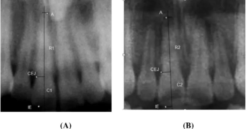
To determine root length of each incisor, perpendicular projections from the points representing the incisal edge, cementoenamel junction (CEJ), and the apex on the long axis of the tooth (Figure 4) were used. The most distinct CEJ landmark, either mesial or distal, was used, with the same aspect being used both for pre- and post-treatment DPTs. A correction factor (C) was estimated in order for the pre- and post-treatment panoramic views to be com-parable, ensuring differences were not attributable to une-ven magnification or distortion. Assuming that the crown
Figure 1 Assessment of canine position, including angle α, angle β, and
length was unchanged from initial to final radiographic examination, the correction factor was calculated as the ratio of radiographic crown length before treatment (C1) to radiographic crown length after treatment (C2). OIRR was defined as the difference in root lengths before and after treatment, after accounting for differences in magni-fication. The formula used to calculate the degree of OIRR was as follows:
OIRR = R1 − (R2 × C), where R1 = root length before treatment and R2 = root length after treatment.
All measurements were performed for each incisor sepa-rately and rounded up to the nearest 0.01 mm. The primary outcome (OIRR) was assessed per tooth in millimetres and as a percentage of initial root length.
The intra-examiner reliability of the measurements of OIRR and tooth location (including angular and linear measurements) was assessed by re-examining 10 randomly selected panoramic radiographs 3 weeks after initial evalu-ation. To quantify random error, the Dahlberg formula (Dalhberg, 1940) was used: τ = √(∑D2 /2N), where D is the
difference between duplicate measurements and N is the remeasured sample size. The systematic error was evaluated using intra-class correlation coefficient (ICC).
Statistical analysis
Statistical analyses were performed at patient and tooth levels. Descriptive statistical analysis for quantitative variables was performed and frequency tables for both impaction and control groups were performed for quali-tative variables. Mann–Whitney U-test was performed to compare the extent of OIRR between the two treatment groups for each tooth separately. Additionally, univari-ate and multivariunivari-ate linear mixed models were used to investigate the influence of maxillary canine impaction (MCI) and other variables, such as age, gender, habits, and the clinical and therapeutic characteristics (inde-pendent variables), on OIRR (de(inde-pendent variable) for each patient.
The association of impaction variables, such as angle
α, angle β, height, and follicle/tooth ratio for OIRR in the
impaction group was investigated using a multiple regres-sion model. The level of significance was set at α = 0.05.
All analyses were conducted with STATA, version 12.1 (StataCorp LP, College Station, Texas, USA).
Figure 2 Antero-posterior assessment of canine position, based on the
study of Ericson and Kurol (1988).
Figure 3 Cropped panoramic view showing measurement of follicle/
tooth ratio.
Figure 4 Cropped panoramic radiographs (pre-treatment and post-treatment) of the same patient. Measurement of root length before and after treatment
for maxillary central incisor right. IE, incisal edge; CEJ, cementoenamel junction; Α, apex. (Α) C1, crown length before treatment; R1, root length before treatment; (Β) C2, crown length after treatment; R2, root length after treatment.
Results
The intra-examiner reliability of the method was found to be excellent (ICC > 0.90) for all parameters. The random error ranged from 0.15 to 0.3 mm for crown length measurement and from 0.03 to 0.6 mm for root length. Each patient was considered as a cluster contributing four maxillary incisors, with the exception of one patient having agenesis of a right lateral incisor and one patient who had the left lateral inci-sor extracted for orthodontic reasons, i.e. in total, 190 teeth were investigated in terms of OIRR. In the impaction group, 17 subjects had unilateral and 7 had bilateral impaction.
The two treatment groups were well matched for all investigated risk factors of OIRR, with the exceptions of overjet and treatment duration (Table 1); the impaction group had a smaller overjet (2.16 mm, SD = 1.76) compared to the mean of the control group (3.50 mm, SD = 2.89;
P = 0.05). The duration of treatment with fixed appliances
was significantly longer (P =0.001) for the MCI group (41.2 months, SD = 11.9) than was the case in the control group (29.5 months, SD = 11.9).
The mean amount of root resorption during treatment ranged from 0.48 to 1.17 mm (4.5–10% of initial root length). OIRR of the maxillary left central incisor was shown to be significantly more likely in the impaction group, with a mean difference in resorption of 0.57 mm (P = 0.04; Table 2). Overall, subjects in the impaction group suffered from an average of 0.38 mm more resorption than their counterparts without impacted teeth (95% confidence interval, CI: 0.03, 0.74; P = 0.04; Table 3). However, the multivariate analysis revealed no difference in the amount of OIRR between the impaction and non-impaction groups overall (β = 0.25, 95% CI: 0.27, 0.78; P = 0.35). Similar
findings were obtained when the percentage of initial root length was used as a dependent variable (Table 4).
In addition, age, gender, malocclusion characteristics, treat-ment variables, and the initial degree of displacetreat-ment of the canine (Table 5) were not shown to be reliable predictors of root shortening. However, a positive association between OIRR and initial root length was observed (95% CI: 0.08, 0.27; P < 0.001).
Discussion
Severe root resorption is one of the most significant and common potential adverse consequences of fixed-appliance orthodontic treatment, with root length loss of more than 20% of all four maxillary incisors shown in almost 3% of orthodontic patients (Sameshima and Sinclair, 2004). A combination of biological and mechanical factors is impli-cated in inflammatory OIRR (Brezniak and Wasserstein, 2002). Specifically, known biological predictors include genetic susceptibility (Al-Qawasmi et al., 2003; Viecilli et al., 2009), and important mechanical factors include the degree of required tooth movement, force levels, nature and direction of forces, torque movements, and prolonged treat-ment (Segal et al., 2004; Weltman et al., 2010).
Although biological factors are beyond the control of a clinician, mechanical factors increase the susceptibility to root resorption during forced eruption of ectopic canines. In particular, significant tooth movement is necessary, torque is usually important, and treatment tends to be lengthy. In the present study, univariate analysis showed that the impaction group experienced slightly more (0.38 mm) OIRR than the control group. However, in the multivariate analysis, canine impaction was found to be a weak risk predictor of OIRR during orthodontics. This finding relates to the fact that the maxillary incisors effectively act as anchorage units and are subjected to high intrusive forces during canine eruption. Intrusive forces are believed to place higher compressive
Table 1 Comparison of the two groups in terms of variables associated with orthodontically induced root resorption.
Variables Unit/category Non-impaction
(n = 24; mean (SD) or %) Impaction (n = 24; mean (SD) or %) P value
* Age Years 20.4 (9.2) 20.0 (8.2) 0.90 NS Gender Male 6 (25%) 7 (29.2%) 0.75 NS Female 18 (75%) 17 (70.8%) Angle malocclusion I 11 (45.8%) 9 (37.5%) 0.82 NS II 10 (41.7%) 11 (45.8%) III 3 (12.5%) 4 (16.7%) Habits/trauma/general factors No 10 (41.7%) 7 (29.2%) 0.37 NS Yes 14 (58.3%) 17 (70.8%) Agenesis/extraction No 15 (62.5%) 19 (79.2%) 0.20 NS Yes 9 (37.5%) 5 (20.8%)
Duration of treatment with fixed appliances Months 29.5 (11.9) 41.2 (11.9) 0.001
Initial root length Millimetres 11.33 (1.99) 11.02 (1.84) 0.57 NS
Overjet Millimetres 3.5 (2.89) 2.16 (1.76) 0.05
Overbite Millimetres 2.98 (2.79) 4.17 (2.24) 0.11 NS
Duration of elastics Months 5.45 (5.50) 6.67 (6.43) 0.49 NS
Duration of contraction Months 2.18 (3.76) 1.73 (2.41) 0.63 NS
forces on the periodontal ligaments in the apical region, risking root resorption (Han et al., 2005; Harris et al., 2006;
Weltman et al., 2010). Furthermore, during orthodontic alignment of impacted canines, torque is required to align the canine; and torquing moments are therefore transmit-ted to the maxillary incisors. Another factor implicatransmit-ted in the increase in OIRR in the presence of canine impaction is the requirement for more prolonged orthodontic treatment (Mavreas and Athanasiou, 2008). In view of the limited dif-ference observed between the groups in the present study, it may be that certain genetic profiles that may override the importance of the afore-mentioned factors exist, thus con-cealing the deleterious effect of treatment-related factors. It has previously been identified that genetic factors may account for almost two-thirds of the observed variability in the extent of OIRR (Harris et al., 1997; Hartsfield et al., 2004).
Age, gender, malocclusion characteristics, and treatment variables were not found to be significant predictors of OIRR; however, a positive association between OIRR and initial root length was observed. Based on the adjusted analysis, for each millimetre increase in tooth length, 0.17 mm greater OIRR can be expected. This finding is in agreement with previ-ous studies (Mirabella and Årtun, 1995; Sameshima and Sinclair, 2001) and may stem from the possible requirement for heavier forces to move teeth with longer roots, and the fact that the magnitude of displacement of the root apex is larger during tipping or torquing when teeth are longer. While apical shortening arising in teeth with initially shorter roots may be of greater concern, neither clear pattern nor increased propensity has emerged from previous research (Levander and Malmgren, 1988; Lund et al., 2012). However, in other studies an increased susceptibility to resorption has been described (Taithongchai et al., 1996; Harris et al., 1997).
Table 2 Orthodontically induced root resorption in millimetres and in percentage reduction of initial root length in the two comparison groups.
Maxillary incisor Maxillary canine impaction group Control group
Mean SD Minimum Maximum Median Range Mean SD Minimum Maximum Median Range
12 n = 23 n = 24 Millimetres — 0.93 1.24 –1.00 4.16 0.87 5.16 — 0.48 0.74 –0.56 2.77 0.29 3.33 % — 8.77 11.74 –9.40 39.58 7.53 48.98 — 4.49 7.23 –6.47 21.58 2.46 28.05 11 n = 24 n = 24 Millimetres — 1.08 1.29 –1.97 3.55 0.87 5.53 — 0.65 0.99 –1.64 2.98 0.45 4.62 % — 9.63 10.95 –16.39 27.84 8.36 44.23 — 5.42 8.29 –13.26 23.46 3.14 36.73 21 n = 24 n = 24 Millimetres — 1.17 1.18 –0.53 4.20 0.93 4.73 — 0.60 0.75 –0.37 2.43 0.35 2.80 % — 9.97 8.93 –3.80 28.28 8.39 32.07 — 5.33 7.11 –3.43 24.42 6.45 27.85 22 n = 23 n = 24 Millimetres — 0.86 1.47 –0.65 4.66 0.33 5.32 — 0.78 0.86 –0.33 3.71 0.65 4.03 % — 7.53 12.58 –7.16 36.40 3.41 43.56 — 7.50 7.76 –3.84 28.56 6.45 32.40 SD, standard deviation.
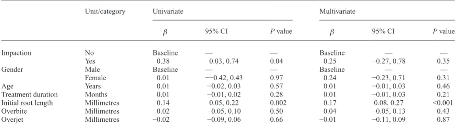
Table 3 Results of univariate and multivariate regression analyses of the influence of the independent variables maxillary canine impaction (0 = no, 1 = yes), gender (0 = male, 1 = female), age (years), treatment duration (months), initial root length (millimetres), overjet (millimetres), and overbite (millimetres) on the dependent variable orthodontically induced root resorption (millimetres).
Unit/category Univariate Multivariate
β 95% CI P value β 95% CI P value
Impaction No Baseline — — Baseline — —
Yes 0.38 0.03, 0.74 0.04 0.25 −0.27, 0.78 0.35
Gender Male Baseline — — Baseline — —
Female 0.01 −−0.42, 0.43 0.97 0.24 −0.23, 0.71 0.31
Age Years 0.01 −0.02, 0.03 0.57 0.01 −0.01, 0.03 0.46
Treatment duration Months 0.01 −0.01, 0.02 0.28 0.01 −0.01, 0.03 0.21
Initial root length Millimetres 0.14 0.05, 0.22 0.002 0.17 0.08, 0.27 <0.001
Overbite Millimetres 0.02 −0.05, 0.10 0.50 0.04 −0.05, 0.13 0.43
Overjet Millimetres −0.02 −0.09, 0.06 0.66 −0.01 −0.11, 0.09 0.87
Selection of the control group was performed by match-ing the impaction group in respect of age, gender, and angle malocclusion. Statistical evaluation revealed a shorter treat-ment duration and larger pre-treattreat-ment overjet in the control group. Although the treatment duration could have been used as a covariate in the analysis because it may have an effect on the extent of resorption, covariate matching methods have two major practical limitations: the number of confounding variables must be relatively small in respect to the sample size (24 impacted patients) and participants must be available for both groups. Thus matching more than three confounding variables was considered prohibitive. Multivariate analysis accounted for these between-groups differences, thus decreas-ing possible confounddecreas-ing effects. Ideally, a randomized clini-cal trial would be most appropriate to eliminate any known and unknown confounders; however, such a design to address this research question is unfeasible. Therefore, knowledge of this topic can only be based upon observational studies,
particularly either case–control or cohort studies in which the unavoidable possibility of selection bias issues exist.
In the impaction group, treatment time was considerably longer than that in the control group; this is in agreement with other studies. Stewart et al. (2001) compared retrospectively treatment time between young patients (aged 20 years or younger) with palatally displaced canines and a control group without impactions. Orthodontic treatment was 5.9 months longer on average in the impaction group. In addition, ortho-dontic treatment of cases with bilaterally impacted canines was 6.5 months longer on average versus cases with unilaterally impacted canines. Similar findings were observed in the pre-sent study, with the mean between-groups difference in treat-ment time being almost 12 months. Both unilateral (n = 17) and bilateral (n = 7) impaction cases were considered in the present research to increase sample size and external validity. A pro-spective study including only cases of unilateral impaction may be a better approach because retrospective studies have several limitations when undertaken in an available sample.
When studying OIRR, it is important to distinguish between the patient-level and the tooth-level analysis. Measurements of different teeth derived from the same subject are correlated with each other. It would be intuitive to expect similar levels of resorption of contralateral incisors to occur during ortho-dontic treatment on the same patient. Based on the tooth-level analysis, it was found that the maxillary central left incisor underwent more OIRR in the impaction group than the same tooth type in the non-impaction group. This finding should be interpreted with caution because these statistical tests do not account for the clustering effects of correlated data, lead-ing to the possibility of findlead-ing statistically significant results that are not genuine (Koletsi et al., 2012). Therefore, further statistical analysis that considered the correlated nature of the data was implemented .
Panoramic radiographs were used to assess OIRR in the present study. This technique is in keeping with previous
Table 5 Results of multivariate regression analysis of the influence of the independent variables angle α (degrees), angle β (degrees), height (millimetres), follicle/tooth ratio, and sector (1, 2, 3, 4, 5) on the dependent variable orthodontically induced root resorption (millimetres).
Unit/category β 95% CI P value
Angle α Degrees 0.01 −0.004, 0.03 0.12
Angle β Degrees 0.01 −0.003, 0.03 0.11
Height Millimetres 0.04 −0.06, 0.13 0.43
Follicle/tooth ratio Ratio 0.4 −0.78, 1.57 0.51
Sector 1 Reference Baseline —
2 0.47 −0.24, 1.18 0.2
3 0.6 −0.23, 1.42 0.16
4 0.67 −0.63, 1.4 0.07
5 0.47 −0.36, 1.31 0.27
CI, confidence intervals.
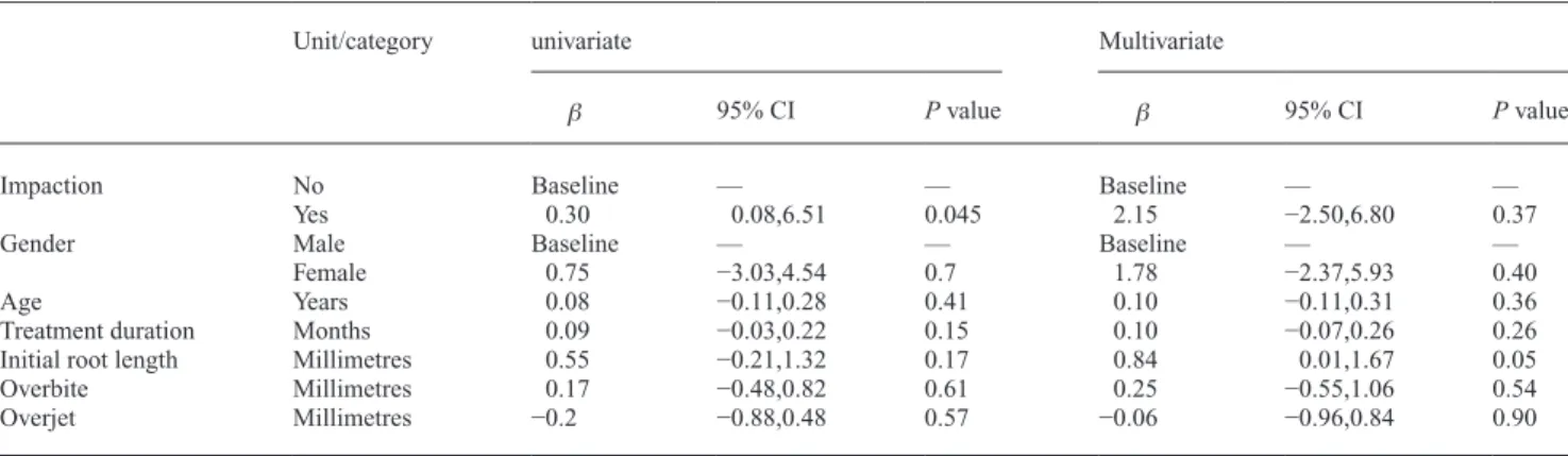
Table 4 Results of univariate and multivariate regression analyses of the influence of the independent variables maxillary canine impaction (0 = no, 1 = yes), gender (0 = male, 1 = female), age (years), treatment duration (months), initial root length (millimetres), overjet (millimetres), and overbite (millimetres) on the dependent variable percentage reduction of initial root length (%).
Unit/category univariate Multivariate
β 95% CI P value β 95% CI P value
Impaction No Baseline — — Baseline — —
Yes 0.30 0.08,6.51 0.045 2.15 −2.50,6.80 0.37
Gender Male Baseline — — Baseline — —
Female 0.75 −3.03,4.54 0.7 1.78 −2.37,5.93 0.40
Age Years 0.08 −0.11,0.28 0.41 0.10 −0.11,0.31 0.36
Treatment duration Months 0.09 −0.03,0.22 0.15 0.10 −0.07,0.26 0.26
Initial root length Millimetres 0.55 −0.21,1.32 0.17 0.84 0.01,1.67 0.05
Overbite Millimetres 0.17 −0.48,0.82 0.61 0.25 −0.55,1.06 0.54
Overjet Millimetres −0.2 −0.88,0.48 0.57 −0.06 −0.96,0.84 0.90
studies (Apajalahti and Peltola, 2007; Pandis et al., 2008;
Dudic et al., 2009; Huang et al., 2010). The study of external lateral root resorption as a consequence of impaction was beyond the scope of this investigation, which was limited to root shortening during orthodontic treatment. The latter may introduce three-dimensional root resorption of the apex. It is accepted that panoramic views may be insensitive to very minor changes in root lengths and may be less accurate than periapical radiographs in studying the severity of OIRR. However, the validity of the periapical films in accurately depicting OIRR has been also questioned because of the influence of tooth shape and morphology (Katona, 2006), inconsistent film and patient positioning, and bending of the film intra-orally. In a direct comparison of panoramic and periapical films of OIRR, however, the differences accru-ing between the recorded lengths of the maxillary incisors was less than 0.2 mm (Sameshima and Asgarifar, 2001). It is, therefore, reasonable to infer that the use of repeated panoramic films resulted in a valid assessment of incisor length in the present investigation. Comparing DPT with cone-beam computed tomography (CBCT) in orthodontic patients, OIRR was found to be underestimated on pano-ramic films (Dudic et al., 2009). Alqerban et al. (2009) in their study demonstrated high sensitivity and specificity for CBCT. Although CBCT is considered a more precise tool in terms of registration of landmarks and OIRR estima-tion, there is still the concern of high radiation exposure, particularly in adolescence (SEDENTEXCT, 2012; http:// www.sedentexct.eu/files/radiation_protection_172.pdf)]. In the present study, the aim was to compare the extent of OIRR in two groups rather than to determine the absolute values of root shortening; consequently, panoramic views were deemed appropriate for this purpose.
The occurrence of negative values for OIRR could be attributed either to immaturity of certain teeth or to meas-urement issues relating to DPTs (Mavragani et al., 2002). The youngest patients were 10.3 years old, having root growth potential at the start of treatment. Another source of potential inaccuracy could relate to differences in incisor torque. Planned alteration of incisor inclination may alter apparent tooth lengths radiographically (Apajalahti and Peltola, 2007). Baseline imbalances in overjet between the groups may be indicative of differences in tooth inclinations, although skeletal relationships are also important. However, multivariate analysis involving multiple predictor variables was undertaken to account for this potential difference.
Conclusions
Patients with at least one impacted maxillary canine ortho-dontically treated with surgical exposure and traction experienced similar levels of root resorption compared to orthodontic patients with normally erupting canines. Further research is required to validate the findings from this investigation.
References
Al-Qawasmi R A et al. 2003 Genetic predisposition to external apical root resorption. American Journal of Orthodontics and Dentofacial Orthopedics 123: 242–252
Alqerban A, Jacobs R, Souza P C, Willems G 2009 In-vitro comparison of 2 cone-beam computed tomography systems and panoramic imaging for detecting simulated canine impaction-induced external root resorp-tion in maxillary lateral incisors. American Journal of Orthodontics and Dentofacial Orthopedics 136: 764.e1–764.e11
Apajalahti S, Peltola J S 2007 Apical root resorption after orthodontic treatment—a retrospective study. European Journal of Orthodontics 29: 408–412
Blake M, Woodside D G, Pharoah M J 1995 A radiographic comparison of apical root resorption after orthodontic treatment with the edgewise and Speed appliances. American Journal of Orthodontics and Dentofacial Orthopedics 108: 76–84
Brezniak N, Wasserstein A 2002 Orthodontically induced inflammatory root resorption. Part I: The basic science aspects. Angle Orthodontist 72: 175–179
Dalhberg G 1940 Errors of estimation. In: Statistical methods for medical and biological students. George Allen and Unwin, London, pp. 122–132 Dudic A, Giannopoulou C, Leuzinger M, Kiliaridis S 2009 Detection of
apical root resorption after orthodontic treatment by using panoramic radiography and cone-beam computed tomography of super-high reso-lution. American Journal of Orthodontics and Dentofacial Orthopedics 135: 434–437
Ericson S, Kurol J 1986 Longitudinal study and analysis of clinical super-vision of maxillary canine eruption. Community Dentistry and Oral Epidemiology 14: 172–176
Ericson S, Kurol J 1988 Early treatment of palatally erupting maxil-lary canines by extraction of the primary canines. European Journal Orthodontist 10: 283–295
Fleming P S, Scott P, Heidari N, Dibiase A T 2009 Influence of radio-graphic position of ectopic canines on the duration of orthodontic treat-ment. Angle Orthodontist 79: 442–446
Han G, Huang S, Von den Hoff J W, Zeng X, Kuijpers-Jagtman A M 2005 Root resorption after orthodontic intrusion and extrusion: an intraindi-vidual study. Angle Orthodontist 75: 912–918
Harris D A, Jones A S, Darendeliler M A 2006 Physical properties of root cementum: part 8. Volumetric analysis of root resorption craters after application of controlled intrusive light and heavy orthodontic forces: a microcomputed tomography scan study. American Journal of Orthodontics and Dentofacial Orthopedics 130: 639–647
Harris E F, Kineret S E, Tolley E A 1997 A heritable component for exter-nal apical root resorption in patients treated orthodontically. American Journal of Orthodontics and Dentofacial Orthopedics 111: 301–309 Hartsfield J K, Everett E T, Al-Qawasmi R A 2004 Genetic factors in
exter-nal apical root resorption and orthodontic treatment. Critical Reviews in Oral Biology and Medicine 15: 115–122
Huang Y, Wang X X, Zhang J, Liu C 2010 Root shortening in patients treated with two-step and en masse space closure procedures with slid-ing mechanics. Angle Orthodontist 80: 492–497
Katona T R 2006 Flaws in root resorption assessment algorithms: role of tooth shape. American Journal of Orthodontics and Dentofacial Orthopedics 130: 698.e19–698.e27
Killiany D M 1999 Root resorption caused by orthodontic treatment: an evi-dence-based review of literature. Seminars in Orthodontics 5: 128–133 Koletsi D, Pandis N, Polychronopoulou A, Eliades T 2012 Does published
orthodontic research account for clustering effects during statistical data analysis? European Journal of Orthodontics 34: 287–292
Levander E, Malmgren O 1988 Evaluation of the risk of root resorp-tion during orthodontic treatment: a study of upper incisors. European Journal of Orthodontics 10: 30–38
Levander E, Malmgren O 2000 Long-term follow-up of maxillary incisors with severe apical root resorption. European Journal of Orthodontics 22: 85–92
Linge B O, Linge L 1983 Apical root resorption in upper anterior teeth. European Journal of Orthodontics 5: 173–183
Linge L, Linge B O 1991 Patient characteristics and treatment variables asso-ciated with apical root resorption during orthodontic treatment. American Journal of Orthodontics and Dentofacial Orthopedics 99: 35–43 Lund H, Gröndahl K, Hansen K, Gröndahl H G 2012 Apical root
resorp-tion during orthodontic treatment. A prospective study using cone beam CT. Angle Orthodontist 82: 480–487
Mavragani M, Boe O E, Wisth P J, Selvig K A 2002 Changes in root length during orthodontic treatment: advantages for immature teeth. European Journal of Orthodontics 24: 91–97
Mavreas D, Athanasiou A E 2008 Factors affecting the duration of ortho-dontic treatment: a systematic review. European Journal of Orthoortho-dontics 30: 386–395
Mirabella A D, Årtun J 1995 Prevalence and severity of apical root resorp-tion of maxillary anterior teeth in adult orthodontic patients. European Journal of Orthodontics 17: 93–99
Pandis N, Nasika M, Polychronopoulou A, Eliades T 2008 External api-cal root resorption in patients treated with conventional and self-ligating brackets. American Journal of Orthodontics and Dentofacial Orthopedics 134: 646–651
Sameshima G T, Asgarifar K O 2001 Assessment of root resorption and root shape: periapical vs panoramic films. Angle Orthodontist 71: 185–189 Sameshima G T, Sinclair P M 2001 Predicting and preventing root
resorp-tion: part II. Treatment factors. American Journal of Orthodontics and Dentofacial Orthopedics 119: 511–515
Sameshima G T, Sinclair P M 2004 Characteristics of patients with severe root resorption. Orthodontics and Craniofacial Research 7: 108–114
SEDENTEXCT 2012 Cone beam CT for dental and maxillofacial radi-ology. Evidence-based guidelines. European Commission’s Radiation Protection No 172. ( http://www.sedentexct.eu/files/radiation_protec-tion_172.pdf) (13 December 2013, date last accessed)
Segal G R, Schiffman P H, Tuncay O C 2004 Meta analysis of the treat-ment-related factors of external apical root resorption. Orthodontics and Craniofacial Research 7: 71–78
Stewart J A, Heo G, Glover K E, Williamson P C, Lam E W, Major P W 2001 Factors that relate to treatment duration for patients with pala-tally impacted maxillary canines. American Journal of Orthodontics and Dentofacial Orthopedics 119: 216–225
Taithongchai R, Sookkorn K, Killiany D M 1996 Facial and dentoalveolar structure and the prediction of apical root shortening. American Journal of Orthodontics and Dentofacial Orthopedics 110: 296–302
Viecilli R F, Katona T R, Chen J, Hartsfield J K Jr, Roberts W E Jr 2009 Orthodontic mechanotransduction and the role of the P2X7 receptor. American Journal of Orthodontics and Dentofacial Orthopedics 135: 694.e1–694.e16
Weltman B, Vig K W, Fields H W, Shanker S, Kaizar E E 2010 Root resorp-tion associated with orthodontic tooth movement: a systematic review. American Journal of Orthodontics and Dentofacial Orthopedics 137: 462–476
Woloshyn H, Årtun J, Kennedy D B, Joondeph D R 1994 Pulpal and perio-dontal reactions to orthodontic alignment of palatally impacted canines. Angle Orthodontist 64: 257–264
Zuccatti G, Ghobadlu J, Nieri M, Clauser C 2006 Factors associated with the duration of forced eruption of impacted maxillary canines: a ret-rospective study. American Journal of Orthodontics and Dentofacial Orthopedics 130: 349–356