. . . .
. . . .
BASIC SCIENCE
CC chemokine CCL5 plays a central role
impacting infarct size and post-infarction heart
failure in mice
Fabrizio Montecucco
1*
†, Vincent Braunersreuther
1†, Se´bastien Lenglet
1,
Benedicte M.A. Delattre
2, Graziano Pelli
1, Vanessa Buatois
3, Florence Guilhot
3,
Katia Galan, Nicolas Vuilleumier
4, Walter Ferlin
3, Nicolas Fischer
3,
Jean-Paul Valle´e
2, Marie Kosco-Vilbois
3, and Franc¸ois Mach
11
Division of Cardiology, Foundation for Medical Researches, Department of Internal Medicine, University Hospital, Geneva, Switzerland;2
Department of Radiology – CIBM, Geneva University Hospital, Geneva, Switzerland;3
Novimmune S.A., Plan-les-Ouates, Geneva, Switzerland; and4
Division of Laboratory Medicine, Department of Genetics and Laboratory Medicine, Geneva University Hospitals, Switzerland
Received 4 November 2010; revised 22 February 2011; accepted 24 March 2011; online publish-ahead-of-print 23 May 2011
Aims
The chemokine CCL5 plays a critical role as neutrophil and macrophage activator do in atherosclerosis andmyocar-dial infarction. Thus, we investigated whether the treatment with a neutralizing monoclonal antibody (mAb) to mouse CCL5 would provide therapeutic benefit when provoking a coronary-associated ischaemic event.
Methods
and Results
C57Bl/6 mice were submitted to left coronary artery permanent ligature. Then, various parameters were monitored for up to 21 days. At5 min and 3 days after coronary occlusion, mice received one intravenous injection of the rat anti-mouse CCL5 mAb or isotype IgG control. Infarct size was assessed histologically and by measuring serum cardiac troponin I levels. Kinetics of CCL5 tissue expression, leucocyte infiltration, matrix metalloproteinase (MMP) levels, and collagen deposition were histologically assessed. Serum chemokine levels were measured by enzyme-linked immunosorbent assay. Cardiac function and dimensions were assessed by magnetic resonance imaging (MRI). Chronic ischaemia increased both circulating and intracardiac levels of CCL5. At 24 h, treatment with the anti-CCL5 mAb resulted in a smaller infarct size and reduced circulating levels of chemokines. This effect was associated with reduction of neutrophil and macrophage infiltration within the infarcted myocardium. After 3 days of chronic ischaemia, anti-CCL5 mAb treatment reduced cardiac MMP-9. At 7 days, collagen content was sig-nificantly lower. At 21 days, neutralizing CCL5 improved mouse survival, cardiac myocyte size, and cardiac function.
Conclusion
Treatment with anti-CCL5 mAb significantly reduced both infarct size and post-infarction heart failure in a mouse model of chronic cardiac ischaemia. Cardioprotective effects were associated with the reduction of leucocyte recruitment within infarcted hearts.-Keywords
Myocardial infarction † Leucocytes † Heart failureIntroduction
Myocardial infarction is frequently complicated by pathological cardiac remodelling that diminishes contractile function and reduces stroke volume. Cardiac remodelling characterized by the
alteration of ventricular mass, form, and function, may potentially lead to ventricular arrhythmias, heart failure, and ultimately
increases cardiovascular mortality.1,2 Acute myocardial infarction
initiates a large inflammatory response resulting in the clearance of dead cardiomyocytes, matrix debris, and replacement of the
*Corresponding author: Cardiology Division, Department of Medicine, Geneva University Hospital, Foundation for Medical Researches, 64 Avenue Roseraie, 1211 Geneva, Switzerland. Tel:+41 22 382 7238, Fax: +41 22 382 7245, Email:[email protected]
†Contributed equally.
Published on behalf of the European Society of Cardiology. All rights reserved.&The Author 2011. For permissions please email: [email protected] European Heart Journal (2012) 33, 1964–1974
damaged tissue with a collagen-based scar. The activation of the complement system and toll-like receptors, as well as the presence of oxidative stress induce cytokine and chemokine secretion within
the injured myocardium.3–5Subsequent leucocyte accumulation in
the infarcted tissue is associated with potent cytotoxic effects through the release of proteolytic enzymes and degradation of collagen and extracellular matrix in cardiac tissue, resulting in ventricular dilatation and adverse remodelling. Thus, targeting chemokine-induced leucocyte recruitment during myocardial repair may represent an attractive therapeutic strategy to improve cardiac function following an acute myocardial infarct. Human and animal studies suggest a crucial role of chemokines and their cognate receptors (e.g. CCL2/MCP-1, CCL4/MIP-1b, CXCL8/IL-8 and CCR1, CCR5, or CXCR2) in post-infarction
remodelling.6–9 Another chemokine CCL5/RANTES (Regulated
on Activation Normal T cell Expressed and Secreted) has been shown to orchestrate the recruitment to inflammatory sites of several inflammatory cell subsets, such as monocytes, neutrophils, dendritic cells, and lymphocytes through the binding to CCR1,
CCR3, or CCR5.10,11
Given the pathophysiological relevance of CCL5 in several immuno-inflammatory diseases such as atherosclerosis, stroke,
and myocardial ischaemia/reperfusion, we investigated the
potential role of CCL5/RANTES in post-infarction cardiac
remodelling.12–14
To better understand the inflammatory processes governing ischaemic myocardial injury and repair, we selected to block CCL5 during the early phases of chronic ischaemia. Thus, develop-ment of post-infarction heart failure as well as cardiac and systemic inflammation was assessed at different times in mice treated immediately and 3 days post-occlusion with either an anti-mouse CCL5 monoclonal antibody (mAb) or the isotype control.
Methods
Antibodies
The rat anti-mouse CCL5 IgG2a MAB478 was purified using protein G affinity chromatography from supernatants of hybridoma (R&D Systems Inc, Minneapolis, MN, USA). In an in vitro chemotaxis assay, MAB478 specifically neutralizes migration of CCR5 expressing cells induced by 36 ng/mL of mouse CCL5 with an IC50 of 0.2 mg/mL (data not shown). MAB478 half-life time in vivo in adult male C57Bl/6 mice is 211 h. The rat anti-bovine calf acetylcholine receptor alpha subunit IgG2a (clone mAb64) was used as an isotype control. All anti-bodies were tested using a Limulus Amoebocyte Lysate-based assay and endotoxin levels were found undetectable (,0.005 EU/mL, from Charles River Laboratories International, Wilmington, MA, USA).
In vivo cardiac chronic ischaemia
and ischaemia/reperfusion protocols
Male C57Bl/6 mice (8 – 12 weeks of age) were obtained from the University Medical Centre (CMU) animal facility, Medical Faculty, University of Geneva, Geneva, Switzerland. The investigation conforms to the Guide for the Care and Use of Laboratory Animals published by the US National Institutes of Health (NIH Publication No. 85-23, revised 1996) and has been approved by local authorities.
In both protocols, mice were initially anaesthetized with 4% isoflur-ane and intubated. After starting mechanical ventilation (tidal volume
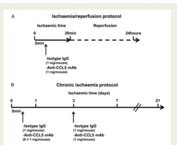
of 150 mL, 120 breaths/min) by supplementation with 100% oxygen, anaesthesia was maintained with 2% isoflurane. A thoracotomy was performed in the left third intercostal space and the pericardium was removed. Ligation of the left anterior coronary artery at the inferior edge of the left atrium was performed using an 8-0 Prolene suture. In ischaemia/reperfusion protocol, a small piece of polyethylene tubing was used to secure the ligature without damaging the artery. After 30 min of ischaemia, the occlusion of the left anterior coronary artery occlusion was released and reperfusion occurred. Reperfusion was confirmed by visible restoration of colour to the ischaemic tissue (Figure1A). In chronic ischaemia protocol, ligation was main-tained till animal sacrifice (Figure1B). In both protocols, after 5 min of ischaemia onset, either the rat anti-mouse CCL5 mAb (at doses ranging 0.1 – 1.0 mg/mouse) or the isotype control (1.0 mg/mouse) was administered intravenously to neutralize serum and cardiac CCL5 bioactivity in vivo. Next, the chest was closed and the ventilator removed to restore normal respiration. Sham-operated animals were submitted to the same surgical protocol as described but without arterial occlusion. In the chronic ischaemia protocol, a second intrave-nous injection of the anti-CCL5 mAb (1 mg/mouse) or the isotype control (1 mg/mouse) was administered after 3 days of chronic ischae-mia (Figure1A). At different time points of chronic ischaemia (from 8 h to 21 days) or reperfusion (1 day), animals were sacrificed for infarct size determination or immunohistochemical analysis.
Area at risk (AAR) and infarct size (I)
assessment
Area at risk and infarct size were assessed as described in the Supplementary material online.
Serum cardiac troponin I and
chemoattractant level detection
Levels of serum cardiac Troponin I (cTnI) and chemokines were measured as described in detail in the Supplementary material online.
Immunostaining
Immunostainings were performed as described in detail in the Supplementary material online.
Figure 1 In vivo myocardial infarction protocols. (A) Ischaemia/ reperfusion protocol. (B) Chronic ischaemia protocol.
Sirius Red staining for collagen content
Sirius Red staining was performed as described in detail in the Supplementary material online.
Magnetic resonance imaging (MRI)
MRI was performed at days 1 and 21 of chronic myocardial ischaemia in the same mice, as described in the Supplementary material online.
Statistical analysis
Statistical analysis was performed as described in detail in the Supplementary material online.
Results
Circulating and cardiac levels of CCL5
are up-regulated during chronic ischaemia
In order to investigate the possible role of CCL5 during chronic myocardial ischaemia, we assessed CCL5 expression within the
myocardium and in the serum of sham-operated and infarcted untreated mice (permanent ligation protocol). Chronic ischaemia significantly increased the cardiac expression of CCL5 after 3 and 7 days as compared to sham-operated mice (Figures2A and B). On days 1 and 3 of chronic ischaemia, serum levels of CCL5 were sig-nificantly increased as compared to sham-operated mice (Figure2C). At 1 day, CCL5 expression was detected in ventricular regions rich in infiltrating inflammatory cells (Supplementary material online Figure S1A), indicating that these cells have to be considered as the major candidates mediating the release of CCL5 in infarcted hearts. At 3 days of chronic ischaemia, CCL5 staining was particularly detected in macrophage-rich areas (Supplementary material online Figure S1B).
Blocking CCL5 reduces infarct size at 24 h
in I/R and chronic ischaemia protocols
In order to confirm and further validate the potential therapeutic benefit of CCL5 neutralization in myocardial infarction,14 we first assessed treatment with an anti-CCL5 mAb in a mouse model of
Figure 2 Chronic myocardial ischaemia after myocardial infarction increases cardiac and circulating levels of CCL5. Left anterior coronary artery permanent ligation was performed in male untreated C57Bl/6 mice. Intracardiac and serum levels of CCL5 were assessed at different time points of chronic ischaemia in untreated and sham-operated (Sham, sacrificed at 24 h) mice. Data are expressed as mean + SEM. (A) Quantification of CCL5 stained area within the infarcted myocardium (n ¼ 5 per group). (B) Representative images of CCL5-stained middle heart sections of infarcted and sham-operated hearts. (C) Serum CCL5 levels of sham-operated and chronic ischaemic untreated mice at differ-ent time points (n ¼ 7 – 9 per group).
F. Montecucco et al.
myocardial ischaemia/reperfusion. Similar to our results obtained with another CCL5 antagonist (44AANA47-RANTES),14the single
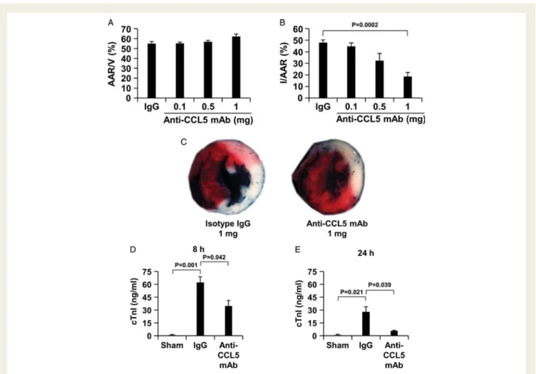
adminis-tration of the anti-CCL5 mAb reduced the infarct size (n ¼ 10, average + SEM, I/area at risk: 24.06% + 1.28) as compared to controls (n ¼ 10, average + SEM, I/area at risk: 50.76% + 6.1, P ¼ 0.0005). The benefit of the anti-CCL5 mAb treatment was also associated with a decrease in the serum cTnI levels at 24 h of reperfusion (n ¼ 7 per group, average + SEM, anti-CCL5 mAb vs. isotype control: 15.67 + 6.63 vs. 46.34 + 5.76 ng/ml, P ¼ 0.004). After in vivo validation of the anti-CCL5 mAb neutralizing activity, we investigated the role of CCL5 neutralization in the mouse model of chronic myocardial ischae-mia. The anti-CCL5 mAb or isotype control was administered via a single intravenous administration 5 min after the ischaemic period onset and infarct size was assessed after 24 h. In the chronic ischaemia protocol, area at risk (AAR) was similar in all treatment groups, indi-cating that ligature was reproducibly performed at the same level of the left anterior coronary artery (Figure 3A). Treatment with the anti-CCL5 mAb dose-dependently reduced the infarct as compared to the isotype control (Figures3B and C ). The anti-CCL5 mAb treat-ment significantly reduced serum cTnI levels (a marker of myocardial
necrosis) levels as compared to isotype-treated mice after 8 h and 24 h of reperfusion (Figures3D and E).15,16
Blocking CCL5 reduces neutrophil and
macrophage recruitment in infarcted hearts
during chronic ischaemia
To investigate whether neutralization of CCL5 modified leucocyte recruitment during chronic ischaemia, neutrophil, and macrophage infiltration was analysed at different time points (from 8 h through 21 days). Considering the kinetics of the serum levels and cardiac expression of CCL5 during chronic ischaemia (Figure2C) and half-life time (211 h), we administered two injections of the anti-CCL5 mAb or isotype control, i.e. at 5 min and 3 days after onset of chronic ischaemia. The anti-CCL5 mAb treatment significantly reduced neu-trophil infiltration on days 1 and 3 as compared to isotype controls (Figures 4A and B). The anti-CCL5 mAb also reduced macrophage (CD68+ cell) recruitment at 3 days of chronic ischaemia (Figures4C and D).
Figure 3 Single administration of anti-CCL5 mAb reduces infarct size and serum cardiac troponin I release after myocardial infarction. Anti-CCL5 mAb (0.1 – 1mg/mouse) or isotype IgG (1 mg/mouse) was administered 5 min after ischaemia onset. Data are expressed as mean + SEM. (A) Quantification of area at risk (AAR) per ventricle surface (V) after 24 h of chronic ischaemia (n ¼ 10 per group). (B) Quanti-fication of infarct size (I) per AAR after 24 h of chronic ischaemia (n ¼ 10 per group). (C) Representative images of TTC-stained middle heart sections of isotype IgG- or anti-CCL5 mAb-treated mice. Serum cardiac troponin I (cTnI) levels of sham-operated (Sham), 1 mg isotype- or 1 mg anti-CCL5 mAb-treated mice after 8 h (D) and 24 h (E) of chronic ischaemia (8 h: n ¼ 7 per group; 24 h: n ¼ 10 per group).
Blocking CCL5 reduces serum levels of
neutrophil and monocyte chemoattractants
during chronic myocardial ischaemia
CCL5 has been shown to induce neutrophil and monocyte chemotaxis in vivo and in vitro.17,18In order to investigate if CCL5 neutralization might influence neutrophil and macrophage recruitment within infarcted hearts, we analysed the possible reduction of serum levels of other chemoattractants (i.e. CXCL1, CXCL2, and CCL2).19,20 These chemokines were markedly up-regulated in mouse serum of isotype-treated mice after 8 h and 1 day of chronic ischaemia as com-pared to sham-operated mice (Figures 5A – C ). The anti-CCL5 mAb
treatment significantly reduced CXCL1 and CXCL2 serum levels at 8 h of chronic ischaemia as compared to isotype-treated mice (Figure 5A and B). The anti-CCL5 mAb also reduced CCL2 serum levels at 1 and 3 days of chronic ischaemia as compared to isotype-treated mice (Figure5C).
Blocking CCL5 transiently reduced MMP-9
and collagen amounts, but not MMP-8 in
infarcted hearts during chronic ischaemia
In order to investigate if the anti-CCL5 mAb treatment could modulate some molecular mediators of post-infarction cardiac remodelling
Figure 4 Treatment with anti-CCL5 mAb reduces neutrophil and macrophage infiltration in infarcted hearts during chronic ischaemia. (A) Quantification of infiltrated neutrophils per area in frozen sections of infarcted hearts at different time points of chronic ischaemia (0.3 – 21 days). (B) Representative images of neutrophil infiltration at 1 and 3 days of chronic ischaemia are shown. (C) Quantification of infiltrated macrophages per area in frozen sections of infarcted hearts at different time points of chronic ischaemia (0.3 – 21 days). (D) Representative images of macrophage (CD68+ cell) infiltration at 3 and 7 days of chronic ischaemia are shown. Data are expressed as mean + SEM (n ¼ 7 – 9 per group).
F. Montecucco et al.
during chronic ischaemia, we assessed the cardiac content of MMP-8, MMP-9, and different collagen types. Treatment with the anti-CCL5 mAb did not affect MMP-8 amounts at time points measured (from 1 to 21 days) as compared to isotype-treated mice (Figure 6A and data not shown). In contrast, MMP-9 content was significantly reduced in infarcted hearts after 3 days of chronic ischaemia as compared to the isotype-treated mice (Figure6B) and remained low. In addition, the anti-CCL5 mAb treatment also initially impaired total collagen production after 7 days of chronic ischaemia as compared to isotype-treated mice (Figures 7A and D). This effect was mainly associated with the reduction of collagen type III (Figure 7C), as collagen type I was not affected (Figure7B).
Blocking CCL5 improves survival and cardiac
function in infarcted mice during chronic
ischaemia
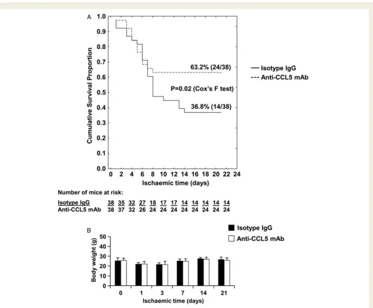
Mice treated with the anti-CCL5 mAb exhibited an overall survival rate of 63.2% (24/38) during 21 days of chronic ischaemia compared with mice treated with isotype IgG (survival rate 36.8%, 14/38, P ¼ 0.02, Figure 8A). During chronic ischaemia, neither the anti-CCL5
mAb-nor the isotype IgG-treated mice presented a decrease in body weight (Figure8B). No significant benefit in cardiac function (left ven-tricle ejection fraction [EF%]) was observed after 1 day of chronic ischaemia (Figures9A and B). At 21 days following chronic ischaemia, the EF% was markedly improved in the anti-CCL5 mAb- as compared to the isotype IgG-treated mice (Figure 9A and C ). Furthermore, cardiac end-systolic volume (ESV) and end-diastolic volume were improved by the anti-CCL5 mAb treatment, although without reaching statistical significance (Figure 9C, isotype-treated group [n ¼ 5] vs. anti-CCL5 mAb-treated group [n ¼ 14], average + SEM: ESV: 133.97 + 19.66 vs. 105.49 + 9.69 ml, P ¼ 0.171; EDV: 165.34 + 18.25 vs. 147.57 + 11.14 ml, P ¼ 0.422). Post-infarction adverse cardiac remodelling was also assessed by mouse heart weight, body weight, MRI determination of left ventricular dimensions and wall thickness, and histological assessment of cardiac myocyte size. After 21 days of chronic ischaemia, no difference between anti-CCL5 mAb- and isotype IgG-treated mice was observed in all parameters assessed (Table 1 and Supplementary material online Table S1), except for cardiac myocyte size that was increased in anti-CCL5 mAb-treated mice as compared to controls (Figure9D and E). Accordingly, at 21 days of chronic ischaemia, cardiac myocyte size positively
Figure 5 Treatment with anti-CCL5 mAb reduces neutrophil and macrophage chemoattractant serum levels during chronic ischaemia. Serum levels of CXCL1, CXCL2 (neutrophil chemoattractants) and CCL2 (monocyte/macrophage chemoattractant) were measured in sham-operated (Sham, sacrificed at 24 h), isotype IgG (1 mg/mouse)- or anti-CCL5 mAb (1 mg/mouse)-treated mice, respectively. Data are expressed as mean + SEM (n ¼ 7 – 12). (A) CXCL1; (B) CXCL2; and (C) CCL2.
correlated with left ventricle EF% (r ¼ 0.776, P ¼ 0.001). These data suggest that, after 21 days of chronic ischaemia, selective treatment neutralizing CCL5 improves infarct size and post-infarction with a marginal effect on left ventricular adverse remodelling.
Discussion
In the present study, we demonstrate that CCL5 is up-regulated at both systemic (blood stream) and local (within the infarcted myo-cardium) sites in animals during chronic ischaemia. In particular, a marked peak in CCL5 production was observed in the period between 3 and 7 days after myocardial infarction. Significant elevation of CCL5 serum levels have been reported in patients after acute myocardial infarction complicated by heart failure and
severe left ventricular dysfunction.21 Increased CCL5 serum
levels were also observed in patients with severe congestive heart failure (NYHA Class IV), independently of the cause of the
disease.23 At a local level, we have recently documented an
increase in CCL5 production within a few minutes of the initiating reperfusion in another model of myocardial ischaemia/reperfusion
using ApoE2/2 mice.14Thus, our results, that confirm the
obser-vations of others, imply that CCL5 would be an appropriate protein to target to modulate the inflammation underlying adverse cardiac remodelling. Due to the published mechanism of
action for CCL5,10,14,17,18we hypothesized that the selective
neu-tralization of CCL5 during the early phases (e.g. in the first 3 days) of permanent ischaemia would directly reduce neutrophil and
monocyte recruitment to myocardial infarcted areas. Indeed, we observed a potent inhibitory effect of the anti-CCL5 mAb was observed at days 1 and 3 for neutrophils and at 3 days for macro-phages. However, our data also suggest either an additional or an alternative mechanism, which could indirectly inhibit leucocyte recruitment within the infarcted hearts. The anti-CCL5 mAb treat-ment significantly reduced serum levels of other neutrophil (CXCL1 and CXCL2) and monocyte/macrophage (CCL2) che-moattractants as compared to the isotype-treated control group. These data indicate that the neutralization of CCL5 can be associ-ated with the reduction of serum levels of other classical leucocyte chemoattractants, thus impairing leucocyte migration via other
receptors.23
Such a precedent has been recently reported by Mei and colleagues, who demonstrated that removing the kine CXCL5 from the system leads to a decrease in other chemo-kines, such as CXCL1 and CXCL2, thereby altering their
gradient.23 Administration of the anti-CCL5 mAb not only
reduced leucocyte recruitment, but also infarct size. Thus, we have been able to reinforce our previous results using a different CCL5 antagonist in the murine model of cardiac I/R. In this study, the beneficial effect of CCL5 neutralization was also
shown at 24 h in the permanent ligation protocol.14 Taking all
results together then indicate a potential use of anti-CCL5 mAb to reduce infarct size, also mechanical or pharmacological myocardial reperfusion therapies are not performed.
Interestingly, we also showed that the anti-CCL5 mAb treat-ment improved post-infarction ‘clinical’ outcomes, such as survival
Figure 6 Treatment with anti-CCL5 mAb reduces MMP-9, but not MMP-8 release in infarcted hearts during chronic ischaemia. (A) Quanti-fication of MMP-8 stained area in frozen sections of sham-operated (Sham, sacrificed at 3 days) and infarcted hearts at different time points of chronic ischaemia (3 – 7 days). Data are expressed as mean + SEM (n ¼ 7 – 9 per group). (B) Representative images of MMP-8 stainings at 3 days of chronic ischaemia are shown. (C) Quantification of infiltrated MMP-9 stained areas in frozen sections of sham-operated (Sham, sacrificed at 3 days) and infarcted hearts at different time points of chronic ischaemia (3 and 7 days). Data are expressed as mean + SEM (n ¼ 7 – 9 per group). D. Representative images of MMP-9 stainings at 3 days of chronic ischaemia are shown.
F. Montecucco et al.
and cardiac function. Since the mice did not modify their body weight during permanent myocardial ischaemia, their death was most probably associated with the severity of cardiac heart failure or possibly with other post-infarction complications, such as cardiac arrhythmias. These long-term positive effects of anti-CCL5 mAb treatment were also observed on molecular mediators (such as MMP-9 and collagen) of cardiac remodelling
during chronic ischaemia.24,25These effects might actively
contrib-ute to the anti-CCL5 mAb-mediated improvement on cardiac function. The reduction in cardiac collagen content we observed
was a surprising result. In fact, these data are not in accordance with the classical paradigm showing an inverse association
between collagen and its degrading enzymes MMPs.23
Further-more, reduction in cardiac collagen content during post-infarction remodelling is not widely considered as a beneficial event, mainly
because of the increased risk in cardiac rupture.23Despite being
potentially adverse, the effect of anti-CCL5 mAb on collagen was relatively weak and certainly transient. In addition, it could be explained as a consequence of the reduced infarct size and corresponding scar surface in the anti-CCL5 mAb-treated mice,
Figure 7 Treatment with anti-CCL5 mAb transiently reduces collagen in infarcted hearts during chronic myocardial ischaemia. Quantifi-cations of total collagen (A), collagen I (B), and collagen III (C ) in frozen sections of sham-operated (Sham, sacrificed at 7 days) and infarcted hearts at different time points of chronic ischaemia (7 – 21 days). Data are expressed as mean + SEM (n ¼ 7 – 9 per group). (D) Representative microphotographs of infarcted hearts showing bright-field sections without polarization (figures on the left) and corresponding sections under polarized light illumination (figures on the right).
instead of an altered collagen deposition in scar tissue. This specu-lation was also suggested by the absence of post-infarction myo-cardial rupture events observed in the entire animal study cohort during autopsy. Interestingly, treatment with anti-CCL5 mAb did not induce any significant improvement in certain cardiac remodelling parameters (such as the measurement of heart weight and by MRI determination of ventricular dimensions and diastolic wall thickness at mid-papillary muscle level) at 21 days of chronic ischaemia. On the other hand, in the same mice submitted to MRI protocol, cardiac myocyte size was increased in anti-CCL5 mAb-treated mice as compared to controls and it positively correlated with left ventricle EF% (r ¼ 0.776, P ¼ 0.001). Although treatment with anti-CCL5 mAb induced only marginal effects on regional adverse cardiac remodelling (assessed by MRI), the increase of viable cardiomyocyte surface at 21 days in
the same mice should be considered as a crucial determinant of the improvement in cardiac function. To summarize, our results suggest that anti-CCL5 mAb treatment was enough to reduce the infarct size and leucocyte infiltration in the first few days post-myocardial infarction. These beneficial effects resulted in a delayed increase in cardiomyocyte size and left ventricular function at 21 days. Conversely, acute anti-CCL5 treatment was not sufficient to significantly modify cardiac adverse remodelling. Since infarct size and adverse cardiac remodelling have been shown as strongly associated with the development of post-infarction heart failure,
our results might appear as apparently discordant.26 However,
our treatment approach was planned to selectively neutralize CCL5-mediated leucocyte inflammation in the first few days post-myocardial infarction, thus potentially interfering with early cardiac
repair and healing.4
Figure 8 Treatment with anti-CCL5 mAb improves survival in mice after acute myocardial infarction followed by chronic myocardial ischae-mia. (A) Kaplan – Meier survival curve following myocardial infarction and permanent ischaemia in isotype IgG- and anti-CCL5 mAb-treated mice (n ¼ 38 per group). (B) Body weight of isotype IgG- and anti-CCL5 mAb-treated mice during permanent ischaemia (from the day of surgery [time 0] till 21 days of chronic ischaemia). Data are expressed as mean + SEM (n ¼ 11 in isotype-treated mouse group and n ¼ 23 in anti-CCL5 mAb-treated mouse group). Differences in body weight at any time point investigated and also between treatment groups were not statistically significant.
F. Montecucco et al.
In conclusion, we have shown that CCL5 is highly expressed during chronic ischaemia following myocardial infarction. Adminis-tration of a neutralizing anti-CCL5 mAb during early phases of
ischaemia reduced infarct size in vivo in two mouse models (myo-cardial ischaemia/reperfusion and permanent ischaemia). The car-dioprotective effects of CCL5 neutralization during early phases of chronic ischaemia were associated with the decrease of neutro-phil and macrophage recruitment in the infarcted myocardium. Furthermore, treatment with anti-CCL5 mAb induced a reduction in MMP-9 levels yet, only a transient impairment of collagen content and an increase in cardiac myocyte size. These partial changes on well-accepted molecular mediators of cardiac remodel-ling resulted in marginal effects on regional adverse cardiac remo-delling (as assessed by MRI). However, anti-CCL5 mAb-mediated increase in cardiomyocyte size at 21 days was strongly associated with the improvement of left ventricle ejection fraction. Indeed, after 21 days, treatment with the anti-CCL5 mAb also improved the survival rate. Therefore, neutralization of CCL5 bioactivity
Figure 9 Treatment with anti-CCL5 mAb improves left ventricular function after 21 days of chronic ischaemia. (A) Left ventricular function was evaluated in the same animals subjected to left coronary permanent ligation at days 1 and 21 after myocardial infarction. Ejection fraction (EF %) is expressed as mean + SEM (n ¼ 5 in isotype IgG-treated mouse group and n ¼ 14 in anti-CCL5 mAb-treated mouse group). (B) Repre-sentative magnetic resonance images at mid papillary muscle level of end-systolic volume (ESV) and end-diastolic volume (EDV) of isotype IgG-and anti-CCL5 mAb-treated mice at 1 day of chronic ischaemia are shown. (C) Representative magnetic resonance images at mid-papillary muscle level of end-systolic volume (ESV) and end-diastolic volume (EDV) of the same isotype IgG- and anti-CCL5 mAb-treated mice after 21 days of chronic ischaemia are shown. (D) Quantitative analysis of cardiac myocyte cross-sectional area (heavy chain cardiac myosin staining) at 21 days post-myocardial infarction. Results are expressed as mean + SEM of positive pixels (n ¼ 5 in isotype IgG-treated mouse group and n ¼ 8 in anti-CCL5 mAb-treated mouse group). (E) Representative images of heavy chain cardiac myosin staining of cardiac sections at mid-papillary muscle level.
. . . . Table 1 Heart and body weights at 21 days of chronic ischaemia of mice submitted to MRI
Measurements Isotype IgG-treated mice (n 5 5) Anti-CCL5 Ab-treated mice (n 5 14) P-value Heart weight (mg) 178.5 + 6.4 185.8 + 3.3 0.2906 Body weight (g) 28.4 + 0.4 28.1 + 0.4 0.5932 Data are expressed as mean + SEM.
during the early phase of chronic ischaemia following myocardial infarction could represent a promising therapy to reduce infarct size, post-infarction heart failure, and related mortality.
Supplementary material
Supplementary material is available at European Heart Journal online.
Funding
This research work was funded by EU FP7, Grant number 201668, AtheroRemo, a grant from the Swiss National Science Foundation (310030-118245) and a grant Foundation Novartis Foundation to Dr. F. Mach. This work was funded by the ‘Sir Jules Thorn Trust Reg’ fund and Gustave and Simone Pre´vot fund to Dr. F. Montecucco. This work was supported by a grant from the Foundation Ernst et Lucie Schmidheiny to Dr. Braunersreuther. Conflict of interest: None to be declared.
References
1. Pfeffer MA, Braunwald E. Ventricular remodeling after myocardial infarction. Experimental observations and clinical implications. Circulation 1990;81: 1161 – 1172.
2. St John Sutton M, Lee D, Rouleau JL, Goldman S, Plappert T, Braunwald E, Pfeffer MA. Left ventricular remodeling and ventricular arrhythmias after myocar-dial infarction. Circulation 2003;107:2577 – 2582.
3. Nian M, Lee P, Khaper N, Liu P. Inflammatory cytokines and postmyocardial infarction remodeling. Circ Res. 2004;94:1543 – 1553.
4. Frangogiannis NG. The immune system and cardiac repair. Pharmacol Res 2008;58: 88 – 111.
5. Frantz S, Bauersachs J, Ertl G. Post-infarct remodelling: contribution of wound healing and inflammation. Cardiovasc Res 2009;81:474 – 481.
6. Weir RA, Murphy CA, Petrie CJ, Martin TN, Clements S, Steedman T, Wagner GS, McMurray JJ, Dargie HJ. Monocyte chemoattractant protein-1: a dichotomous role in cardiac remodeling following acute myocardial infarction in man? Cytokine 2010;50:158 – 162.
7. Orn S, Breland UM, Mollnes TE, Manhenke C, Dickstein K, Aukrust P, Ueland T. The chemokine network in relation to infarct size and left ventricular remodeling following acute myocardial infarction. Am J Cardiol 2009;104:1179 – 1183. 8. Liehn EA, Merx MW, Postea O, Becher S, Djalali-Talab Y, Shagdarsuren E,
Kelm M, Zernecke A, Weber C. Ccr1 deficiency reduces inflammatory remodel-ling and preserves left ventricular function after myocardial infarction. J Cell Mol Med 2008;12:496 – 506.
9. Dobaczewski M, Xia Y, Bujak M, Gonzalez-Quesada C, Frangogiannis NG. CCR5 signaling suppresses inflammation and reduces adverse remodeling of the infarcted heart, mediating recruitment of regulatory T cells. Am J Pathol 2010; 176:2177 – 2187.
10. Ferrandi C, Ardissone V, Ferro P, Ruckle T, Zaratin P, Ammannati E, Hauben E, Rommel C, Cirillo R. Phosphoinositide 3-kinase gamma inhibition plays a crucial
role in early steps of inflammation by blocking neutrophil recruitment. J Pharmacol Exp Ther 2007;322:923 – 930.
11. Houard X, Touat Z, Ollivier V, Louedec L, Philippe M, Sebbag U, Meilhac O, Rossignol P, Michel JB. Mediators of neutrophil recruitment in human abdominal aortic aneurysms. Cardiovasc Res 2009;82:532 – 541.
12. Braunersreuther V, Steffens S, Arnaud C, Pelli G, Burger F, Proudfoot A, Mach F. A novel RANTES antagonist prevents progression of established atherosclerotic lesions in mice. Arterioscler Thromb Vasc Biol. 2008;28:1090 – 1096.
13. Terao S, Yilmaz G, Stokes KY, Russell J, Ishikawa M, Kawase T, Granger DN. Blood cell-derived RANTES mediates cerebral microvascular dysfunction, inflam-mation, and tissue injury after focal ischemia-reperfusion. Stroke 2008;39: 2560 – 2570.
14. Braunersreuther V, Pellieux C, Pelli G, Burger F, Steffens S, Montessuit C, Weber C, Proudfoot A, Mach F, Arnaud C. Chemokine CCL5/RANTES inhibition reduces myocardial reperfusion injury in atherosclerotic mice. J Mol Cell Cardiol 2010;48:789 – 798.
15. Montecucco F, Lenglet S, Braunersreuther V, Burger F, Pelli G, Bertolotto M, Mach F, Steffens S. CB(2) cannabinoid receptor activation is cardioprotective in a mouse model of ischemia/reperfusion. J Mol Cell Cardiol 2009;46:612 – 620. 16. Eckle T, Grenz A, Ko¨hler D, Redel A, Falk M, Rolauffs B, Osswald H, Kehl F,
Eltzschig HK. Systematic evaluation of a novel model for cardiac ischemic precon-ditioning in mice. Am J Physiol Heart Circ Physiol 2006;291:H2533 – H2540. 17. Jan MS, Huang YH, Shieh B, Teng RH, Yan YP, Lee YT, Liao KK, Li C. CC
chemo-kines induce neutrophils to chemotaxis, degranulation, and alpha-defensin release. J Acquir Immune Defic Syndr 2006;41:6 – 16.
18. Furuichi K, Gao JL, Horuk R, Wada T, Kaneko S, Murphy PM. Chemokine recep-tor CCR1 regulates inflammarecep-tory cell infiltration after renal ischemia-reperfusion injury. J Immunol 2008;181:8670 – 8676.
19. McColl SR, Clark-Lewis I. Inhibition of murine neutrophil recruitment in vivo by CXC chemokine receptor antagonists. J Immunol. 1999;163:2829 – 2835. 20. Rollins BJ. Chemokines. Blood 1997;90:909 – 928.
21. Parissis JT, Adamopoulos S, Venetsanou KF, Mentzikof DG, Karas SM, Kremastinos DT. Serum profiles of C-C chemokines in acute myocardial infarc-tion: possible implication in postinfarction left ventricular remodeling. J Interferon Cytokine Res 2002;22:223 – 239.
22. Aukrust P, Ueland T, Mu¨ller F, Andreassen AK, Nordøy I, Aas H, Kjekshus J, Simonsen S, Frøland SS, Gullestad L. Elevated circulating levels of C-C chemo-kines in patients with congestive heart failure. Circulation 1998;97:1136 – 1143. 23. Mei J, Liu Y, Dai N, Favara M, Greene T, Jeyaseelan S, Poncz M, Lee JS,
Worthen GS. CXCL5 regulates chemokine scavenging and pulmonary host defense to bacterial infection. Immunity 2010;33:106 – 117.
24. Fang L, Gao XM, Moore XL, Kiriazis H, Su Y, Ming Z, Lim YL, Dart AM, Du XJ. Differences in inflammation, MMP activation and collagen damage account for gender difference in murine cardiac rupture following myocardial infarction. J Mol Cell Cardiol 2007;43:535 – 544.
25. Moshal KS, Rodriguez WE, Sen U, Tyagi SC. Targeted deletion of MMP-9 attenu-ates myocardial contractile dysfunction in heart failure. Physiol Res 2008;57: 379 – 384.
26. Parodi G, Memisha G, Carrabba N, Signorini U, Migliorini A, Cerisano G, Antoniucci D. Prevalence, predictors, time course, and long-term clinical impli-cations of left ventricular functional recovery after mechanical reperfusion for acute myocardial infarction. Am J Cardiol. 2007;100:1718 – 1722.
F. Montecucco et al.