HAL Id: hal-03108284
https://hal.archives-ouvertes.fr/hal-03108284
Submitted on 1 May 2021HAL is a multi-disciplinary open access archive for the deposit and dissemination of sci-entific research documents, whether they are pub-lished or not. The documents may come from teaching and research institutions in France or abroad, or from public or private research centers.
L’archive ouverte pluridisciplinaire HAL, est destinée au dépôt et à la diffusion de documents scientifiques de niveau recherche, publiés ou non, émanant des établissements d’enseignement et de recherche français ou étrangers, des laboratoires publics ou privés.
Ontogenetic dynamics of the subepidermal spicule
complex in Nudibranchia (Gastropoda): the case of
Onchidoris muricata
Ekaterina Nikitenko, Alexander Ereskovsky, Elena Vortsepneva
To cite this version:
Ekaterina Nikitenko, Alexander Ereskovsky, Elena Vortsepneva. Ontogenetic dynamics of the subepi-dermal spicule complex in Nudibranchia (Gastropoda): the case of Onchidoris muricata. Zoology, Elsevier, 2021, 144, pp.125886. �10.1016/j.zool.2020.125886�. �hal-03108284�
Ontogenetic dynamics of the subepidermal spicule complex in Nudibranchia (Gastropoda): the case of Onchidoris muricata
Nikitenko Ekaterina*1, Ereskovsky Alexander 2,3,4, Vortsepneva Elena 1
1
Invertebrate Zoology Department, Faculty of Biology, Lomonosov Moscow State University, Leninskie Gory 1/12, 119234, Moscow, Russia
2
Embryology Department, Faculty of Biology, Saint-Petersburg State University, Universitetskaya emb. 7/9, 199034, Saint-Petersburg, Russia
3
Institut Méditerranéen de Biodiversité et d’Ecologie marine et continentale (IMBE), Aix Marseille University, CNRS, IRD, Avignon University, Station Marine d’Endoume, Rue de la Batterie des Lions, 13007, Marseille, France
4
Koltzov Institute of Developmental Biology of Russian Academy of Sciences, Moscow, Russia
* Corresponding author: Invertebrate Zoology Department, Biological Faculty, M. V. Lomonosov Moscow State University, Leninskie Gory 1/12, 119234 Moscow, Russia, e-mail:
nikitenkocatia@yandex.ru
alexander.ereskovsky@imbe.fr vortcepneva@gmail.com
Abstract
Spicules are mineral-based biocomposites skeletal structures that are widely distributed among phylogenetically distant groups of invertebrates (Porifera, Cnidaria, Mollusca, Echinodermata). Subepidermal spicules are formed under the ectodermal epithelium and are characterized for all groups except mollusks (Aplacophora, Polyplacophora, Bivalvia), their spicules are located on the surface of the body. However, one group of mollusks (Gastropoda: Heterobranchia) have unique subepidermal spicules that have never been detected above the ectodermal epithelium and similarly to those characterized for Porifera, Cnidaria and Echinodermata. Understanding subepidermal spicule formation in mollusks could help solve the question on the origin of spicules. Spicules in nudibranchs have been described for more than 150 years, yet ontogenetic dynamics of spicules have never been studied and the full mechanism of their formation remains unknown. Herein we investigate the spicule formation in different stages of postlarval development of the nudibranch Onchidoris muricata (O.F. Müller, 1776). For the first time, ontogenetic transformations of the spicule complex are described using experiments and different morphological methods. Our studies demonstrate that spicules of O.
muricata form in the subepidermal space in early developmental stages immediately after veliger
settlement. A single spicule forms inside a huge vacuole within a sclerocyte and remains there throughout the entire life of the specimen. Signs of spicule or sclerocyte migration under the epithelium in postlarval development was not found. Spicules only form during larval settlement, increasing only in size as development furthers. For the first time, spicule mineralization zones were detected at the tips of the spicules as well as the presence of collagen I in the overall composition of the spicules. Thus, our findings suggest that spicules form by an ectodermal cell that emerged under the ectodermal epithelium during the earliest stages of postlarval development.
Keywords
3D-reconstruction; micro-CT; sclerocyte; calcareous spicules; spiculogenesis; ultrastructure
1. Introduction
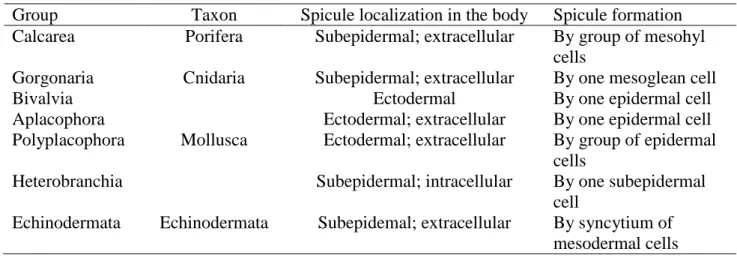
Calcareous spicules are the as common morphological form of mineral-based biocomposites to be found in different metazoans groups including Porifera, Cnidaria, Mollusca and Echinodermata (Morse, 1976; Ledger and Jones, 1977; Kingsley and Watabe, 1984; Willmer, 1990) (Table 1).
Table 1. Calcareous spicule formation in invertebrates base on literature data
Group Taxon Spicule localization in the body Spicule formation Calcarea Porifera Subepidermal; extracellular By group of mesohyl
cells
Gorgonaria Cnidaria Subepidermal; extracellular By one mesoglean cell Bivalvia
Mollusca
Ectodermal By one epidermal cell Aplacophora Ectodermal; extracellular By one epidermal cell Polyplacophora Ectodermal; extracellular By group of epidermal
cells
Heterobranchia Subepidermal; intracellular By one subepidermal cell
Echinodermata Echinodermata Subepidemal; extracellular By syncytium of mesodermal cells
All eight major lineages of Mollusca produce calcified spicules, that vary by localization and formation mode in ectodermal epithelium or in subepidermal space (Salvini-Plawen, 1972; Leise, 1984; Harper et al., 2006; Kocot et al., 2016) (Table 1). Epidermal spicules meet in Bivalvia, Aplacophora, Polyplacophora formed by single (Bivalvia, Aplacophora) or group (Polyplacophora) of epidermal cells (Beedham and Trueman, 1968; Salvini-Plawen, 1972; Carter and Aller, 1975; Leise, 1984; Kocot et al., 2016) (Table 1). Calcareous subepidermal spicules are unique among mollusks (Morse, 1976; Brenzinger et al., 2011) characterize only for Heterobranchia (Acochlidiimorpha, Rhodopoidea and Nudibranchia) (Kocot et al., 2016; Sigwart, 2017). Heterobranchs spicules have been studied for over 100 year (Lankester, 1883), but the way of formation was described only once (Prenant, 1924). Spicules have a mesodermal mitochondrial origin, which was described using only the light microscopy method (Prenant, 1924). . Spicule or secondary-shell formation in the body wall is associated with shell loss (Kocot et al., 2016). Spicules take on a supporting function instead of a shell (Thompson, 1960; Ros, 1976; Todd, 1981; Cataneo-Vietti, 1993; Wägele and Willan, 2000). The spicules may be a remainder of the shells of gastropods (Ros, 1976) or their synapomorphy features (Cattaneo-Vietti et al., 1993).
Suborder Doridina (Nudibranchia) is a worldwide distributed exclusively free-living marine mollusks group with more than 2000 species (Wägele and Willan, 2000; Hallas et al.,
2017). Many Doridina have internalized calcareous spicules. Their spicules consist of insignificant amounts of organic material (Woodland, 1907), multiple forms of calcium carbonate (Odum, 1951; Ehrlich, 2019), fluorite and various magnesium compounds (Lowenstam and Weiner 1983; Cattaneo-Vietti et al., 1995; Weiner et al., 2003).
While these spicules have been previously described several times over (Alder and Hancock, 1855; Woodland, 1907; Prenant, 1924; Edmunds, 1968; Kress, 1981; García et al., 1986; Foale and Willan, 1987; Cattaneo-Vietti et al., 1993, 1995; Kress, 1981; Wägele and Cervera, 2001; Penney, 2006), description of the morphology of the spicules was confined solely to external characters of shape and size (Chang et al., 2013; Sánchez-Tocino et al., 2014) or general internal morphology without any ultrastructural details (Cattaneo-Vietti et al., 1995). Ultrastructural micrographs from transmission electron microscopy (TEM) are generally lacking, however, studies dedicated to epithelium morphology (Àvila and Durfort, 1996) or surface structures, i.e., caryophyllidia have inadvertently revealed accompanying spicule characteristics (Foale, 1987). Yet, there are still no published accounts of the spicule ultrathin morphology.
More recently, studies have focused on spicule localization in the body (Penney, 2006, 2008; Kasamesiri et al., 2011; Alba-Tercedor and Sánchez-Tocino, 2011; Chang et al., 2013; Sánchez-Tocino et al., 2014; Nikitenko and Vortsepneva, 2020; Penney et al., 2020). Spicules not lying chaotically in the body, they formed the spicule tracts which are organized into spicule networks (Penney 2006, 2008; Kasamesiri et al., 2011, Chang et al., 2013; Nikitenko and Vortsepneva, 2020; Penney et al., 2020)
In the foot and notum of mollusks spicules lies along the body wall in arterior-posterior axis and form two independed horizontal networks: notum horizontal network and foot horizontal network respectively. Horizontal networks can be forming by different tracts, because orientation of spicules in central part of notum and edge of notum or foot can be different (Penney, 2008). Vertical network located in the tubercles, when spicules lie perpendicular to spicules of notum in dorso-ventral axis and can form rossete, ring, rod, mound like structures (Penney, 2008). Tubercles are supporting by layer of spicules, that located under them. These spicules form stellular tract (Nikitenko and Vortsepneva, 2020). Spicules lie at an angle to the spicules of vertical and horizontal tracts in intermediate zone between tubercles and notum.
Previously correlation between the thickness of the integument and the thickness of the spicular tracts was noted (Nikitenko and Vortsepneva, 2020). The density of the integument depends not on the number of spicules, but on the nature of their localization relative to each other and the thickness of the layer in which they are located. This correlation was explained ecological differences between species (Nikitenko and Vortsepneva, 2020).
Spicule networks predominantly contain four spicule types: monoactine, diactine, triactine and tetractine (Thompson, 1958; Kress, 1981; Chang et al., 2013; Nikitenko and Vortsepneva, 2020). Spherical spicules, spherical spicules with small tubercles and v-shape spicules are rarely founded (Sánchez-Tocino et al., 2014). The spicules have different internal structures which have also been described: empty, concentric, mixed and monolithic (Foale and Willan, 1987; Cattaneo-Vietti et al., 1995; Nikitenko and Vortsepneva, 2020). Internal structures of spicules do not depend on spicules shape (Nikitenko and Vortsepneva, 2020).
At present, two types hypotheses of nudibranch spicule formation exist: epidermal formation then sink and mesodermal formation. The first has been based only on histological sections. According to this hypothesis, one cell of the veliger mantle forms a single spicule from a mineral granule formed by calcareous sedimentation. After the settlement and shading of the veliger shell, the spicule is absorbed into the epidermis and held in a subepidermal position (Woodland, 1907; Thompson, 1958; Rose, 1983). The second has been based only on light microscopy observation. The second hypothesis presumes spicule formation in the subepidermal space initially, where all adult spicules (Prenant, 1924; Àvila and Durfort, 1996). Spicules form by mitochondria of scleroblasts in subepidermal space (Prenant, 1924). The mitochondria stretch, take a rod-shaped form and contains primary calcium particles (Prenant, 1924). Spicules grow due to the contents of other mitochondries in the scleroblast, which is mineralized on the surface of the growing spicule (Prenant, 1924).
Despite the detailed information on spicule morphology, the questions of spicule origination, biomineralization mode, spicule morphogenesis and chemical composition during development still remain. Anatomical observations of living animals are critically important to understanding the evolution of mollusks and indeed any organism. Considering, that spicules and scale armour have evolved convergently in multiple gastropods and also cephalopods. Study of spicule anlage may provide a better understanding towards the origin of mollusk spicules. This work is the first detailed study of the ontogenetic dynamics of the spicule complex during postlarval development of the dorid nudibranch Onchidoris muricata (O.F. Müller, 1776).
O. muricata is a widespread common species, including the White Sea. Adults inhabit
intertidal and subtidal zones, preferentially grazing the cheilostome polyzoan Electra pilosa (Linnaeus, 1767). O. muricata is a hermaphrodite mollusk, strictly annual. Spawning begins in late winter or early spring (Todd, 1979), laying small eggs that hatch as poorly-developed planktotrophic veliger larvae. Juveniles have been detected on the shells of dead horsemussels,
Modiolus modiolus (Linnaeus, 1758).
O. muricata spicule complex was detailed described previously by using complex method
Nikitenko and Vortsepneva, 2020). All needle-like spicules forms occur in the O. muricata body, but dominantly having tetractine spicules (Nikitenko and Vortsepneva, 2020). Dense clusters of spicules form the horizontal tract of the notum. Density of spicules in the central part of notum little decrease (Penney et al., 2020). In the marginal zone, the spicules lie parallel to each other, but perpendicular to the body axis. The marginal zones of notum contain predominantly multiaxial spicules. The secondary axes of spicules are located one below the other. The horizontal tract of the foot is formed by spicules arranged in layers. The spicules of vertical tract in the tubercles form a rosette-like structure. Spicules under the tubercle diverge radially from the base and form the stellular tract (Nikitenko and Vortsepneva, 2020). Spicules at networks are localized orderly and very density to each other. The arrangement of spicules allows for efficient coverage, increase the strength of the body wall while using fewer spicules. The strength of the covers depends not on the number of spicules, but on the nature of their localization relative to each other.
2. Material and methods
Specimens of Onchidoris muricata were collected at 12 – 15 m depth in the White Sea (Kandalaksha Bay; 66°34 N, 33°08 E), near the Pertsov White Sea Biological Station, Moscow State University by SCUBA diving. The smallest just settled juveniles (100 – 1000 µm length) were sampled from the meiobenthos, specifically large, well-washed shell from 20 – 22 m depth. A total of 168 specimens of O. muricata from 100 μm to 12 mm length were studied using light (n = 69) and fluorescence microscopy (n = 28), scanning (n = 41) and transmission electron (n = 9) microscopy, confocal laser scanning microscopy (n = 12) and computer microtomography (n = 9).
2.1. Light microscopy (LM)
Different pieces of the foot, the notum and the tubercles of O. muricata were placed in a bleach (10% NaOCl solution) after anesthetized in order to study individual spicules. Soft tissue dissolution occurred at room temperature after 10 – 15 minutes. After that, spicules were washed with distilled water. Total spicule preparations were studied with a Leica DM 2500 (Leica, Germany) light microscope, Nikon eclipse 200 (Nikon, Japan).
Spicules measurements were carried out under a light microscope or scanning electron microscope using a scale segment or an electronic ruler. The length of multiaxial spicules was estimated by the length of the main (largest) axis.
All specimens for electron microscopy were quickly studied (maximum per month) and did not store in a fixator a lot of time for avoid the artifacts.
Scanning electron microscopy (SEM). Specimens of O. muricata were relaxed using a
4% magnesium chloride (MgCl2) solution isotonic to seawater for 2 – 12 hours (depending on
size). Specimens were fixed for EM in 2.5% glutaraldehyde in 0.1 М phosphate buffered saline (PBS) (or PB Millonig's pH 7.4 (0.1 M; Millonig, 1964 (unpublished data)) for one hour at 4ºC. Afterwards, the initial solution was replaced with new, and the fixation continued for an additional hour. The mollusks were then washed in the same buffer 3 times for a total of 1.5 hours, then carried through a series of increasing concentrations (10%, 30%, 50%) of ethyl alcohol up to 96%. Solutions were changed twice at each stage after 20 minutes. Fixations were carried out similarly in a mixture of alcohol and acetone with ratios of 3:1, 1:1, 1:3, respectively. Immediately following, mollusks were transferred to pure acetone and critical point dried.
Dried objects were divided into fragments of notum, foot and tubercles with tweezers and dissecting needles. The internal structure of the body and spicules was studied on scrappings using a JEOL JSM – 6380L (JEOL, USA), CamScan S2 (Cambridge Instrument Scientific Company, Great Britain) and Hitachi S405A SEM (Hitachi, Japan).
Transmission electron microscopy (TEM). Internal structures and mutual arrangement of
spicules were studied by TEM followed by three-dimensional reconstruction. Specimens were postfixed with 1% osmium tetroxide in 0.1 M PBS for 2 – 6 hours in the dark after fixation with 2.5% glutaraldehyde in 0.1 M PBS as described above. A saturated solution of chelator (ethylenediaminetetraacetate, EDTA) was used for decalcification, at pH = 7 at room temperature after 3-8 hours. Decalcified specimens were washed with distilled water, which was replaced every 20 min for 3 hours. Washed specimens were then treated with an ascending ethanol series (10%, 30%, 50%, 70%, 85%, 96%), twice changing the solution in each stage after 20 min. After dehydrating in ethanol, specimens were placed in a resin embedding series of acetone and resin (3:1, 1:1, 1:3, respectively), each stage lasting 24 hours. Final embedding was in pure Spurr’s epoxy resin (Sigma Aldrich).
Semithin and ultrathin section. We used 1 µm thick semithin sections for 3D
reconstruction. Sections were obtained using LKB Bromma 2088 Ultratome V, Dupont MT -5000 microtomes (DuPont Company, USA). Ultrathin 70 nanometers thick sections were obtained using a Leica EM UC6 ultratome using a diamond knife (Histo Jumbo, Diatome, Biel, Switzerland and PELCO 12785SU, Ted Pella, USA). Semithin sections were stained by a solution of methylene blue for 30 – 60 sec. Then the sections were photographed using a Nikon Eclipse - 200 microscope. All ultrathin sections were stained using 1% uranyl acetate for 40 min at 35°C, followed by 0.4% lead citrate for 10 min at room temperature. Micrographs of ultrathin
sections were made using transmission electron microscopes JEM - 1011, JEM 100B, JEOL JEM – 1400 Flash (JEOL, Japan).
2.3. Three-dimensional reconstruction (3D reconstruction)
Three-dimensional (3D) reconstruction of the photographed sections was done using a Leica DM 2500 digital camera coupled to a light microscope (Nikon Eclipse e200). Three-dimensional (3D) reconstructions were made from a series of semithin sections 1 µm in thickness as described by Ruthensteiner and Heß (2008). The digitized images of the slices were converted to 8-bit format. Image stacks were aligned using AMIRA 5.2.2 (Amira Visaging GmbH, Germany) and reconstructed using Imaris 7.0.0 (BitplaneAG, Zurich, Switzerland).
2.4. Confocal laser scanning microscopy (CLSM)
Specimens of O. muricata were fixed in 4% paraformaldehyde (PFA) in 0.1M phosphate buffer for 4 hours at 4°C after relaxing specimens in 4% MgCl2. Specimens were then washed
with 0.1 M PBS (or PB Millonig's pH 7.4 (0.1 M; Millonig, 1964 (unpublished data))). Next, specimens were washed in the PBS or PB Millonig's buffer 3 – 5 times for 30 min. Specimens were fixed in PBT solution for 4 hours. After that, collagen was detected with immunohistochemical staining and performed using antibodies Anti-Collagen I (rabbit) (DAR 647, Invitrogen)). Stained in antibodies I and II was for 24 hours. After each steps specimens were washed in the Blocking solution for 8 hours. The nucleus was stained using Propidium Iodide (PI, 5 μg / mL in PBS, Sigma) and DAPI (10 μg / mL in PBS, Sigma). The samples were cleared with glycerol or Murrey Clear. Calcein (calcein disodium salt (1 μg / mL in SW; Honeywell Fluka, Santa Cruz Biotechnology) was used to study calcification processes.
The prepared samples were examined using a Nikon A1R – A1 confocal microscope (Nikon Corporation, Japan) and a Leika DM2500 fluorescence microscope with an ebq-100 lamp (Leica Biosystems Division of Leica Microsystems Inc., USA).
2.5. Computer microtomography (µCT)
Quantitatively counting of spicules in the central part of the body was held by means of X-ray microtomography using microtomograph SkyScan 1172 (Bruker MicroCT, Belgium) at the Resource Centre for X-ray Diffraction Studies of Saint Petersburg State University (Saint Petersburg, Russia). Samples were prepared similarly to the scanning microscopy method. Dried specimens were placed in a plastic cylinder or in Eppendorf tube size 1.5 ml. The cylinder was closed at both ends to prevent the movement of the object. Best scans were obtained by placing
The SkyScan 1172 settings were as follows: Cu radiation, acceleration voltage – 91 kV, resolution – 1.66 μm, 2 μm, and 2.69 μm, crystal rotation angle – 0.2 grad., 4 scans in one position, exposure – 1.27, 1.42, and 1.47 sec. For reconstruction of the shadow image array, a NRecon software package (BrukerMicroCT) was used, since it is capable of neutralizing the instrumental artefacts and can provide a gray scale diapason corresponding to X-ray absorption, and, consequently, to chemical composition of the sample. Microtomographic sections obtained were analyzed using DataViewer and CTVox software packages.
2.6. Experiment with calcein
Calcein (calcium disodium salt) was used to detect calcium deposition zones. Three replicates of the experiment were performed, lasting a total of 21 days. We used 28 specimens of
O. muricata, ranging from 1 to 12 mm length. All individuals were kept in plastic Petri dishes at
a temperature of 8°C in a solution of calcein with seawater (saturated solution of calcein in a ratio of 1:10 with seawater). The solution was changed every 2 days. Individuals were monitored using a Leica DM 2500 microscope with appropriate fluorescence filters. Mollusks were washed before use in microscopy using clean sea water with shaking for 2 – 24 hours (time is an empiric parameter: washed until mollusks look clear and the autofluorescence from calcein disappeared). After that, specimens were placed in depression slides and examined from both the dorsal and ventral sides. Once finished, specimens were returned to the calcein solution with the appropriate concentration. In addition to detecting spicules inside the body, spicules were extracted from the body using dissecting needles and photographed using a Nikon A1R – A1 confocal microscope.
2.7. Terminology
Network – complex of tracts in a particular compartment of the body.
Sclerocyte (synonymous to «spicule cell» (Foale, 1987), «spicule forming cell» (Beniash
et al., 1999)) – the cell forming spicule inside of the vacuole. The main feature of this cell is a big vacuole whereby the spicule nearly occupies the entire cell. This is the first time this term has been used for Nudibranchia by analogy to other invertebrates (Porifera, Cnidaria).
Stellular tract («tubercle base network» after Penney (2006)) – newly described tract
(Nikitenko and Vortsepneva, 2020) located between tubercle and notum; provides a supportive function for the tubercle.
Tract – a cluster of spicules oriented in a similar way.
Tubercle (synonymous to papillae (Alder and Hancock, 1855; Foale, 1987)) – outgrowth
3. Results
Based on the details of the general and internal morphology, we defined the three ontogenetic groups of Onchidoris muricata at postlarval development: group I has a length from 100 µm to 2 mm; group II from 2 to 6 mm; and group III from 6 to 12 mm. Body wall of all stages of postlarval development consists of ectodermal epithelium, subepidermal spicules and a collagen matrix with fibroblasts and other cells resembling muscle cells and neurons (Fig. 1). Differences between each group include the transparency of the body wall, stage of the tubercle formation, form and size of the spicules and organization of tracts and spicule networks.
3.1. General morphology of the spicule complex
Spicules form organized spicule tracts and networks in the O. muricata body (Figs. 2, 3).
Specimens of group I (Fig. 2) have short spicules, that do not organize in the spicule networks in the earliest stages. The organization of tracts is characterized by high variability (Fig. 2), consisting of rapid increases in the number of spicules and their relative position. First, spicules form in the central part of the notum (Fig. 2A, B, G), then in the peripheral zone (Fig. 2C, E, H, I) and lastly in the tubercles (Fig. 2D, F, H, I). Spicules of group II and III are elongate and begin to form spicule tracts (Fig. 3). Group II (Fig. 3A – D) is characterized by the presence of horizontal tracts forming in the notum (Fig. 3B), the foot (Fig. 3D) and the vertical tracts in the tubercles (Fig. 3C). The spicules are arranged in several layers and form the densest cluster in the edge of the body. But density of the spicules is lower in the center and the spicules arranged the only one layer. Spicules orientation is perpendicular to edge along the marginal part the body and are perpendicular to anterior-posterior axis in the body center (Fig. 3B, D). The stellular tract forms in the base of the tubercle (Figs. 3C, 4B). The spicules extend radially in the intermediate zone between tubercle and notum. Adjacent stellular tracts are at a distance from each other (Fig. 3C).
The spicules networks organization of group III similar to that of group II (Fig. 3E – H). A degree of difference is visible in the organization of the tracts: spicules of group II are located less frequently relative to each other (Fig. 3A, C, D) when compared to tracts of group III, where the spicules are more densely concentrated (Fig. 3E – H). The densest arrangement of spicules is also typical for the edge of the body, where spicules lie in several layers (Fig. 4D). Vertical tract of group III has rossete like form and contains two spicule layers (Figs. 3E, 4A, C). Longer spicules are located along the entire tubercles. The shorter spicules of which are displaced to their edge, approximately from the middle of the tubercle's length, shorter spicules located between the longer spicules and form the second layer of the tract. The tips of the shorter spicules in the apical part of the tubercle are displaced laterally, and the longer spicules – in the
central part (Fig. 4C). Stellular tracts under the tubercles (Fig. 4C, F) located more closely to each other (Fig. 4E).
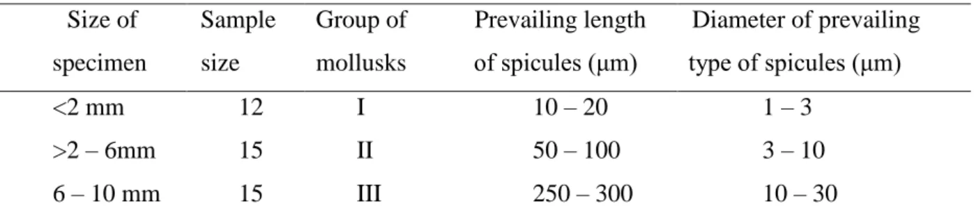
Four types of spicules were identified in the body wall of O. muricata: monoactine (Fig. 5A, B), diactine (Fig. 5C, D), triactine (Fig. 5E, F) and tetractine, being the predominant type (Fig. 5G). Lineage size and diameter of spicules are increase with body size (Table 2). Notum spicules in group I were between 10 – 20 μm in length and 1 – 3 μm in diameter; in group II group they ranged from 50 – 100 μm in length and 3 – 10 μm in diameter; and in group III they were between 250 – 300 μm in length and 10 – 30 in diameter.
Table 2. Length and diameter of spicules between the three groups Size of specimen Sample size Group of mollusks Prevailing length of spicules (μm) Diameter of prevailing type of spicules (μm) <2 mm 12 I 10 – 20 1 – 3 >2 – 6mm 15 II 50 – 100 3 – 10 6 – 10 mm 15 III 250 – 300 10 – 30
However, generally, the larger the specimen the greater the length and diameter of the spicule.
In group I the number of spicules increases very rapidly, mainly during the period when specimens are between 100 μm to 1 mm in length. Spicules first emerge in the central part of the notum, gradually increasing in size. New spicules then form on the edge of the notum, then in the foot and tubercles (Fig. 2A, B, G). Specimens that are 1 mm in size have larger spicules and their arrangement begins to resemble the spicule tracts of group II and III, but the spicules are still spaced far apart.
The number of spicules in area proportional to the total animal body size is the same for groups II and III, having 12 – 14 spicules in the central part of the notum (Fig. 3B, F), 14 – 17 in the foot (Fig. 3D, H) and 10 in the 180µm diameter tubercle and 18 – 20 in 300 µm diameter tubercle (Fig. 3C, G) (Table 3).
Table 3. Number of spicules in different compartments of the central body part in
Onchidoris muricata (based on µCT data).
Size of specimen Group of mollusks Amount of spicules Sample size Notum Tubercles (100 – 180 µm diameter) Foot
3mm 2 II 12 10 15
4 mm 2 II 12 10 17
6 mm 2 III 15 10 14
7 mm 2 III 14 9 15
There are six types of spicules based on internal morphology from SEM micrographs of the spicule fractures (Fig. 5H – P): hollow spicules, spicules with spongy contents in the center (Fig. 5H), spicules with concentric internal structures (Fig. 5I, J), spicules with layered internal structures (Fig. 5K), spicules with a mixed structure (Fig. 5L – N) and those spicules that are monolithic (Fig. 5O, P). The internal morphology of the spicules is characterized by size of the animals. Hollow and concentric (Fig. 5I, J) spicules are dominant for group I. For specimens larger than 1.5 mm in length, spongy centered spicules (Fig. 5H) have also been found. Group II had the highest diversity of spicule type, including layered (Fig. 5K), concentric, monolithic (Fig. 5O) and mixed (Fig. 5L – N). Ultrastructural investigations of the internal morphology of the mixed spicule has shown three different structures: 1) spicules consist of concentric layers up to 1 μm with a thin bridge between them (Fig. 5L); 2) spicules have a central monolithic rod with concentric layers on the periphery (Fig. 5M) and 3) spicules with a central monolithic rod with a thin periphery layer consisting of radially dispersed rods (Fig. 5N). The diameter of the central rod depends on the juvenile group; in group II it is less 5 μm (with a spicule diameter of 10 μm) and for group III it is 20 μm, with a spicule diameter of 30 μm. Monolithic spicules (Fig. 5O, P) have a homogeneous internal structure and are characterized mostly from group III.
According to three-dimensional tubercle reconstructions from groups III group (Fig. 4C), one spicule is located inside the vacuole of a mononuclear cell – sclerocyte (Fig. 4G). The size of the vacuole corresponds to the size of the spicule, and also takes the shape characteristic of each spicule, respectively. Four-axis spicules remain covered with a single vacuole membrane, giving it a branched shape (Fig. 4G).
3.2. Ultrastructure of the sclerocyte and collagen matrix
Subepidermal spicules of three groups were located close to the epithelium. Epithelium cells are flattened areas adjacent to the spicules, but there are no find any contacts between sclerocytes and epithelial cells. The subepidermal space of O. muricata contains sclerocytes and fibroblasts (Fig. 6), which synthesize the collagen. The collagen matrix (Fig. 6A – C) differs between the groups by density (Figs. 1; 6A – C).
Group I has a weakly developed matrix in the central part of notum, but denser towards the notums edge and near the spicules (Figs. 1A, B; 8A). Fibroblasts are more closely associated
with sclerocytes. The collagen matrix of groups II and III is developed throughout the body, not only near the spicules (Figs. 1C – F; 6B, C; 8). Fibroblasts are less associated with sclerocytes in group II. In group III, only outgrowths of the fibroblast are detected near the sclerocytes. In all groups, collagen fibers are concentrated near the sclerocytes, forming a sheath (Figs. 1; 8G – I).
The ultrathin morphology of the amoeboid fibroblasts is similar for all groups (Figs. 6; 7), having several long pseudopodia (Fig. 7F, G) with electron-dense granules (0.1 – 0.3 μm diameter) (Fig. 7F). The central part of the cell has an oval nucleus (maximum 2 – 6 μm), a rough endoplasmic reticulum (Fig. 7B), oval mitochondria (Fig. 7B) and electron-transparent vesicles (Fig. 7A, C, D, F).
Fibroblasts and sclerocytes were in close contact with each other, especially in specimens of group I. There are numerous vesicles, collagen fibers and membrane fold in this area between fibroblast and sclerocyte (Fig. 7A – D). As the body size increases, the number of fibroblasts in contact with sclerocytes becomes fewer, while the distance between them increases.
Sclerocytes are located under the epithelium throughout the body. These cells have an irregular shape and form spicules inside the large vacuole (Figs. 4G; 8A, D, G). Cell organelles, including mitochondria, endoplasmic reticulum and Golgi apparatus are concentrated near the nucleus. The vacuole membrane may lie close inside the sclerocyte membrane, but never fused. Sclerocyte vacuoles of group II are mostly filled with collagen fibers (Fig. 8D) while sclerocytes in groups I and III lack any contents (Fig. 8A, G). Single-membrane vesicles (0.1 – 1 μm in diameter) (Fig. 8B, C, E, F, H, I) are located near the nucleus, along the cell membrane and especially in contact with the fibroblasts.
Differences between the sclerocytes of group II and III include signs of apoptosis and autolysis, including vesicles with two membranes (0.5 μm diameter) (Fig. 8E, H) and electron-dense mitochondria with flattened cristae and very electron-dense nuclei.
3.3. Spicule mineralization
Counting spicules on identical parts of the body in relation to the total body volume in different groups showed that the main quantity of spicules is formed during the veliger’s settlement. During juvenile development, spicules only increase in size. Experiments to detect spicule calcification zones showed that calcification is most active in groups I and II (Fig. 9A – D) while practically absent in group III (Fig. 9E, F). Strongest signals were detected in the tips, with less intensity on surfaces of the spicule (Fig. 9).
4.1. Sclero- and spiculogenesis
Subepidermal spicule of Onchidoris muricata is formed in the vacuole of the specialized
cell (sclerocyte) that is not characterized for other groups of Mollusca. Spicules are laid at the early stage of development and keep the subepidermal position during the life. The sclerocytes have well-developed synthetic apparatus at this period. They are apoptosis with time, but not destroyed. This type of spicule formation significantly differs from other mollusks in such characters as subepidermal localization and intracellular formation (Table 1), that is apomorphic character for all Heterobranchia (Acochlidiimorpha, Rhodopoidea and Doridina). In meiofauna heterobranchs (Acochlidiimorpha, Rhodopoidea) spicules considered as an adaptation to the interstitial habitat, probably serving to stabilize certain body parts during movements through the interstices (Jörger et al., 2008). The second supposed function for all heterobranchs is protection (Thompson, 1960; Kocot et al., 2016).
Formation of epidermal spicules of Aplacophora and Polyplacophora are similar. The spicules being secreted within a thin cup-like membrane which exhibits somewhat similar properties to the inner cuticle. The cuticle contains an appreciable number of hollow, acicular calcified spines or spicules, each borne internally on a thin, cup-like organic base. Most of aplacophoran spicules formed by a one cell, but polyplacophoran spicules formed by group of cells: calcium carbonate-secreting cells, cuticle-secreting cells, and papillae (Beedham and Trueman, 1968; Kocot et al., 2016). Initially spicule formation process is intracellular, but further growth of the spicule occurs inside a crystal chamber composed of organic macromolecules secreted by a number of cells surrounding the spicule (Treves et al., 2003). This way of spicule formation is not comparable with O. muricata.
Similar pattern of nudibranchs subepidermal spicule formation is found in distant groups including Cnidaria, Porifera and Echinodermata and should be considered as a convergence (Table 1). Intracellular formation of calcareous spicule is only knowing in Octocorallia (Cnidaria) (Kingsley and Watabe, 1984), where the spicule is formed in one of the Golgi apparatus vacuoles of the spicule forming cells. Growing spicules extend into the vacuole until it fills the volume of the cell, merging both the vacuole and cell membranes. After spicule formation, the membrane of the spicule forming cell is destroyed and the mature spicule is localized in the mesoglea outside the cell (Kingsley and Watabe, 1984). Individual spicules contain mineralized forms of calcium and collagen. The ratio of organic to mineral components may vary depending on the season of the year (Kingsley et al., 1990). Spicules that formed extracellularly between the sclerocytes are characterized for Calcarea (Porifera) (Ledger and Jones, 1977; Sethmann and Worheide, 2008). Spicules of calcareous sponges form in an intercellular cavity within a group of sclerocytes. Each monaxon arises within a pair of
sclerocytes and each three-rayed triactine is formed within a group of six sclerocytes. Sclerocytes secrete both mineral and organic matter into the intercellular space, where calcite crystallizes (Sethmann and Worheide, 2008). Most of the sclerocyte organoids are located in the nuclear zone, with most of the spicules surface being covered with elongated “processes” of sclerocyte. Fully formed spicules are surrounded by collagen fibrils in the mesohyl (Ledger and Jones, 1977). In Echinodermata (Echinoidea), spicules are laid during stages of gastrula development in the extracellular space in membrane-defined compartments. These compartments are formed by syncytium of primary mesenchymal cells from a calcite crystal (Beniash et al., 1999).
The exact way of spicule laid in nudibranch is unknowns. But now exists two hypotheses of their laid in ontogenesis. According to the first one, which was proposed by Woodland (1907) (Thompson 1958; Ehrlich, 2019), spicules formed in the mantle of veliger and migrate under the epithelium. The second hypothesis was proposed by Prenant (1924), wich supposes the spicule's original formation from mitochondria of scleroblast in mesoderm. Subepidermal spicules origination was assumed one more time by Àvila and Durfort (1996), but without elaborations of the sclerocytes origination. Spicules were not detected in the surface of the body of O. muricata, even in the smallest, just settled juveniles with several spicules in our studies. We could not confirm the hypothesis stating that spicule formation was on the surface of body wall and then migrated into the subepidermal space. Our results are more consistent with the second hypothesis of the subepidermal sclerocytes origination.
We discovered, that one of the spicules of O. muricata is located inside of one sclerocyte vacuoles during postlarval development in subepidermal space. The statement “The only one spicule inside the one sclerocyte” agrees with Woodland’s (1907) and Prenant’s (1924) data. But the position about mitochondrial origin of spicule raises questions: what happens with mitochondrial features like specific proteins, DNA, etc. We need to check this position using set of modern methods.
We suspect that the spicule forms by an ectodermal cell that migrate under the ectodermal epithelium in the earlier stages. Furthermore, our results didn’t show any signs of spicule or cell migrations under the epithelium. However, more researches are needed to reach a conclusion.
4.2. General morphology and development of the spicule complex
For the first time in Nudibranchia, the ontogenetic dynamics of spicules and the spicule complex in O. muricata has been studied. Significant findings from this study include, that subepidermal intracellular spicules are located in the subepidermal space throughout
development. The first indication of spicules was detected immediately after larval metamorphosis in the central part of the body, similar to the dorid Adalaria proxima (Alder and Hancock, 1855) (Thompson, 1958). Short spicules are occupied in a transverse position along the anterior-posterior axis (Fig. 2A, G). As the tubercles develop, the spicules are simultaneously forming alongside. Subsequently, these spicules given rise to the vertical tract. We additionally note that the rate of formation and transformation of spicules in closely related species is different, which may be due to differences between the overall adult sizes, i.e., A. proxima reaches 20 mm while O. muricata only 12 mm (Todd, 1979). Spicules in Rostanga pulchra MacFarland, 1905 (Discodorididae) was detected in tubercles immediately after settlement (Chia and Koss, 1978), but we have not information about localization in notum and foot and cannot compare formation of networks in O. muricata and R. pulchra. With an increase in the body size of O. muricata, the size of the spicules also increases (Table 2); a similar pattern was also noted for representatives of the Chromodorididae family (Sánchez-Tocino et al., 2014). With an increase in the size of the spicules, the distance between them decreases, and the overall density of the arrangement of spicules and integuments increases.
Our mCT data findings of organization of spicule networks in group III are agree with data Penney (Penney et al., 2020) and our 3D-reconstruction are complemented information about vertical tract organization. The vertical tract is composed of two layers of spicules. Tetractine spicules are located along the entire tubercles. The secondary axes of which are displaced to their edge; approximately from the middle of the tubercle's length. Diactine spicules located between the tetractine spicules and form the second layer of the tract. The tips of the diactine spicules in the apical part of the tubercle are displaced laterally, and the tetraactine spicules - in the central part. For the first time in O. muricata, we have shown constant spicule number throughout the development starting from the 200 μm size (Table 2). Beginning from the 200 μm size, the main differences within the morphology of the spicule complex in O.
muricata are the spicule size and the manner of spicule localization.
In O. muricata, empty spicules are predominant in group I, with the spicule with mixed internal structures prevailing in group II and the monolithic type in group III. Interestingly, we found the largest number of structural types in group II, indicating a high rate of transformation between the internal structure of the spicules and their mineralization. While the internal morphology of the spicules does not depend on their overall shape (Nikitenko and Vortsepneva, 2020), the noted differences are likely related to formation stage of the spicules and the postlarval developmental stage. All spicules appear in the early development stages, afterwards, they and becoming more densely situated and form the spicule complex.
4.3. Ultrastructure of the sclerocytes and collagen matrix
O. muricata subepidermal spaces contain spicules, nerves, muscle fibers and fibroblasts
that synthesize the collagen. The morphology of the sclerocytes, spicules and collagen matrix are transformed during the developmental stage. The smallest juvenile (group I) is characterized by the following features: sclerocytes distant to each other, numerous fibroblasts associated with a single sclerocyte and a weakly developed collagen matrix near the spicules. The density of the collagen matrix near and between the sclerocytes increases, while the number of fibroblasts decreases with growth (Figs. 2; 6A – C). The collagen matrix of the largest studied juvenile (group III) well already developed, occupying the entire space between the spicules, especially near the sclerocyte, fibroblasts located in the distance to spicule.
The first description of sclerocytes morphology was made in the first half of the 20th century using light microscopy (Woodland, 1907; Prenant, 1924). Spicules are intracellular structures during all live. The sclerocytes are strongly elongate, have expanded cell part most often with one nucleus and the flattened edge part (Woodland, 1907; Prenant, 1924). Our study contains the first ultrastructural description of sclerocytes in nudibranchs using transmission electron microscopy. In particular, we have reliably shown that sclerocytes have only a single nucleus, surrounded by numerous other cellular elements. A vacuole, with a spicule, is located in the central part of the cell, with the remainder of the cell being filled by cytoplasm and vesicles (Fig. 8). Sclerocytes of group I are characterized by a high content of vesicles, suggesting a high level of synthetic activity. Signs of apoptosis and autolysis were also noted for individuals in groups II and III (Fig. 8E, H).
The spicule arises inside the scleroblast from mitochondria, which lose their outer membrane according to the description of the Prenant (1924). However, our study failed to establish the subsidence of the membrane surrounding the spicule and we need more data.
Collagen matrix comprised of collagen type I is firstly found in the sclerocyte vacuole of group II (Figs. 6E; 8D). This indicates the variability of the chemical composition of the spicule. Therefore, the statement about the content of high concentrations of calcite with impurities of fluorite and vaterite (Odum, 1951; Cattaneo-Vietti et al., 1995 only applies to early larval stages.
4.4. Spicule mineralization
Collagen, type I, was found in the central axis of spicules in group II (Figs. 1; 6E; 8D), suggesting that it is an organic matrix used for mineralization. Experiments with O. muricata in calcium disodium salt shows that the most active mineralization occurs in group I and II (Fig. 9C), with practically no expression in group III (Fig. 9F). The most active area of mineralization was detected at the distal end of the spicule’s central axis, being less active over spicules surface,
similar to what was previously described in spicules of Neomeniomorpha (Aplacophora, Mollusca) (Woodland, 1907). Mineralization of the aplacophoran spicule is as follows: the mineral granule forms as a result of deposition of calcareous matter from the cytoplasm of the sclerocyte, taking the form of an elongated rod that resembles the shape of a formed uniaxial spicule. The matrix for mineralization is a nanoscale organic matrix. The middle part of the adult spicule is formed first, followed by growth towards both ends (Woodland, 1907; Ehrlich 2010, 2019). According to the Prenant (1924), the spicule arises after the elongation of the mitochondria and the appearance of a calcium crystal in it. The spicule growth occurs when the contents of other mitochondria of the scleroblast enter the surface of the growing spicule. It is this content that is subsequently mineralized. We cannot compare our data about mineralization with Prenant’s (1924) data and need additional experiments on the cellular level.
5. Conclusion
Detailed studies of the ontogenetic dynamic of the spicule complex of Onchidoris
muricata allows us to assume the hypothesis of formation and maturation of both spicules and
spicule networks.
1) Subepidermal position of the spicule is kept from the early stage of postlarval development.
2) New spicules actively form in the postlarval stage until juveniles reach 500 μm in length. Increases to the number of spicules past this stage was not observed. Evidence of this was based off the following: sclerocytes with well-developed synthetic apparatus were detected in group I with apoptotic sclerocyte processes starting in group II. During this time, the number of spicules stays constant in all groups and into adults.
3) Spicule start to form the networks immediately after their emerging. The main features of the adult spicule networks are characterized for the group I at the end of the spicule forming process.
4) The internal morphology of spicules based on SEM micrographs corresponds to the stages of postlarval development. Thus, spicule maturation (Fig. 10) may follow this progression: hollow spicules of group I are a mineralized “shell” with no internal matrix. In group II, fibroblasts actively form collagen and begin to form a spicule axis resembling sponge-like structures. As a result of calcification, the internal structure of the spicules changes to concentric, with greater mineralization to mixed internal structure. In group III, the spicules are completely mineralized, resulting in a monolithic internal structure. Active calcification process is present in groups I and II.
Thus, the data generated and observed on the transformation of the spicule apparatus in nudibranchs has substantially supplemented the available information on postlarval development of subepidermal spicules. However, many open questions remain, including how spicules appear in the subepidermal space, the mechanism of spicule mineralization, their exact chemical composition and the homology of the supporting structures in Gastropoda and throughout Mollusca.
Acknowledgements
We are grateful to Professor Alexander B. Tzetlin for the fruitful discussions and to Dr. B.C. Gonzalez for the invaluable help with the English. We express our gratitude to our colleagues of the Pertsov White Sea Biological Station and the Zoology Department of Lomonosov Moscow State University. We thank A.A. Semenov, D. Ozerov, A.L. Mikhlina, T. Antochina, A.A. Prudkovsky and A. Koynova for helping with sampling. We want to thank the three unknown reviewers for most helpful comments on the typescript. We are very thankful to I.A. Kosevich, B.V. Osadchenko, G.D. Kolbasova, A.I. Lavrov, F.V. Bolshakov and I.A. Ekimova assistance with the morphological methods and experimental protocols. Sincere appreciation goes to G. Davidovich, A. Bogdanov and the Electron Microscopy Laboratory of the Shared Facilities Center of Lomonosov Moscow State University, sponsored by the RF Ministry of Education, Science and Research. The authors would also like to thank S. Mitelev, G. Bykov and I.D. Papanin from the Institute for the Biology of Inland Waters, Russian Academy of Sciences for the help with the electron microscopy. We are appreciative to L.Y. Kruchkova (Resource Centre for X-ray Diffraction Studies, Saint Petersburg State University, Saint Petersburg, Russia) and D.V. Korost (MSU) for assisting with the X-ray microtomography. Light microscopy was made possible at the Center of microscopy, WSBS, MSU.
Funding
This work was supported by Moscow State University Grant for Leading Scientific Schools "Depository of the Living Systems" in the frame of the MSU Development Program and by the Russian Foundation for Basic Researches, Grants nos. 18-05-60158 and 19-04-00501.
Declaration of interests
The authors declare that they have no known competing financial interests or personal relationships that could have appeared to influence the work reported in this paper.
Alba-Tercedor, J., Sánchez-Tocino, L., 2011. The use of the SkyScan 1172 high-resolution micro-CT to elucidate if the spicules of the sea slugs (Mollusca: Nudibranchia, Opisthobranchia) have a structural or a defensive function. In SkyScan Users Meeting. 2011, 113–121.
Alder, J., Hancock, A., 1855. A Monograph of the British Nudibranchiate Mollusca: with Figures of All the Species. The Ray Society, London, England.
Àvila, C., Durfort, M., 1996. Histology of epithelia and mantle glands of selected species of doridacean mollusks with chemical defensive strategies. The Veliger. 39, 148–163.
Beedham, G.E., Trueman, E.R., 1968. The cuticle of the Aplacophora and its evolutionary significance in the Mollusca. J. Zool. 154, 443–451.
Beniash, E., Addadi, L., Weiner, S., 1999. Cellular control over spicule formation in sea urchin embryos: A structural approach. J. Struct. Biol. 125, 50–62.
Brenzinger, B., Wilson, N.G., Schrödl, M., 2011. 3D microanatomy of a gastropod ‘worm’,
Rhodope rousei n. sp. (Heterobranchia) from southern Australia. J. Molluscan Stud. 77, 375–
387.
Brenzinger, B., Haszprunar, G., Schrödl, M., 2013. At the limits of a successful body plan – 3D microanatomy, histology and evolution of Helminthope (Mollusca: Heterobranchia: Rhodopemorpha), the most worm-like gastropod. Front. Zool. 10, 37.
Carter, J.G., Aller, R.C., 1975. Calcification in the bivalve periostracum. Lethaia 8, 315–320. Cattaneo-Vietti, R., Angelini, S., Bavastrello, G., 1993. Skin and gut spicules in Discodoris
atromaculata (Bergh, 1880) (Mollusca: Nudibranchia). Bollettino Macalogico 28, 173–180.
Cattaneo-Vietti, R., Angelini, S., Gaggero, L., Lucchetti, G., 1995. Mineral composition of nudibranch spicules. J. Molluscan Stud. 61, 331–337.
Chang, Y.W., Willan, R.C., Mok, H.K., 2013. Can the morphology of the integumentary spicules be used to distinguish genera and species of phyllidiid nudibranchs (Porostomata: Phyllidiidae)? Molluscan Res. 33, 14–23.
Checa, A.G., Vendrasco, M.J., Salas, C., 2017. Cuticle of Polyplacophora: structure, secretion, and homology with the periostracum of conchiferans. Mar. Biol. 164, 64.
Chia, F.S., Koss, R., 1989. The fine structure of the newly discovered propodial ganglia of the veliger larva of the nudibranch Onchidoris bilamellata. Cell Tissue Res. 256, 17–26.
Edmunds, M., 1968. Acid secretion in some species of Doridacea (Mollusca, Nudibranchia). J. Molluscan Stud. 38, 121–133.
Ehrlich, H., 2010. Molluscs Spicules. In: H. Ehrlich Biological Materials of Marine Origin. Invertebrates, Springer, New York, NY, USA, pp. 211–237.
Ehrlich, H., 2019. Spicular structures in Molluscs. In: H. Ehrlich Marine Biological Materials of Invertebrate, Origin, Springer, Cham, pp. 133–157.
Foale, S.J., Willan, R.C., 1987. Scanning and transmission electron microscope study of specialized mantle structures in dorid nudibranchs (Gastropoda: Opisthobranchia: Anthobranchia). Mar. Biol. 95, 547–557.
García, F.J., Garcia, J.C., Cervera, J.L., 1986. Estudio morfológico de las espículas de
Doriopsilla areolata (Gastropoda: Nudibranchia). Malacologia 27, 83–96.
Hallas, J.M., Chichvarkhin, A., Gosliner, T.M., 2017. Aligning evidence: Concerns regarding multiple sequence alignments in estimating the phylogeny of the Nudibranchia suborder Doridina. R. Soc. Open Sci. 4, 171095.
Harper, E.M., Dreyer, H., Steiner, G., 2006. Reconstructing the Anomalodesmata (Mollusca: Bivalvia): morphology and molecules. Zool. J. Linn. Soc. 148, 395–420.
Jörger, K.M., Neusser, T.P., Haszprunar, G., Schrödl, M., 2008. Undersized and underestimated: 3D visualization of the mediterranean interstitial acochlidian gastropod Pontohedyle
milaschewitchii (Kowalevsky, 1901). Org. Divers. Evol. 8, 194–214.
Kasamesiria, P., Meksumpuna, S., Meksumpunc, C., 2011. Spicule network patterns of Phyllidia
varicosa. Sci. Asia 37, 160–164.
Kingsley, R.J., Watabe, N., 1984. Synthesis and transport of the organic matrix of the spicules in the gorgonian Leptogorgia virgulata (Lamarck) (Coelenterata: Gorgonacea). Cell Tissue Res. 235, 533–538.
Kingsley, R.J., Tsuzaki, M., Watabe, N., Mechanic, G.L., 1990. Collagen in the spicule organic matrix of the gorgonian Leptogorgia virgulata. Biol. Bull. 179, 207–213.
Kocot, K.M., McDougall, C., Degnan, B.M., 2016. Developing perspectives on molluscan shells, part 1: introduction and molecular biology. Physiology of molluscs, a collection of selected reviews 1–42.
Kress, A., 1981. A scanning electron microscope study of notum structures in some dorid nudibranchs (Gastropoda: Opisthobranchia). J. Mar. Biol. Assoc. U. K. 61, 177–191.
Lankester E.R., 1883. Mollusca. Encyclopedia Britannica 9.
Ledger, P.W., Jones, W.C., 1977. Spicule formation in the calcareous sponge Sycon
ciliatum. Cell Tissue Res. 181, 553–567.
Leise, E.M., 1984. Chiton integument: metamorphic changes in Mopalia muscosa (Mollusca, Polyplacophora). Zoomorphology 104, 337–343.
Lowenstam, H.A., Weiner S., 1983. Mineralization by organisms and the evolution of biomineralization, in: Westbroek, P., de Jong, E.W. (Eds.), Biomineralization and Biological Metal Accumulation. Springer, Dordrecht, pp. 191–203.
Morse, M.P., 1976. Hedylopsis risen sp. n., a New Interstitial Mollusc from the New England Coast (Opisthobranchia, Acochlidiacea). Zool. Scr. 5, 221–229.
Nikitenko, E.D., Vortsepneva, E.V., 2020. Spicule complex of three Onchidorididae species (Gastropoda: Doridina) from the White Sea. Invert. Zool. 17, 1–15.
Odum, H.T., 1951. Nudibranch spicules made of amorphous calcium carbonate. Science 114, 395–395.
Penney, B.K., 2006. Morphology and biological roles of spicule networks in Cadlina
luteomarginata (Nudibranchia, Doridina). Invertebr. Biol. 125, 222–232.
Penney, B.K., 2008. Phylogenetic comparison of spicule networks in cryptobranchiate dorid nudibranchs (Gastropoda, Euthyneura, Nudibranchia, Doridina). Acta Zool. 89, 311–329.
Penney, B.K., Ehresmann, K.R., Jordan, K.J., Rufo, G., 2020. Micro-computed tomography of spicule networks in three genera of dorid sea-slugs (Gastropoda: Nudipleura: Doridina) shows patterns of phylogenetic significance. Acta Zool. 101, 5–23.
Prenant, M., 1924. Contributions L’étude Cytologique du Calcaire. Bull. Biol. Fr. Belg. 58, 331– 378.
Ros, J., 1976. Sistemas de defensa en los opistobranquios. Oecologia aquatica 2, 41–77.
Rose, R.A., 1983. Lecithotrophic development of Hoplodoris nodulosa (Angas) (Opisthobranchia, Gastropoda). J. Malac. Soc. Aust. 6, 63–70.
Ruthensteiner, B., Heß, M., 2008. Embedding 3D models of biological specimens in PDF publications. Microsc. Res. Tech. 71, 778–786.
Salvini-Plawen, L.V., 1972. Zur Morphologie und Phylogenie der Mollusken: die Beziehungen der Caudofoveata und der Solenogastres als Aculifera, als Mollusca und als Spiralia. Z. wiss. Zool. 184, 205–394.
Sánchez-Tocino, L., Tierno de Figueroa, J.M., Cervera, J.L., 2014. Ontogenetic changes in the spicule formation and their possible role in chromodorid opisthobranchs (Mollusca, Chromodorididae). Mar. Biol. Res. 10, 357–373.
Scheltema, A.H., 1993. Aplacophora as progenetic aculiferans and the coelomate origin of mollusks as the sister taxon of Sipuncula. Biol. Bull. 184, 57–78.
Sethmann, I., Wörheide, G., 2008. Structure and composition of calcareous sponge spicules: a review and comparison to structurally related biominerals. Micron 39, 209–228.
Sigwart, J.D., 2017. Molluscs all beneath the sun, one shell, two shells, more, or none. Curr. Biol. 27, 708–710.
Thompson, T.E., 1958. The natural history, embryology, larval biology and post-larval development of Adalaria proxima (Alder and Hancock) (Gastropoda: Opisthobranchia). Philos. Trans. R. Soc. Lond. B. Biol. Sci. 242, 1–58.
Thompson, T.E., 1960. Defensive adaptations in opisthobranchs. J. Mar. Biol. Ass. U. K. 39, 123–134.
Todd, C.D., 1979. Reproductive energetics of two species of dorid nudibranchs with planktotrophic and lecithotrophic larval strategies. Mar. Biol. 53, 57–68.
Todd, C.D., 1981. The ecology of nudibranch molluscs. Oceanogr. Mar. Biol. 19, 141–234. Treves, K., Traub, W., Weiner, S., Addadi, L., 2003. Aragonite formation in the chiton (Mollusca) girdle. Helv. Chim. Acta. 86, 1101–1112.
Wägele, H., Willan, R.C., 2000. Phylogeny of the Nudibranchia. Zool. J. Linnean Soc. 130, 82– 181.
Wägele, H., Cervera, J.L., 2001. Histological study of Goniodoris castanea Alder and Hancock, 1845 (Nudibranchia, Doridoidea, Goniodorididae). J. Morphol. 250, 61–69.
Wägele, H., Klussmann-Kolb, A., 2005. Opisthobranchia (Mollusca, Gastropoda) – more than just slimy slugs. Shell reduction and its implications on defence and foraging. Front. Zool. 2, 3. Weiner, S., Levi-Kalisman, Y., Raz, S., Addadi, L., 2003. Biologically formed amorphous calcium carbonate. Connect. Tissue Res. 44, 214–218.
Willmer, P., 1990. Invertebrate Relationships: Patterns in Animal Evolution. Cambridge University Press.
Woodland, W.,1907. Studies in spicule formation. Quart Journ. miscrosc. Sci. 51, 31.
Wysokowski, M., Jesionowski, T., Ehrlich, H., 2018. Biosilica as a source for inspiration in biological materials science. American Mineralogist: Journal of Earth and Planetary Materials 103, 665–691.
Figure legends
Fig. 1. General morphology of the body wall organization between three groups Onchidoris
muricata (A, C, E – light microscopy of transverse histological section; B, D, F – general
scheme based on TEM micrographs). A, C, E. Subepidermal spicules surrounded by collagen matrix in notum and tubercles in the group I (A), II (C) and III (E). C. The lines in right side of the pictures are cutting artefacts. B. Group I. The collagen matrix develops only near the spicules. The fibroblasts and their outgrowth located close to sclerocyte. D. Group II. The collagen matrix develops near the spicules and in extracellular space. Intracellular collagen matrix found in sclerocyte. Sclerocyte has first apoptosis sings. Fibroblasts located at some distance from sclerocyte. F. Group III. Collagen matrix density develops in everything extracellular space. Fibroblasts are not found near the spicule, only fibroblasts outgrowths are found. Abbreviations: amt – apoptotic mitochondria; ep – epithelium; cm – collagen matrix; fb – fibroblast; fbo – fibroblasts outgrowth; icm – intracellular collagen matrix; m – muscles; msc – membrane of sclerocytes; mt – mitochondria; mvs – membrane of the spicule vacuole; n – nucleus; sc – subepidermal sclerocytes; sp – spicule; t – tubercle; tvs – two membrane vesicles; vs- vesicles;
Fig. 2. Transformation of the spicules complex in the notum of group I in Onchidoris muricata. A. Diagram of 200 μm length O. muricata with visible eyes and spicules, without tubercles; short and wide spicules locate perpendicularly to the anterio-posterior axis and parallel to each other on the dorsal side of the notum, edge of the notum without visible spicules. B. Diagram of the dorsal part of the notum in specimens of 200 μm length with spicules. C. Diagram 500 μm length specimens with a single layer of spicules. Elongate spicules chaotically located in the central part of the body and oriented at the distance to each other, spicules under tubercles are radially; spicules in the edge of the notum are perpendicular to the edge. D. Diagram of tubercles of 500 μm length in O. muricata. Elongate spicules form rosette-shaped vertical tracts. E. Diagram of 1 mm length O. muricata; spicules are perpendicular to the anteroposterior axis in the central part of the notum; radially located spicules beginning to form stellular tracts under the tubercles; spicules located on the edge of the body, similar to those found in specimens of 500 μm length but more densely arranged. F. Tubercles with spicules from specimens 1 mm in length, forming the vertical tract. The ends of the spicules from the stellular tract are located on the base of the tubercle. G – I. Light microscopy of live specimens of O. muricata with translucent spicules, 200 μm, 500 μm and 1 mm lengths. Abbreviations: cn – center of notum; e – eyes; en – edge of notum; sp – spicule; st – stellular tract; t – tubercle. Scale bars: A, C, E – 100 μm.
Fig. 3. Different views of the Micro-CT volume rendering reconstruction of Onchidoris muricata group II and III. A – D. Group II; E – H. Group III; A. Optical cross section through the front half of body; spicules of horizontal tract are visible in the notum and foot; spicules form vertical tract in tubercles. B. Horizontal tract of notum group II contains spicules that located parallel to each other but perpendicular to anterio-posterior body axis (A – P). C. Spicules of stellular tract located radially in the basal part of tubercles on the distance to each other. D. Dorsal view on horizontal tract of the foot; spicules located not parallel on the distant to each other in the center
of foot. E. Optical cross section through the front half of body; internal organs are well detected in compare with the Fig. 3A because another parameter of scanning have been used. F. Spicules of horizontal tract in the central part of the notum (dorsal view). G. Tubercles of the central part of body; spicules formed stellular tract. H. Spicules of the foot in horizontal tract located parallel to each other; in the central part spicules form more densely structure than in the same tract of group II. Abbreviations: f – foot; h – head; htf – horizontal tract of foot; htn – horizontal tract of notum; io – internal organs; sp – spicule; sst – spicules of stellular tract; vt – vertical tract. Scale bar: A – 45 µm; B – 50 µm; C – 20 µm; D – 250 µm; E – 70 µm; F – 110 µm; G – 35 µm; H – 700 µm.
Fig. 4. Structure of spicule complex of Onchidoris muricata. A, D – F. SEM micrograph. B – micro-CT. C, G – 3D reconstruction. A. Longitudinal section through the central part of notum with tubercles in group III. B. Optical section through the central part of notum in group II; spicules of vertical tract form rosette-like structure; spicules of stellular tract located under the vertical tract. C. The tubercle with the spicule’s vertical tract. Spicules located in two layers, short diactine and triactine spicules (light blue) located between long di-, tri- and tetractine spicules (dark blue); spicules of stellular tract (corral) support them. D. The notum’s edge without epithelium. Spicules are densely packed in several layers. E. General view on the notum without epithelium (view from dorsal side). Spicules of the stellular tract located radially under them. F. Spicules of stellular and vertical tracts in the base of the tubercle. G. Tetractine spicule from vertical tract; sclerocyte contain only one large vacuole with one spicule, surface of the cell membrane is semitransparent. Abbreviations: ct – cover of tubercle; msc – membrane of spicule cell; n – nucleus of sclerocytes.
Fig. 5. External and internal morphology of the spicules of three groups Onchidoris muricata. A – G: light microscopy, H – P: SEM micrograph (H – outside view; I – P – cross section). A. Monoaxone spicules of group I. B. Monoaxone spicule of group III. C. Diactine spicule of group II. D. Diactine spicule of group III. E. Triactine spicule with long secondary axis of group II. F. Triactine spicule with short secondary axis of group II. G. Tetractine spicule of group III; H. Internal spicule morphology of the 1.5 mm length specimensinternal layer of spicules is spongy; spicule shell does not differ from other groups. I. Internal spicule morphology of the 2 mm length specimen; central part of the spicule is spongy; the peripheral part consists of concentric layers. J. Structure of the concentric layers of 2 mm length specimen. K. The layered inner structure of 2 mm specimen’s spicules. L. Internal spicule morphology 3 mm length specimen; the spicule contains concentric layers and radial structure. M. Mixed internal structures of spicules group II; the spicule has monolithic middle part and peripheral concentric layers. N. Mixed internal structures of spicules group III; monolithic layer occupies most of the spicule; the radial striation located only on periphery. O. Monolithic spicule in group II. P. Monolithic spicule in group III. Abbreviation: cl – concentric layer; cs – spicules center; il – internal layer; ma – main axis of the spicule; ml – monolithic layer; rs – radial structures; sa – secondary axis of the spicule; ss – spicule shell. Scale bars: A – 2 μm; B – 25 μm; C – 7 μm; D – E – 20 μm; F –10 μm; G – 20 μm.