CASE REPORT
Cystic lesion of the parotid following drug-induced toxic
epidermal necrolysis (Lyell’s syndrome)
P. Paquet
1, E. Jacob
2, G. E. Pie´rard
11Department of Dermatopathology University Hospital Sart Tilman, Lie`ge, Belgium;2Intensive Care Medicine Burn Center, University Hospital Sart Tilman, Lie`ge, Belgium
A 32-year-old woman developed a unilateral cyst of the duct of parotid gland 4 months after severe oral involvement of drug-induced toxic epidermal necrolysis (TEN). The pathomechanism leading to the TEN epi-dermal destruction had presumably involved the salivary epithelium as well, leading to the development of the cystic lesion. The patient had low serum lipase levels, but high serum amylase levels at the time of TEN. These serological markers could represent a clue for the risk of developing cystic lesions of the large salivary glands fol-lowing TEN.
J Oral Pathol Med (2005) 34: 380–2
Keywords: cyst; Lyell’s syndrome; parotid; toxic epidermal necrolysis
A 32-year-old woman developed drug-induced toxic epidermal necrolysis (TEN) 48 h after oral intake of ibuprofen for headache. An erythematous rash covered by blisters spread over 64% of the body surface. Severe oral lesions were present associating oropharyngeal erosions and bullae on the palate, tongue and cheek mucosa. Ocular involvement consisted of conjunctival synechiae and corneal ulcerations. The diagnosis of TEN was confirmed by histological examination of a skin biopsy taken 72 h after the initial cutaneous lesions. A subepidermal blister was present with rare inflamma-tory mononuclear cells scattered between confluent necrotic keratinocytes. The patient was hospitalized for 3 weeks in a burn unit where she was placed on a fluidized bed. She benefited from supportive and anti-septic measures, including daily bath. She was also treated with cyclosporin IV (5 mg/kg/day) for five consecutive days. Corticosteroids were not adminis-tered. During that period of time, the amylase serum values were severely and repeatedly increased during her
three-week hospitalization reaching a maximum of 998 IU/ml (normal upper limit: 90 IU/ml). By contrast, the lipase serum values remained near to the upmost normal range limit, occasionally and moderately above it with a peak value of 225 IU/ml (upper limit of normal values: 200 IU/ml). The patient was discharged from the burn unit following complete cutaneous reepidermiza-tion and healing of the oropharyngeal ulcerareepidermiza-tions.
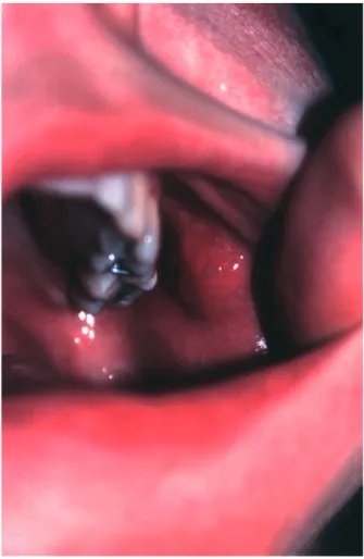
Four months after TEN, she progressively developed an oral swelling lesion nearby the left Stenon’s duct orifice (Fig. 1). Histological examination of the surgi-cally removed lesion showed a unilocular 0.9 cm dia-meter well circumscribed cyst filled by some amorphous material. The cyst wall was surrounded by a dense fibrous connective tissue containing a mild inflamma-tory cell infiltrate. The luminal surface of the cyst was lined by an epithelium that exhibited a stratified, squamous or columnar aspect according to the different areas. A salivary duct cyst of the parotid was diagnosed.
Comments
Toxic epidermal necrolysis is a rare potentially lethal disease characterized by sudden necrosis of the epider-mis, and of the genital and oropharyngeal mucosae (1). The only recognized cause of the disease is an adverse reaction to drugs, especially sulfonamides, phenytoin, non-steroidal antiinflammatory drugs and allopurinol. Death rate due to TEN reaches about 30%. Pulmonary, myocardial, osseous, renal and gastrointestinal long-term sequelae have been reported (1). However, the most commonly recognized long-term alterations involve the eyes, skin, nails and the genital mucosae (1). Conjunctival retraction, scars and sicca syndrome with severe corneal lesions represent the main ocular complications, sometimes resulting in a major handicap for the patient. Cutaneous sequelae include keloidal scars, alopecia, speckled eruptive melanocytic naevi and hyper- or hypomelanosis. Residual nail dystrophies are frequent. Genital involvement can lead to chronic vulvo-vaginal synechiae. Oral and lip lesions usually heal without complication, but strictures may develop in the throat and œsophagus (1).
Correspondence: Dr Philippe Paquet, Department of
Dermatopatho-logy, CHU Sart Tilman, B-4000 Lie`ge, Belgium. E-mail: P.Paquet@chu. ulg.ac.be
Accepted for publication December 14, 2004
J Oral Pathol Med (2005) 34: 380–2
ª Blackwell Munksgaard 2005 Æ All rights reserved
Sjo¨gren-like syndrome with xerostomia is also a recognized complication of TEN (2). This condition results from a chronic lymphocytic infiltration of small salivary glands. However, post-TEN cystic lesion of the large salivary glands has not been described so far. We report one adult patient who developed unilateral cystic lesion of the parotid duct following severe oral TEN involvement. Ductal obstruction is thought to be the main etiologic factor of this type of salivary cysts (3). No malignant neoplasm, calculi or mucus plugs were found in the presently reported patient. Hence, it is likely that TEN sequelae were responsible for the ductal obstruc-tion. The inflammatory infiltration of the cystic wall was discrete and undistinguishable from that found in non-TEN salivary ductal cysts. This suggests that the formation of the cystic lesion probably resulted from mechanical obstruction rather than from a post-TEN persistent immunologic process.
Several destructive or cell-modifying immunopatho-logical mechanisms found in TEN epidermis are prob-ably also operative in the epithelial component of the salivary glands. First, the L1 antigen (calprotectin), a calcium-binding protein, is overexpressed in TEN epi-dermis. It is thought to play an important role in TEN
epidermal apoptosis by disturbing Ca2+homeostasis (4). Normal or modified salivary gland duct cells also express high amounts of calprotectin (5). In addition, the pro-apoptotic CD 95 system (CD 95 receptor/FasR-CD 95 ligand/FasL) has been reported to be involved in TEN (4). Accordingly, the apoptosis-promoting mole-cules FasR and FasL are strongly expressed in inflam-matory conditions affecting ductal and acinar salivary epithelial cells (6). Finally, dysregulation in tumor necrosis factor a (TNF-a) system is likely involved in cutaneous TEN pathogenesis (4). Ductal epithelial cells also express TNF-a and its receptors in inflammatory salivary glands (7). Moreover, TNF-a is able to upreg-ulate FasL and FasR in these conditions (8).
Sustained high serum amylase levels combined with low serum lipase levels were found in our patient. The salivary glands and the pancreas are the two major sources of amylase in humans. As the lipase levels were close to normal in our TEN patient, a pancreatic involvement is unlikely. Hence, the prolonged elevation in serum amylase was probably due to a salivary involvement. In TEN, the incidence of hyperamylasemia is over 30% of the patients, and salivary hyperamy-plasemia is predominant (9). Hyperamylasemia reflects the extent in mucosal membrane involvement. It seems to have a predictive value of post-TEN Sjoegren-like sicca syndrome (9). It is also possible that high serum amylase levels coupled with low lipase serum levels could have a predictive value for the development of post-TEN lesions of the large salivary glands. Indeed, we have another record in our files of a Warthin tumor of the parotid gland diagnosed 10 years following severe TEN. The serum amylase dosages were elevated at the time of skin blistering while serum lipase were in the normal range of values.
In conclusion, we report a TEN patient in whom a cystic lesion developed subsequently in the parotid gland apparatus. It is suggested that such a lesion represents a possible long-term consequence of TEN severely affect-ing the oral cavity.
References
1. Fritsch PO, Sidoroff A. Drug-induced Stevens-Johnson syndrome/toxic epidermal necrolysis. Am J Clin Dermatol 2000; 1: 349–60.
2. Roujeau JC, Koso M, Andre C, et al. Sjo¨gren-like syndrome after drug-induced toxic epidermal necrolysis. Lancet1985; i: 609–11.
3. Ellis GL, Auclair PL. Tumors of the salivary glands. Atlas of Tumor pathology, 3rd series, fascicle 17. Washington, DC: Armed Forces Institute of Pathology, 1996; 426–7. 4. Paquet P, Pie´rard GE. Would cyclosporine A be beneficial
to mitigate drug-induced toxic epidermal necrolysis? Der-matology1999; 198: 198–202.
5. Huang JW, Ming Z, Parshanta S, et al. Immunohistochemi-cal evaluation of the Ca2+-binding S-100 proteins S-100A1, S100A2, S-100A4, S-100A6 and S-100B in salivary gland tumors. J Oral Pathol Med 1996; 25: 547–55.
6. Matsumura R, Kagami M, Tanioka H, et al. Expression of ductal Fas antigen in sialoadenitis of Sjo¨gren’s syndrome. Clin Exp Rheumatol1996; 14: 309–11.
Figure 1 Post-toxic epidermal necrolysis duct cyst of the parotid.
Parotid cyst in Lyell’s syndrome Paquet et al.
381
7. Koski H, Janin A, Humphreys-Beker MG, Sorsa T, Malinstro¨m M, Konttinen YT. Tumor necrosis factor-alpha and receptors for it in labial salivary glands in Sjo¨gren’s syndrome. Clin Exp Rheumatol 2001; 19: 131–7. 8. Abu-Helu R, Dimitriou I, Kapsogeorgou EK,
Moutsopo-ulos HM, Manoussakis MN. Induction of salivary gland
epithelial cell injury in Sjo¨gren syndrome: in vitro assess-ment of T cell-derived cytokines and Fas protein expression. J Autoimmunity2001; 17: 141–53.
9. Chosidow O, El Wady Z, Devanlay M, Jaffray P, Revuz J, Roujeau JC. Hyperamylasemia in toxic epidermal necro-lysis. Arch Dermatol 1993; 129: 792–3.
Parotid cyst in Lyell’s syndrome Paquet et al. 382