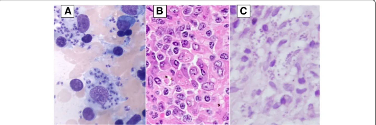
Recurrence of visceral and muco-cutaneous leishmaniasis in a patient under immunosuppressive therapy
Texte intégral
Figure

Documents relatifs
Constatant que tous les élèves ne partent pas avec des compétences équivalentes, la préoc- cupation centrale des enseignants était de ren- dre leur enseignement profitable tant dans
All patients with parasitologically confirmed cutaneous or mucosal leishmaniasis who received at least one infusion of liposomal AmB (Ambisome 1 ) were included.. Only patients
Here, cellular and humoral immune responses of healed CL (hCL) and Mediterranean visceral leishmaniasis (MVL) patients were evaluated against results for Leishmania major
The meetings were attended by expert CL trialists representing 10 clinical study sites from 7 OWCL countries (Afghanistan, Burkina Faso, Ethiopia, Iran (Islamic Republic of),
First report on natural infection of Phlebotomus sergenti with Leishmania promastigotes in the cutaneous leishmaniasis focus in southeastern Tunisia. Tabbabi A, Ghrab J, Aoun K,
Citation: Benabid M, Ghrab J, Rhim A, Ben- romdhane R, Aoun K, Bouratbine A (2017) Temporal dynamics and Leishmania infantum infection prevalence of Phlebotomus perniciosus
and the similarities of the immune responses between the healed and the asymptomatic populations in CL endemic areas, it is plausible that Leishmania species persist inside the skin
The full-length rLmlRAB as well as its divergent part rLmlRABC in- duced high significant levels of IL-10 in immune as well as in naïve groups, indicating that IL-10 production was
