Rôle des contractures lors de la marche des enfants atteints de
dystrophie musculaire de Duchenne
par
Nathaly Gaudreault
École de réadaptation Faculté de médecine
Thèse présentée à la Faculté des études supérieures en vue de l’obtention du grade de Philosophiae Doctor (Ph.D.)
en Sciences Biomédicales (option réadaptation)
Mai, 2007
o
Direction des bibhothèques
AVIS
L’auteur a autorisé l’Université de Montréal à reproduire et diffuser, en totalité ou en partie, par quelque moyen que ce soit et sur quelque support que ce soit, et exclusivement à des fins non lucratives d’enseignement et de recherche, des copies de ce mémoire ou de cette thèse.
L’auteur et les coauteurs le cas échéant conservent la propriété du droit d’auteur et des droits moraux qui protègent ce document. Ni la thèse ou le mémoire, ni des extraits substantiels de ce document, ne doivent être imprimés ou autrement reproduits sans l’autorisation de l’auteur.
Afin de se conformer à la Loi canadienne sur la protection des renseignements personnels, quelques formulaires secondaires, coordonnées ou signatures intégrées au texte ont pu être enlevés de ce document. Bien que cela ait pu affecter la pagination, il n’y a aucun contenu manquant. NOTICE
The author of this thesis or dissertation has granted a nonexclusive license allowing Université de Montréal to reproduce and publish the document, in part or in whole, and in any format, solely for noncommercial educational and research purposes.
The author and co-authors if applicable retain copyright ownership and moral rights in this document. Neither the whole thesis or dissertation, flot substantial extracts from it, may be printed or otherwise reproduced without the author’s permission.
In compliance with the Canadian Privacy Act some supporting forms, contact
information or signatures may have been removed from the document. While this may affect the document page count, it does not represent any loss of content from the document.
Cette thèse intitulée
Rôle des contractures lors de la marche des enfants atteints de dystrophie musculaire de Duchenne
présentée par: Nathaly Gaudreault
a été évaluée par un jury composé des personnes suivantes:
Dre Debbie Feldman, président-rapporteur Dr Denis Gravel, directeur de recherche
Dre Sylvie Nadeau, co-directeur Dr Hubert Labelle, membre interne du jury Dre Brenda Brouwer, examinateur externe Dr Paul Allard, représentant du doyen de la FES
o
III
Résumé
La dystrophie musculaire de Duchenne (DMD) est caractérisée par un affaiblissement progressif des muscles et l’apparition de contractures. Ce sont les deux facteurs principaux qui conduisent à la perte de la capacité de marcher chez les enfants atteints de cette maladie. Toutefois, le rôle de ces facteurs lors de la marche n’est pas établi précisément. L’objectif général de la thèse était de quantifier les moments passifs de flexion à la hanche et à la cheville et de déterminer leur contribution aux moments nets de flexion produits lors de la marche chez des enfants atteints de DMD. L’hypothèse générale stipulait qu’en raison de la présence de contractures en flexion à la hanche et à la cheville, les moments passifs en flexion seraient plus élevés chez les enfants atteints de DMD que chez des enfants en santé. Aussi, la contribution des moments passifs aux moments nets
de flexion produits lors de la marche serait plus importante chez les enfants
dystrophiques.
Onze enfants atteints de DMD et quatorze enfants en santé ayant les mêmes caractéristiques anthropométriques ont participé à l’étude. Les procédures expérimentales ont été conduites au laboratoire de pathokinésiologie de l’institut de réadaptation de Montréal. La marche des enfants ainsi que les moments de forces passives de flexion à la hanche et à la cheville ont été évalués, lors d’une même session expérimentale, à l’aide d’un système d’analyse de mouvement combiné à des plateformes de forces.
Le premier objectif de la thèse était de comparer les profils de marche des enfants atteints de DMD à ceux d’enfants en santé afin de déterminer si les changements observés étaient dus à l’effet de la maladie ou à celui de la vitesse de marche, qui est moindre chez les enfants dystrophiques. Les résultats ont indiqué que les enfants atteints de DMD avaient une enjambée plus courte, une amplitude en extension réduite à la hanche et des moments nets en extension à la hanche, au genou et à la cheville inférieurs à ceux des enfants en santé. L’analyse des centres de pression et des forces de réaction du sol a permis d’expliquer certaines différences, dont l’intensité des moments et
l’inversion de ceux-ci, chez certains enfants dystrophiques. Des différences ont aussi été observées dans les profils de puissances musculaires et dans le décours temporel de certaines variables de la marche. Ces changements n’étaient pas attribuables à l’effet de la vitesse de marche, mais à la faiblesse musculaire provoquée par la maladie. Le deuxième objectif de l’étude visait à mesurer les moments passifs de flexion à la hanche et à Ta cheville et de déterminer si, en raison de la présence de contractures, ces moments de force étaient plus élevés chez les enfants atteints de DMD. Étant donné que les enfants dystrophiques n’étaient pas contracturés à la hanche, les résultats n’ont pas montré de différence entre les deux groupes d’enfants dans les valeurs de moments passifs de flexion.
À
la cheville, la présence de contractures en flexion plantaire chez les enfants atteints de DMD est reflétée par un coefficient de rigidité qui était plus élevé chez ces enfants. Le troisième objectif était d’estimer la contribution des moments passifs aux moments nets de flexion produits à la hanche et à la cheville lors de la marche.À
la hanche, les résultats ont révélé un apport important des moments passifs aux moments nets de flexion chez les deux groupes d’enfants. Par contre à la cheville, une contribution plus élevée des moments passifs de flexion plantaire a été observée chez les enfants atteints de DMD à la fin de la phase d’appui.Les résultats de cette thèse confirment que certains changements observés chez les enfants dystrophiques lors de la marche sont attribuables aux déficiences associées à la maladie, soit la faiblesse et les contractures en flexion plantaire. Par ailleurs, la raideur à la hanche n’est pas aussi importante que prévu chez les enfants dystrophiques évalués. Les résultats de nos travaux suggèrent que dans les premiers stades d’évolution de la maladie, les moments de forces passives, associés ou non à la présence de contractures, ne nuisent pas à la marche des enfants dystrophiques.
Mots-clés Dystrophie musculaire de Duchenne, faiblesse, contracture, marche, cinématique, cinétique
Summary
Muscle weakness and joint contractures are the major impairments that affect the locomotor system cf children with Duchenne muscular dysttophy (DMD). These impairments contribute to the loss cf autonomous ambulatory capacity in these chiidren. However, the interaction between muscle weakness and contractures during gait is not clearly understood. We believe that in the early stages cf the disease, passive moments produced by joint contractures could potentially compensate for muscle weakness, and therefore be beneficial to the gait of these chiidren. The main objective of this study was to quantify the contractures experimentally and subsequently, estimate their mechanical contribution to the net moment production during the gait of children with DMD.
A group of eleven children with DMD and fourteen heathy chiidren were evaluated at the pathokinesiology laboratory located at the Montreal Rehabilitation lnstitute. Kinematic and kinetic variables were measured with a 3D motion analysis system combined with force plates. The experimentation was conducted on the same day and under the same experimental conditions during the gait and during the dynamometric passive flexion moments assessments.
The first objective was to compare the gait patterns of DMD children to those of the control children while considering the effect cf gait velocity. The results revealed differences in some gait parameters regardless of the gait velocity comparison: the DMD children had a shorter step length, decreased hip extension angle and lower hip, knee and ankle extension moments. The lower moments observed were explained by force plate and center of pressure data analyses. These results suggest that most of the modifications observed in the DMD children’s gait pattern are not related to the effect cf gait velocity but appear to be disease-specific.The second objective was to quantify passive flexion moments that may be associated with the presence cf hip and ankle flexion contractures. The results demonstrated that since the DMD children did not have contractures at the hip, no significant group differences in hip flexion passive moment were observed. At the ankle
o
vithe presence of plantar flexion contractures in children with DMD was reflected by a higher rigidity coefficient value. The third objective was to estimate the contribution of the passive moment to the net flexion moment produced at the hip and ankle joints during gait. The results indicated that the contribution of the hip passive moment to the netflexion moment is worth noting in both groups of children. At the ankle, the contribution of the passive moment to the net plantar flexion moment was higher for DMD children at the end of the stance phase of the gait cycle.
In general, the results confirmed that DMD chiidren evaluated in this study show
gait adaptations that are associated with the effects of the disease. In addition, these results support that the passive moments, whether associated with the presence of contractures or not, can assist DMD children with gait in the early stages of the disease evolution.
Keywords:
Duchenne muscular dystrophy, weakness, contracture, gait, kinematics, kineticsTable des matières
Résumé iii
Summary y
Table des matières vii
Liste des tableaux xiv
Liste des figures xv
Liste des abréviations xx
Dédicace xxi
Remerciements xxii
Chapitre 1: Introduction 1
Chapitre 2 : Recension des écrits scientifiques 3
2.1. Premier article: Motor function in Duchenne muscular dystrophy children: A review
of the literature 4
2.1.1.Abstract 5
2.1.2. Introduction 6
2.1.2.1. Overviewofthe pathophysiological processes 6
2.1.2.2. Physical clinical status 7
2.1 .3. Muscle weakness in DMD children 8
2.1.3.1. Muscle weakness and its assessment 8
2.1.3.2. Strength profile of DMD children 11
2.1.4. Contracture in DMD children 16
2.1.4.2. Contractures in DMD children 22
2.1.5. Functional performance, muscle weakness and joint contracture in DMD
chiidren 23
2.1.5.1. Evaluation of functional performance 23
2.1.5.2. DMD chiidren functional performance profile 24
2.1.6. Gait performance in DMD chiidren 27
2.1.6.1. Spatio-temporal parameters 27
2.1.6.2. Kinematics and kinetics of gait in DMD chiidren 28
2.1.7. Conclusion 32
2.1 .8. References 34
2.2. Les avenues thérapeutiques proposées et explorées pour traiter les enfants atteints
UeDMD 43
2.2.1. Les traitements proposés en réadaptation 43
2.2.1.1. Les programmes d’activité physique 43
2.2.1.2. Les exercices de renforcement 43
2.2.1.3. La stimulation électrique 44
2.2.1.4. Les exercices d’étirement 44
2.2.1.5. Les orthèses de correction et de stabilisation 45
2.2.2. Les traitements chirurgicaux 45
2.2.3. Les traitements pharmacologiques 46
2.2.3.1. La production de dystrophine 46
2.2.3.2. Les inhibiteurs de la réponse inflammatoire 47
o
ix2.2.5. Le transfert de myoblastes .49
2.3. Modèle théorique reliant les contractures à la performance de la marche des
enfants afteints de DMD 50
2.3.1. Rôle des contractures en flexion plantaire et en flexion de la hanche lors de
sous tâches de la marche 53
2.3.1.1. Contribution des contractures en flexion plantaire dans le contrôle de
l’effondrement du corps 53
2.3.1.2. Contribution des contractures en flexion de la hanche sur le maintien de
l’équilibre du tronc 54
2.4. Objectifs et hypothèses de la thèse 55
2.4.1. Objectif général et hypothèse générale de la thèse 55
2.4.2. Objectifs spécifiques de la thèse 55
Chapitre 3: Méthodologie 57
3.1. Deuxième article: Evaluation of joint stiffness component in the gait of children with
Duchenne dystrophy 58
3.1.1. Abstract 59
3.1.2. Introduction 60
3.1.3. Materials and methods 62
3.1.4. Results 69
3.1.5. Discussion 75
3.1.6. References 78
3.2. Section complémentaire à la méthodologie 81
3.2.2. Participants, critères d’inclusions et procédure de recrutement 81 3.2.3. La mesure des variables à l’étude dans la thèse 82
3.2.3.1. Les variables cliniques 82
3.2.3.2. Les variables de la marche 83
3.2.3.3. Les variables de moments passifs de flexion à la cheville et à la hanche. 84 3.2.3.4. Les variables de contribution du moment passif de flexion 85
3.2.4. Analyses statistiques des données 85
3.2.4.1. Analyses statistiques utilisées pour répondre au premier objectif de la
thèse 85
3.2.4.2. Analyses statistiques utilisées pour répondre au deuxième et au troisième
objectif de la thèse 86
Chapitre 4: Résultats 87
4.1. Troisième article: Gait parameter comparison of Duchenne muscular dystrophy children to control subjects walking at natural and slow velocities 87
4.1.1. Avant-propos 88 4.1.2. Abstract 89 4.1.3. Introduction 90 4.1.4. Methods 91 4.1.4.1. Participants 91 4.1.4.2. Gait analysis 92 4.1.4.3. Statistical analysis 94 4.1.5.Results 94 4.1.6. Discussion 101
o
xi4.1.6.1. Study limitations.103
4.1.7. Conclusion 104
4.1.8. Reference 105
4.2. Quatrième article: Contribution of hip joint passive moments to the gait of Duchenne
muscular dystrophy chiidren 107
4.2.1. Avant-propos 108 4.2.2. Abstract 109 4.2.3. Introduction 110 4.2.4. Methods 111 4.2.4.1. Clinical assessment 111 4.2.4.2. Gait assessment 111
4.2.4.3 Hip passive flexion moments assessment 112
4.2.4.4. Estimation of hip passive flexion moment contribution 113
4.2.4.5. Statistical analysis 114
4.2.5. Results 114
4.2.6. Discussion 118
4.2.7. References 121
4.3. Cinquième article: Mechanical contribution of ankle plantar flexion passive moments
during the gait of Duchenne muscular dystrophy chiidren 123
4.3.1. Avant-propos 124
4.3.2. Abstract 125
4.3.3. Introduction 126
4.3.4.1. Subjects. 128
4.3.4.2. Clinical assessment 128
4.3.4.3. Gait assessment 129
4.3.4.4. Dynamometric assessment of passive plantar flexion moments 130 4.3.4.5. Passive moment contribution calculation 132
4.3.4.6. Statistical analysis 133
4.3.5. Results 134
4.3.5.1. Clinical variables 134
4.3.5.2. Passive plantar flexion moments variables 134 4.3.5.3. Gait parameters and passive moment contribution 136
4.3.6. Discussion 139
4.3.7. Conclusion 140
4.3.8. Références 142
4.4. Résultats complémentaires 145
4.4.1. Analyses des données enregistrées sur les des plateformes de forces lors de la
marche 145
4.4.1.1. Composante verticale de la force de réaction du sol 145 4.4.1.2. Composante antéro-postérieure de la force de réaction du sol 147 4.4.1.3. Composante médio-latérale de la force de réaction du sol 147 4.4.1.4. Résultante totale des forces de réaction du sol 148
4.4.1.5. Orientation antéro-postérieure du vecteur résultant de la force de réaction
du sol 148
4.4.2. Résultats de l’analyse de fidélité de l’évaluation dynamométrique des variables de moments passifs de flexion plantaire à la cheville 151
Chapitre 5 : Discussion 153
5.1. Les effets de la vitesse de marche sur les paramètres cinématiques et cinétiques
des enfants DMD 154
51.1. Les variables spatio-temporelles 154
5.1.2. Les variables cinématiques et cinétiques 155 5.2. Comparaison des moments passifs de flexion à la hanche et à la cheville entre les
deux groupes d’enfants 160
5.2.1. Moments passifs de flexion à la hanche 161 5.2.2. Moments passifs de flexion à la cheville 162 5.3. La contribution des moments passifs de flexion lors de la marche 164 5.4. Implication des résultats sur la pratique clinique 167
5.5. Les limites de l’étude 169
5.6. Les avenues de recherche proposées en réadaptation 170
Conclusion 171
Bibliographie 173
Annexe 1: Courbes cinématiques et cinétiques individuelles Annexe 2 : Certificat d’éthique
Annexe 3: Formulaires de consentement Annexe 4: Déclaration des coauteurs
Annexe 5 : Preuve de soumission et d’acceptation pour publication d’articles Annexe 6: Permissions pour l’insertion d’articles publiés dans la thèse
Liste des tableaux
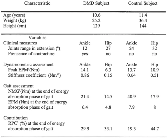
Table 1 : Subjects characteristics and selected variables values 71
Table 2 : Clinical characteristics of children with DMD 95
Table 3 : Time-distance variables of DMD and control children 96
Table 4: Selected clinical, dynamometric and gait variable values 116
Table 5: Passive moment variable values for each group (Mean ± SD) 136
Tableau 6 : Indices de fidélité pour les 4 variables de moments passifs de flexion plantaire
C
xv
Liste des figures
Figure 1: Mean knee extensor strength scores in 45 healthy boys and 43 boys with DMD. Ordinates represent combined scores in pounds from left and right sides. (Reprinted and adapted from Arch Phys Med Rehabll 48, Fowler WM, Jr.,Gardner GW. Quantitative strength measurements in muscular dystrophy.pp. 629-64,© 1964 with permission from The American Congress of Rehabilitation Medicine and the
American Academy of Physical Medicine and Rehabilitation) 13
Figure 2: Serial knee extensor strength scores in 11 boys with DMD. Ordinates represent combined scores in pounds from the left and right sides. Measurements were obtained at four-month intervals over a two-year period. (Reprinted and adapted from Arch Phys Med Rehabil, 48, Fowler WM, Jr.,Gardner GW. Quantitative strength measurements in muscular dystrophy.pp. 629-64,© 196% with permission from The American Congress of Rehabilitation Medicine and the American Academy of
Physical Medicine and Rehabilitation) 14
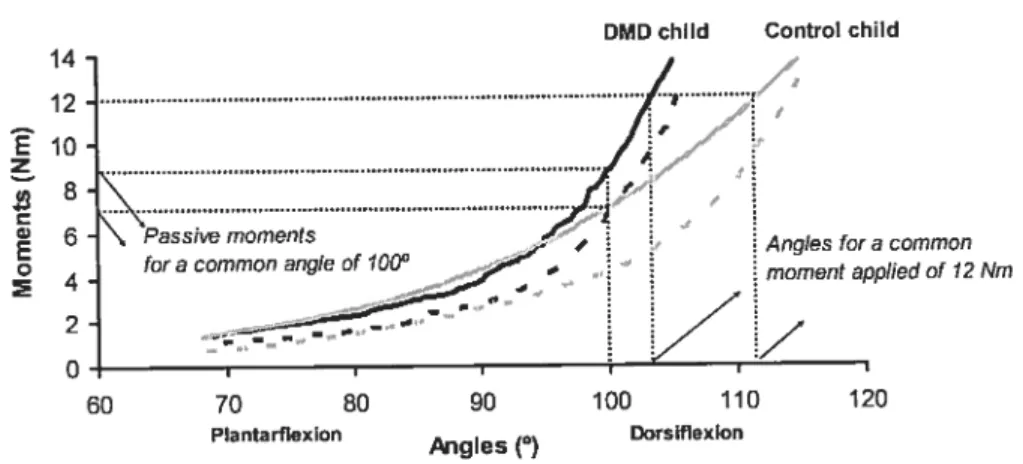
Figure 3: Moment-angle curves for the ankle joint of one healthy child and one DMD child. Ordinates represent the moment values calculated for an ankle angle common to
both subjects as the joint is moyeU from plantar flexion to dors iflexion 22
Figure 4: Scale showing grades for lower extremity function as proposed by Vignos et al.
(1963) 24
Figure 5: Changes in the ground reaction force vector and joints alignment during loading response, mid-stance and terminal stance in DMD children as the disease progresses. (Reprinted and adapted from Clinical Orthopaedics and Related Research, 288, Hsu J D, Furumasu J, Gait and posture changes in the Duchenne muscular dystrophy child . pp.l22-25, © 1993, with permission from Lippincott,
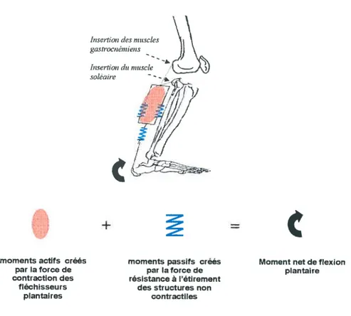
Figure 6: Schématisation du moment net de flexion plantaire créé par la sommation algébrique des moments produits par les forces actives et passives qui agissent sur
la cheville lors de la phase d’appui 51
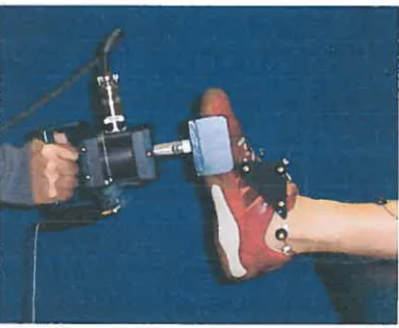
Figure 7: The evaluator applied a tangential force on the foot with a triaxial dynamometer
during EPM assessment of the ankle 65
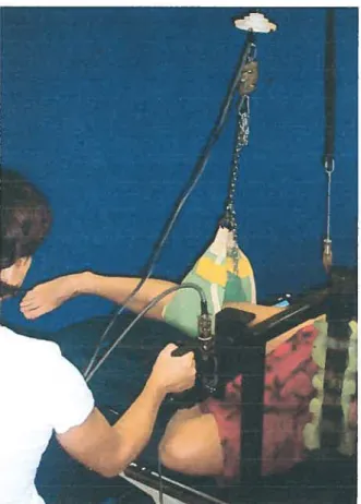
Figure 8: For the EPM assessment of the hip, the subject was lying on his left side and the
right lower limb was supported in a suspension system to counteract gravity 67
Figure 9: This figure shows moment-angle curves obtained from the EPM assessment of the ankle of the two subjects. The curve of the child with DMD is steeper than the curve of the control child, reflecting an increased passive resistance at the DM0 child’s ankle. The hysteresis phenomenon (the difference between the energy absorbed during the stretching phase [dorsiflexion movement] and the energy released during the return phase [plantar flexion movement] can be observed 70
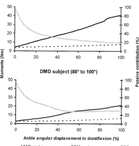
Figure 10 : This figure illustrates the NMG, EPM and RPC for both subjects as a function of the energy absorption phase identified during gait. The ankle angles corresponding to the energy absorption phase of each subject are provided parenthetically. From 50% to 100% of the absorption phase of gait, the plantar flexion contracture of the child with DMD contributes considerably (29.9%) to the net moment production at the
ankle 73
Figure 11: This figure illustrates the NMG, EPM and RPC for both subjects as a function of the energy absorption phase identified during gait. The hip angles corresponding to the energy absorption phase of each subject are provided parenthetically. The RPC profile of the control subject is similar to the RPC profile of the DMD subject. No contractures were found at the hip of either child on clinical examination 74
Figure 12: Markers were placed on the following body segments: the pelvis (A); right lateral aspect cf the iliac crest, the left and right postero-superior iliac spines (PSIS), the right thigh (B), 3 markers pre-positioned on a rigid triangle positioned on the postero-lateral surface of the thigh, the right leg (C), lateral malleolus, lateral and anterior surface of the shank and the right foot (D), 3 markers pre-positioned on a rigid triangle placed on the lateral side cf the foot 93
Figure 13: Graphical representation cf the angular displacements, moments and power data estimated at the hip, the knee and the ankle around the transverse axis and referring to flexion/extension movement. The mean data profile cf the selected gait variables is presented for one complete gait cycle at a natural velocity for the DMD children and at a natural and slow gait velocity for the control group 97
Figure 14: Graphical representation cf the angular displacements, moments and power data estimated at the hip around the antero-posterior axis and referring te abduction adduction movements. The mean data profile cf the selected gait variables is presented for one complete gait cycle at a natural velocity for the DMD chiidren and at a natural and slow gait velocity for the control group 99
Figure 15: The mean data profile for forward displacement cf the COP under the supporting foot during one complete gait cycle is depicted here. The zero value on the ordinate corresponds to the position cf the ankle articular center; therefore, positive values refer to an anterior position cf the COP relative to the ankle joint, whereas negative values tefer to a postericr location cf the COP. A significant difference
(p<O.05)
was found in the COP location between DMD children and control chiidrenat a natural and at slcwt gait velocity for these percentages cf the gait cycle 100
Figure 16: Passive flexion moment evaluation set up. The right lower limb was supported in a suspension system. A triaxial dynammeter (A) was used to measure the passive flexion moments. The force acting on the suspension cable was measured by a uniaxial force transducer, as shcwn by the arrow 113
Figure 17: Graphical representation of the angular displacements, moments and power data estimated at the hip around the transverse axis and referring to flexion!extension movement. The mean data profile of the selected gait variables is presented for one complete gait cycle at a natural velocity for the DMD children and for the control group. The 3 power events are represented as followed: HI: first hip power event (energy generation), H2: second hip power event (energy absorption), H3: third hip
power event (energy generation) 115
Figure 18: Moment-angle curves obtained from hip passive flexion moment assessment of
the children with DMD and the control children 117
Figure 19: Illustration showing the position of the triaxial dynamometer on the foot segment during the dynamometric assessment of passive plantar flexion moment at the ankle. The markers were in the same position as during the gait assessment 131
Figure 20: Passive moment-angle curies obtained from the dynamometric assessment of the passive ankle plantar flexion moments of both groups. The Ieft side cf the figure shows the mean moment-angle values (1 SD) for both groups estimated between —5° to +5°. The right upper corner of the figure illustrates the passive resistance variables that were estimated at 7 Nm. The points represent the mean angle for a given moment whereas the small une segments illustrate the rigidity coefficients’ slopes. 135
Figure 21: Ankle joint kinematic profiles of both groups during the stance phase of gait. 137
Figure 22: Gait moment profile curies showing the absolute passive moments contribution of the plantarflexor non contractile structures. Note that the Y axis scale is the same
Figure 23 Représentation graphique de la force globale de réaction du sol A) et de ses différentes composantes; B) composante verticale, C) composante antéro-postérieure et D) composante médio-latérale. Les forces sont présentées pour la période du cycle
de marche allant de 0 ¾ à 60 ¾ 146
Figure 24 Orientation antéro-postérieure du vecteur de la force de réaction du sol présentée pour une période du cycle de marche allant de O ¾ à 60 ¾. Les valeurs négatives indiquent que le vecteur est incliné vers l’arrière, la valeur O indique que le vecteur est vertical et les valeurs positives indiquent que le vecteur est incliné vers
l’avant 149
Figure 25 Situation du centre de pression sous le pied de la jambe d’appui. La distance a été normalisée pour la longueur du pied. Les données sont présentées pour la période du cycle de marche allant de O % à 60 % 150
Liste des abréviations
DMD Duchenne muscular dysttophy ROM Range of motion
PROM Passive range of motion AF Change in the passive force A L Change in the length
A M Change in the passive moment A A Change in the angular displacement AKE Active knee extension test
RPC Relative passive contribution EPM Estimated passive moment HEPM Flip estimated passive moment NMG Net moment during gait
EMG Electromyographic
ASIS Anterior superior iliac spine PSIS Posterior superior iliac spine COP Center of pressure
Al First ankle power event, energy absorption A2 Second ankle power event, energy generation
Ki First knee power event, energy absorption K2 Second knee power event, energy generation K3 Third knee power event, energy absorption K4 Fourth knee power event, energy absorption HI First hip power event, energy generation H2 Second hip power event, energy absorption H3 Third hip power event, energy generation ESM Écart-type de l’erreur de mesure
À
ma mère et à mon père. Pour leur force et leur immense courage face à l’adversité.Remerciements
Je remercie tout d’abord mes directeurs de recherche, Dr Denis Gravel et Dre Sylvie Nadeau, de m’avoir témoigné leur confiance en sachant très bien que je venais de loin. Ils ont su partager leur savoir et leur passion pour la recherche à chacune des étapes de cette thèse. En m’incitant à aller toujours un peu plus loin, ils m’ont appris la rigueur et la persévérance. Mais surtout, je tiens à les remercier pour leur soutien moral dans les moments difficiles.
Je tiens à exprimer ma gratitude à tous les enfants qui ont participé à l’étude ainsi qu’à leurs parents. Le courage et la détermination dont les enfants dystrophiques font preuve au quotidien m’ont grandement touchée et inspirée. Sans leur collaboration, ce projet n’aurait pu être possible.
J’adresse mes remerciements à deux précieux collaborateurs de la clinique des maladies neuromusculaires du CHU Mère-Enfant de l’hôpital Ste-Justine de Montréal merci au Dre Sylvie Houde, physiatre, d’avoir partagé ses connaissances; merci à Mme Sylvie D’Arcy, physiothérapeute, pour son aide lors du recrutement des sujets.
Je tiens à témoigner ma reconnaissance envers le personnel du Centre de recherche interdisciplinaire en réadaptation du Montréal métropolitain, Institut de réadaptation de Montréal. Je remercie particuiièrement MM. Pierre Desjardins et Michel Goyette, ingénieurs, qui ont contribué au développement de programmes d’analyse. Merci également à M. Daniel Marineau, pour son implication à la mise au point du montage expérimental. Je voudrais aussi souligner l’excellent travail et la patience d’Alexandra Duranceau, assistante de recherche, lots du traitement des données.
Je remercie tous mes collègues étudiants du centre de recherche d’avoir été disponibles pour échanger ou pour m’aider au cours des quatre dernières années. Je remercie particulièrement mes amis de
fin
de parcours: France Piotte, Marie-Hélène Milot,Dany Gagnon, Cyril Duclos, Anabèle Brière, Julie Lecours et Mélanie Morin. Nos discussions et nos fous tires me manqueront beaucoup.
Je tiens à exprimer toute ma reconnaissance envers mon directeur de recherche, la faculté des études supérieures de l’Université de Montréal, le Réseau de recherche provincial en adaptation-réadaptation et les Instituts de recherche en santé du Canada pour l’octroi de bourses d’études. Ce projet a été réalisé grâce à une subvention des Instituts de recherche en santé du Canada, en partenariat avec Dystrophie Musculaire Canada.
Enfin, je remercie toute ma famille et mes amis qui, de près ou de loin, m’ont encouragée dans la réalisation de ce projet. Merci de me pardonner de vous avoir négligés. Merci à mon conjoint Vincent, mon rayon de soleil. Son assiduité et sa persévérance dans la recherche de la perfection de son art, m’ont appris qu’il ne faut pas baisser les bras devant les obstacles qui nous semblent insurmontables.
e
Chapitre
1:
Introduction
Les dystrophies musculaires sont un groupe de maladies héréditaires qui
provoquent un affaiblissement progressif des muscles. La forme de Duchenne, l’une des plus sévères, atteint principalement les garçons. Selon Dystrophie Musculaire Canada, son incidence serait de I cas sur 3500 nouveaux nés et sa prévalence serait de 3 cas sur 100 000 habitants. Le caractère pathologique de cette myopathie se traduit par l’absence d’une protéine musculaire, la dystrophine, responsable de la stabilité et de l’intégrité de la membrane de la cellule musculaire. Sans cette protéine, une dégénérescence progressive des muscles squelettiques et du muscle cardiaque est observée ce qui amène le déclin du statut fonctionnel des enfants. Ces derniers perdent leur capacité ambulatoire autonome vers l’âge de dix ans.
Le confinement prématuré au fauteuil roulant n’est aucunement souhaitable compte tenu des effets néfastes de l’immobilisation sur la santé psychologique et physique des enfants. Ainsi, les traitements proposés à ces enfants en réadaptation ont comme principal objectif de maintenir leur autonomie à la marche et par le fait même, de prolonger leur indépendance fonctionnelle. Cet objectif ne peut être atteint sans une meilleure compréhension des mécanismes biomécaniques inhérents à la marche des enfants atteints de dystrophie musculaire de Duchenne (DMD). Sachant que la force musculaire est un facteur déterminant de la performance de la marche, la question suivante s’est donc imposée à nous comment ces enfants parviennent-ils à marcher en dépit d’une faiblesse marquée des muscles extenseurs des membres inférieurs?
Au cours des dernières années, la démarche typique des enfants atteints de DMD a fait l’objet de quelques études. Ces dernières concluent que des ajustements de certains paramètres de la marche comme la longueur de l’enjambée, la vitesse de marche, la position des forces de réaction du sol, feraient en sorte que la demande mécanique imposée aux muscles extenseurs serait moindre. Ceci apporterait une partie de la réponse à la question posée. Nous croyons qu’une autre piste d’explication possible réside dans l’influence qu’ont les torces passives associées à la présence d’une contracture en flexion sur les moments articulaires créés lors de la marche. Ces forces passives permettraient
de compenser la faiblesse des muscles fléchisseurs, tant à la hanche qu’à la cheville. Afin d’aller plus loin dans cette voie d’investigation, la mise au point d’un protocole de mesure permettant de quantifier les forces passives associées aux contractures en flexion de même que l’analyse de l’impact mécanique de ces forces passives sur la marche des enfants atteints de DMD sont deux étapes incontournables qui ont conduit aux travaux qui sont présentés dans cette thèse.
Les contractures sont perçues d’emblée comme étant nuisibles à la performance motrice des enfants dystrophiques et leur réduction fait parti des traitements proposés en réadaptation. Sans une connaissance exacte de leur rôle dans la locomotion, il devient difficile pour les cliniciens de prédite l’effet de la réduction d’une contracture sur la capacité de marcher des enfants. Ces travaux apporteront une contribution essentielle à cette problématique et ils aideront les cliniciens à prendre des décisions plus rationnelles dans le choix des thérapies envisagées pour traiter cette déficience.
Cette recension des écrits est présentée sous la forme d’un article intitulé « Motor function in Duchenne muscular dystrophy chiidren : A review of the literature ». Cet article est publié dans le journal Critical Reviews in Physical and Rehabilitation Medicine, 2005, 17(3) :231 -248. Les auteurs sont, Nathaly Gaudreault, Denis Gravel, Sylvie Nadeau et Sylvie Houde. Trois grands thèmes sont abordés dans ce manuscrit. Le premier thème décrit les mécanismes pathologiques de la maladie. Ensuite, un deuxième thème définit les principales déficiences du système locomoteur rencontrées chez les enfants DMD, soit la faiblesse et les contractures, et présente les différentes méthodes d’évaluation de ces déficiences. Enfin, un troisième thème se rapporte à l’influence de la faiblesse et des contractures sur le développement moteur des enfants atteints de DMD. Ce thème cible plus particulièrement la marche. Dans le but de compléter celle section, les avenues thérapeutiques proposées pour contrer cette maladie seront abordées à la suite de l’article.
La recension des écrits se terminera par la présentation du modèle théorique reliant les contractures articulaires à la performance de la marche des enfants atteints de DMD. Enfin, les objectifs et les hypothèses de la thèse seront énoncés afin de clore ce chapitre.
o
4
2.1.
Premier
article:
Motor function
in
Duchenne
muscular
dystrophy chiidren: A review of the literature
Nathaly Gaudreault,
Denis Gravel, 1,2
Sylvie Nadeau, 1,2
Sylvie Houde,
1
Centre de recherche interdisciplinaire en réadaptation du Montréal Métropolitain (CRIR), Site Institut de réadaptation de Montréal, Montréal, Canada 2 École de réadaptation, Faculté de médecine, Université de Montréal, Montréal, Canada; 3CHU Mère-Enfant Ste Justine, Montréal, Canada.
En tant que première auteure, je confirme ma participation à toutes les étapes qui ont mené à l’écriture de ce manuscrit, de la recension des écrits scientifiques à la critique de ces derniers. Dt Denis Gravel et Dre Sylvie Nadeau, experts en biomécanique et directeurs de la thèse et Dr Sylvie Houde, physiatre en pédiatrie, ont également collaboré à l’écriture et à la révision de l’article.
2.1.1. Abstract
Duchenne muscular dystrophy (DMD) is an X-iinked disease affecting the muscular system(s) of young boys. Currently, no cure exists and the goals of treatment are to reduce disabilities and to prolong the chiidren’s functîonal independence. The two major physicai impairments associated with this disease are muscle weakness and joint contractures. The purpose of the present paper is to review the profite cf muscle weakness and joint contractures in DMD chiidren as weii as the means by which these impairments have been evaiuated. The impact cf muscle weakness and joint contractures on functional activities and the interaction between muscle weakness and joint contractures during gait
wili aise be covered.
2.1.2. Introduction
2.1.2.1. Overview of the pathophysiologïcal processes
Duchenne muscular dysttophy (DMD) is an X-linked recessive disorder affecting approximately 1 in 3500 males born in the world (Biggar et al., 2002; Roland, 2000).Although most childreninherit the disease from cartier mothers, approximately one thitd of cases are due to a new genetic mutation (Biggar et al., 2002; Roland, 2000). DMD has been shown to be related to a deletion within the xp2l gene leading to a lack of dystrophin, an important protein involved in membrane stability of the catdiac and skeletal muscles (Ibraghimov-Beskrovnaya et al., 1992; Rybakova et al.,2000). Without dystrophin, muscle membrane damage is enhanced by contractions, resulting in the activation of an inflammatory ptocess within the muscle. The fibers undergo regeneration-degeneration cycles until the regeneration process can no longer take place. As early as 1851, microscopic examination of the muscles of affected boys revealed irreversible necrosis of the muscle fibers, which were replaced by connective tissue and fat (Meryon, 1852). This phenomenon was related to pseudohypertrophy of the muscle (Dubowitz, 1995), more particularly the calves, and these changes in muscle tissue can now be confitmed using more advanced technical instrumentation such as ultrasound and magnetic resonance imaging (MRI) (Marden et al., 2004; Reimers et al., 1996). Moreover, the leaky muscle membrane allows creatine kinase (CK) to escape and enter the bloodstream, leading to an increase in serum OK in the blood of affected children. Laboratory investigations using molecular genetic techniques, muscle biopsies, MRI, chest X-rays and serum CK levels are used to confirm the diagnosis. Impairment has been defined as a loss or abnormality in the otgan or at the organ system level of the body (US department of Health and Services, 1993). Thus, muscle weakness as well as joint contractures, muscle fatigue and reduced cardiopulmonary function are impairments that may result from pathophysiological modifications of the muscle tissue.
o
7
2.1.2.2. Physical clinical status
According to some authors (Brooke et al., 1989; Vanasse, 1993), the clinical signs and symptoms of DMD are first characterized by the children’s difficulty in achieving motor milestones and a delayed independent ambulation during early infancy. Secondary to
proximal muscle weakness and the development of joint contractures, DMD children show
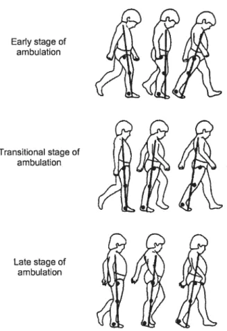
modifications in the performance of locomotor tasks. A waddling gait is observed between ages 4 and 6 and some children experience frequent falls (Sutherland et al., 1981; Vanasse, 1993). The DMD child can raise himself from the floor using a stereotyped method described by Gowers (see Tyler, 2003). He rolis himself over on his stomach, straighten his legs and arms, to finally put one hand at a time on his knees and move them alternately doser to his hips to straighten his trunk. As the disease progresses, the disability, which is defined as a limitation in performing tasks, activities and rotes at levels expected within physical and social contexts (US department of Health and Services, 1993), becomes increasingly severe. The inability to climb stairs and to run occurs, and most children lose ambulation before 13 years of age (average 10.5 years) (Brooke et al., 1989). Once the chiidren are confined to a wheelchair following the ambulatory phase, musculoskeletal deformities such as equinovarus of the feet and scoliosis develop quite rapidly. Cardiomyopathies and respiratory failure are the leading cause of death, which occurs somewhere in the second decade (Darras et al., 2003; Roland, 2000).
Although clinical trials investigating the effect of medication such as Prednisone and Deflazacor on muscle weakness have shown positive results, no curable treatment currently exists for this disease. The proposed rehabilitation treatments share a common goal, which is to delay functional limitation. Functional limitation is the restriction or the lack of ability to perform an action in the manner or within the range consistent with the purpose of an organ or an organ system (US department of Health and Services, 1993). For instance, impairments related to DMD can eaU to functional limitation. It is well recognized that muscle weakness and muscle fatigue affect the children’s overali physical performance during the course of the disease (Scott et al., 1982). For example, DMD boys show a decreased self-selected walking speed and they cover shorter distances per unit
time than normal boys. The impacts of joint contractures on gait have been mentioned in the literature but the underlying mechanisms of their action are stili uncleat (Lee et al., 1997; Messier et al., 1992). Furthermore, the interaction between muscle weakness and contractures as weII as the Iink between these impairments and the decline of Iocomotor skills during the whole course of the ambulatory stage of DMD boys needs to be clarified. The purpose of this paper is to review muscle weakness and joint contracture evaluation and development in DMD children and to try to shed light on the impact of these impairments on functional activities, more specifically on gait. Aspects regarding the treatment of these impairments as well as the effects of these treatments on gait will not be covered in the present paper.
2.1.3. Muscle weakness in 0MO chiidren
2.1.3.1. Muscle weakness and its assessment
Muscular strength can be defined as the force or tension a muscle or
a
muscle group can exert against a resistance in one maximal effort (Fox et al., 1989). In the case of very weak muscles, the resistance can be the segment weight. Muscle strength is an essentiai component of the production of a movement (Blimkie, 1991). When it is decreased, the children’s performance in activities of daily living or in sports activities can be affected (Blimkie, 1991). Classic literature from the century reports thatthe major clinical characteristic of DMD is the progressive loss of muscle strength without any sensory deficit (Tyler, 2003). As previously mentioned, the absence of dystrophin in the muscles of DMD children is the primary underlying pathophysiology explaining muscle weakness. Other secondary mechanisms such as overwork muscle injury and disuse can enhance a decrease in the muscle strength of chiidren with neuromuscular diseases (Johnson, 1971; Vignos, 1983; Appell, 1990). Assessment of muscle strength is thus considered an important part of the children’s overall examination process. Strength measurements are not only important in describing the natural clinical course of the disease, but these measures are also important in studying the effects of therapeuticinterventions (Fowler & Gardner, 1967; Brooke, et al., 1981a; Brooke et al., 1983; Mendell et al., 1987). They have also been used to try to explain the deterioration of the boys’ functional independence (Uchikawa et al., 2004).
Static muscle strength in children can be evaluated with grading systems using an ordinal scale such as manual muscle testing (MMT) as well as with instrumented dynamometry (Medical Research Council, 1943; Horvat et al., 1992). MMT appears to be the most widely used system for estimating muscle strength in clinical settings (Kendall & McCreary, 1983). It is easy to employ, it requires no sophisticated equipment, and it shows good reliability in the grades below “Fair” or “3”, depending on the grading system used (Lilienfeld et al., 1954; Rothstein, 1985). However, some limitations with regard to its utilization should be highlighted. Beasly (1961) reports that in adults, MMT overestimates strength in the higher grades. In the author’s opinion, grade “5”, which reflects the maximal strength of a muscle, corresponds to a range of 50% to 75% of the normal strength values measured with more objective techniques. Also, grades “4” and “5” cover a very wide range of strength and it has been demonstrated in children and adults that individuals who obtained the same strength measurements for a muscle on a dynamometer could be graded either 4 or 5 for that muscle on MMT (Aitkens et al., 1989; Schwartz et al., 1992). Lilienfeld et al. (1954) and Rothstein (1985) find that in adults, gravity-eliminated grades (grades from “0” to “2”) are the most reliable. Their results are corroborated by those of Florence et aI. (1992) in an investigation involving DMD children. Florence et al. (1992) also report that intratester reliability is beller for proximal muscles than for distal muscles and it is also beller for lower extremity muscles than for upper extremity muscles. These findings should be taken into consideration when monitoring strength gain with MMT following intervention in clinical trials, especially for higher grade scores. According to Struberg and Metcalf (1988) who studied strength in normal and DMD children, no operational definition of “Normal” muscle strength for various subject characteristics such as age, sex and body build exists so far, and this could contribute to the poor reliability of grades “4” and “5”. Other factors such as the weight cf the tested limb and the examiner’s strength can influence the scoring in the grades from “3” to “5” and explain their poor rel iabil ity.
o
10
Instrumented dynamometry has been used to assess static muscle strength in bath
normal and DMD chiidren, and these methods seemed ta eliminate some issues
encountered with MMT (Stuberg et al., 1988; Brussock et al., 1992; Horvat et al., 1992). Some studies have investigated the reliability cf quantitative static muscle testing in normal and DMD children with a hand-held dynamometer (Stuberg et al., 1988; Harvat et al., 1992). Stuberg and Metcalt (1998) report Pearsan correlation coefficient for intratester reliability tanging from 0.74 ta 0.99 for healthy chiidren and tram 0.83 ta 0.99 far DMD children. Regarding intertester reliability, Horvat et al. (1992) conclude that a handheld dynamometer is a reliable taol, with 1CC ranging tram 0.49 ta 0.95, but their study involved healthy children anly. This point is important ta cansider since different clinicians are often invalved in the follaw-up evaluations cf DMD children, and therefore intertester reliability remains ta be determined for hand-held dynamometry strength measurements for these patients. In addition ta praviding more objective measurements than MMT, the hand-held dynamometer has the advantage cf being easy te operate and it can be employed in a clinical setting as an alternative ta methads using a cable tensiometer such as seen in laboratary set-up. Brussock at al. (1992) have evaluated DMD children’s muscle strength using a strain gauge system and they also report that intertester reliability was Iower than intratester reliability (lCCs between 0.74 and 0.97 for intertester and between 0.88 and 0.99 for intratester). Accarding ta these authors, the strain gauge system has the ability ta test stronger muscle groups in non-disabled children as apposed ta a hand-held dynamometer.
A point ta consider in sttength assessment is the variation in strength with the joint angle (Williams & Goldspink, 1984). In MMT and dynamametric testing, it is comman ta measure farce at ane angle. Thus, if one muscle group is tested at an angle where t is naturally weak, as when the muscle is in a shartened position, and another muscle group is tested at an angle where it is naturally strong, such as when the muscle is at its optimal length, it may be erraneausly cancluded that the former is weaker than the latter. The same applies if contraI subjects and impaired subjects are being campared. For example, if a muscle group in impaired chiidren is evaluated in a joint position where this muscle group is weak and if the same muscle group in the children cf the contraI group is
evaluated in a position where it is strong, it can be incorrectly concluded that the muscles of the impaired children are weaker than the muscles of the unaffected children. To resolve this issue, muscle force across a range of motion as obtained by isokinetic dynamometry should be recorded, or some criterion should be applied to determine a comparable angle of measurement in static testing.
Thus, muscle performance can also be estimated through dynamic measurements, which means that strength is evaluated during movement. When using these measurements, many factors should be considered. For instance, it is necessary to consider angular velocity because strength depends on the muscles shortening velocity (Hill, 1970). Also, muscle strength can be assessed as the muscles shorten (concentrically) or as they lengthen (eccentrically) and strength measures for the same muscle group will be different depending on the method chosen. Moreover, muscle strength can also be expressed in terms of joint moment. Joint moment is estimated from the product of the muscle strength and the lever arm, which is the perpendicular distance between the force direction and the joint center, and is expressed in Newton meter units (Nm). Stuberg et al. (1988) and Brussock et al. (1992) have evaluated strength in DMD children and they both concluded that moment measurements would be more appropriate than strength measurements because lever arms are considered. Lever arms would be an important factor in a case where, for example, two children generate the same amount of strength for a given muscle group on a dynamometer located at the distal part of a limb segment. Moment values could be different if the length of the limbs is not the same in both children. Lever arms would also be essential to consider in longitudinal studies on strength involving children who are growing up.
2.1.3.2. Strength profile of DMD chiidren
The strength profile of DMD children is characterized by an initial gain in early childhood so that they can achieve motor milestones normally. However, one must keep in mmd that quantification of strength under the age of 5 s difficult, so interpretation of
strength data under that age cculd be biased (Stocke et al., 1981a; Scott et al., 1982; Brooke et al., 1983). As the child grows older, discrepancies occur in the literature regarding the strength profile. MMI and instrumented dynamometry were both used in the evaluation cf DMD children and the results cbtained with these approaches wete flot always consistent. Some studies where MMI was used te monitor strength demonstrate that for mcst cf the muscle groups, strength cf the DMD children decreases linearly from the age cf 5 until the loss cf ambulaticn (Scott et al., 1982; McDonald et al., 1995). Other studies using quantitative strength measures indicate minimal changes
in
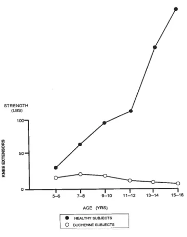
strength up te 7-8 years cf age and even showed an increase in the mean force up te 10 years cf age in many muscle groups (Fcwler et al., 1967; McDonald et al., 1995). On the cne hand, this discrepancy can be explained by the pccr reliability cf the MMT methcd for the higher quctations. On the other hand, it may be possible that scme DMD children effectively increase their strength after the initial evaluaticn because strength increases with age (Mclnar & Alexander, 1974; Backman & Henriksson, 1988; McDonald et al., 1995). Thus, the lcss cf sttength asscciated with the progression cf the disease may be masked by the maturation phenomenon. The balance between maturation and disease effects ccutd be determinant cf the rate cf change in strength at the beginning cf the disease. Nevertheless, the difference in strength between normal and DMD children increases with age since normal children ccntinuously get stronger until their adult years, while detericration is cbserved in DMD persons as they age. Another possible explanaticn for the inccnsistency between MMI and dynamometric approaches in the abcve studies is the fact that DMD clinical manifestations may prcgress differently from child te chïld and the initial clinical evaluaticn may occur at varicus ages as well. If this is the case, the mean force value calculated at a specific age is made up cf strcng and weak patients resulting in minimal changes in the early stage cf the disease.Ihis last interpretaticn cculd be supported by the results cf Fowier and Gardner (1967) whc demonstrated a decline in strength in serial measurements for the same subject and an increase in strength when group data were ccnsidered. Iheir study included 43 children with muscular dystrcphy between the ages cf 6 and 16, and strength was measured using a strain gauge. Figure I represents mean strength score data for the
o
13
knee extensors of ail the chiidren who participated in the study as a function of age. Figure
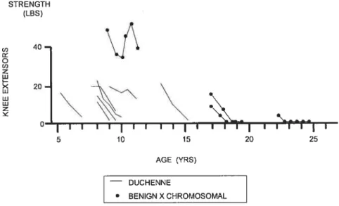
2 illustrates the knee extensor strength score of 11 boys with Duchenne muscular
dystrophy (4 of these boys had the benign X-chromosomai type) obtained at 4 four-month intervais over a two-year period.
STRENGTH (LBS) 100rn o, o o, 50 0— I I I I I 5—6 7—8 9--10 11—12 13--14 15--16 AGE (YRS)
Figure 1: Mean knee extensor strength scores in 45 healthy boys and 43 boys with 0MO. Ordinates represent combined scores in pounds from Ieft and right sides. (Reprinted and ada pted from Arch Phys Med Rehabil, 48, Fowler WM, Jr.,Gardner GW. Quantitative strength measurements in muscular dystrophy.pp. 629-64,© 1967 with permission from The American Congress of Rehabilitation Medicine and the American Academy of Physical Medicine and Rehabilitation).
ppco
• HEALTKY
STRENGTH (LBS) U) 40-o U) z w I— 20.\
:
0• I I I I I I I I I I I I I I 5 10 15 20 25 AGE (YRS) DUCHENNE • BENIGN X CHROMOSOMALFigure 2: Serial knee extensor strength scores in 11 boys with DMD. Ordinates represent combined scores in pounds from the Ieft and right sides. Measurements were obtained at four-month intervals over a two-year period. (Reprinted and adapted from Arch Phys Med Rehabil, 48, Fowier WM, Jr., Gardner GW Quantitative strength measurements in muscular dystrophy. pp. 629-64, © 1967, with permission from The American Congress of Rehabilitation Medicine and the American Academy of Physical Medicine and Rehabilitation).
When one looks at the relationship between strength and age in the strength profile of DMD boys, the results of this study show that when mean strength scores are plotted against age, strength increases slightly up to age 10 and then starts to decrease (Figure 1). On the other hand, strength score data for the same child measured over a 2-year period of time show that strength decreases after obtainment of the first measure even in
young boys (Figure 2). Although older children (up to 10 years old) demonstrate higher mean strength scores than younger children, the decline in strength starts as early as 5 years of age (Fowler et al., 1967; Ziter et al., 1977; Brooke et al., 1981a; Scott et al., 1982). McDonald et al. (1995) report that the rate of strength decline, as measured with manual muscle testing (MMT), is linear and the decline is greater between 5 and 13 years otage.
Quantitative static strength measurements for different muscle groups expressed as a percent of normal control strength showed that the strength of DMD children was 35-50% of normal control values (McDonald et al., 1995). Muscles of DMD children are not evenly affected by weakness. Some studies have reported that weakness was greater in proximal muscle groups than in muscles located distally (Scott et al., 1982; Brooke et al., 1989; McDonald et al., 1995). Moreover, the muscles of the pelvic girdle are involved first, followed several years later by the muscles of the shoulder girdle (Fowler et al., 1967; McDonald et al., 1995). Quantitative data from Fowler and Gardner (1967) indicate thatthe rate of strength decrease is about the same for both lower and upper extremities. Lord et al. (1987) found that the coefficient of determination for the upper extremity and lower extremity strength comparison was 0.62. This means that 62% of upper extremity strength can be explained by lower extremity strength. Thus a significant portion of the relationship remains unexplained. They concluded that although upper extremity loss of strength parallels lower extremity loss of strength, interpretations based on the results of the former should not be applied to the latter. Regarding side difference, some studies have reported that strength was gteater on the right side than on the left in DMD chiidren as well as in normal boys, and this could be associated with the effect of side dominance (Fowler et al., 1967; Brussock et al., 1992). However, the effect of side dominance was not observed in other studies (Scoif et al., 1982; McDonald et al., 1995). Finally, Scott et al. (1982) report that the extensor groups are aiways weaker than the flexor groups as assessed with MMT and myometry. This imbalance between flexors and extensors was also reported for the trunk muscles (McDonald et al., 1995).
As far as weakness distribution in the muscles of the lower extremities is concerned, Fowler and Gardner (1967) as well as McDonald et al. (1995) state that
o
16
weakness affects first the hip flexors (iliopsoas), knee extensors (quadriceps) and hip extensors (gluteus maximus). Other studies reported an early weakness of the dorsiflexor muscles (Sadjadpour, 1975; Berrol, 1976) of the ankle but quantitative measurement by Scott et al. (1982) indicated a stable force value for this muscle group with the evolution of the disease. As already mentioned, comparison of strength between muscle groups can be misinterpreted if the measurements are obtained at angles where the muscle is in a shortened position. The distribution of weakness in the upper extremities resembles that of the lower extremities. Fowler and Gatdnet report that when arm strength scores for each muscle group are expressed as a percentage of total upper extremity strength, the hand grip of DMD children conttibuted 45 to 65% of the total upper extremity scores. In normal boys, the proportion of hand grip strength was 30 to 38%. These data suggest that proximal muscle strength represents a lower percent of total upper extremity strength. This was confirmed by the proximal strength score, where shoulder abductors made up 3-13% of the total upper extremity scores in affected children, whereas in normal chiidren, the percentage for this muscle group was 12 to 19%.
2.1.4. Contracture in DMD chiidren
2.1.4.1. Defïnition and measurements of contractures
After muscle weakness, joint contractures represent the second major clinical impairment affecting the locomotor system of DMD children. As for muscle strength, measurement of joint contracture in a DMD population is useful for monitoring the progression of the disease or for evaluating the effects of a treatment. However, proper measurement of joint and muscle extensibility in humans represents a great challenge. As early as 1959, attempts were made to define the term contractures from both the physiological and clinical points of view. Physiologically, they were related to a prolonged depolarization of the muscle fiber membrane potential, whereas clinically, they were simply defined as a decrease range of motion (Archibald & Vignos, 1959). More recently, contracture was defined as an excessive resistance to passive joint mobilization not
associated with any musculat activity when this joint is mobilized into its available range, which may be decreased (Tardieu et al., 1982; Llorens, 1996). This resistance can be attributed ta multiple causes such as muscle retraction, tendinous adherence, loss cf skin or subcutaneous tissues, joint capsule thickening or a combination of these factors
f
Roberson & Giurintano, 1995).In order ta understand the impact of joint contracture on locomotor functions, one must look at the structures and mechanisms contributing to passive properties cf the muscle. Passive extensibility cf skeletal muscle can be defined as the ability cf a skeletal muscle to lengthen without muscle activation fGajdosik, 2001). Maximal muscle length can contribute to maximal joint range of motion, which is generally believed ta influence functional activities and athletic performances (Gajdosik, 2001). Resistance to passive stretch is influenced by the amount of deformation of the epymysium, perimysium and endomysium, which are connective tissues around and within the muscle. lt has been proposed that from its architectural arrangement, the perimysium contributes mcst ta the resistance ta stretch fPurslow, 1989). Sub-cellular components within muscle fibers can aIse provide resistance to passive stretch. It has been shown that resistance can occur from the filamentous connection within the endosarcomeric and exosarcomeric cytoskeletons (Wang & Ramirez-Mitchell, 1983; Magid & Law, 1985). Finally, Hill (1968) proposes that resistance to passive stretch could also be attributed to the crcss-links between actin and myosin. Under certain conditions, such as immobilization in a shcrtened position, changes in these structures and mechanisms can corne about and adaptation cf muscle length and extensibility can occur. Archibald and Vignos (1959) report that muscle imbalance across a joint can contribute te the development cf a ccntracture in DMD boys. The stronger muscles would tend to move the limb segment in the direction cf their action, and as a consequence, joint range cf motion cpposite to that direction decreases and the strcnger muscles adapt to this new length. In fact, in their model cf prcgressive functional deterioraticn cf DMD children, these authors go further and prcpose that contracture development cornes from multiple interactions between muscle imbalance, muscle weakness, muscle tightness and decreased functional capacity. Moreover, Archibald and Vignos (1959) agree that jcint contracture can be initiated cr aggravated by muscle
imbalance occurring at other joints. Although Brooke et al. (1983) also report muscle imbalance as a possible cause of joint contracture in DMD children, this proposai was flot supported by McDonald et al. (1995). Other causes, such as proionged static positioning of the Hmb as seen in DMD children who are confined to wheelchairs, can enhance joint contractures of hip and knee flexors as well as elbow flexors(Scott et al., 1982; McDonald et al., 1995). Animal studies show thatthe decrease in muscle length is brought about by a reduction in the number of sarcomeres and changes in the connective tissues of the muscle when the muscle is immobilized in a shortened position (Tabary et al., 1972; Wiiliams et aI., 1984).
Some tests are currently used by health care professionals in various clinical settings to assess joint contracture in DMD children. One of these tests is the straight-leg raising test (SLR) to measure hamstring muscle length and flexibility. The angle of hip flexion in relation to the horizontal plane represents the outcome measure. However, hamstring muscle tightness is not the sole factor influencing hip flexion angle during SLR. Pain induced by stretching other structures such as the fascia, dura and sciatic nerve can limit hip flexion excursion (Gajdosik et al., 1985; Smith et al., 1993). Also, there is some evidence that pelvic rotation occurs during SLR, meaning that the hamstring muscles are not fixed at their insertion on the ilium (Bohannon, 1982; Bohannon et al., 1985; Gajdosik, 2001). Therefore, the hip flexion angle cannot be considered a valid assessment of hamstring muscle length and flexibility. Other tests, for instance the standing toe-touch test and the sit-and-reach test, are used to evaluate hamstring muscle length and flexibility. However, movements of the pelvis, lumbar spine and other joints of the iower extremities can not be avoided when performing these tests, resulting in measures of doubtful value (Gajdosik & Lusin, 1983). In an effort to address these issues and to design a more objective tool, the active-knee extension test (AKE) was developed and showed very good intratester reliability (Pearson product-moment correlation = 0.99) (Gajdosik et al., 1983).
Rakos et al. (2001) report good intertester reiiability of the AKE in school-age children (1CC 2,1 = 0.79). Thus, the AKE seems to be the most apptoptiate test to evaluate hamstring
contractures in DMD children in clinical practice as long as a standardized protocol is followed.
o
19Regarding evaluation of hip flexors muscle tightness, fout methods developed many years ago are stili being used: the prone hip extension test, the Thomas test, the Mundale test, and the pelvifemoral angle test (Milch, 1942; Staheli, 1977). Bartlett et al. (1985) compared the reliability of these four techniques in different populations, such as spastic diplegia and meningomyelocele subjects, as well as healthy controls. They concluded that no single measurement technique was superiorto the others and that the technique should be chosen based on different factors and on the specificity of the population evaluated. The Thomas and the prone extension tests were more reliable (intertester reliability) for the meningomyelocele group (r = 0.90 and r = 0.92 respectively) than the Mundale and pelvifemoral angle tests (r = 0.79 and r =0.73 respectively). For the spastic diplegia group, the Thomas test showed high variability and a moderate correlation value (r = 0.70). Among healthy subjects, the prone extension test and the Thomas test demonstrated good reliability and the authors suggest that these tests should be considered for this population since they are quick and easy to perform. As far as DMD chiidren are concerned, the severity of hip flexion contractures would probably determine which of these tests would be the most appropriate for evaluating the deformity. In the early stages of the disease, the prone extension test and the Thomas test would probably be the best choices, but as the deformity becomes more severe and as comfort becomes jeopardized, the pelvifemoral angle test would probably be a beller choice.
Joint passive flexibility has been quantified with the measurement of angular passive joint range of motion (PROM) through goniometry. Although goniometric measurements have been shown to be reliable, important factors to consider when using these measures to quantify joint contractures in a clinical setting were identified. First, one must consider the structure, function and location of the joint being evaluated. Boone et al. (1978) report that both intertester and intratester reliability is greater for upper extremity joints than for lower extremity joints. This finding is corroborated by Pandya et al. (1985)
who studied the reliability of goniometric measurements in DMD children. Difficulty
identifying bony landmarks accurately as welI as an inability to afign and manipulate the
11mb segment properly are possible explanations for the decreased reliability score for
simple hinge joint, such as the elbow, shows less variation in PROM measurements than more complex joints like the wrist (Low, 1976). This is an important point because clinicians have to deal with contractures of complex joints such as the hip. Thus, they must be careful when interpreting their results if goniometric measurements are used as an objective tool to document the progression of the disease. The same advice applies when these measures are used to evaluate the effect of an intervention aiming to reduce joint contractures of DMD patients. Regarding intratester reliability versus intertester reliability, repeated goniometric measurements of a joint performed by the same evaluator are more reliable than repeated measurements of that joint obtained from different evaluators (Low, 1976; Boone et al., 1978; Pandya et al., 1985). As for strength measurements, this point is an important one from a clinical point of view because it is often unrealistic to assume that the patient will consistently be assessed by the same clinician.
When using goniometry to assess joint contracture, as has been done in many studies involving DMD children, the measurement results must be correctly interpreted. Degree units are used to record goniometric measurements, and therefore range of motion (ROM) can be measured. As a consequence, PROM should be used as a measure of joint excursion, and flot as a measure of absolute muscle length and flexibility. This does not imply that joint ROM cannot be a direct measure of muscle flexibility. The association between absolute muscle length and joint angle has been demonstrated for the triceps sutae (Tardieu et al., 1977), but to our knowledge, no other study has compared absolute muscle length and related it to joint ROM measurements.
In a review of the literature on passive extensibility, Gajdosik (2001) describes passive extensibility as the distance between initial muscle length, the point at which the first passive resistance to stretch can be measured, and the maximal length, which is the point of maximal passive resistance. On the other hand, the terms flexibility and stiffness refer to a physiologic relationship between passive resistive forces and the length of the muscle as it undergoes stretching. These terms are thus defined as the ratio (i\) of the change in the passive resistance or passive force fAF) to the change in the length displacement (AL) measured as the muscle is being stretched (Gajdosik et al., 1987). Since these direct measurements are invasive in human muscles, passive stiffness
corresponds to the ratio of the change in passive moment as the joint is being stretched (iM expressed in N/m) to the change in angular displacement 0fthat joint (zA expressed in degtee) (Magnusson, 1998). Therefore, one way to quantify muscle stiffness would be through the interpretation of the moment-angle relationship. If the moment values are plotted against the angle values for a given joint while it is stretched, the slope of the moment-angle relationship should reflect the stiffness of that joint. Thus, the steeper is the siope, stiffer is the joint. This method has been used to develop a biomechanical model describing passive joint stiffness characteristics among healthy and physically impaired subjects (Riener & Edrich, 1999; Edrich et aI., 2000; Harlaar et aI., 2000). Other parameters such as the passive moment calculated for a common angle and the angle for a common estimated moment can also be used to compare passive joint behavior among individuals.
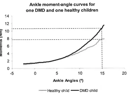
Figure 3 illustrates the ankle joint passive plantar flexion moment of one DMD child and one healthy control subject measured while their ankle joint is moved from plantar flexion to dorsiflexion. This figure shows that for both children, as ankle dorsiflexion angle increases, the moment increases as well. If one looks at an angle common to both subjects in the second portion of the curie, the corresponding moment values are higher for the DMD child, thus reflecting more resistance to stretch for this child in that portion of the movement. These data come from preliminary results of a study actually in progress conducted by Gaudreault et al. (2004).
The effect of interventions such as stretching on muscle flexibility and joint ROM has also been explored using the moment-angle relationship (Halbertsma et al., 1996; Magnusson et al., 1998). The challenges encountered with such measurements are that few clinical settings are equipped with the technical instrumentation necessary to collect reliable passive torque and joint ROM data. The lack of a standardized evaluation protocol is another issue.