TEST YOURSELF: ANSWER
Bone mass of the calvarium
Paolo Simoni
&Jennifer Stulko
&Selma Ben Mustapha
&Laurent Schoysman
&Elettra Bianchi
&Nadia Withofs
&Tudor Racaru
&Jo Caers
&Victoria Alvarez-Miezentseva
Published online: 19 December 2012 # ISS 2012
Plasmacytoma of the calvarium is very rare, accounting for
the 0.7 % of all plasmacytomas [
1
–
5
]. The most common
locations are the thoracic and lumbar spine (30–40 % of
cases). Involvement of the hip, femurs, and ribs is less
frequent [
6
].
Plasmacytoma of the calvarium can be
indistinguish-able from other lytic lesions, especially metastatic
dis-ease. Meningioma, eosinophilic granuloma, and
dermoid cysts can have similar appearances, although
usually in a different clinical context [
10
,
11
].
Histo-pathological examination and laboratory tests are
nec-essary to make a definitive diagnosis of plasmacytoma
(Fig.
1
).
The typical features on imaging are a lytic lesion on
X-ray examination (Fig.
1
question) [
1
,
6
]. On computed
tomography (CT), plasmacytoma presents as a lytic
punched-out lesion with interspersed residual thickened bony
trabeculae. Cortical thinning and focal destruction are typical
(Fig. 2 question) [
2
]. On magnetic resonance imaging (MRI),
bone plasmacytoma of the calvarium presents as an
intra-medullary lesion of low to intermediate intensity on
T1-weighted images and high intensity on T2-T1-weighted images.
Contrast enhancement is usually mild [
4
–
9
].
In the presented case, the calvarium plasmacytoma
presents a
“mini-brain” appearance, due to the presence of
low-intensity hypertrophic residual trabeculae interspersed
in a relatively homogeneous mass, mimicking the brain
sulci.
Major et al. first described the
“mini-brain” appearance
of plasmacytoma in a series of ten patients with spinal
The case presentation can be found at doi: 10.1007/s00256-012-1548-5 Answer: Plasmacytoma of the calvarium with evolution into multiple myeloma (MM).
P. Simoni
:
J. Stulko:
S. Ben Mustapha:
L. Schoysman:
V. Alvarez-MiezentsevaService of MSK imaging, University Hospital of Liège (CHU), Domaine du Sart Tilman Bât. 35,
4000 Liège, Belgium E. Bianchi
Service of Pathology, University Hospital of Liège (CHU), Domaine du Sart Tilman Bât. 35,
4000 Liège, Belgium N. Withofs
Service of Nuclear Medicine, University Hospital of Liège (CHU), Domaine du Sart Tilman Bât. 35,
4000 Liège, Belgium
T. Racaru
Service of Neurosurgery, University Hospital of Liège (CHU), Domaine du Sart Tilman Bât. 35,
4000 Liège, Belgium J. Caers
Service of Haematology, University Hospital of Liège (CHU), Domaine du Sart Tilman Bât. 35,
4000 Liège, Belgium P. Simoni (*)
Service of MSK Imaging, University Hospital of Liège, Domain du Sart Tilman Bât. 45,
4000 Liège, Belgium
e-mail: [email protected] Skeletal Radiol (2013) 42:1185–1187
lesions [
2
]. Since this appearance had not been observed in
other bony lesions, they concluded that the
“mini-brain”
ap-pearance is virtually pathognomonic for bone plasmacytoma
of the spine [
3
]. They argued that thickened trabeculae might
be due to mechanical stress from the lytic process [
2
].
However, the
“mini-brain” on MRI has also been
de-scribed in plasmacytoma of the appendicular skeleton [
3
];
it has not previously been described in the calvarium.
Nev-ertheless, a
“mini-brain” appearance can be retrospectively
observed on MRI images of most bone plasmacytomas of
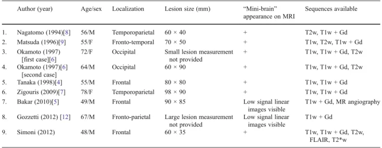
the calvarium reported in the literature (see Table
1
).
We think that the finding of the characteristic
“mini
brain” appearance can help to narrow the differential
diag-nosis of lytic lesions of the skull.
Fig. 1 Surgical biopsy- histo-logical and immunohistochemi-cal specimens. a H&E stain (original magnification 40×); plasma cells exhibit abundant cytoplasm, eccentric nuclei with coarse chromatin. b Immunos-tain showing plasma cell marker CD138 (brown, Syndecan-1), original magnification 20× ; Serotec , B-B4 clone, 1/100 di-lution. c Ig Kappa expression (brown) showing a strongly pos-itive monotypic light-chain staining compared to the absence of plasma cell staining for Ig Lambda (d); (original magnifi-cation 40×, Dako, 1/8,000 dilu-tion). d Ig Lambda-negative immunostaining (brown); (origi-nal magnification 40×, Dako, 1/ 8,000 dilution)
Table 1 Imaging features of BP of the calvarium reported in the English literature
Author (year) Age/sex Localization Lesion size (mm) “Mini-brain” appearance on MRI Sequences available 1. Nagatomo (1994)[8] 56/M Temporoparietal 60 × 40 + T2w, T1w + Gd 2. Matsuda (1996)[9] 55/F Fronto-temporal 70 × 50 + T1w, T2w, T1w + Gd 3. Okamoto (1997) [first case][6]
72/F Occipital Small lesion measurement not provided + T1w, T1w + Gd, T2w 4. Okamoto (1997)[6] [second case] 64/M Occipital 60 × 90 + T1w, T1w + Gd, T2w 5. Tanaka (1998)[4] 55/M Frontal 80 × 80 + T1w, T1w + Gd 6. Zigouris (2009)[7] 78/F Temporoparietal 98 × 90 + T1w, T1w + Gd 7. Bakar (2010)[5] 49/M Frontal 90 × 85 Low signal linear
images visible
T1w + Gd, MR angiography 8. Gozzetti (2012) [12] 67/M Fronto-parietal Large lesion measurement
not provided
Low signal linear images visible
T1w + Gd
9. Simoni (2012) 48/M Frontal 60 × 35 + T1w, T1w + Gd, T2w, FLAIR, T2*w +0 presence of a “mini-brain” appearance
w0 weighted
References
1. Kuzeyli K, Duru S, Ceylan S, Aktürk F. Plasmacytoma of the calvarium. A case report. Neurosurg Rev. 1995;18(2):139–42. 2. Major NM, Helms CA, Richardson WJ. The“mini brain”
plasma-cytoma in a vertebral body on MR imaging. AJR. 2000;175 (1):261–3.
3. Subhas N, Bauer TW, Joyce MJ, Sundaram M. The“mini brain” appearance of plasmacytoma in the appendicular skeleton. Skeletal Radiol. 2008;37(8):771–4.
4. Tanaka M, Shibui S, Nomura K, Nakanishi Y. Plasmacytoma of the calvarium: a case report. Jpn J Clin Oncol. 1998;28(10):626–30. 5. Bakar B, Tekkok IH. Plasmacytoma of the calvarium vault.
Turk-ish Neurosurgery. 2010;22(1):S095–8.
6. Okamoto K, Ito J, Furusawa T, Sakai K, Tokiguchi S, Sato M, Tanaka R, Nemoto K, Oyanagi K. Plasmacytomas of the occipital bone: a report of two cases. Eur Radiol. 1997;7(4):503–6.
7. Zigouris A, Drosos D, Alexiou GA, Fotakopoulos G, Mihos E, Pahatouridis D, Tsiouris S, Fotopoulos AD, Voulgaris S. Primary plasmacytoma of the cranial vault: a case report. Cases J. 2009;2 (1):9154.
8. Nagatomo Y, Uno H, Maeda K, Matsuoka H, Tsuruda T, Okayama A, Tachibana N, Tsubouchi H. Bulky plasmacytoma of the bone with intracranial invasion. Intern Med. 1994;33(6):376–9. 9. Matsuda M, Nakazawa T, Kizuki H, Matsumura K, Nakasu S,
Handa J. Plasmacytoma of the calvarium vault–case report. Neurol Med Chir (Tokyo). 1996;36(6):388–92.
10. Simoni P, Meunier B, Deprez M, Racaru T, Martin D. 36-year-old man with sudden severe headache. Skeletal Radiol. 2011;40 (10):1361–2.
11. Meyer JR, Roychowdhury S, Cybulski G, Russell EJ. Intramedul-lary plasmacytoma of the calvarium base mimicking aggressive meningioma. Calvarium Base Surg. 1997;7(2):101–5.
12. Gozzeti A, Cerase A, Defina M, Bocchia M. Plasmacytoma of the calvarium. Eur J Haematol. 2012;88:369.