L'UTILISATION D'UN ALGOMÈTRE MANUEL
AFIN D'ÉVALUER LE SEUIL DE LA DOULEUR
AU NIVEAU DU TRONC ET DES MEMBRES SUPÉRIEURS
CHEZ DES JEUNES FEMMES DE NIVEAU COLLÉGIAL
MÉMOIRE
PRÉSENTÉ
COMME EXIGENCE PARTIELLE
DE LA MAÎTRISE EN KINANTHROPOLOGIE
PAR
DAVID H JONES
THE USE OF A HAND-HELD ALGOMETER
TO EVALUATE PRESSURE PAIN THRESHOLDS
IN THE TORSO AND THE UPPER EXTREMITY
OF COLLEGE AGED WOMEN
MASTER'S THESIS
AS PART OF THE MASTER'S PROGRAM
THROUGHTHE
DEPARTMENT OF KINANTHROPOLOGIE
BY
DAVID H JONES
Avertissement
La diffusion de ce mémoire se fait dans le respect des droits de son auteur, qui a signé le formulaire Autorisation de reproduire et de diffuser un travail de recherche de cycles
supérieurs (SDU-522 - Rév.01-2006). Cette autorisation stipule que «conformément à l'article 11 du Règlement no 8 des études de cycles supérieurs, [l'auteur] concède à l'Université du Québec à Montréal une licence non exclusive d'utilisation et de publication de la totalité ou d'une partie importante de [son] travail de recherche pour des fins pédagogiques et non commerciales. Plus précisément, [l'auteur] autorise l'Université du Québec à Montréal à reproduire, diffuser, prêter, distribuer ou vendre des copies de [son] travail de recherche à des fins non commerciales sur quelque support que ce soit, y compris 1'1 nternet. Cette licence et cette autorisation n'entraînent pas une renonciation de [la] part [de l'auteur] à [ses] droits moraux ni à [ses] droits de propriété intellectuelle. Sauf entente contraire, [l'auteur] conserve la liberté de diffuser et de commercialiser ou non ce travail dont [il] possède un exemplaire.»
Pain in the upper ann, shoulder, upper back and neck region commonly occurs in women who have undergone or are undergoing breast cancer therapy. The original goal of this project was to see if applying a therapeutic modality, such as ice, would be effective at controlling the post operative pain in women who had undergone a partial mastectomy with an axillary node dissection, as part of the breast cancer therapy. It was decided that several preliminary projects would have to be performed before the effectiveness of this therapeutic modality on shoulder pain could be properly evaluated. The preliminary projects included 1) Identifying an appropriate measurement tool and testing protocol to quantify the subjective changes in perceived pain of different individuals; 2) Identify the potentially problematic locations on the body where the measurement tool and testing protocol would be used; 3) Evaluating the testing protocol on populations that do not presently have any dysfunction, to understand what may be considered "normal" in the locations to be tested; 4) Using the testing measurement tool and testing protocol on patients that are going to have breast sparing surgery to note any trends that may occur; and 5) Finally using the measurement tool and testing protocol to collect baseline values on women undergoing a partial mastectomy with an axillary node dissection, having the women utilize ice as part of their post operative treatment plan and then retest the locations after a fixed period of time.
In this project only stages one (measurement tool and testing protocol), two (detennining the testing locations) and three (using the testing protocol on a normal population) were completed. The material being presented in this manuscript includes the data that was collected during the experimental trials of these stages. It will be speculated that the measuring device and the testing protocol maybe also used for various groups dealing with upper body dysfunction as weB as women undergoing breast cancer therapy.
Preliminary work has begun with a local breast cancer clinic to implement projects four (the use of the testing protocol with breast cancer patients) and five (use the testing protocol to measure the effectiveness of a therapeutic intervention to reduce pain).
It would have been extremely difficult to complete this project with out the tremendous support of my supervisors Dr. Alain Steve Comtois and Dr. Robert Kilgour and my fellow staff members Mr. Ron Rehel, Ms Tracy Griffiths and Ms. Christina Grace in the Department of Exercise Science at Concordia University. My two thesis supervisors played a significant role in helping to shape and define a strong research project. My three colleagues from the Department of Exercise Science who were always ready and wi11ing to help when needed and did make a significant contribution to the project. 1 would also like to thank Dr. Karen Messing for the use of the hand-held algometer to this project and her comments to help make a stronger thesis, Mr. Jean-Bapiste Laporte for his help in translating the abstract of the thesis, as well as to Dr. Marc Belanger for a11 his constructive criticism on the thesis.
1 say goodbye to Alex, Moe, my Dad, Bill, Steve, Anita, Maize, Jean and Helvi.
A final thank you goes to my family, to my Mom and Dad for helping to give me direction and to my wonderful wife and two super sons for a11 of their patience and encouragement.
TABLE OF CONTENTS
LIST OF FIGURES
IXLIST OF TABLES
XIRESUME
XIIABSTRACT
XVCHAPTERI
INTRODUCTION -REVIEW OF THE CURRENT LITERATURE 1
1.01. Breast cancer 1
1.02. Adjuvant therapy 2
1.03. Breast cancer, breast surgery and shoulder dysfunction 5
1.04. Work related injuries 9
1.05. Cinderella theory 13
1.06. Perception of pain 15
1.07. The role of a hand-held algometer 17
1.08. Factors that affect the use of the algometer 20
1.10. Testing locations on the neck shoulder and arm region 25
1.11. Trigger points 27
1.12. Why does a trigger point develop? 33
1.13. The rationale for investigating trigger points in this project 36
1.14. Summary ofliterature review 37
1.15. Implementing the literature review into this protocol 38
1.16. Main hypothesis 39
1.17. Additional questions 39
CHAPTERII
METHODOLOGY 41
2.01. Subject selection 41
2.02. The calibration and application of the algometer. .42
2.03. Application 43
2.04. Experimental procedure 46
2.05. Detennination of the PPTs .47
2.06. The locations that were tested .48
2.08. The skin fold measures 60
2.09. The skin-fold measures for the eight locations 61 2.10. Breakdown of tasks perforrned by individuals in the research project.. 64
2.11. Statistical analysis 66
CHAPTER
III
MANUSCRIP'f NUMBER 1 68
3.01. Overview of the accepted manuscript 68
3.02. Methodology paper - Letter of acceptance 69
3.03. Letter to the editor concerning revisions to manuscript 70 3.04. Revised manuscript - Test-retest reliability of pressure pain threshold
measurements of the upper limb and torso in young healthy women 83
CHAPTERIV
MANUSCRIPT NlJNIBER 2 ; 110
4.01. Overview of the second manuscript 110
4.02. Letter of acceptance for poster presentation competition from the
measurements in the upper arm and torso in young healthy women 112
CHAPTER V
RESULTS PERTAINING TO ADDITIONAL QUESTIONS 146
5.01. Additional question 1: Relationship between skin folds values
and PPT values 146
5.02. Additional question 2: Consistency in determining nodule status 151
5.03. Additional question 3: Presence of nodule and PPT values 158
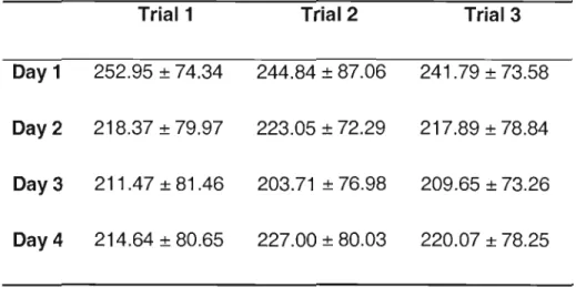
5.04. Additional question 4: Variation in PPT values 161
CHAPTER VI
DISCUSSION CONCERNING ADDITIONAL QUESTIONS 165
6.01. Additional question 1: Relationship between skin folds values and
PPT values 165
6.02. Additional question 2: Consistency in determining nodule status 167
6.03. Additional question 3: Presence of nodule and PPT values 170
6.05. Conclusion and potential benefits of the project 174
6.06. Precautions 175
BIBLIOGRAPHY
176ANNEX
199Annex 1: Fonne de consentement à participer à la recherche 199 Annex 2: Consent Form ta Participate in Research Project.. 204
LIST OF FIGURES
Figure 1: Upper torso and lymph nodes 6
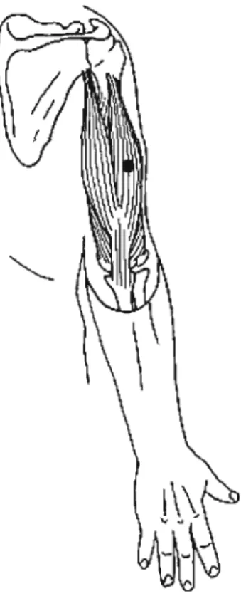
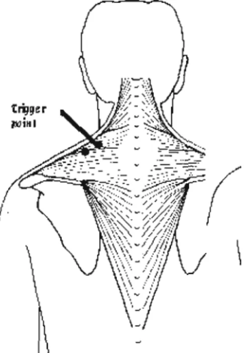
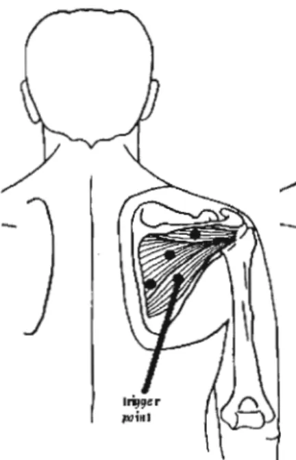
Figure 2: Electronic algometer with hand-held patient switch 19 Figure 3: Diagram of trigger point nodule (contraction knot) in a muscle 30 Figure 4: Diagram of the neuromuscular junction 32 Figure 5: Theoretical model of the development of a trigger point 35 Figure 6: Long head of the biceps trigger point location .48 Figure 7: The anterior fibers on the deltoid trigger point location .49 Figure 8: The lateral head of the triceps trigger point location 50 Figure 9: The rhomboids major trigger point location 51 Figure 10: The posterior fi bers on the deltoid trigger point location 52 Figure 11: The supraspinatus trigger point location 53
Figure 12: The trapezius trigger point location 54 Figure 13: The infraspinatus trigger point location 55 Figure 14: Testing locations on the anterior surface of the upper arm and shoulder 58 Figure 15: Testing locations on the posterior surface of the upper ann and shoulder.. 59 Figure 16: PPT values correlated to skin fold values 148 Figure 17: Pre test agreement on nodule status at each location 152 Figure 18: Post test agreements on nodule status at each location 154 Figure 19: Pre-test to post-test comparison of nodule status for Athletic Therapist # 1 156 Figure 20: Pre test to post test comparison of nodule status for Athletic Therapist # 2 157 Figure 21: Pre-test evaluations of the 8 locations for nodule status 159 Figure 22: Post test evaluation of the 8 locations for nodule status 160 Figure 23: PPT values obtained at location 3, the triceps 162 Figure 24: PPT values obtained at location 7, the trapezius 163 Figure 25: PPT values obtained at location 7, the infraspinatus 164
LIST OF TABLES
Table 1: Sk.în-fold values of the 8 different locations 146
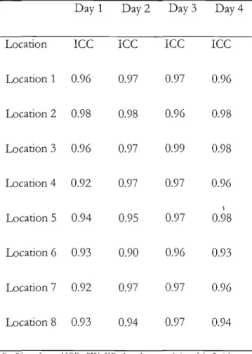
Table 2: The interclass correlation scores of the two athletic therapists,
RÉSUMÉ:
Objectifs:
le but de ce projet fut d'utiliser un algomètre manuel afin de pouvoir apprécier l'intensité du seuil de douleur à la pression (appelé PPT pour pain pressure threshold) sur huit locations cutanées différentes au niveau des membres supérieurs et du tronc chez des jeunes femmes en bonne santé, durant quatre jours consécutifs d'évaluation. Les différentes études sur ce sujetà
ce jour nous permettaient de croire que les huit locations ne présenteraient pas de "PPT" même intensité. Le fait de noter des variations de "PPT" pourrait alors être une première étape dans le développement d'un protocole diagnostique rigoureux qui aurait un intérêt dans l'approche de différents groupes de patients présentant des maux et souffrances au niveau de la nuque, des épaules et des bras.Méthode:
les mesures de l'intensité du "PPT", furent obtenues à partir de huit sites différents sur le bras et le tronc (coté droit seulement) de dix-neuf jeunes femmes. Tous les sujets testés étaient droitières, avaient des cycles menstruels réguliers et n'avaient présenté aucun problème musculo-squelettique relié à l'épaule au cours des six mois précédant la période d'évaluation. Chacune d'entre elle fut recrutée au sein de la population de l'Université Concordia.Deux thérapeutes du sport expérimentés eurent à identifier, valider et marquer les huit différents sites sur la peau de chaque sujet. Les deux thérapeutes du sport palpèrent
musculaire (contracture) pouvant s'associer à un point gachette " trigger point" potentiel
à chacun des emplacements. Cette palpation fut effectuée en début et en fin de chaque session de tests, Au cours de quatre jours consécutifs d'évaluation, un algomètre manuel fut employé sur chacun des emplacements marqués afin de mesurer l'intensité du "PPT" sur chaque sujet. Après la collecte des mesures enregistrées par l' algomètre et l'évaluation palpatoire des deux thérapeutes du sport, une mesure des plis cutanés fut également prise pour chacune des huit sites.
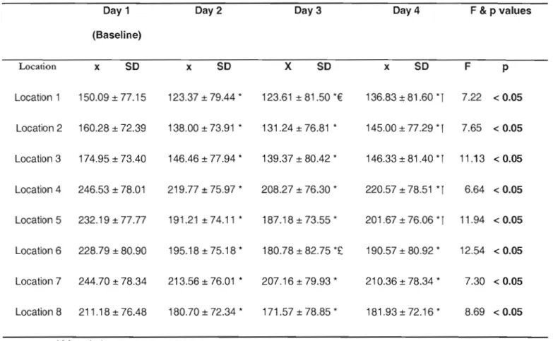
Résultats: la principale conséquence de cette recherche fut de révéler un abaissement significatif de l'intensité du "PPT" pour les huit emplacements lors des quatre jours d'évaluation. Une différence significative de l'intensité du seuil de douleur fut également démontrée au niveau du bras comparé aux valeurs du tronc. Les emplacements de la partie supérieure du bras et de la partie antérieure de l'épaule montrèrent des valeurs de PPT significativement plus faibles lorsque qu'ils furent comparés aux cinq autres emplacements du tronc et de la partie postérieure de l'épaule.
Aucune corrélation ne fut trouvée entre l'épaisseur du pli cutané d'un emplacement et l'intensité du "PPT" de ce même emplacement chez les sujets testés. Les régions ayant une épaisseur du pli cutané plus importante n'ont pas forcément montré d'intensités de "PPT" plus élevées ou faibles.
La mesure statistique « interclass correlation scores» qui pennet d'évaluer le degré d'accord entre les deux thérapeutes du sport sur la présence ou l'absence d'un nodule musculaire associé à un point gachette fut de 0,54. Cette valeur peut être considérée comme basse statistiquement mais se révèle consistante et en rapport avec les autres valeurs du même type retrouvées dans d'autres études évaluant les habilités manuelles de praticiens à des fins diagnostiques.
Cette étude peut donc fournir aux praticiens et chercheurs des indications claires à propos d'un protocole d'évaluation des mesures de "PPT" qui pennettrait d'obtenir des valeurs initiales de base au niveau des régions du bras et du tronc de jeunes femmes ne présentant pas de dysfonctions scapulo-humérales. Des études futures auraient donc comme intérêt d'utiliser ce même protocole d'évaluation chez des femmes souffrant effectivement de douleurs à l'épaule ou chez des femmes pouvant présenter des facteurs de risques quant au développement de ces mêmes douleurs lors d'interventions associées au traitement du cancer du sein.
healthy female subjects over four consecutive days of testing. Based on previous research, it was expected that not ail eight locations wou Id have the same PPT level. Noting the differences in PPT levels at the eight locations would be one of the first steps in the development of an effective diagnostic protocol which may be used on different groups who are experiencing pain in the neck, shoulder and arm region.
Methodology: PPT measures were obtained from eight different locations in the upper arm and torso (right side only) of 19 female subjects. Ali subjects were right hand dominant, had regulaI' menstrual cycles and had not experienced any musculoskeletal problems related to the shoulder in the 6 months prior to being tested. Ali subjects were recruited from the Concordia University community.
Two experienced athletic therapists identified, agreed upon and marked the eight different skin surface locations on each subject. The two athletic therapists also palpated the eight locations to determine the presence or absence of a nodule associated with a potential trigger point at each of the locations. Palpation occurred at the beginning of the testing
session and at the end of the testing session. Over 4 consecutive days of testing, a hand held algometer was applied to each location to detennine the subject's pressure pain threshold.
Upon completion of the algometer measurements by the data collector and the palpation evaluation by the two athletic therapists, skin-fold measurements were taken at each of the 8 locations.
Results: The main effect of this research project showed a significant decline in the PPT values across all 8 locations over the 4 days of testing. A significant difference was also seen in the PPT values of the upper extremity compared to the PPT values of the torso. The locations in the upper arm and anterior shoulder were found to have significantly lower PPT values when compared to the other 5 locations on the torso and posterior shoulder.
No correlation was found between the skin-fold thickness of a location and the PPT value obtained at the same location in the subjects that were tested. Regions that had higher levels of skin-fold thickness did not necessarily have higher or lower PPT values.
on the presence or absence of a nodule associated with a trigger point was 0.54, which is considered low, but is consistent with scores reported in other studies evaluating diagnostic manual skills of clinicians.
This project gives clear guidelines as to the testing protocol that may be used by clinicians and researchers on how to obtain baseline PPT measures in the upper arm and torso region of young women who are not experiencing any shoulder dysfunction. The next step in this project will be to utilize this testing protocol on women who maybe experiencing shoulder problems or who are at risk at developing shoulder problems such as women undergoing adjuvant therapy associated with breast cancer.
CHAPTERI
INTRODUCTION -REVIEW OF THE CURRENT LITERATURE
1.01 Breast cancer
The advancement in diagnostic tools has led to improvements in the early detection of suspicious lumps in breast tissue that are associated with breast cancer (Ugnat et al., 2004). This has led to an increase in of the number of women being identified as being at risk for developing breast cancer, along with a significant decrease in morbidity in this same group (Jatoi and Miller, 2003, Miller et al., 2002, Ellison and Gibbons, 2004). The primary goal of the oncology team is to remove the cancerous tissue and preserve the patient's life, which certainly should be paramount (Harris et al., 1996). Unfortunately there are sorne negative side effects that are associated with breast cancer therapy, which include fatigue, reduction in the ability to complete sorne activities of daily living, swelling, edema along with the possibility of devèloping prolonged shoulder dysfunction (Harris et al., 1996). The following thesis will be focusing on the shoulder dysfunction that occurs for breast cancer patients and how sorne women may have certain characteristics that place them at an increased risk for developing pain in the upper arm, shoulder, upper back and neck region.
1.02 Adjuvant therapy
When breast cancer is suspected, the medical team often recommends sorne form of adjuvant therapy. Adjuvant therapy for breast cancer typically involves four major areas 1) surgery 2) chemotherapy, 3) radiation therapy, and 4) hormonal therapy (Ugnat et al., 2004).
It may be recommended to patients that at least one and sometimes ail four of the therapies he used to treat the breast cancer (Ugnat et al., 2004). As with many interventions, whether surgical or non-surgical, various adverse effects may occur (Giordani et al., 2005, Varabi, 2003).
When surgery takes place, an area slightly larger that the tumour itself (that includes the tumour) is removed from the breast region to ensure obvious cancerous tissue has been excised. A sampling ofaxillary Iymph nodes also takes place, to see if any migration of cancerous cells to other parts of the body has occurred. The sampling of lymph nodes may include a removal of a single lymph node or several nodes (Arnaud et al., 2004).
Chemotherapy involves the introduction of toxic agents to the body. The cancerous cells that created the initial tumour in the breast of the patient are no longer recognized as being foreign and dangerous to the immune system of the individual. The body has begun to accept their presence in the body, making them a significant threat. The goal behind chemotherapy is to introduce toxic agents into the system and stimulate cell death, defined
as "apoptosis". This systemic approach kills off any cancerous cells that may have migrated from the original tumour location (Gajdos et al., 2002). The cell death that is initiated through chemotherapy not only occurs to the cancerous cells but also to the healthy cells of the individual. It becomes easy to see why often patients undergoing chemotherapy experience a significant amount of fatigue that may last weil after the chemotherapy has been completed. This fatigue often affects the patient's activities of daily living (Cella et al., 2007).
Radiotherapy or radiation therapy involves the delivery of small but concentrated amounts of radiation to the breast area to kill any cancerous cells that may still be in close proximity to the site where the tumour was removed. This local approach will cause a reddening of the breast area, similar to having a significant sunbum. Present day refinement of radiation therapy has reduced the impact on nerve endings and blood vessels in close proximity to the treatment area. As with chemotherapy, fatigue is a reoccurring problem for the patient (Lee et al., 2(07).
One approach in honnonal therapy involves the suppression of the honnone oestrogen within the woman's body. Medications such as Tamoxifen try to suppress the oestrogen uptake by the oestrogen receptors within the breast tissue, preventing accumulation, and without disrupting oestrogen homeostasis elsewhere in the body (Harris et al., 1996). Drugs
such as Tamoxifen are also used to help maintain bone density in women, which is nonnally the role of oestrogen. Honnonal therapy along with chemotherapy has been shown to move pre-menopausal women who are undergoing treatment into menopause (Harris et al., 1996). Women do periodically experience hot flashes with Tamoxifen medication along with other side effects (Cella et al., 2007). Other problems that are associated with various fonns of adjuvant therapy may include cognitive symptoms, musculoskeletal pain, vasomotor symptoms, nausea, sexual problems, bladder problems, body image, and vaginal symptoms (Cella et al., 2007).
The role of chemotherapy, radiotherapy and honnonal therapy does seem to be directly related to women initiating shoulder dysfunction although they do seem to aggravate any initial shoulder problems and cause them to last much longer than they should. It is important then to take a closer look at the surgical intervention.
1.03 Breast cancer, breast surgery and shoulder dysfunction
During surgery for a partial mastectomy the tumour is identified as being in one of four quadrants of the breast, upper medial, upper lateral, lower medial and lower lateral. To remove the tumour an incision is typically made along the lateral border of the breast and the tumour is removed (Harris, 1996).
As mentioned previously, a sampling of lymph nodes takes place to verify if any migration of the cancer cells to other regions of the body has occurred. Figure 1 outlines the upper torso and the lymphatic channels in that region; the axillary node dissection associated with a partial mastectomy involves the excising of tissue that will harvest lymph nodes from the axillary lymph node at levels 1 ( point B) and 2 (point C) and occasionally level 3 (point D). During the removal of the tumor, the surgeon avoids cutting into the pectoralis major muscle, which is in close proximity to the breast, unless it is in the area that is affected by the tumor. Cutting into the muscle wou Id increase the risk of shoulder dysfunction for the patient. The surgery does require that incisions be made into the pectoral fascia, the thin layer of tissue which sUlTounds the chest area, since these incisions are considered less invasive for the patient (Harris et al., 1996). The surgeon will then try to harvest the lymph nodes through the same incision site where the tumour was removed. Occasionally a second incision will need to made, if the first incision site is far away from where the axillary lymph nodes may be harvested (Harris et al., 1996).
F
Il
Figure 1: Upper torso and lymph nodes (www.breastcancer.org )
A Pectoraiis major muscle, B Axillary Iymph nodes: Ieveis l,
C Axillary Iymph nodes: Ieveis II, D Axillary Iymph nodes: Ieveis III E Supraclavicular Iymph nodes, F InternaI mammary nodes Iymph
The Iymphatic system of the body is used to recover fluid in the interstitial spaces that has not been taken up by the ve!10us system (Guyton and Hall 2000). The harvesting of Iymph nodes during a partial mastectomy diminishes this uptake process, which may lead to pooling of the Iymph in the upper ex
tremi
ty (Taylor 2004).Following a partial mastectomy with an axillary Iymph node dissection, women may experience pain, swelling in the upper extremity, loss of shoulder mobility and a decrease in their ability to perfonn activities of daily living (Swenson et al., 2002).
After having undergone surgery, it is often recornrnended that women receive addition al therapy, which may include chemotherapy and radiotherapy. Literature reviews indicate that women who enter radiotherapy with shoulder dysfunction are much more likely to maintain these problems when re-evaluated two years post surgery. (Bendz, Fagevik and Olsen, 2002)
A study by Maunsell, Brisson and Deschene (1993) found that 82 % of respondents reported at least one problem, 3 months after receiving a surgical intervention that required a partial mastectomy with an axillary Iymph node dissection. In this study 55 % of the women (n=233) reported experiencing pain in their shoulder or arm. A follow up on the women 18 months after surgery found that 79% of the respondents (n=21O) reported having at least one
shoulder problem. The women reported experiencing pain, swelling or numbness in the shoulder and arm regions.
It is c1ear that the need to regain shoulder mobility exists for this cohort of women, to avoid prolonged shoulder problems. One approach that maybe utilized to address this problem is to control the amount of postoperative pain the women are enduring so that they may begin to move their arm.
There has been some work by different groups (Kil gour, Jones and Keyserlingk, 2007) to try and minimize the amount of post operative shoulder dysfunction that does take place through the use of a home based exercise program, but there is still much work to be done.
Certainly having a portion of one's body removed is a very traumatic event but in comparison to many other types of orthopedie surgery in and around the shoulder, this intervention is considered minor surgery (Leidenius et aL, 2003). Oncology physicians are still not certain why so many women run into problems, yet many of the women who do undergo this regime of treatment are at risk for developing chronic arm, shoulder and neck problems (Leidenius et aL, 2003).
It is possible that women may be predisposed to developing problems in the upper body. Thus, it may also be important to look at other areas of daily living where women may be at risk for developing pain in the neck, shoulder and arm regions. These are outlined in the following section.
1.04 Work-related injuries
Women seem to be at greater risk for developing pain in the neck, shoulder and arm regions in comparison to men (Chesterton et al., 2003). Women often comprise a significant portion of different occupations (Sorock and Courtney, 1996) that are most at risk to upper body injuries. Men have also been shown to have repetitive injury syndrome, but not to the same extent as women. When evaluating the work of men and women who work on production lines, there is a similarity in the type of chronic injuries that occur (Westgaard and Winkel, 1996), although women seem to be affected to a greater extent.
In sorne instances there is a misunderstanding as to the actual demands of the tasks that are being petformed. Activities, specifically jobs that maybe perceived at being easy and not being physically demanding may in reality tum out to be quite physically demanding. A good example of this involved a study looking at women who worked at sewing machines sewing clothes. An activity that may take a short period of time to complete (on one piece of clothing) but because of the repetitive demands and the materials being used (such as denim
in jeans) it tums into a more physically challenging activity (Vezina, Tiemey and Messing, 1992)
Lindman et al., (1990, 1991) evaluated pain in the neck and shoulder region in women and noted differences in morphologie al development of the trapezius muscle between men and women. Lindman believes this may explain why women seem to be at greater risk for developing neck, shoulder and arm problems. When Lindman et al., (1990, 1991) performed muscle biopsies on both groups he found that women had a significantly greater proportion of type 1 muscle fi bers when compared to men. Lindman et al., (1990, 1991) hypothesized that the difference in fiber composition may explain why the two genders may have ended up moving towards specifie type of jobs. Women who have a greater composition of type 1 (slow twitch fatigue resistant) fibers in their trapezius muscle have a greater aerobic capacity and do not fatigue as quickly, as compared to type 2 fibers (Guyton and Hall, 2000; Lindman, Eriksson and, Thomell, 1991). Men who have a greater composition of type 2 fibers (fast twitch non-resistant to fatigue) fibers in their trapezius muscle, are able to generate greater explosive contractions/force, but will fatigue faster, as compared to type 1 fi bers (Guyton and Hall, 2000; Lindman, Eriksson and, Thomell, 1991). Lindeman et al., (1991) also looked at the way women and men perform the same the activity, specifically data entry. The women involved in the study were shown to use shoulder; arm and wrist motions in a different manner compared to men and were expending significantly more
energy. The women were also often found to be much more effective at entering large volumes of data compared to men. Entering more data and expending more energy may also be two factors that have contributed to women experiencing more pain while performing this task in comparison to men, who used less energy as weil as entered less data.
Work by Hooftman et al., (2004) found gender differences in relation to musculoskeletal pain where men were more likely to complain of pain in the low back and women were more likely to complain of pain in the shoulder. Hooftman et al., (2004) did not find any gender relationship with neck pain.
Karlqvist, Leijon and Harenstam (2003) believe that workers that are in poor physical condition are at greater risk for developing pain and dysfunction in different regions of the body. Their work has shown that workers, both men and women, but in particular females, are not in good physical condition and that the jobs they perforrn on a regular basis do not improve their fitness level. If anything, the jobs tend to be deleterious towards the workers health.
Many different occupational groups, such cashiers, dental hygienist and people who work at computer terminaIs have been found to have significant problems with pain in the neck,
shoulder and ann regions (Johansson et aL, 2003; Luime et aL, 2004). The problems experienced by workers have included different forms of neck and shoulder pam, tendonitis/tendinosis in the forearm; lateral epicondylitis (Johansson et al., 2003), and shoulder; supraspinatus tendonitis (Lundberg et aL, 1999), shoulder bursitis (Luime et al., 2004), periodic numbness; thoracic outlet, brachial plexopathies (Mense and Simons, 2001), nerve entrapment (Pascarelli and Hsu, 2001) and carpel tunnel syndrome (Rice, Nindl and Pentikis, 1996).
Workers who experience pain in the neck, upper torso and ann regions are often asked to perfOlm a task(s) that require a muscle or group of muscles to maintain a static position for a prolonged period of time (Mense and Simons, 2001). The tasks that are being performed often donot require a significant amount of muscular strength or force, usually having low biomechanical demands, but are repetitive in nature (Mense and Simons, 2001). Workers having pain in the neck shoulder and atm region include people who operate computers terminaIs, super-market cashiers (Lundberg et aL, 1999), dental personnel (Rice, Nindl and Pentikis, 1996; Rising et al., 2005), nursing home workers (Luime et al., 2004) and musicians (pascarelli and Hsu, 2001; Zaza and Farewell, 1997). A corrunon trend that is seen in many of these studies is the high prevalence of neck-shoulder pain in females (Chesterton et al., 2003).
There is growmg consensus that musculoskeletal disorders may be related to the occupational activities of the individuals (Punnett and Wegman, 2004) but it is still unclear how much predisposing factors play a role in a worker developing musculoskeletal disorders.
As Messing et al., (2003) point out in their literature review of work demands and gender differences, the actual number of women who suffer injury that is associated with the tasks that they perfonn at their jobs may be much larger than what has been recorded to date.
1.05 Cinderella theory
The Cinderella syndrome theory developed by Hagg (1991) explains how repetitive movements may have a negative impact on the body and why women may be at greater risk for certain musculoskeletal disorders (Johansson et al., 2003; Ge, Madeleine and Arendt Nielsen, 2005). ,During active movement (whether static or dynamic) where force is required, motor units associated with small muscle fibers will be the first muscle fibers to be recruited and as the intensity of the work increases and greater force is required, motor units associated with larger muscle fibers will be recruited. This concept was outlined in Henneman's principle (Henneman, Somjen and Carpenter, 1965) concerning the order of recruitment of motor units. The Cinderella syndrome (Ge, Madeleine and Arendt-Nielsen, 2005) attempts to utilize Henneman's principle (Henneman, Somjen and Carpenter, 1965)
of the order of muscle fiber recruitment where small muscle fibers are being asked to perforrn work at a low iniensity for a prolonged period of time and the larger fast twitch fibers being sporadically recruited. This situation may lead to excessive shearing forces at the level of the small slow twitch fibers and sorne forrn of dysfunction at the motor endplates (Mense and Simons, 2001).The shearing forces would lead to a disorganization of the actin and myosin cross bridges. In the story of Cinderella, Cinderella was always the first to start working in the moming and the last to stop working at night, similar to what maybe happening to the smaller fibers. One of the drawbacks with the Cinderella theory is the concept that during active muscle contraction the same muscle fibers (Type 1 muscle fiber motor units) will be continually recruited and will have limited recovery time leading to the same motor units of muscle fi bers being continuously contracted. We know from the physiology literature that during active movement, motor units of small and large muscle fibers will be contracted and relaxed continuously and that not ail motor units may be recruited at one time (Gardiner 2001). It is only through the use of electrical stimulation that ail motors units of small and large muscle fibers may be stimulated leading ta a muscle contraction (Gardiner 2001). This continuous recruitment and relaxation of the motor units is not really addressed in the explanation of the Cinderella theory. Nonetheless, the Cinderella theory is a promising theory that has led researchers to question Henneman's principle of recruitment especially as it pertains to repetitive work related syndromes.
The connection with the Cinderella hypothesis as it pertains to women is that if women have a greater composition of type l fibers in trapezius muscle compared to men, then it may explain why women appear to develop neck pain and shoulder more frequently than men. When perfonning a low intensity task for a long period of time, men would fatigue more quickly with type 2 fi bers and may be less likely to develop chronic pain in this region; because before the problem develops the men are more likely stop performing the required task. Women with a larger composition of type l fi bers in the trapezius would be able to perform the low intensity tasks for longer periods of time before fatigue would set in and might only notice the area becoming painfullong after tissue damage started to occur.
1.06 Perception of pain
Why women may likely experience more pain in the neck, shoulder and arm region maybe related to the way in which men and women process pain on a cognitive level (Nie et al., 2005; Wahlstrom et al., 2000). A study by Sarlani et al. (2004) that evaluated gendered response to chronic painful stimulation found women to be less likely to habituate to the chronic painful stimulus in comparison to men. In the study, both groups received a continuous low level stimulation which was irritating but did not cause any type of tissue damage. Over time, men were able to adapt to the chronic painful stimulus, eventually being able to habituate to and! or block out the stimulus. The women in this study were less able to accommodate to the chronic stimulus and were less likely to develop a coping strategy (Sarlani et al., 2004). This lack of adaptation may also apply to the neck and shoulder
region, where women may have been less likely to ignore pain or develop a coping strategy so that they may continue to perform their job (Sarlani et al., 2004; Greenspan and McGillis,
1994).
A final reason why more women than men have been identified as having pain in this region may be related to the fact that women tend to be more verbal and forthcoming about pain and issues they are experiencing (Sarlani et al., 2004; Sarlani, Farooq and Greenspan, 2003; Sarlani and Greenspan, 2002). There is evidence that women are conscientious about taking care of their body and may seek out medical attention more readily than men (Sarlani et al., 2004; Sarlani, Farooq and Greenspan, 2003; Sarlani and Greenspan, 2002).
To summarize this section, women who work outside of the home seem to make up a disproportionate number of the work force that ends up coping with pain in the neck, shoulder and arrn. The number of women who make up this group maybe underreported. The risk of injury maybe related to the tasks the women perform at their jobs and there appears to be an underestimation of the physical demands placed on women to complete the different jobs. The muscle fiber composition of the trapezius muscle and possibly other muscles in the region may place women at greater risk for developing neck pain. Women may be more aware of how their body handles stress, especially when stress provokes pain, compared to men.
1.07 The role of a hand-held algometer
One of the first challenges for this praject was to validate the efficacy of the measuring tool that could quantify the subjective impression of pain of an individual. Measuring devices such as algometers have been used to give a quantitative measure of the subjective pain threshold of individuals (Messing and Kilbom, 2001). The algometer has been used on mammals to evaluate the pressure pain threshold (PPT) of the paw, noting what pressure has to be applied before the mammal attempts to remove its paw fram the area (Garell, McGillis and Greenspan, 1996). The algometer has also been used on humans, looking at pain thresholds in different groups of individuals and body locations (Itoh, Okada and Kawakita, 2004; Kosek and Ordeberg, 2000; Messing and Kilbom, 2001; Svendsen et al, 2005).
There are at least 3 different measurements that may be derived from the algometer: 1) the
perception of pressure when the subject first feels pressure being applied fram the algometer to a location on the individual; 2) Pressure pain threshold (PPT) when the sensation of pressure to the skin surface changes to the sensation of pain; and 3) Maximal pain tolerance or the maximum amount of force the individual is able to tolerate (Nussbaum and Downes, 1998; Persson et al., 2000; Vanderweeen et al., 1996). For this study, we used the PPT as a standard measure.
Algometers have evolved over time, moving from manual devices to electronic and computer assisted devices. The original algometer was a spnng loaded gauge unit that reguired verbal feedback from the subject as to when different thresholds have been attained. (Nussbaum and Downes, 1998) Electronic algometers followed, allowing subjects to depress hand-held switches that connect to the algometer and indicate the threshold. The electronic algometer also has a digital read out, which improves the testing accuracy. This change has lead to studies comparing the PPT val ues obtained using electronic algometers and manu al algometers on the same locations and same subjects (Atkins et al., 1992) Electronic algometers have been shown to be more sensitive than manual algometers and may give a more accurate reflection of the subject's PPT (Atkins et al., 1992). Electronic algometers were also found to record lower PPT values than manual algometers. The third generation of algometers have computer links and are attached to support stands so that progressive loads maybe carefully applied and monitored (Stohler and Ashton-Miller, 2007). The electronic algometer was used for this study on multiple locations to be tested on each subject and in two different testing positions.
Figure: 2 Electronic algometer (hand with wristwatch) with hand-held patient switch
1.08 Factors that affect the use of the Algometer
As researchers leamed how to use the algometer certain factors became apparent. When utilizing an algometer, there is a leaming curve the subject must undergo before understanding what sensations the data collector wants to record (Nussbaum and Downes, 1998; Persson, Brogargh and Sjolund, 2004). The practice in sorne studies involves applying the algometer to the designated landmarks, discarding the first set of trials and calculating the mean of the subsequent tIials (Persson, Brogargh and Sjolund, 2004). For this thesis project, the algometer was applied to a skin surface landmark four different times. Details of the algometer application are presented in the methods section.
A number of studies have looked at the reli abi lity of the algometer, between different evaluators. The interclass correlation of the testers has been found to be high (Nussbaum and Downes, 1998). This type of testing has usually occurred on the same day or days. Earlier studies with manual algometers used mixed groups (males and females) over consecutive days of testing and found little variation in PPT values or in sorne instances elevation of values over the testing period (Persson et al., 2000 ).
Handedness appears to play a role in sensitivity to shoulder pain (Ozcan et al., 2004). Right hand dominant subjects have been shown to have a significant difference between their dominant and non-dominant sides in PPT scores, compared to left hand dominant subjects
(Ozcan et al., 2004). The non -dominant side of right handed dominant subjects was shown to have significantly lower pressure pain threshold values. This is in contrast to left hand dominant subjects who did not show as much variation in PPT scores between their dominant side and non-dominant sides (Ozcan et al., 2004).
The female menstrual cycle has been recently indicated as having an impact on values obtained using an algometer (Bajaj et al., 2001; Sherman et al., 2005). Studies evaluating women's PPT values at different stages of their menstrual cycle noted variations in the subject's level of pain tolerance. Women showed stable palpation pain intensity ratings at menses, ovulatory, and midluteal phases, with increased intensity at the late luteal phase (Bajaj et al., 2001; Sherman et al., 2005). Many older studies have failed to include this fact or have not mentioned it in their methodology. It is therefore important to take into consideration the female subjects' menstrual cycle and standardize the starting time for aIl subjects, in order to collect valid data.
To summarize this section, implementing a leaming phase for the subjects has been shown to be important. Mixed gender groups show different values compared to single gender groups, males show different values from females. Rand dominance has to be considered when testing ipsilateral and contralateral sides. The female menstrual cycle has only begun to be considered when using the algometer and may influence results.
1.09 Types
of
PainFor this project, we have focused on the physiological and subjective responses to pain. Pain is norrnally thought of as the body's early waming system to prevent further injury to an already damaged structure. A protective tissue response to stress exists in the body in response to stress, strain on the tissue. Should any of the protective mechanisms that are in place become over stressed or exceed the tissue's tolerance for injury, pain may result. These mechanisms include level of tissue stiffness, viscoelasticity, creep of tissue, uncrimping of tissue, and stress relaxation (Mcgee, Zachazewski and Quillen, 2007).
Crimp: under a microscope the collagen fibers have a wavy appearance in a relaxed state which is known as crimp. The uncrimping of tissue occurs as the tissue is being lengthened and the wavy appearance of the collagen fibers will disappear. This slack area known as crimp is one of the first Iines in response to stress (Mcgee, Zachazewski and Quillen, 2007).
Viscoelasticity: is the primary mechanism used by tissues which include ligaments,
capsules and muscles to increase their length. The elasticity component enables the tissue to retum to its original shape when stress has been removed (Mcgee, Zachazewski and Quillen, 2007).
Creep: also considers part of viscoelasticity and is related to the continuous defOlmation of the tissue. The creep of the collagen fibers occurs aIl the way along the fiber. Creep allows for the constant lengthening of tissue in response to stress (Mcgee, Zachazewski and Quillen, 2007).
Stress or force relaxation occurs when the tissue is stretched to its pathophysiological end of range. The tissue that has been stressed (or injured) will progressively shorten after the stress has been removed, with the greatest amount of stress relaxation (shortening) taking place between 6-8 hours after a trauma (Mcgee, Zachazewski and Quillen, 2007).
When movements are slow and controlled (small load) or the tissue is WaIm, there is a plastic type of flow of the tissue where creep and stress relaxation is followed by graduaI tissue lengthening and eventual tissue remodeling (Mcgee, Zachazewski and Quillen, 2007).
Tissue injury may occur with a quick movement, an uncontrolled movement, a large load being applied to the tissue, or nOlmalload being applied to cold tissue. When one of these four mechanisms occurs, there is deforrnation of the tissue and trauma, which may lead to injury. (Mcgee, Zachazewski and Quillen, 2007).
The pain that occurs with tissue injury may be classified as being transient, acute or chronic (Mense and Simons, 2001).
Transient pain that lasts a few seconds to a few minutes (Mense and Simons, 2001) is an experience that many individuals feel on a daily basis. This is the pain associated with bumping into an object or an overenthusiastic handshake. This type of pain quickly disappears after the incident has taken place.
Acute pain is often associated with but not exclusive to acute trauma (Mense and Simons, 2001). Acute pain may be seen when the cell walls of tissue within the body are damaged, causing the release of a number of chemical agents from dopamine and norepinephrine as precursors, to the later release of prostagladins, which will increase nerve fiber sensitivity and bradykinin, which increases vascular permeability during the inflammatory response of the body (Guyton and Hall, 2000; Simons, 2004). This type of pain may last from 12 hours to 72 hours and may be perceived as intense.
Chronic Pain may occur when the early warning system that was in place for acute pain has lost its function. The responses that were in place for acute pain, to prevent further damage and diminish symptoms, continue with chronic pain, but they do not alleviate symptoms; instead they continually aggravate a painful condition (Mense and Simons, 2001). Chronic
pain maybe associated with a specifie trauma or a systernic problem and may last a series of months or a whole lifetime.
In this present thesis, we have focused on the transient pain that occurs through the use of the algometer being applied ta the different locations in the neck, shoulder and arrn. We hope ta eventually implement this testing protocol with people who are suffering with acute and/or chronic pain.
1.10 Testing locations on the neck shoulder and arm region
PPT values obtained on bony prorninences tend ta be more variable compared ta PPT values obtained over muscular areas (Baker, Kelly and Eston, 1997). Bony areas have been found ta be more sensitive and have lower PPT values compared ta muscular areas on the sarne individual (Baker, Kelly and Eston, 1997). Sorne studies have used bony areas (locations of the proximal tibia) as a benchmark to compare sensitivity at different locations. In this thesis, the algometer was ta be used over muscle and not bone.
A number of different locations have been used in other research projects evaluating torso and upper extrernity dysfunction (Nussbaum and Downes, 1998; Persson et al., 2000;
Persson, Brogargh and Sjolund, 2004). In some instances, single locations have been used to identify PPT (Nussbaum and Downes, 1998). In other instances, researchers have tested multiple locations along the same muscle (Persson et al., 2000), or have done comparisons between muscle and bone (Baker, Kelly and Eston, 1997; Kelly and Eston, 2001; Falla and Farina 2005). Researchers have found that more consistent PPT values are obtained when an examiner has applied the algometer to a muscle. This is in comparison to applying the algometer to a bony location where PPT values were found to be more variable between the same examiner (Kosek, Ekholm and Nordemar', 1993).
When applying the algometer ta a muscle it is important to recognize that the PPT values would be different if the algometer was to be applied to the mid point of the muscle compared to the distal or proximal ends of a muscle. In this study, the algometer was to be applied to the mid point of muscles for consistency.
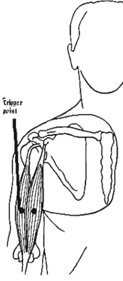
Sorne of the locations (trapezius, rhomboids, supraspinatus) selected for the project are frequently cited in other studies (Falla and Farina, 2005) and we have included other locations (biceps, triceps, deltoid) that are often found to be painful after a partial mastectomy with an axillary node dissection (McCredie et al., 2001). The clinicians who treat people with musculoskeletal disorders often describe a location in the middle of a muscle as being hypersensiti ve and thickened. This thickened structure is often thought to be
the cause of some of the pain and discomfort. These locations are known as trigger points (Davies and Davies, 2004; Mense and Simons, 2001; Takahashi et al, 2005).
1.11 Trigger points
A trigger point, as illustrated in Figure 3, is thought to be a hypersensitive area in a muscle which may cause pain (Simons, 2004). When an individual is in pain, whether acute or chronic pain, the person may seek advice from a doctor and treatment from a rehabilitation therapist. The medical professionals who evaluate patients with musculoskeletal problems will often try to palpate the involved area to note whether a trigger point nodule is present and may be a component of the patient's underlying problem (TraveJi and Simons, 1999). The presence of a nodule at the location of pain in muscular tissue is further confirmation of a potential trigger point.
Different types of trigger points may be found by the therapist on the patient, including active trigger points, latent trigger points (Davies and Davies, 2004; Mense and Simons, 2001; Travell and Siroons, 1999; Simons, 2(04), and satellite trigger points (Davies and Davies, 2004; Mense and Simons, 2001; Simons, 2004). An active trigger point is a location on the muscle that is presently causing pain which may be either local or referred (Mense and Simons, 2001). Referred pain is pain in a different location from where the original problem is located, such that pain in the trapezius may refer pain to the head region or down
the arm (Travell and Simons, 1999). The active trigger point may also restrict joint movement and cause the individual to seek out a remedy to reduce the pain, such as applying ice to the location or consulting a therapist. An active trigger point is thought to always provoke pain.
A latent trigger point is only painful when pressure is applied to it, and is not considered as significant a problem as the active trigger point. It is common for an individual to not experience any pain in the area of a latent trigger point unless someone or something applies pressure to the area (Simons, 2004). At that point the person with the latent trigger point will feel pain.
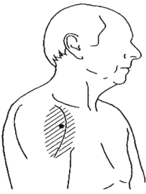
A satellite trigger point is a trigger point that will cause referred pain to multiple locations on the individual (Simons, 2004). When pressure is applied to the location of the trigger point, pain is felt in more than one location and may stay present for a period of time long after the stimulus has been removed from the original location. An example of this would be applying pressure to a trigger point in the rniddle of the upper trapezius fibers and noting how it may provoke pain in the back of the neck up to the occipitus ( the back of the head) and down to the shoulder and upper arm regions (Travel1 and Simons, 1999).
A trigger point is thought to develop from a muscle perfonning a single movement or a series of repeated movements leading to a contracture in the tissue (Simons, 2004). A contracture takes place when the muscle fibers have not return to a resting position but have remained in a shortened or contracted position. The trigger point is described as being a nodule-like structure attached to or resting within the muscle. The nodule structure is thought to occur because of thickening of muscle fibers. When the therapist palpates the nodular structure, a cord or rope-like formation is found underneath their finger tips (Davies and Davies, 2004; Mense and Simons, 2001; Travell and Simons, 1999).
(a) Nodule
Local Pain
(b)
Normal ~'ffiT1rmv'/ Conlracliof
libers knot
Figure 3: Diagram of trigger point nodule (contraction knot) in a muscle (Adapted from Simons, 2004)
In diagram 3, CTrP is defined as the contraction knot of the trigger point and ATrP is the active trigger point location on the muscle where the person would be experiencing pain.
It is not uncommon for a trigger point ta be called a myofascial trigger point (Stecco, 2004). The name, myofascial trigger point, describes the tissue"fascia" that envelopes a muscle,
becoming adherent at the same location where a trigger point has developed (Figure 3). Fascia is known to be adherent at different points (Stecco, 2004) in the body. If the fascia was not adherent at different locations throughout the body, it would be continuously moving and become displaced. The location where the fascia is adherent to the muscle is often in close proximity to where potential trigger point nodules may develop. If a trigger . point nodule develops at the site where the fascia is adherent to the muscle the location will become thickened and less pliable potentially aggravating an underlying problem. This would be in contrast to no nodule developing at the location where the fascia is adhererit to the muscle or if the nodule develops in another location other than where the fascia adheres to the muscle (Stecco, 2004). The fascial adherence and the trigger point may lead to a common problem, the myofascial trigger point.
a,-motor nerve
~Motor end plate region
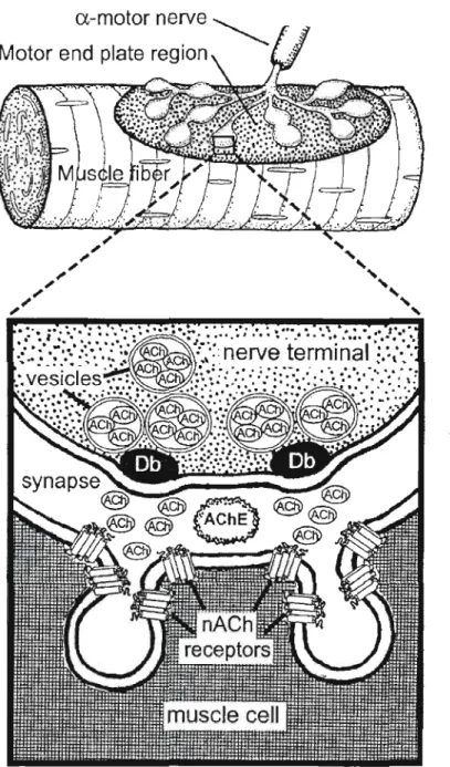
1.12 Why does a trigger point develop?
A trigger point is thought to develop because of sorne form of dysfunction that takes place at either the presynaptic or the post synaptic end plate of the neuromuscular junction. The release of acetylcholine (ACh) across the neuromuscular junction allows for action potentials to occur and for a muscle contraction to take place (Guyton and Hall, 2000). An integrated hypothesis developed by Simons (2004) explaining the etiology of the myofascial trigger point, speculates that excessive levels of acetylcholine released from the presynaptic end plate to the post synaptic end plate c1eft may be one of the reasons why a trigger point develops. Simons along with Travell (1999) have done extensive investigation into the role of trigger points and pain over the last 40 years.
Acetylcholinesterase, (AChE) is the enzyme that would normally breakdown the acetylcholine at the neuromuscular junction (Figure 4). When a problem occurs, sorne type of dysfunction may prevent the acetylcholinesterase from being released to break down the acetylcholine. As weil, the release of acetylcholine at the presynaptic deft (Point 1 in Figure 5) is dependent on the voltage-gated calcium Ca2 + channels (L-type and N' type) (Guyton and Hall, 2000) . If there is a defect/dysfunction of one of these Ca2 + channels there will be a continued release of acetylcholine (Simons, 2004). Thus, two mechanisms leading to a dysfunctional motor end plate causing muscle contracture may cause the development of trigger point nodules (Figure 3).
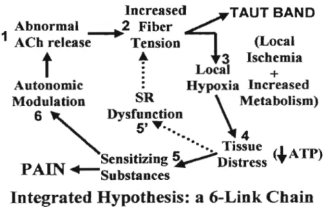
As mentioned above, dysfunction at the motor end plate may lead to a sustained contraction of muscle contraction. A sustained muscle contraction, as illustrated in Figure 5 (points 2 to 6) will lead to compression of local sensory nerves, reduced local blood supply, decrease local supply of circulation oxygen and eventual energy crises within the tissue (Simons, 2004). A resulting lack of ATP (Points 4 and 5' in Figure 5) may lead to impairment in the Ca2 + reuptake by the sacroplasmic reticulum, maintaining elevated cytoplasmic Ca2 + concentrations, which may continue contractile activity. As weil, the reduction in ATP may lead to an increase in chemicals (Point 5 in Figure 5) being secreted by the sustained muscle contractures that stimulate free nerve endings. These chemicals include bradykinin, cytokines, serotonin and histamine. This continued dysfunction rnay lead to sorne form of disorganization of the mitochondria. (Sirnons, 2004)
Increasedi:TAUT BAND
Abnormal
.2 Fiber
.
1
ACb release
Tension
(Local
~
3
Iscbemia
t
:
Local
+
Autonomie
:
Hypoxia Increased
Modulation
SR
\
Metabolism)
6 "
Dysfunction
5' •.
..
4
... ... Tissue (1 ATP)
PAIN
Sensitizing
~
Distress
:'f'
"-Substanees
Integrated Hypothesis: a 6-Link Chain
Figure 5: Theoretical model of the development of a trigger point (adapted from
Simons, 2004)
Different components of the integrated model put forward by Simons (2004) have been looked at by researchers using animal models. Hou et al., (2002) has looked at the raIe of calcium blockers and trigger points. Chen et al, (1998) has looked at the raIe of the autonomie nervous system and trigger points. Kuan et al., (2002) has looked at the raIe of excessive acetylcholine release and trigger points. Each of the studies has confirmed Simons's integrated hypothesis so far.
At least three other structures may be found in tissue that are somewhat similar to a trigger point nodule and should be identified. These structures include: 1) calcification of tissue
leading to calcium deposits which are bone-Iike structures hard to the touch (myositis ossificans), 2) a fatty nodule (often seen in the low back) which is soft and pliable and 3) a ganglion which is closely linked to superficial nervous tissue. What differentiates the calcium deposit and the fatty nodule from a trigger point is the mobility of the structures. Both the calcium deposit and the fatty nodule have a slight mobility, and both structures may be moved in close proximity to where they are found. This is in contrast to a trigger point, which is not mobile and will remain in the same location until sorne form of intervention takes place (Davies C, Davies, 2004; Travell and Simons, 1992). A ganglion is similar to the trigger point in that it does not move, but the ganglion is more likely to be attached to a superficiaJ structure such commonly palpated around the anterior aspect of the distal forearm. The ganglion often does not elicit pain when palpated and a common therapeutic approach to remove the ganglion has been to quickly apply a compressive force to flatten the ganglion, or to have the ganglion surgically removed (Dumontier et aL, 2006)
1.13 The rationale for investigating trigger points in this project
A trigger point is often found near the mid point of the muscle in close proximity to the motor points of that muscle. Knowing that trigger point nodules are commonly used by c!inicians to interpret pain for their patients and having a general idea of where to locate a potential trigger point nodule on the muscle, it was deemed appropriate the same locations be used in this project. Also, knowing what is not considered a trigger point nodule gave the athletic therapists clear guidelines as ta what to look for when palpating the subjects.
1.14 Summary of literature review
Pain in the upper arm, shoulder and neck are common problems after breast cancer therapy.
Women seem to be at greater risk for developing shoulder and neck problems, but it is unclear whether it is because of their muscle composition in that region, the jobs that they are required to perform on a daily basis or, as in men, due to their lack of physical activity.
The algometer has been used to quantify the subjective pain of an individual through the use of PPT values. PPT measured at the mid point of a muscle tends to be more consistent compared to other locations on the body. Studies that have used the algometers to evaluate pain have only recently taken into consideration the idea that a women's menstrual cycle may have an effect on the values obtained.
Clinician's that work with people experiencing musculoskeletal pain believe that trigger point nodules found in the muscle tissue contribute to a patient's musculoskeletal problem. A number of charts have been developed and books written outlining the locations of potential trigger point nodules in the body.
1.15 Implementing the literature review into this protocol
A hand-held algometer was used to give a quantifiable measure of pain in the upper arm, shoulder and neck region. The algometer was applied to the mid point of muscles where potential trigger point nodule rnay be located. The muscles that were to be tested were locations that are consistent with problematic areas associated with breast cancer surgery or cornmon musculoskeletal disorders.
The protocol was to be first used with individuals who are not experiencing any musculoskeletal dysfunction as to limit the number confounding variables. A women' s rnenstrual cycle is known to influence her perception of pain. The subjects who were selected for the project would have to be pre rnenopausal, with regular menstrual cycles. (A decision to limit the number of confounding variables) Ali of the women participating in the study should be tested at the same stage of their menstrual cycle.